Embed Size (px)
Citation preview

Figure 4.0 Protein

Figure 4.1 Abiotic synthesis of organic compounds under “early Earth” conditions

Figure 4.2 The shapes of three simple organic molecules

Figure 4.2x Shapes of Molecules
Methane
Ethane
Ethene

Figure 4.3 Valences for the major elements of organic molecules

Figure 4.x1 Urea

Figure 4.4 Variations in carbon skeletons

Figure 4.4x Hydrocarbons: molecular models
Butane
Isobutane
Hexane
Cyclohexane

Figure 4.5 The role of hydrocarbons in fats

Figure 4.6 Three types of isomers

Figure 4.6ax Structural isomers
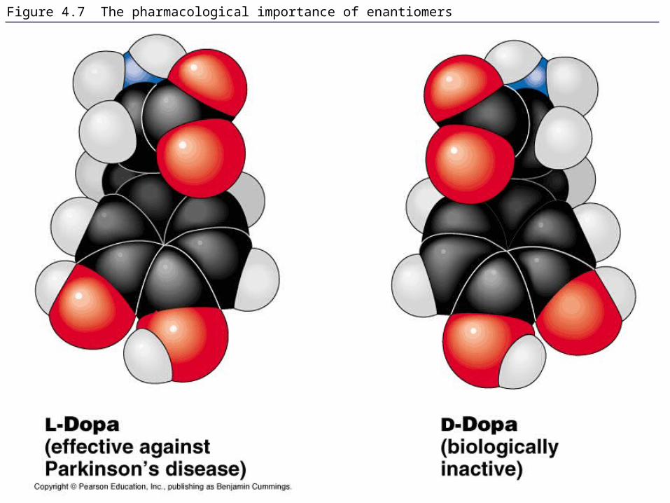
Figure 4.7 The pharmacological importance of enantiomers

Table 4.1 Functional Groups of Organic Compounds

Figure 4.8 A comparison of functional groups of female (estradiol) and male (testosterone) sex hormones

Figure 4.8x1 Estrone and testosterone

Figure 4.8x2 Male and female mallards

Figure 4.8x3 Male and female peacocks

Figure 4.8x4 Male and female sage grouse
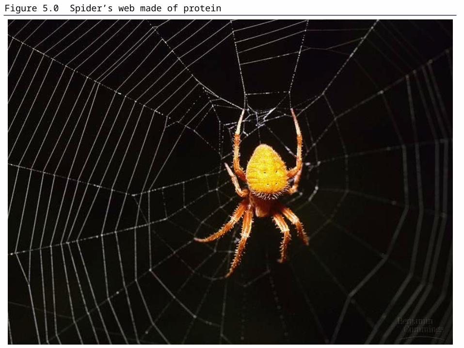
Figure 5.0 Spider’s web made of protein

Figure 5.1 Building models to study the structure and function of macromolecules

Figure 5.2 The synthesis and breakdown of polymers

Figure 5.3 The structure and classification of some monosaccharides

Figure 5.3x Hexose sugars
Glucose Galactose

Figure 5.4 Linear and ring forms of glucose

Figure 5.5 Examples of disaccharide synthesis

Figure 5.5x Glucose monomer and disaccharides
Glucose monomer
Sucrose
Maltose

Figure 5.6 Storage polysaccharides

Figure 5.7a Starch and cellulose structures

Figure 5.7b,c Starch and cellulose structures

Figure 5.7x Starch and cellulose molecular models
Glucose Glucose
Starch
Cellulose

Figure 5.8 The arrangement of cellulose in plant cell walls

Figure 5.x1 Cellulose digestion: termite and Trichonympha
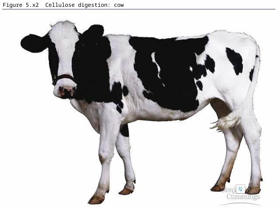
Figure 5.x2 Cellulose digestion: cow

Figure 5.9 Chitin, a structural polysaccharide: exoskeleton and surgical thread

Figure 5.10 The synthesis and structure of a fat, or triacylglycerol
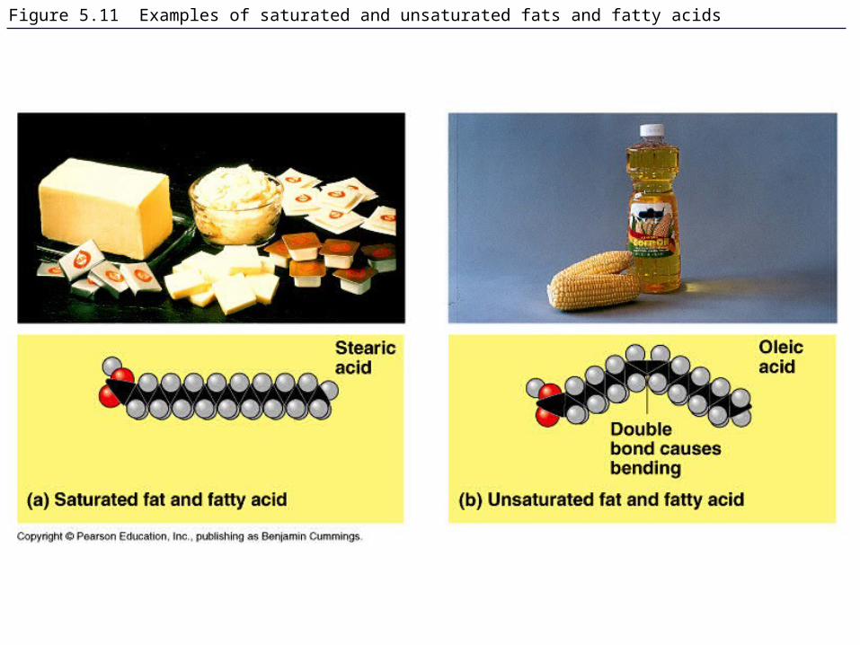
Figure 5.11 Examples of saturated and unsaturated fats and fatty acids

Figure 5.11x Saturated and unsaturated fats and fatty acids: butter and oil

Figure 5.12 The structure of a phospholipid

Figure 5.12x Phospholipid

Figure 5.13 Two structures formed by self-assembly of phospholipids in aqueous environments

Figure 5.14 Cholesterol, a steroid

Figure 5.14x Cholesterol

Table 5.1 An Overview of Protein Functions

Figure 5.15 The 20 amino acids of proteins: nonpolar

Figure 5.15 The 20 amino acids of proteins: polar and electrically charged

Figure 5.16 Making a polypeptide chain

Figure 5.17 Conformation of a protein, the enzyme lysozyme

Figure 5.18 The primary structure of a protein

Figure 5.19 A single amino acid substitution in a protein causes sickle-cell disease

Figure 5.19x Sickled cells

Figure 5.20 The secondary structure of a protein
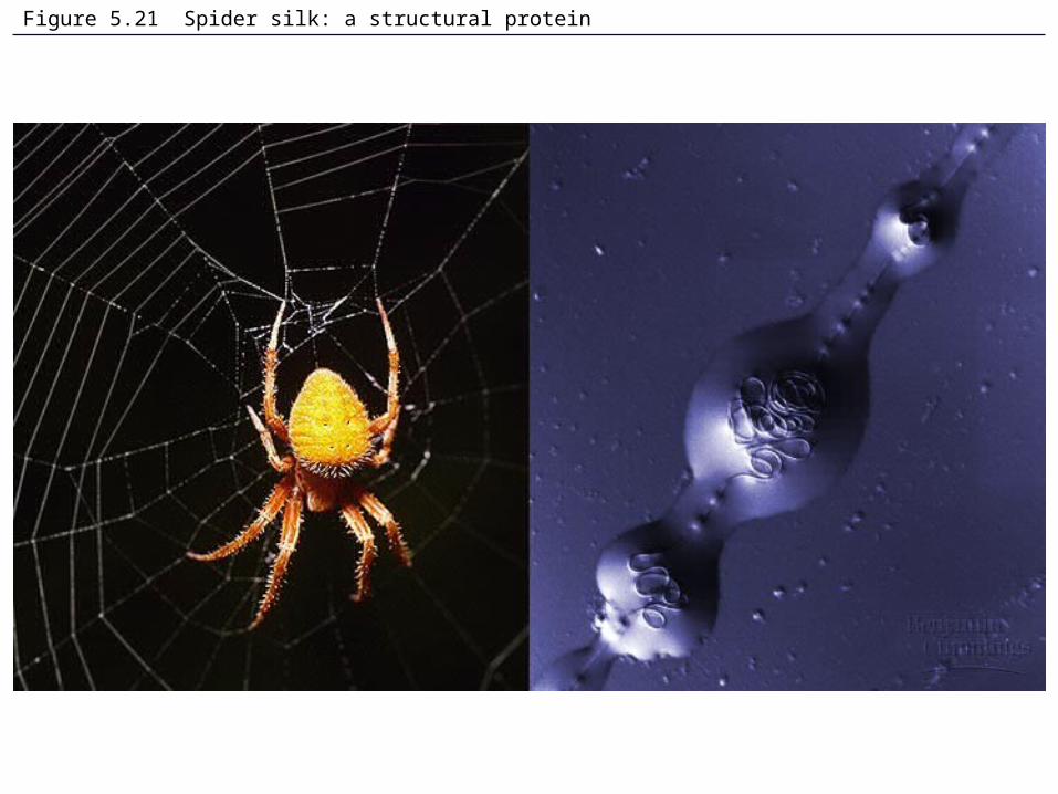
Figure 5.21 Spider silk: a structural protein

Figure 5.21x Silk drawn from the spinnerets at the rear of a spider

Figure 5.22 Examples of interactions contributing to the tertiary structure of a protein

Figure 5.23 The quaternary structure of proteins

Figure 5.24 Review: the four levels of protein structure

Figure 5.25 Denaturation and renaturation of a protein

Figure 5.26 A chaperonin in action

Figure 5.27 X-ray crystallography

Figure 5.x3 James Watson and Francis Crick

Figure 5.x4 Rosalind Franklin

Figure 5.28 DNA RNA protein: a diagrammatic overview of information flow in a cell

Figure 5.29 The components of nucleic acids

Figure 5.30 The DNA double helix and its replication

Table 5.2 Polypeptide Sequence as Evidence for Evolutionary Relationships