Embed Size (px)
Citation preview
FINAL REPORT ON THE SAFETY ASSESSMENT OF p-CHLORO-m-CRESOL’
p-Chloro-m-Cresol is a substituted phenol that functions as a cosmetic biocide preservative in skin care and suntan cosmetic formulations. While p-chloro-m- Cresol is approved for preservative use b.y the European Union at concentra- tions of up to 0.296, it can be used at other concentrations wh.en not intended as a preservative, and zt is prohibited from use in products intended to contact mucous membranes. p-Chloro-m-Cresol is approved for use as an indirect food additive in the United States. Acute, short-term, and subchronic tests in ani- mals indicate no dose-related toxicity other than reduced body weight gain. A chronic feeding stud?/ in rats identified kidney damage in high-dose males and an increase in pituitary adenomas in m.id- and high-dose females. No evidence of mutagenicity u?as seen in Ames tests, but there was an increase in SOS-DNA repair synthesis in Escherichia coli. A solution of p-Chloro-m-Cresol at concen- trations as low as O.Ofi%produced ocular irritation in rabbits. Some evidence of dermal irritation and sensitization ulas found in animal studies. In clinical studies, 2% p-Chloro-m-Cresol seems to be a skin irritant. Predictive patch tests using .5% p-Chioro-m-Cresol as the challenge were negative, but provocative patch tests yielded positive responses in a few individuals. Other data pertinent to the safety assessment of this ingredient are not available, including the con- centration of use in cosmetics, UV absorption data (photosensitization data also are needed if the ingredient absorbs in the WA or UVB region), dermal developmental toxicity data and a mutagenicity study using a mammalian s.ys- tern (a B-year dermal carcinogenicity studv is also needed if the mutagenesis study is positivei. Based on the presently available data, the safety of this ingre- dient in cosmetic. formulations cannot be supported.
p-Chloro-m-C resol (PCMC) is a substituted phenol that functions as a cosmetic biocide and as a preservative (Wenninger and McEwen, 1995a). This review assesses the safety of this ingredient as used in cosmetic formulations.
1 Reviewed by the Cosmetic Ingredient Review Expert Panel. Monice Zondlo Fiume, Scientific Analyst/Report Management Coordinator, prepared
this report. Address correspondence to Dr. F. A. Andersen, Cosmetic Ingredient Review, 1101
17th Street, NW, Suite 310, Washington, DC 20036, USA.
International Journal of Toxicology, l&235-268, 1997 Copyright 0 1997 Cosmetic Ingredient Review
1091-5813/97 $12.00 + .oo
235
236 COSMETIC INGREDIENT REVIEW
CHEMISTRY
Definition and Structure
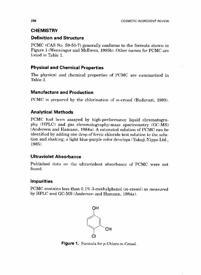
PCMC (CAS No. 59-50-7) generally conforms to the formula shown in Figure 1 (Wenninger and McEwen, 1995b). Other names for PCMC are listed in Table 1.
Physical and Chemical Properties
The physical and chemical properties of PCMC are summarized in Table 2.
Manufacture and Production
PCMC is prepared by the chlorination of m-cresol (Budavari, 1989).
Analytical Methods
PCMC had been assayed by high-performance liquid chromatogra- phy (HPLC) and gas chromatography-mass spectrometry (GC-MS) (Andersen and Hamann, 1984a). A saturated solution of PCMC can be identified by adding one drop of ferric chloride test solution to the solu- tion and shaking; a light blue-purple color develops (Yakuji Nippo Ltd., 1985).
Ultraviolet Absorbance
Published data on the ultraviolent absorbance of PCMC were not found.
Impurities
PCMC contains less than 0.1% 3-methylphenol (m-cresol) as measured by HPLC and GC-MS (Andersen and Hamann, 1984a).
CH
Figure 1. Formula for p-Chloro-m-Cresol.
pCHLORO-m-CRESOL
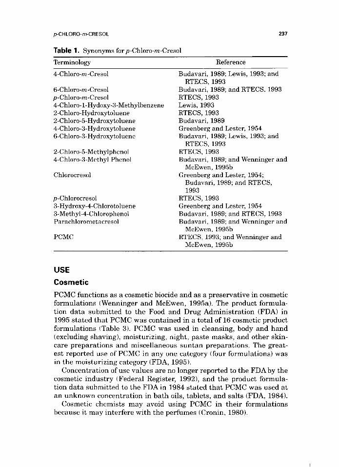
Table 1. Synonyms for p-Chloro-m-Cresol
Terminology Reference
237
4-Chloro-m-Cresol
6-Chloro-m-Cresol p-Chloro-m-Cresol 4-Chloro-1-Hydoxy-3-Methylbenzene 2-Chloro-Hydroxytoluene 2-Chloro-5Hydroxytoluene 4-Chloro-3-Hydroxytoluene 6-Chloro-3-Hydroxytoluene
2-Chloro-5-Methylphenol 4-Chloro-3-Methyl Phenol
Chlorocresol
p-Chlorocresol 3-Hydroxy-4-Chlorotoluene 3-Methyl-4-Chlorophenol Parachlorometacresol
PCMC
Budavari, 1989; Lewis, 1993; and RTECS, 1993
Budavari, 1989; and RTECS, 1993 RTECS, 1993 Lewis, 1993 RTECS, 1993 Budavari, 1989 Greenberg and Lester, 1954 Budavari, 1989; Lewis, 1993; and
RTECS, 1993 RTECS, 1993 Budavari, 1989; and Wenninger and
McEwen, 199513 Greenberg and Lester, 1954;
Budavari, 1989; and RTECS, 1993
RTECS, 1993 Greenberg and Lester, 1954 Budavari, 1989; and RTECS, 1993 Budavari, 1989; and Wenninger and
McEwen, 1995b RTECS, 1993; and Wenninger and
McEwen, 1995b
USE
Cosmetic
PCMC functions as a cosmetic biocide and as a preservative in cosmetic
formulations (Wenninger and McEwen, 1995a). The product formula-
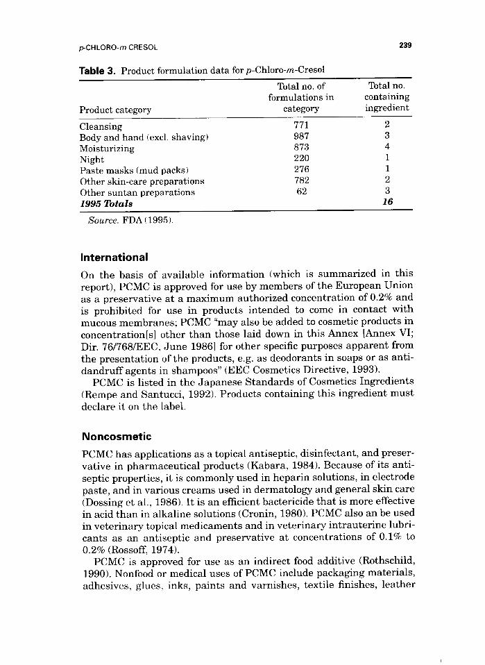
tion data submitted to the Food and Drug Administration (FDA) in 1995 stated that PCMC was contained in a total of 16 cosmetic product formulations (Table 3). PCMC was used in cleansing, body and hand (excluding shaving), moisturizing, night, paste masks, and other skin- care preparations and miscellaneous suntan preparations. The great- est reported use of PCMC in any one category (four formulations) was in the moisturizing category (FDA, 1995).
Concentration of use values are no longer reported to the FDA by the cosmetic industry (Federal Register, 19921, and the product formula- tion data submitted to the FDA in 1984 stated that PCMC was used at an unknown concentration in bath oils, tablets, and salts (FDA, 1984).
Cosmetic chemists may avoid using PCMC in their formulations because it may interfere with the perfumes (Cronin, 1980).
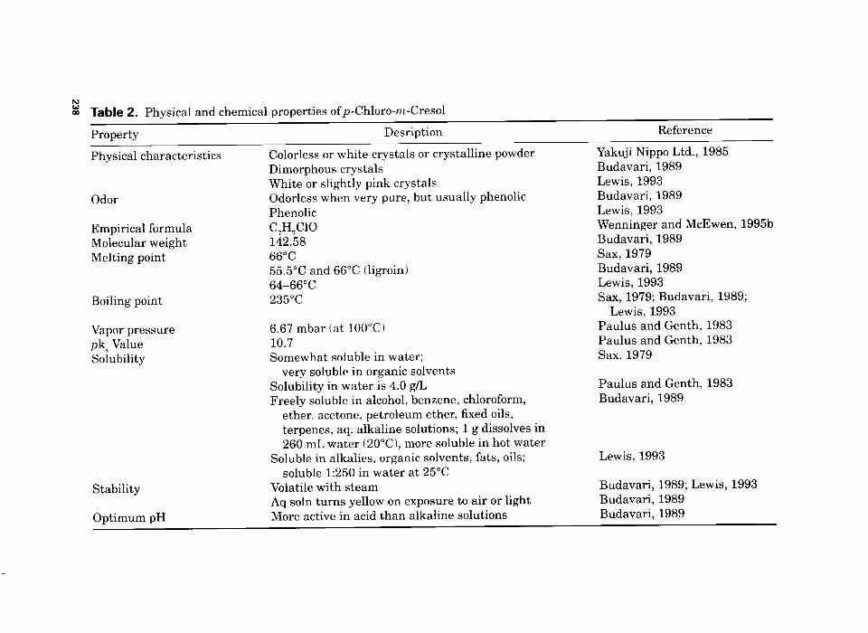
E co Table 2. Physical and chemical properties of p-Chloro-m-Cresol
Property Desription Reference
Physical characteristics
Odor
Empirical formula Molecular weight Melting point
Boiling point
Vapor pressure pks Value Solubility
Stability
Optimum pH
Colorless or white crystals or crystalline powder Dimorphous crystals White or slightly pink crystals Odorless when very pure, but usually phenolic Phenolic C,H,ClO 142.58 66°C 55.5”C and 66°C (ligroin) 64-66°C 235°C
6.67 mbar (at 100°C) 10.7 Somewhat soluble in water;
very soluble in organic solvents Solubility in water is 4.0 g/L Freely soluble in alcohol, benzene, chloroform,
ether, acetone, petroleum ether, fixed oils, terpenes, aq. alkaline solutions; 1 g dissolves in 260 mL water (2O”C), more soluble in hot water
Soluble in alkalies, organic solvents, fats, oils; soluble 1:250 in water at 25°C
Volatile with steam Aq soln turns yellow on exposure to air or light More active in acid than alkaline solutions
Yakuji Nippo Ltd., 1985 Budavari, 1989 Lewis, 1993 Budavari, 1989 Lewis, 1993 Wenninger and McEwen, 1995b Budavari, 1989 Sax, 1979 Budavari, 1989 Lewis, 1993 Sax, 1979; Budavari, 1989;
Lewis, 1993 Paulus and Genth, 1983 Paulus and Genth, 1983 Sax, 1979
Paulus and Genth, 1983 Budavari, 1989
Lewis, 1993
Budavari, 1989; Lewis, 1993 Budavari, 1989 Budavari, 1989
pCHLORO-m-CRESOL
Table 3. Product formulation data for p-Chloro-m-Cresol
Total no. of formulations in
Product category category
239
Total no. containing ingredient
Cleansing 771 2 Body and hand (excl. shaving) 987 3 Moisturizing 873 4 Night 220 1 Paste masks (mud packs) 276 1 Other skin-care preparations 782 2 Other suntan preparations 62 3 1995 Totals 16
Source. FDA (1995).
International
On the basis of available information (which is summarized in this report), PCMC is approved for use by members of the European Union as a preservative at a maximum authorized concentration of 0.2% and is prohibited for use in products intended to come in contact with mucous membranes; PCMC “may also be added to cosmetic products in concentration[s] other than those laid down in this Annex [Annex VI; Dir. 76/768/EEC, June 19861 for other specific purposes apparent from the presentation of the products, e.g. as deodorants in soaps or as anti- dandruff agents in shampoos” (EEC Cosmetics Directive, 1993).
PCMC is listed in the Japanese Standards of Cosmetics Ingredients (Rempe and Santucci, 1992). Products containing this ingredient must declare it on the label.
Noncosmetic
PCMC has applications as a topical antiseptic, disinfectant, and preser- vative in pharmaceutical products (Kabara, 1984). Because of its anti- septic properties, it is commonly used in heparin solutions, in electrode paste, and in various creams used in dermatology and general skin care (Dossing et al., 1986). It is an efficient bactericide that is more effective in acid than in alkaline solutions (Cronin, 1980). PCMC also an be used in veterinary topical medicaments and in veterinary intrauterine lubri- cants as an antiseptic and preservative at concentrations of 0.1% to 0.2% (Rossoff, 1974).
PCMC is approved for use as an indirect food additive (Rothschild, 1990). Nonfood or medical uses of PCMC include packaging materials, adhesives, glues, inks, paints and varnishes, textile finishes, leather
240 COSMETIC INGREDIENT REVIEW
and tanning agents, and industrial oils and emulsions (Dooms- Goossens et al., 1981).
The use concentration for most noncosmetic applications of PCMC ranges from 0.05% to 0.5% (Andersen and Hamann, 198413); aqueous drugs for parenteral use are sometimes preserved with 0.05% to 0.1% PCMC (Cronin, 1980).
GENERAL BIOLOGY
Absorption, Distribution, Metabolism, and Excretion
A pharmacokinetic study was performed in which rats were dosed orally with 300 mg/kg PCMC. PCMC reportedly was eliminated rapidly through the kidneys. In addition, there is no likelihood of cumulation effects. A corresponding examination of fatty and hepatic tissues from rats that were fed 150 to 1500-ppm PCMC for up to 13 wk reported no indication of an accumulation of PCMC in these tissues (Paulus and Genth, 1983).
Four groups of conventional female albino guinea pigs, three per group, were used to determine the bioavailability of PCMC (Andersen et al., 1985). Occlusive patches of 0.2 mL of a 5% PCMC aqueous sus- pension stabilized with Carbomer 941, a saturated aqueous solution of 0.38% PCMC, 5.0% PCMC in olive oil/acetone (4/l), or 5.0% PCMC in propylene glycol were applied for 24 h. After 96 h, the animals were killed and the skin at the site of patch testing was removed for analysis (the patches were kept for analysis to determine the amount of PCMC remaining in the patch material). Fractional sampling of the urine and feces was performed to determine the rate of absorption of PCMC. An additional three animals had been injected with PCMC intraperi- toneally to determine the excretion rate; however, no free PCMC was found, indicating rapid metabolism. The investigators stated that fur- ther analysis would be required before PCMC absorption can be esti- mated by this method (‘“C-PCMC was not available).
In determining bioavailability, the calculations were based on the assumption that the saturated PCMC solution is 0.4% (w/v), corre- sponding to 0.8 mg in 0.2 mL, and that 0.2 mL of the 5% PCMC prepa- rations contained 10 mg of the chemical. The results indicated that 25% of the aqueous PCMC (stabilized with carbomer 941) and 46% of the saturated aqueous PCMC solution remained in the patches. Only 0.2% of the aqueous PCMC (stabilized with carbomer 941) and 0.5% of the saturated aqueous PCMC solution were found in the skin at the patch site. These findings were compared with 65% of the PCMC in propylene glycol and 66%~ of the PCMC in olive oil/acetone solutions remaining in
p-CHLORO-m-CRESOL 241
the patch; 0.7% and 1.6%, respectively, were found in the skin at the patch site. The investigators concluded that PCMC was more bioavail- able from the aqueous preparations. After 96 h, 0.2% and 0.5% PCMC was detected at the patch test site in the animals dosed with 5.0% and saturated aqueous PCMC, respectively, and 0.7% and 1.6% PCMC were found in the skin of the animals patch-tested with 5% PCMC in olive oil/acetone and propylene glycol, respectively.
Abdominal skin from SKH-hr-1 mice was used to determine the per- meability of PCMC (Huq et al., 1986). The permeability was evaluated, both through whole skin and skin that was stripped repeatedly (20 times) with cellophane tape to remove the stratum corneum, by mount- ing the excised samples in a two-compartment diffusion cell, with the two half-cells being filled with normal saline. Samples were withdrawn and assayed spectrophotometrically; the absorbance was measured at 245 nm. Permeability coefficients were assessed for both stripped and whole skin under conditions that kept ionization to a minimum.
In assessing the permeability of PCMC through whole skin, the con- centration used was 0.05 g/100 mL, the pKa of PCMC was 9.56, the donor pH was 6.18, and the receiver pH was 6.2. Under these condi- tions, the apparant permeability coefficient for whole skin was 119 + 1.8 X lo-” cm/h, and the average lag time was 35.8 min. For stripped skin, 0.055 g/100 mL PCMC was used, and the donor and receiver pH was 6.2. The permeability coefficient of stripped skin was 241 2 22 X lo-” cm/h, and the lag time was 14.6 min. The estimated permeability coefficients of PCMC for viable tissue and stratum corneum were 302 X lo-” and 235 X 10 ‘$, respectively.
Hepatic Effects
Male Wistar rats were dosed orally with 400 mg/kg PCMC to determine the effect on the development of hepatocellular vacuoles (Meiss et al., 1981). Hepatic tissue taken from the left lobe of the liver 7 days after dosing was examined by electron microscopy. Markedly widened intra- cellular spaces, intracytoplasmic vacuoles of different sizes, and alter- ations of the cell organelles and nuclei were observed. Cells from older animals contained more invaginations and vacuoles than those of younger animals. When younger animals were fasted prior to being killed, the vacuolic formation was similar to that of older animals.
The investigators considered the origin of the hepatic cell vacuoles to be the intracellular space. They observed the following stages of vacuo- lar formation: dilated cell surface, often limited to a restricted region; the plasmalemma-formed vesicular invaginations; and the invagina- tions extended to form large vacuoles with an open connection to inter-
242 COSMETIC INGREDIENT REVIEW
cellular space. The investigators theorized that increasing blood pres- sure caused by PCMC intoxication of the liver was the reason for the pathologic invaginations in the region of the contacts between the hepatocytes.
Neurologic Effects
A single dose of 0.2% PCMC (to correspond with 0.25 mg/kg and 0.5 mg/kg) was injected into the cisterna magna of three rabbits per dose after withdrawal of an appropriate amount of cerebrospinal fluid; two animals were used as controls (Gray and Naim, 1972). One animal from each dose group was killed after 24 h, 48 h, and 5 days. All animals sur- vived. None of the animals developed any functional disability sugges- tive of neurologic damage. No evidence of pathologic lesions caused by PCMC was present, and the brains had no evidence of meningeal parenchymatous damage.
Antimicrobial Activity
A study was conducted to determine whether PCMC affected the enzy- matic activity or the use of amino acids in the inhibition of germination of Bacillus subtilis spores (Sierra, 1970). The initiation of germination of B. subtilis in complete medium and in phosphate buffer plus the enzyme subtilopeptidase is inhibited by PCMC, but the activity of the enzyme is not, which suggests that the proteolytic enzyme is not the primary site of inhibition. Amino acid-initiated germination was reversibly inhibited by PCMC, suggesting that the inhibitor prevents the initiation of germination of bacterial spores at the level of amino acid use, in both complete medium and with subtilopeptidase.
The antimicrobial spectrum of 0.02% PCMC included gram-positive and gram-negative bacteria, including tubercle bacilli, and also yeasts and fungi (Paulus and Genth, 1983).
The effectiveness of PCMC as a preservative in aqueous cream was examined using the Test for Efficacy of Preservatives (British Pharmacopoeia, 1982) modified to include membrane filtration (Brown et al., 1986). Ten-gram samples of the cream were inoculated with 0.1 mL of a suspension containing either Candida albicans or AspergiZZus niger to give concentrations of approximately 10” organisms/g. PCMC did meet the British Pharmacopoeia requirements for the efficacy of antimicrobial preservatives [the number of molds or yeasts recovered per gram is reduced by a factor of not less than 100 within 14 days of challenge and there is no increase thereafter] when tested against A. niger, but not when tested against C. albicans, using membrane filtra- tion to isolate the microorganisms.
p-CHLORO-rr-CRESOL 243
ANIMAL TOXICOLOGY
Acute Toxicity
Oral
Five groups of male Wistar II rats were dosed with 1.0 to 5.0 g/kg PCMC by stomach tube (BAYER AG, 1978). Intensified diuresis, seda- tion, disturbed respiration, trembling, and convulsions were observed (specific doses not identified). The oral LD,, of PCMC for Wistar II rats was 1830 mg/kg.
Groups of male Wistar rats were given a single oral dose of 400 mg/kg PCMC in peanut oil; controls were dosed with an equivalent amount of peanut oil only (Robenek et al., 1980). All animals were killed 60 h after dosing, and hepatic tissue was removed from the cen- ter of the right lobe of the liver for examination by electron microscopy.
After dosing, the animals’ behavior changed: after 30 min, the animals were uneasy and had “ruffled-up” coats. These signs diminished after 1 h but were replaced by long “apathetic motions.” After 24 h until study ter- mination, the haircoats were altered again. At necropsy, the liver appeared slightly enlarged and was a pale red color with pale gray spots.
Light microscopy findings included a distinct dilation of the sinusoids with an activation of the Kupffer cells. The intercellular spaces were enlarged, and there were numerous vacuoles found in the cytoplasm.
In electron micrographs, outpouchings of cell membranes were observed. A greater-than-normal number of lysosomes were found around the bile canaliculi after dosing. Also, there was an increase in the number of mitochondria, many membrane-surrounded vacuoles, alterations in the intercellular space and in the rough endoplasmic reticulum, and an increase in the number and size of gap junctions. In addition, the bile canaliculi were dilated and had irregularities and side branches that extended into the cytoplasm of adjacent hepatocytes.
Five groups of Sprague-Dawley rats, 10 per sex per group, were dosed orally with PCMC in carbowax; males were dosed with 2000 to 7683 mg/kg and females with 1500 to 5762 mg/kg (Mobay Chemical Corporation, 1981). Ataxia, wheezing, muscle fasciculations, tremors, convulsions, and salivation were observed in all dosed animals. The oral LD,, of PCMC was 5129 mg/kg for male Sprague-Dawley rats and 3636 mg/kg for female Sprague-Dawley rats. These values are higher than those reported for Wistar II rats, but no explanation for this strain difference was available.
Percutaneous
The percutaneous LD,, of PCMC for rats was more than 500 mg/kg (Paulus and Genth, 1983).
244 COSMETIC INGREDIENT REVIEW
Subcutaneous
Five groups of 10 or 20 mice (sex not specified) were dosed subcuta- neously with 100 to 500 mg/kg PCMC as a 0.4% aqueous solution (Wein, 1939). Toxic signs appeared within 5 min and included severe muscular tremors, especially in the fore and hind limbs. Death from respiratory failure usually occurred within 3 h, with some animals dying within 30 min. The calculated subcutaneous LD,,, of PCMC for mice was 360 mg/kg.
Three groups of 10 albino rats per group (sex not specified) were injected subcutaneously with a 0.4% aqueous solution of 30,400, or 500 mg/kg PCMC (Wein, 1939). Observations included tremors followed by death in approximately 15 min. The calculated subcutaneous LD,, was 400 mg/kg PCMC.
The Robenek et al. (1980) acute toxicity study described in the “Oral” section also included testing in which a single subcutaneous dose of 400 mg/kg PCMC in peanut oil was administered to rats; the same procedures were followed. The same general behavioral and macroscopic hepatic changes and light microscopy findings were observed. In electron microscopic examination of the liver, the reported findings included enlarged sinusoids, an increase in detritus in the Kupffer cells, enlarged intercellular spaces, and an increased number of cytoplasmic vacuoles. An increase also occurred in the number of lysosomes, some of which were comparable in size to mito- chondria. The rough endoplasmic reticulum appeared disorganized, often surrounded by mitochondria and with an increase in the loss of ribosomes. In addition, an increase occurred in mitochondria, which often appeared pleomorphic. Some of the mitochondria had a long shape and showed a tear in the outer membrane. Also, a translucent matrix accompanied by a decrease in mitochondrial cristae was observed.
Inhalation
Upon a 4-h exposure, the inhalation LC,, for rats was more than 583 mg PCMC-NA/m” air (Paulus and Genth, 1983).
Intravenous
Five groups of 10 or 20 mice (sex not specified) were injected intra- venously with 100 to 500 mg/kg PCMC as a 0.4% aqueous solution (Wein, 1939). The calculated intravenous LD,,, of PCMC for mice was 70 mg/kg.
pCHLORO-m-CRESOL 245
Short-Term Toxicity
Dermal
New Zealand White rabbits, 50 per sex, received cutaneous applications of 0, 10, 40, and 160 mg/kg/day PCMC 5 days per week for 3 wk on a shaved area of the back (Mobay Chemical Co., 1980). Irritation and erythema were observed, but no PCMC-related systemic effects were present compared with controls.
Oral
Groups of 20 Wistar SPF rats, 10 males and 10 females per group, were dosed with 50, 200, or 400 mg/kg/day PCMC in food-grade soybean oil by gavage at a volume of 5 ml/kg/day for 28 days (Madsen et al., 1986). A control group was dosed with soybean oil only. The animals were examined twice daily, and body weights and feed consumption were determined weekly. Blood samples were taken from eight males and eight females in each group for hematologic and clinical chemistry analysis after 21 days of dosing. All rats were necropsied at study ter- mination. The only toxicologic sign observed was a statistically signifi- cant decrease in body weight gain for the males and females dosed with 400 mg/kg PCMC. Relative organ weights were comparable for all groups, and the hematologic and clinical chemistry parameters were normal. No dose-related pathologic changes were observed. The no- effect level (NOEL) was 200 mg/kg/day PCMC.
Rats were fed 2500, 5000, or 10,000 ppm PCMC, and controls were fed untreated feed (number per group, sex, and dose duration not stated) (BAYER AG, 1992). Male rats of the lO,OOO-ppm dose group had reduced body-weight gains. Dose-related effects were not observed for males of the 2500-ppm and 5000-ppm dose groups or for any of the female rats.
Subcutaneous
Five albino rats were injected subcutaneously with 72 mg/kg PCMC as a 0.4% solution daily for 2 wk, and five rats were used as controls (Wein, 1939). All animals survived until study terination. The only observation was that of a mild inflammatory reaction, with some leuco- cyte infiltration, at the site of injection.
Three rabbits were injected subcutaneously with 5 ml/day of 0.25% PCMC in saline for 4 wk (Wein, 1939). Survival was 100% and pathol- ogy was noted.
COSMETIC INGREDIENT REVIEW
Subchronic Toxicity
Oral
SPF rats, 20 per sex per group, were fed 150, 500, or 1500 ppm PCMC in feed, and controls were fed untreated feed daily for 13 wk (BAYER AG, 1980a). Body weights of the animals of the 500-ppm and 1500-ppm dose groups were retarded, independent of dose. No dose-related changes were observed at necropsy or on microscopic examination; sur- vival was 100%. The no-toxic effect concentration was 150 ppm.
Chronic Toxicity
Oral
Bor:WISW(SPF Cpb) Wistar rats, 60 animals per sex per group, were fed 400, 2000, or 10,000 ppm PCMC for 2 yr, with the exception of 10 animals per sex per group that were necropsied after 53 wk (BAYER AG, 1992). A control group of 60 animals per sex was fed a diet that did not contain PCMC for 2 yr, again with the exception of 10 animals per sex that were necropsied after 53 wk (dosages were determined based on results of previous short-term and subchronic studies summarized previously in this report).
The animals were observed at least twice daily, with detailed weekly evaluations; body weights also were measured weekly. Feed consump- tion was determined weekly for 13 wk, after which it was determined at 4-wk intervals. Water intake was determined at 4-wk intervals over the entire study. Ophthalmologic evaluations were performed on 20 ani- mals per sex per group prior to study initiation and then on 20 animals per sex of the control and lO,OOO-ppm groups after 52 and 104 wk of dosing. Blood samples were obtained for examination from 10 animals per sex per group after 27, 52, 79, and 104 wk of dosing. Urinalyses were performed during weeks 26/27, 51/52, 78/79, and 103/104.
Averaged over 2 yr, the males and females of each dose group ate the following quantities (per day) of PCMC: 400-ppm group, 21 and 27.7 mg, respectively; 2000-ppm group, 103.1 and 134.3 mg, respectively; and lO,OOO-ppm group, 558.9 and 743.5 mg, respectively. Body weights of female rats of all dose groups and of male rats of the lO,OOO-ppm dose group were significantly lower compared with controls throughout the study. Body weights of the males of the 400-ppm and 2000-ppm dose groups were comparable with control values. Mean feed intake per animal per day was comparable between males of all dose groups and controls but was slightly lower for females of all dose groups compared with controls. When related to body weight, the mean feed intake of males and females of the lO,OOO-ppm dose groups was increased com- pared with controls. Males rats of the lO,OOO-ppm group had increased
pCHLORO-m-CRESOL 247
mean water intake per animal per day, and the water intake per kg body weight for females of the lO,OOO-ppm group also was increased compared with controls.
The incidence of an increased abdominal circumference was signifi- cantly reduced for female animals of the 2000-ppm and lO,OOO-ppm dose groups, and the frequency of a poor general condition was statisti- cally increased for females of the lO,OOO-ppm dose group. Ophthalmologic examination did not indicate any ocular toxicity. No significant difference existed in mortality between animals of the test and control groups.
PCMC-related clinical chemistry or hematologic changes were lim- ited to reduced serum potassium and phosphate concentrations (com- pared with control values) for males and females of the lO,OOO-ppm group. Urinalysis results included a decrease in total protein excretion for males and females fed lO,OOO-ppm PCMC; males of this group also had a reduced urinary density, often in conjunction with a slightly enhanced urinary volume.
No significant findings were reported in the rats at the interim necropsy after 52 wk; absolute and relative organ weights did not differ significantly between test and control groups. At study termination, necropsy findings included deformation of the kidneys of 6 of 44 surviv- ing males of the lO,OOO-ppm group. Relative kidney weights of males of the 2000-ppm group and males and females of the lO,OOO-ppm group were slightly increased compared with control values.
No microscopic lesions were observed at interim necropsy. Females of the 2000-ppm dose group that died during the study had dilated ducts of the mammary gland. At study termination, an increased incidence of papillary necrosis, cortical tubular dilations, and cortical fibrosis of the kidneys was observed in male rats of the lO,OOO-ppm group. Females of the 2000-ppm and lO,OOO-ppm groups that died on study had a greater incidence of pituitary adenoma, a finding that was not observed at interim necropsy (only preliminary microscopic information is avail- able from rats of the final necropsy). The NOEL was 2000 ppm PCMC.
Ocular Irritation
PCMC, 0.05%’ or 0.1% in 0.9% saline, was instilled into the conjunctival sac of the eyes of rabbits (number of animals not stated) by applying 20 drops/min for 10 min; the eyes were rinsed with saline after 15 min (Fiebig et al., 1972). Fluorescein staining was used. In the animal(s) dosed with 0.05%) PCMC, the “cornea” [sic] was reddened for 15 min- 4 h after application, and the eye was normal after 24 h. In the ani- mal(s) dosed with 0.1% PCMC, the eye was inflamed after 48 h and clear after 106 h.
248 COSMETIC INGREDIENT REVIEW
Soft lenses that were stored in bactericidal-strength solution were applied to the eyes of 13 rabbits for 6 h/day and the eyes were not rinsed; the isotonic solution, pH 7.0, contained 0.1% PCMC (Davies, 1973). PCMC produced severe irritation after a few days.
Dermal Irritation
The trypan blue method of Hoppe was used to determine the dermal irritation potential of PCMC (Baichwal and Phadnis, 1968). Groups of rabbits, two per group (sex not specified), were given a single applica- tion of 0.2% PCMC in normal saline or 0.4% or 0.8% PCMC in 1% Tween in normal saline. The site of application was four areas in the abdominal region and the duration of contact was 0.4 mL injected intradermally within 10 to 15 min. nYenty min after dosing, 1 mL/kg of 1% trypan blue was injected intravenously and the color at the injec- tion sites was observed for 3 h. The maximal irritation score (scale not stated) was 4 for 0.2% and 0.4% and 8 for 0.8% PCMC.
Sensitization
The sensitization studies reported here are summarized in Table 4. Groups of 20 female outbred albino guinea pigs were used in a
guinea-pig maximization test (GPMT) (Magnusson and Kligman, 1970) to determine the sensitization potential of two commercial biocides that contained PCMC; one biocide contained pentachlorophenol and PCMC (percentage of each not stated), whereas the other was more than 99.9% PCMC (Andersen and Hamann, 1984a). Acontrol group that was dosed with vehicle only (induction and challenge dose concentrations were determined in a one-application skin irritancy test using at least four guinea pigs 1 also was used. The challenge reactions were scored on a scale of 0 to 3 after 48 h and 72 h. A grade 1 reaction was not regarded as evidence of sensitization.
The induction doses of the pentachlorophenol-PCMC formulation consisted of intradermal administration of 5% of the biocide in propy- lene glycol and a patch containing 0.5 mL of 25% biocide formulation in yellow petrolatum. Animals were challenged using Finn chambers con- taining 0.5 mL of 5.0% of the biocide on day 21, 1.0% pentachlorophenol or 1.0% PCMC on day 35, and 0.5% of the biocide (all in yellow petrola- turn) on day 42.
For the pure-PCMC formulation, the induction doses consisted of intradermal administration of a 5% solution in propylene glycol and a patch of 0.5 mL of 10% PCMC in yellow petrolatum. The animals were challenged with an occlusive patch with 0.5 mL of 1% PCMC in petro- latum on days 21 and 35.
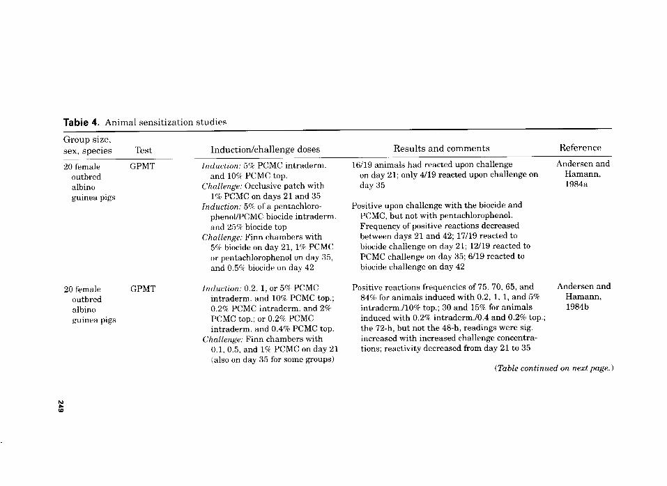
Tabie 4. Animal sensitization studies
Group size,
sex, species Test Induction/challenge doses Results and comments Reference
20 female CPMT Induct~m: 5% PCMC intraderm.
outbred and 10% PCMC top. albino Challenge: Occlusive patch with
guinea pigs 1% PCMC on days 21 and 35 Induction: 5% of a pentachloro-
phenol/PCMC biocide intraderm. and 25% biocide top
Challenge: Finn chambers with 5% biocide on day 21, 1% PCMC or pentachlorophenol on day 35, and 0.5% biocide on day 42
20 female CrPMT Induction: 0.2, 1, or 5% PCMC outbred intraderm. and 10% PCMC top.;
albino 0.2% PCMC intraderm. and 2%
guinea pigs PCMC top.; or 0.2% PCMC intraderm. and 0.4% PCMC top.
Challenge: Finn chambers with 0.1, 0.5, and 1% PCMC on day 21 (also on day 35 for some groups)
1609 animals had reacted upon challenge Andersen and on day 21; only 4119 reacted upon challenge on Hamann, day 35 1984a
Positive upon challenge with the biocide and PCMC, but not with pentachlorophenol. Frequency of positive reactions decreased between days 21 and 42; 17119 reacted to biocide challenge on day 21; 12/19 reacted to PCMC challenge on day 35; 6/19 reacted to biocide challenge on day 42
Positive reactions frequencies of 75, 70, 65, and 84% for animals induced with 0.2, 1, 1, and 5% intraderm./lO% top.; 30 and 15% for animals induced with 0.2% intraderm.lO.4 and 0.2% top.; the 72-h, but not the 48-h, readings were sig. increased with increased challenge concentra- tions; reactivity decreased from day 21 to 35
Andersen and Hamann, 1984b
(Table continued on next page.)
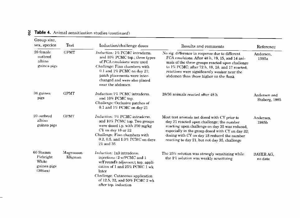
g Table 4. Animal sensitization studies (continued)
Group size, sex, species
20 female outbred albino guinea pigs
36 guinea
Pigs
20 outbred albino guinea pigs
60 Stamm Pirbright White guinea pigs (30hex)
Test Induction/challenge doses Results and comments Reference
GPMT Induction: 1% PCMC intraderm.
GPMT
and 10% PCMC top.; three types of FCA emulsions were used
Challenge: Finn chambers with 0.1 and 1% PCMC on day 21; patch placements were inter- changed and were also placed near the abdomen
Znduction:l% PCMC intraderm. and 10% PCMC top.
Challenge: Occlusive patches of 0.1 and 1% PCMC on day 21
GPMT Induction: 1% PCMC intraderm. and 10% PCMC top. Two groups were dosed i.p. with 250 mg/kg CY on day 18 or 32
Challenge: Finn chambers with 0.2, 0.5, and 1.0% PCMC on days 21 and35
Magnusson- Kligman
Induction: 1x3 intradcrm. injections (2 w/PCMC and 1 w/Freund’s adjuvant); top. appli- cation of 1 and 25% PCMC 1 wk later
Challenge: Cutaneous application of 12.5, 22, and 50% PCMC 2 wk
No sig. difference in response due to different FCA emulsions. After 48 h, 19, 15, and 14 ani- mals of the three groups reacted upon challenge to 1% PCMC; after 72 h, 18, 18, and 17 reacted; reactions were significantly weaker near the abdomen than those higher on the flank
28136 animals reacted after 48 h
Most test animals not dosed with CY prior to day 21 reacted upon challenge; the number reacting upon challenge on day 35 was reduced, especially in the group dosed with CY on day 32; dosing with CY on day 18 reduced the number reacting to day 21, but not day 35, challenge
The 25% solution was strongly sensitizing while the 1% solution was weakly sensitizing
Andersen, 1985a
Andersen and Staberg, 1985
Andersen, 1985b
BAYER AG, no date
after top. induction
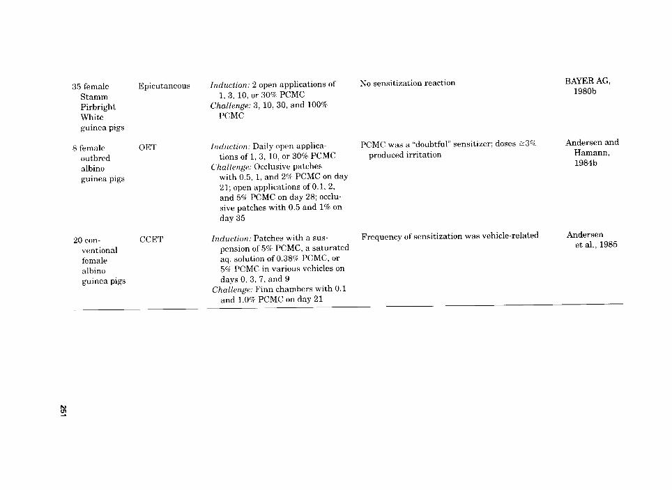
35 female Stamm Pirbright White guinea pigs
8 female outbred albino guinea pigs
20 con- ventional female albino guinea pigs
Epicutaneous
OET
CCET
Induction: 2 open applications of 1, 3, 10, or 30% PCMC
Challenge: 3, 10,30, and 100% PCMC
No sensitization reaction BAYER AG, 1980b
Induction: Daily open applica- PCMC was a “doubtful” sensitizer; doses ~3% Andersen and
tions of 1,3, 10, or 30% PCMC produced irritation Hamann,
Challenge: Occlusive patches 1984b
with 0.5, 1, and 2% PCMC on day 21; open applications of 0.1, 2, and 5% PCMC on day 28; occlu- sive patches with 0.5 and 1% on day 35
Induction: Patches with a sus- Frequency of sensitization was vehicle-related Andersen
pension of 5% PCMC, a saturated et al., 1985
aq. solution of 0.38% PCMC, or 5% PCMC in various vehicles on days 0,3,7, and 9
Challenge: Finn chambers with 0.1 and 1.0% PCMC on day 21
252 COSMETIC INGREDIENT REVIEW
For the animals injected with the pentachlorophenol/PCMC biocide, positive reactions were obtained upon challenge with the biocide and with PCMC but not with pentachlorophenol. The frequency of positive reactions upon challenge decreased between days 21 and 42; on day 21, 17 of 19 animals reacted to challenge with 5% of the biocide; on day 35, 12 of 19 animals reacted to 1% PCMC; on day 42, 6 of 19 animals reacted to challenge with 0.5% of the biocide.
Of the animals induced with PCMC, 16 of 19 had a positive reaction at day 21 of challenge, but only four of 19 animals reacted on day 35. Of the control animals used in determining the sensitizing potential of PCMC, three had slight erythema and one had moderate erythema upon chal- lenge; none of the positive results in controls were clinically explainable.
Six groups of 20 female outbred albino guinea pigs were used in a GPMT to determine the sensitization potential of PCMC (Andersen and Hamann, 198413). Induction consisted of intradermal administra- tion of 0.2%, l.O%, or 5.0% PCMC and topical administration of 10.0% PCMC, intradermal administration of 0.2% PCMC and topical admin- istration of 2.0%) PCMC, and intradermal injection of 0.2% PCMC and topical administration of 0.4% PCMC. The vehicles used for intrader- ma1 administration and topical administration were propylene glycol and petrolatum, respectively. A group of 40 negative control animals was dosed with vehicle only. The challenge performed on day 21 (and day 35 for some groups), used Finn chambers with O.l%, 0.5%, and 1.0% PCMC in yellow petrolatum. The challenge reactions were scored on a scale of 0 to 3 after 48 and 72 h. A grade 1 reaction was not regarded as evidence of sensitization.
The frequencies of positive reactions were 75%, 70%, 65%, and 84% for the animals induced with intradermal administration of 0.2%, l.O%, and 5.0% PCMC and topical administration of 10.0% PCMC, respec- tively, and 30%’ and 15% for animals induced with intradermal admin- istration of 0.2%~ PCMC and topical application of 0.4% or 2.0% PCMC, respectively. The 72-h, but not the 48-h, readings were significantly increased with increased challenge concentrations. Reactivity to PCMC at challenge decreased from day 21 to day 35.
As a follow-up to the study described previously (Andersen and Hamann, 1984133, another GPMT was performed using female outbred albino guinea pigs to determine whether the sensitization rate that was observed could be caused by minor changes in the type of Freund’s com- plete adjuvant (FCA) used or to the sites of the challenge patch appli- cations (Andersen, 1985a). Three groups of 20 animals were dosed intradermally with 1% PCMC in propylene glycol on day 0 and topically with 10% PCMC in yellow petrolatum on day 10. The animals were challenged on day 21 with 0.1% and 1.0% PCMC in petrolatum using Finn chambers that were placed horizontally on the flanks of the ani-
pCHLORO-m-CRESOL 253
mals; the 0.1% patch was placed in front on half the animals and in back on the other half. A parallel pair of patches were placed 2 cm lower on the flank to determine whether the responses near the abdomen were similar to the challenge responses nearer the back. The challenge sites were scored 48 h and 72 h after challenge, and the reactions were scored on a scale of 0 to 3. A grade 1 reaction was not regarded as evi- dence of sensitization.
Three types of FCA emulsions were used: (1) 50% FCA (v/v) and 50% propylene glycol, with (for the test groups) and without (for the control group) 2% PCMC (w/w); (2) 50% FCA(v/v) and 50% saline-ethanol (7+3) (v/v) with and without 2% PCMC (w/w); and (3) 50% saline and 50% FCA (v/v) that contained 2% PCMC (w/w) that was partly dissolved, partly suspended in the FCA prior to mixing with saline.
After 48 h, 19 animals of the FCA-propylene glycol group, 15 ani- mals of the FCA-saline-ethanol group, and 14 animals of the FCA-saline group reacted to challenge with 1% PCMC; after 72 h, the number of animals that reacted was 18, 18, and 17, respectively. Based on the frequency of reaction to PCMC upon challenge, no significant difference was shown between the three FCA emulsions; however, the reactions to challenge patches placed nearer to the abdomen were sig- nificantly weaker than those placed 2 cm higher on the flank.
Skin blood flow determined by laser Doppler flowmetry (LDF) and skin- fold thickness (SFT) were used to quantitate allergic contact dermatitis in a GPMT examining PCMC (Andersen and Staberg, 1985). Thirty-six guinea pigs were induced by intradermal administration of 1% PCMC in propylene glycol on day 0 and topical administration of 10% PCMC in petrolatum on day 7. The animals were challenged on day 21 using occlu- sive patches of 0.1% and 1.0% PCMC in petrolatum. The patches were placed anteroposterior on the mid-flank, with 10 mm of skin between the chambers. The challenge sites were examined visually and scored for reac- tions on a scale of 0 to 3. A grade 1 reaction was not regarded as evidence of sensitization. LDF was performed daily on the animals used to evaluate daily variation and was measured twice to determine the influence of the closed challenge. SFT, measured using calipers, was determined within seconds to avoid the effect of compression.
Upon visual examination 48 h after challenge, 28 of the 36 test ani- mals were sensitized to PCMC. Upon examination of the control ani- mals, it was determined that the occlusive patch test procedure influ- enced LDF and SFT measurements; LDF and SFT were maximal after 24 and 48 h, respectively.
Five groups of 20 female outbred albino guinea pigs were used in a GPMT to determine the sensitization potential of PCMC (Andersen, 1985b). Three groups were given induction doses consisting of 1% PCMC in propylene glycol administered intradermally and 10% PCMC
254 COSMETIC INGREDIENT REVIEW
in yellow petrolatum administered topically. Two of the test groups were given an intraperitoneal injection of 250 mg/kg cyclophosphamide (CY) on days 18 and 32, respectively. A negative control group was treated in a manner similar to the test group that was not given CY, with the exception that vehicle only was used during induction. A posi- tive control group was induced with 1% sodium lauryl sulfate (SLS) in saline given intradermally and 5% SLS in petrolatum administered topically The positive control animals also were dosed with CY by intraperitoneal injection on day 18. The animals of all groups were challenged using Finn chambers on days 21 and 35. The test animals were challenged with 0.2%, 0.5%, and 1.0% PCMC in yellow petrola- turn, the positive control animals were challenged with O.l%, 0.2%, and 0.5% SLS in yellow petrolatum, and the negative control animals were challenged with PCMC and SLS. Challenge reactions were scored on a scale of 0 to 3 after 48 h and 72 h. A grade 1 reaction was not regarded as evidence of sensitization.
Most animals of the test groups that were either not given CY or were dosed with CY on day 32 reacted upon challenge with PCMC on day 21; the number of these animals reacting at challenge on day 35 was reduced, especially in the group dosed with CY on day 32. Administration of CY on day 18 caused a decrease in the number of ani- mals reacting to challenge with PCMC on day 21; however, upon chal- lenge at day 35, almost all animals reacted. CY administration had no effect on reaction to SLS, indicating that the influence of CY was spe- cific. One of the control animals reacted strongly at the day 35 chal- lenge to PCMC; all of the others were negative. It was stated that the decrease in reactions to PCMC at the day-35 challenge “suggests that a chemical may induce effector and suppressor cells which have a differ- ent time course of development.” The investigators proposed that PCMC, “besides being a potential sensitizer[,] significantly induced suppressor cells and, with less probability, antibodies which are responsible for the ‘down regulation’ of sensitivity.”
Sixty Stamm Pirbright White guinea pigs, 30 per sex, were used in a sensitization study performed according to the method of Magnusson and Kligman (BAYERAG, no date). Induction consisted of intradermal injections, two with PCMC and one with FCA, followed 1 wk later with a topical application of 0.1 mL of 1% and 25% PCMC in Lutrol (site of application not stated). The challenge, performed after 2 wk, consisted of cutaneous application of 12.5%, 22.0%, and 50.0% PCMC in Lutrol and 100.0% PCMC to the flank of the animals. A 25% solution of PCMC was “strongly sensitizing,” whereas a 1% solution was “weakly sensitizing.”
An epicutaneous test using 35 female Stamm Pirbright White guinea pigs and performed according to the methods of Klecak et al. (1977) was
PCHLORO-m-CRESOL 255
used to determine the sensitization potential of PCMC (BAYER AG, 1980b). Induction consisted of two O.l-mL open applications of l%, 3%, lo%, and 30% PCMC in Lutrol applied to the left flank, with a negative control group being dosed with vehicle only. Challenge consisted of 3%, lo%, and 30% PCMC in Lutrol and 100% PCMC applied to the right flank (length of time in between induction and challenge was not speci- fied). No sensitization reaction was observed.
The sensitization potential of PCMC was examined in an open epicu- taneous test (OET) using five groups of eight female outbred albino guinea pigs (Andersen and Hamann, 1984b). The induction, performed according to the methods of Klecak et al. (1977), consisted of daily open applications of l%, 3%, lo%, or 30% PCMC in petrolatum; a negative control group was dosed with vehicle only. The animals were chal- lenged using occlusive patches (with a procedure modified from the original method) on the contralateral flank with vehicle, 0.5%, l.O%, and 2.0% PCMC on day 21 and with vehicle, 0.5%, and 1.0% PCMC on day 35. In addition, on day 28, the animals were challenged with open applications of O.l%, 2.0%, and 5.0% PCMC.
PCMC was a “doubtful” sensitizer in the OET (which is regarded as a less sensitive test because it does not use FCA); varying responses were recorded during the successive challenges of the test and control groups. Successive applications of 3% and 10% PCMC in petrolatum resulted in strong irritancy reactions. An irritant reaction was observed in five of eight animals dosed with 30% PCMC after one open applica- tion; induction using 30% PCMC was discontinued after 2 wk because of severe reaction to the test substance. The animals of this group were challenged with the other test groups.
A cumulative contact enhancement test (CCET) was performed according to the methods of Tsuchiya et al. (1982) using groups of 20 conventional female albino guinea pigs to examine the sensitization potential of PCMC (Andersen et al., 1985). Five test groups were induced on days 0, 3, 7, and 9 using patches containing a suspension of 5% PCMC partly dissolved, mainly suspended in water containing Carbomer 941 as a gelling agent, a saturated aqueous solution of 0.38% PCMC, 5.0% PCMC in olive oil/acetone (4/l), 5.0% PCMC in propylene glycol, or 5.0% PCMC in propylene glycol containing 0.15% Carbomer 941. Prior to the third induction patch application, 2 X 0.1 mL of an FCA water-in-oil emulsion (50/50) was injected intrader- mally at the lateral borders of the induction area in the postnuchal region. A negative control group also was used. The animals were challenged using horizontally placed Finn chambers on day 21 with 0.1% and 1.0% PCMC in petrolatum. Challenge reactions were scored 48 h ad 72 h after patch application on a scale of 0 to 3. Agrade 1 reac- tion was not regarded as evidence of sensitization.
256 COSMETIC INGREDIENT REVIEW
During induction, the animals treated with the saturated PCMC aqueous solution had marked erythematous and edematous reactions at the patch-test site. A significant difference was observed between the sensitizing capacity of the preparations. The 5% PCMC suspensions in water and olive oil/acetone solution were the most sensitizing, with 12 of 19 and 11 of 20 animals reacting to 1% PCMC after 48 h, respec- tively; the 5% PCMC in propylene glycol solution had the least sensitiz- ing potential, with only 4 of 20 animals reacting to challenge with 1% after 48 h. Carbomer 941 had no effect on the sensitization rate.
MUTAGENICITY
The mutagenic potential of 500 ug/plate or less PCMC was evaluated in an Ames test that was performed using Salmonella typhimurium strains TA1535, TAlOO, TA1537, and TA98 in the presence of metabolic activation (BAYER AG, 198Oc). Two positive controls, endoxan and try- paflavin, were used. PCMC was not mutagenic.
A single Ames test was performed using S. typhimurium strain TAlOO (Rapson et al., 1980). PCMC, tested at 10-l to 10:’ pg/plate, was not mutagenic.
The mutagenic potential of PCMC was evaluated in a Salmonella/ mammalian microsome test performed according to the methods of Ames et al. (19751, using S. typhimurium strains TAlOO, TA1535, TA1537, and TA98 (Madsen et al., 1986). Doses of 1.28,6.4, 32.0, 160.0, and 800.0 pg PCMC per plate were tested with and without metabolic activation; each dose was tested in five parallel plates and two runs were performed. The vehicle, dimethylsulfoxide (DMSO), served as the negative control. Without metabolic activation, the positive controls were sodium azide (TA1535 and TAlOOO) and 2-nitroflourene (TA1537 and TA98). With metabolic activation, 2-antramine served as the posi- tive control for all strains.
PCMC did not have mutagenic potential in S. typhimurium strains TA1535, TAlOO, TA1537, or TA98, with or without metabolic activation. The number of His+ revertants observed per plate using 1.28 to 32 ug/plate PCMC was similar to the value for the negative control; how- ever, concentrations of 160 and 800 pg/plate generally produced an increasing toxic effect on the tester strains; metabolic activation seemed to decrease this effect.
The mutagenic potential of PCMC in DMSO, 3.3 to 666 pg/plate, was examined using S. typhimurium strains TAlOO, TA1535, TA1537, TA97, and TA98, with and without metabolic activation (Zeiger et al., 1992). The vehicle was used as a negative control. Without metabolic activation, the positive controls were sodium azide (TAlOO and
gCHLORO-m-CRESOL 257
TA1535), 9-aminoacridine (TA1537 and TA97), and 4-nitro-o- phenylenediamine (TA98); with metabolic activation, 2-amino- anthracene served as the positive control for all strains. PCMC was not mutagenic.
A modified SOS chromotest was performed using Escherichia cob strain PQ37 to measure the genotoxic potential of PCMC (Malaveille et al., 1991). At concentrations of 0.1 to 4 mM, PCMC induced SOS-DNA repair synthesis without metabolic activation. The addition of a meta- bolic activation system decreased the genotoxicity of PCMC. Also, the addition of a radical scavenger, Trolox C (a hydrosoluble form of vita- min E), inhibited the genotoxicity of PCMC at 0.3 mM.
CLINICAL ASSESSMENT OF SAFETY
Irritation
Patch testing with 2% PCMC in petrolatum may produce irritant reac- tions, particularly in people with multiple patch-test reactions that are misinterpreted as allergic responses (Lewis and Emmett, 1987). Often retesting several months after the dermatitis had cleared did not result in positive results.
Sensitization
Predictive
In a modified human Draize study using 31 male subjects, 5% PCMC in petrolatum was used for both induction and challenge (Marzulli and Maibach, 1973). Induction consisted of 10 successive applications of 5% PCMC in petrolatum under occlusive patches being applied to the upper lateral portion of the arm; the patches were removed after 48 h or 72 h. After a 2-wk nontreatment period, a challenge patch of 5% PCMC in petrolatum was applied for 72 h. None of the 31 subjects was sensitized.
In another Draize test performed using male subjects, groups of 98, 88, and 66 subjects were induced with 5%, lo%, and 20% PCMC in petrolatum, respectively, for 3 to 5 wk (Marzulli and Maibach, 1974). Ten 48- to 72-h applications of 0.5 g of the test material were made under an occlusive patch to the upper lateral portion of each subject’s arm. Following an approximately 2-wk nontreatment period, subjects of all three groups were challenged with a 72-h patch containing 5% PCMC in petrolatum. None of the subjects in the three test groups responded to the challenge patch.
258 COSMETIC INGREDIENT REVIEW
Provocative
A single occlusive patch of a below-irritation dose of PCMC (concentra- tion not specified) was applied for 48 h to 363 patients with allergic con- tact dermatitis (Burry, 1969). Upon scoring after 96 h, three patients had positive reactions to PCMC.
Two percent PCMC in petrolatum was added to the standard patch series in 1972 (Cronin, 1980). During the time period of 1973 to 1976, the incidence of PCMC sensitivity varied between 0.2% to l.O%, 0.3% to 0.6%, and 0.3% to 0.6% for men, women, and overall, respectively. The total incidence for this time period was 0.5% (17/3189), 0.4% (13/3630), and 0.4% (30/6819) for men, women, and overall, respectively.
Patch tests were performed on patients in two regions of England using allergens, one of which was PCMC, included in a Standard Patch Test Battery because of concern regarding medicament sensitivity (Wilkinson et al., 1980). A total of 651 patients, 267 men and 384 women, were tested in one region and 1029 patients, 373 men and 656 women, were tested in the other region. The occlusive patches were removed after 2 days, and the test sites were scored again after 4 days (the dose concentration was not given). Of the 651 patients in the first region, a total of 0.8% had positive patch-test results to PCMC; 0.5% and 1.1% of the men and women, respectively, reacted to PCMC. In the second region, 0.7% of the patients had positive reactions; 1.3% and 0.3% of the men and women, respectively, reacted to patch testing with PCMC.
An additional 45 patients, 11 men and 34 women, and 128 patients, 36 men and 92 women, with leg ulcers and stasis eczema were patch tested in the two regions. Of the 45 patients tested in the first region, 4.4% had positive reactions to patch testing with PCMC; 9% and 2.9% of men and women, respectively, reacted positively to PCMC. In the second region, 0.8% of 128 patients reacted positively to PCMC; 0.0% and 1.1% of the men and women, respectively, reacted to patch testing with PCMC.
Consecutive eczema patients were tested with the International Contact Dermatitis Research Group (ICDRG) standard patch test series, which included PCMC-containing biocides (Andersen and Hamann, 1984a). Reactions were scored according to the recommenda- tions of the ICDRG (Wilkinson et al., 1970). Of 1462 patients tested with 2% PCMC in petrolatum, only five had positive patch-test results and six had irritant reactions; none of the positive results were clini- cally explainable.
Another study was conducted in which consecutive eczema patients were tested with the ICDRG standard series (Andersen and Veien, 1985). PCMC, 1%> in petrolatum, was tested on 671 patients and no pos-
pCHLORO-m-CRESOL 259
itive reactions were observed. PCMC was withdrawn from the patch- test battery after 3 mo because of the lack of reactions observed.
Case Reports
PCMC is a strong sensitizer in guinea pig maximization tests, but human sensitization has been reported to be fairly rare (Lewis and Emmett, 1987); however, several case studies were reported in the pub- lished literature and are summarized in Table 5.
Cross-Reactivity
Cross-reactivity has been observed between PCMC and chloroxylenol (Hjorth and Tr o e 11 -L assen, 1963); however, the cross-reactivity has only occurred where the initial sensitization was to chloroxylenol, not to PCMC (Lewis and Emmett, 1987).
SUMMARY
PCMC is a substituted phenol that functions as a cosmetic biocide and a preservative in skin-care and suntan preparations. It is soluble in alkalies, organic solvents, fats, oils, and somewhat in water.
In 1995, data submitted to the FDA reported that PCMC was used in 16 cosmetic product formulations. Internationally, PCMC is approved for use by members of the European Union as a preservative at a max- imum authorized concentration of 0.2%; however, it also may be added to cosmetic products at a concentration other than that specified for a preservative for other specific purposes apparent from the presentation of the product. PCMC is prohibited for use in products intended to come in contact with mucous membranes. In Japan, PCMC is listed in the Japanese Standard of Cosmetic Ingredients; products containing this ingredient must declare it on the label. PCMC is approved for use as an indirect food additive. Many of the noncosmetic uses of PCMC use its antiseptic properties. The use concentrations for most noncosmetic applications of PCMC range from 0.05% to 0.5%.
In a pharmacokinetic study in which rats were dosed orally with PCMC, rapid elimination through the kidneys occurred, and no indica- tion of accumulation of PCMC in fatty or hepatic tissues was present, In a study assessing the permeability of PCMC using whole and stripped abdominal skin from mice, the estimated permeability coeffi- cients of PCMC for viable tissue and stratum corneum were 302 X lo-” and 235 X 10 “, respectively.
The antimicrobial spectrum of 0.02% PCMC included gram-positive and gram-negative bacteria, yeast, and fungi. In aqueous cream,
g Table 5. Case reports concerning PCMC-containing substances
Test material Patient Case Testing/results Reference
Steroid cream containing PCMC
Steroid cream containing PCMC
Steroid cream containing PCMC
Glue and steroid cream containing PCMC
Steroid cream containing PCMC
Steroid cream containing PCMC
PCMC as a pre- servative in betamethasone cream
39-yr-old man
Dermatitis recurred on previously affected sites on the ankle and feet due to treatment with the steroid cream; an erythematous reaction with fine vesiculation was observed on the legs
24-yr-old Application of the steroid cream exacerbated existing man dermatitis
40-yr-old woman
Application of the steroid cream caused existing eczema to deteriorate
27.yr-old woman
Handling of the glue caused severe contact eczema; treatment with the steroid cream did not relieve the condition
1%yr-old woman
An eczematous eruption was treated with the cream for 6 mo; the condition worsened and spread
35yr-old woman
Treatment with the steroid cream for acute dermatitis initially resulted in transient improvement but then caused it to spread
56-yr-old woman
Daily applications of dilute cream were being used to treat widespread dermatitis; application was stopped and the dermatitis cleared
Patch testing with 1% PCMC resulted in ++ reactions after 48,72, and 96 h; subsequent testing several months later resulted in +++ reactions with 1 and 5%. tt reaction to 0.1%. and + reaction to O.OlQ PCMC after 96 h
Patch testing with 2% PCMC resulted in ++ and +++ reactions after 48 and 96 h, respectively
Patch testing with 2’Z PCMC (from two sources) resulted in + and ++ reactions and in - and ++ reactions after 48 and 96 h, respectively
Patch testing with 2% PCMC was positive
Initial patch testing with 2% PCMC was negative; patch testing after the condition worsened resulted in + reactions to 2% PCMC after 48 and 96 h
Patch testing with 2% PCMC resulted in ++ reactions after 48 and 96 h
A patch test with PCMC was positive; (previous testing gave positive reactions to chloroxylenol)
Lewis and Emmett, 1987
Oleffe et al., 1979
Oleffe et al., 1979
Dooms- Goossens et al., 1981
Archer and MacDonald, 1984
Goncalo et al., 1987
Burry et al., 1975
PCMC as a pre- servative in betamethasone cream
35yr-old woman
An acute contact dermatitis was treated with the cream; itching and mild dermatitis continued and mild contact dermatitis appeared elsewhere; this cleared with cessation of cream usage
Patch tests with PCMC was positive (previous testing gave positive reactions to chloroxylenol)
Burry et al., 1975
PCMC as a pre- servative in betamethasone cream
32-yr-old man
The cream was used to treat dermatitis: the dermatitis cleared when treatment was stopped
A patch test reaction to 5% PCMC in soft paraffin was greater than the reaction to 1% PCMC in soft pariffin (positive reaction to chloroxylenol was slight)
Burry et al., 1975
PCMC as a pre- servative in betamethasone cream
43-yr-old woman
The cream was used to treat dermatitis; mild contact dermatitis appeared elsewhere; the dermatitis cleared once treatment was stopped
Upon patch testing, there was no reaction to 1% or 5% PCMC in soft pariffin (a strong positive reaction to chloroxylenol was observed )
Burry et al., 1975
PCMC, 0.15%, as a preservative in mucous heparin
35yr-old woman
The patient was given 10,000 units of the heparin i.v. and immediately collapsed after the second dose with pallor. sweating, hypotension, and tachycardia: she recovered spontaneously after 30 min.
Intradermal skin tests gave positive reactions to both PCMC-preserved and preservative-free heparin
Hancock and Naysmith, 1975
PCMC, 0.15%. as a preservative in mucous heparin
55-yr-old man
The patient was given 10.000 U of the heparin iv. and over the next hr developed nasal congestion, profuse sweating, and a generalized urticarial rash
lntradermal skin testing with preservative- free heparin did not produce a reaction
Hancock and Naysmith, 1975
PCMC, 0.15%, as a preservative in mucous
Seven The patients were given 10,000 U prophylactic patients subcutaneous heparin twice daily; within a few hr i sex after the first and subsequent injections, an unspec- indurated erythematous reaction developed at the ified) injection site
Intradermal skin testing with preservative- free heparin in four of these patients produced no response
Hancock and Naysmith, 1975
(Table continued on next page.)
Table 5. Case reports concerning PCMC-containing substances (continued)
Test material Patient Case Testing/results Reference
PCMC, 0.15%, 21-yr-old as a preserva- woman tive in mucous heparin
Cleaning 2%yr-old detergent and woman disinfectant containing PCMC
The patient was given 5000 U heparin 6-hourly: administration of the first two doses resulted in severe burning pain at the injection site, radiating along the veins and up the forearm into the arm, nausea, light-headedness, and drowsiness with pallor and sweating
The patient complained of red swollen eyelids each time she used the detergent disinfectant [of which the two active principles are PCMC and o-benzyl-p- chlorophenol (OBPCP)]
.4 dose of PCMC-free heparin did not result in pain of a systemic reaction; formal intradermal skin testing produced a reaction to the mucous heparin but none to preservative-free heparin
Ainley et al., 1977
PCMC was diluted to 1 and 10% in water: Freitas and ethanol (1: 1) and tested with open and Brandao, prick tests: the results of the open test 1986 were negative and +++ for 1 and lo%, respectively, and of the prick test were +? and +++ for 1 and 109, respectively; open tests for 30 min with the detergent at 20% in water elicited a urticarial wheal; after the wheal, tests with PCMC produced superficial necrosis, which could be observed on the eyelids at the same time; testing with the detergent at 10% in water and of OBPCP were negative
pCHLORO-m-CRESOL 263
PCMC met the British Pharmacopoeia requirements for the efficacy of antimicrobial preservatives when tested against A. niger but not when tested against C. albicans.
The oral LD,,, of PCMC was 1830 mg/kg for Wistar rats and 5129 and 3636 mg/kg for male and female Sprague-Dawley rats, respectively. The percutaneous LD,, of PCMC was more than 500 mg/kg for rats, and the subcutaneous LD,, was 400 mg/kg for rats and 360 mg/kg for mice. The inhalation LC,, for rats, following a 4-h exposure, was more than 583 mg PCMC-NA/m” air.
In a short-term dermal toxicity study, 160 mg/kg or less PCMC pro- duced irritation and erythema but had no systemic effects on rabbits. In two short-term oral toxicity studies, no significant dose-related toxi- city (excluding body weight parameters) was observed when rats were given 400 mg/kg/day or less PCMC by gavage or 10,000 ppm or less PCMC in feed. In short-term subcutaneous toxicity studies, 72 mg/kg and 0.25% PCMC did not have an effect on rats and rabbits, respec- tively. In a subchronic study in which rats were fed 1500 ppm or less PCMC, the only observations made were retardation of body weight.
In a chronic study in which rats were fed 10,000 ppm or less PCMC for 2 yr; the NOEL was 2000 ppm PCMC. No PCMC-related changes in clinical chemistry and hematologic parameters were observed, and no significant difference in mortality was observed between test and con- trol animals. At study termination, necropsy findings included kidney deformation and, microscopically, an increase in papillary necroses and cortical tubular dilations and fibrosis of the kidneys for some of the high-dose male rats. Female rats of the mid-dose and high-dose groups that died on study had a greater incidence of pituitary adenoma.
PCMC, as low as 0.05%, produced ocular irritation in the eyes of rab- bits, and soft lenses that were stored in solution containing 0.1% PCMC produced severe irritation after a few days.
In a dermal irritation study using the trypan blue method, 0.2%, 0.4%, and 0.8%’ PCMC resulted in maximum irritation scores of 4, 4, and 8, respectively.
Numerous sensitization studies were performed with PCMC. Gen- erally, some type of reaction was seen upon challenge; however, the reactivity to PCMC decreased with time. In an epicutaneous test, 30% or less PCMC did not produce a sensitization reaction, and in an OET, PCMC was a “doubtful” sensitizer. In a CCET, the frequency of sensiti- zation was vehicle-dependent.
PCMC produced no evidence of mutagenic potential in Ames tests. PCMC, 0.1 to 4.0 mM, induced SOS-DNA repair synthesis without metabolic activation; the addition of metabolic activation or a radi- cal scavenger decreased or inhibited the genotoxicity of PCMC, respectively.
264 COSMETIC INGREDIENT REVIEW
In clinical studies, patch testing with 2% PCMC in petrolatum may produce irritant reactions, particularly in people with multiple patch- test reactions, that are misinterpreted as allergic responses. In two pre- dictive patch tests, initiation with 20% or less PCMC followed by chal- lenge with 5% PCMC did not produce a sensitization reaction in 31 of 252 subjects. In several provocative patch tests, some reactions to PCMC usually were observed. Cross-reactivity between PCMC and chloroxylenol has been observed, but only when the initial sensitization was to chloroxylenol.
DISCUSSION
Section 1, paragraph (p), of the Cosmetic Ingredient Review (CIR) Procedures states that “a lack of information about an ingredient shall not be enough to justify a determination of safety.” In accor- dance with Section 30(j)(Z)(A) of the Procedures, the Expert Panel informed the public of its decision that the data on PCMC were insuf- ficient to determine whether PCMC, for possible purposes of cosmetic use, is either safe or unsafe. The Expert Panel released a “Notice of Insufficient Data” announcement on September 13, 1994 outlining the data needed to assess the safety of PCMC. The types of data required included:
1. Concentration of use in cosmetics 2. UV absorption data; if absorbed in the WA or UVB range, photo-
sensitization data are needed 3. Dermal developmental toxicity data 4. At least one genotoxicity study using a mammalian system; if posi-
tive, dermal carcinogenicity data performed according to National Toxicology Program methods are required
No offer to supply the necessary data was received. In accordance with Section 45 of the CIR Procedures, the Expert Panel will issue a Final Report-Insufficient Data. When the requested data are available, the Expert Panel will reconsider the Final Report in accordance with Section 46 of the CIR Procedures, Amendment of a Final Report.
CONCLUSION
The Expert Panel concludes that the available data are insufficient to support the safety of p-Chloro-m-Cresol for use in cosmetic products.
p-CHLORO-m-CRESOL 265
REFERENCES
Ainley, E. J., Mackie, I. G., and MacArthur, D. 1977. Adverse reaction to chlorocresol-preserved heparin. Lancet 8013:705.
Ames, B. N., McCann, J., and Yamasaki, E. 1975. Methods for detecting car- cinogens and mutagens with the SaEmonellalmammalian microsome muta- genicity test. Mutat. Res. 31:347-364.
Andersen, K. E. 1985a. Guinea pig maximization test: Effect of type of Freund’s complete adjuvant emulsion and of challenge site location. Dermatosen 33:132-136.
Andersen, K. E. 1985b. Sensitivity and subsequent “down regulation” of sensi- tivity induced by chlorocresol in guinea pigs. Arch. Dermatol. Res. 277184-87.
Andersen, K. E., and Hamann, K. 1984a. The sensitizing potential of metal- working fluid biocides (phenolic and thiazole compounds) in the guinea-pig maximization test in relation to patch-test reactivity in eczema patients. Food Chem. Toxicol. 22:655-660.
Andersen, K. E., and Hamann, K. 1984b. How sensitizing is chlorocresol? Allergy tests in guinea pigs versus the clinical exposure. Contact Derm. ll:ll-20.
Andersen, K. E., and Staberg, B. 1985. Quantification of contact allergy in guinea pigs by measuring changes in skin blood flow and skin fold thick- ness. Acta. Derma. Venereal. (Stockh.) 65:37-42.
Andersen, K. E., and Veien, N. K. 1985. Biocide patch tests. Contact Derm. 12:99-103.
Andersen, K. E., Carlsen, L., Egsgaard, H., et al. 1985. Contact sensitivity and bioavailability of chlorocresol. Contact Derm. 13:246-251.
Archer, C. B., and MacDonald, D. M. 1984. Chlorocresol sensitivity induced by treatment of allergic contact dermatitis with steroid creams. Contact Derm. 11:144-145.
Baichwal, M. R., and Phadnis, A. S. 1968. Preservatives for parenteral prepa- rations II. Indian J. Pharmacy 4:89-91.
BAYER AG. 1978. Acute oral toxicity study using rats. Unpublished data sub- mitted by COLIPA (1 page).*
BAYERAG. 1980a. Thirteen wk study using rats. Unpublished data submitted by COLIPA (1 page).‘k
BAYER AG. 1980b. Epicutaneous sensitization study using guinea pigs. Unpublished data submitted by COLIPA (1 page).*
BAYER AG. 198Oc. SaZmoneZla/microsome test with PCMC. Unpublished data (Report No. 9122) submitted by COLIPA (1 page).*
BAYER AG. 1992. Chronic toxicity and carcinogenicity study in Wistar rats (administration in feed for 104 weeks-interim report) with cover letter dated 03/27/92. NTIS Report No. 01’80535962.
BAYER AG. No date. Sensitization study performed according to Magnusson- Kligman using guinea pigs. Unpublished data submitted by COLIPA (1 page).‘”
266 COSMETIC INGREDIENT REVIEW
British Pharmacopoeia. 1982. Efficacy of antimicrobial preservatives in phar- maceutical products. 1980 Addendum. London: British Pharmacopoeia.
Brown, M. A., Evans, C., Ford, J. L., et al. 1986. A note on the recovery of micro-organisms from an oil-in-water cream. J. Clin. Hosp. Pharm. 11:117-123.
Budavari, S. 1989. The Merck Index: An Encyclopedia of Chemicals, Drugs, and Biologicals, 11th ed. 329. Rahway, NJ: Merck and Co., Inc.
Burry, J. B. 1969. The value of patch testing: A review of 363 cases of allergic contact dermatitis. Med. J. Austral. 1:1226.
Burry, J. N., Kirk, J.. Reid, J. G., et al. 1975. Chlorocresol sensitivity. Contact Derm. 1141-42.
Cronin, E. 1980. Phenolic compounds. In Contact Dermatitis. 673-674. Edinburgh: Churchill.
Davies, M. 1973. Rabbit eye irritation from bactericides in soft lens soaking solutions. J. Pharm. Pharmac. 25:134.
Dooms-Goossens, A., Degreef, H., Vanhee, J., et al. 1981. Chlorocresol and chloracetamide: Allergens in medications, glues, and cosmetics. Contact Derm. 7:51-52.
Dossing, M., Wulff, C. H., and Olsen, P. Z. 1986. Repeated facial palsies after chlorocresol inhalation. J. Neural. Neurosurg. Psychiatry 49:1452-1454.
European Economic Community (EEC). 1993. EEC Cosmetics Directive 76/768/EEC, as amended, Annex IV. 4. Brussels: EEC.
Federal Register. January 28, 1992. Modification in voluntary filing of cos- metic product ingredient and cosmetic raw composition statements. Final rule. 57:3128-3130.
Fiebig, A., Felczak, J., Pankowska, B., et al. 1972. Research into the irritant action of some preservatives used in eye drops. Farmacja. Pot. 28:1145.
Food and Drug Administration (FDA). 1984. Cosmetic product formulation and frequency of use data. FDA Database. Washington, D.C., FDA.
FDA. 1995. Frequency of use of cosmetic ingredients. FDA Database. Washington: FDA.
Freitas, J. P., and Brandao, F. M. 1986. Contact urticaria to chlorocresol. Contact Derm. 15:252.
Goncalo, M., Goncalo, S., and Moreno, A. 1987. Immediate and delayed sensi- tivity to chlorocresol. Contact Derm. 17146-47.
Gray, T. C., and Naim, R. K. 1972. Intrathecal chlorocresol. Anaestesia 7:86. Greenberg, L. A., and Lester, D. 1954, Handbook of Cosmetic Materials. 91.
New York: Interscience Publishers, Inc. Hancock, B. W., and Naysmith, A. 1975. Hypersensitivity to chlorocresol-
preserved heparin. Br Med. J. 3:746-747. Hjorth, N., and Trolle-Lassen, C. 1963. Skin reaction to ointment bases. Trans.
St. Johns Hosp. Sot. 491127-140. Huq, A. S., Ho, N. F. H., Husari, N., et al. 1986. Permeation of water contami-
native phenols through hairless mouse skin. Arch. Environ. Toxicol. 15:557-566.
Kabara, J. J., 1984. Cosmetic and Drug Preservation. Principles and Practice. 683-684. New York: Marcel Dekker, Inc.
pCHLORO-m-CRESOL 267
Klecak, G., Geleick, H., and Frey, J. R. 1977. Screening of fragrance materials for allergenicity in the guinea pig. (I). Comparison of four testing methods. J. Sot. Cosmet. Chem. 2853-64.
Lewis, P. G., and Emmett, E. A. 1987. Irritant dermatitis from tri-butyl tin oxide and contact allergy from chlorocresol. Contact Derm. 17:129-132.
Lewis, R. J., Sr. 1993. Hawley’s Condensed Chemical Dictionary, 11th ed. 268. New York: Van Nostrand Reinhold Co.
Madsen, C., Andersen, P. H., Meyer, O., et al. 1986. 4-Chloro-3-Methylphenol: SaZmonella/mammalian microsome mutagenicity test and subacute toxicity test in rats. Bull. Environ. Contam. Toxicol. 37:651-654.
Magnusson, B., and Kligman, A. G. 1970. Allergic Contact Dermatitis in the Guinea Pig. Springfield, IL: Charles C. Thomas.
Malaveille, C., Brun, G., and Bartsch, H. 1991. Genotoxicity of ochratoxin A and structurally related compounds in Escherichia coli strains: Studies on their mode of action. IARC Monographs 115:261-266.
Marzulli, F. N., and Maibach, H. I. 1973. Antimicrobials: Experimental contact sensitization in man. J, Sot. Cosmet. Chem. 24:399421.
Marzulli, F. N., and Maibach, H. I. 1974. The use of graded concentrations in studying skin sensitizers: Experimental contact sensitization in man. Food Cosmet. Toxicol. 121219-227.
Meiss, R., Robenek, H., Rassat, J., et al. 1981. New aspects of the origin of hepatocellular vacuoles. Exp. Path. 19:239-246.
Mobay Chemical Corporation. 1980. Subchronic dermal study in rabbits. Unpublished data submitted by COLIPA, (1 page).*
Mobay Chemical Corporation. 1981. Acute oral toxicity to rats. Study No. 80- 001-14. Unpublished data submitted by COLIPA, (1 page).“’
Oleffe, J. A., Blondeel, A., and De Coninck, A. 1979. Allergy to chlorocresol and propylene glycol in a steroid cream. Contact Derm. 5:53-54.
Paulus, W., and Genth, H. 1983. Microbiocidal phenolic compounds-A critical examination. In Biodeterioration, Vol 5. eds. T. A, Oxley and S. Barry, 701-702. New York: John Wiley & Sons.
Rapson, W. H., Nazar, M. A., and Butsky, V. V. 1980. Mutagenicity produced by aqueous chlorination of organic compounds. Bull. Environm. Contam. Toxicol. 24:590-596.
Registry Of Toxic Effects Of Chemical Substances (RTECS). 1993. P-Chloro-m- Cresol entry. RTECS Database. Bethesda, MD: National Library of Medicine.
Rempe, J. M., Santucci, L. G. 1992. CTFA List of Japanese Cosm.etic Ingredients, 2nd ed. 11, 141. Washington: Cosmetic, Toiletry, and Fragrance Association.
Robenek, H., Meiss, R., Gehling, J., et al. 1980. Alterations in the rat liver induced by p-Chlor-m-Cresol with emphasis on the intracellular junctions. A thin-section and freeze-fracture study. J. Submicrosc. Cytol. 12:635-646.
Rossof, I. S. 1974. Handbook of Veterinary Drugs: A Compendium for Research and Clinical Use. 104. New York: Springer Publishing Co.
Rothschild, D. L., Jr. 1990. The Food Chemical News Guide to the Current Status of Food Additives and Color Additives. Washington: Food Chemical News.
268 COSMETIC INGREDIENT REVIEW
Sax, N. I. 1979. Dangerous Properties of Industrial Materials, 5th ed. 490. New York: Van Nostrand Reinhold Co.
Sierra, G. 1970. Inhibition of the amino acid induced initiation of germination of bacterial spores by chlorocresol. Can. J. Microbial. 16:51-52.
Tsuchiya, S., Kondo, M., Okamono, K., et al. 1982. Studies on contact hyper- sensitivity in the guinea pig: The cumulative contact enhancement test. Contact Derm.. 8~246-255.
Wein, R. 1939. The toxicity of parachlorometacresol and of phenylmercuric nitrate. Quart. J. Pharm. Pharmac. 12:212-229.
Wenninger, J. A,, and McEwen, G. N., Jr. 1995a. Cosmetic Ingredient Handbook, 3rd ed. 125. Washington: The Cosmetic, Toiletry, and Fragrance Association, Inc.
Wenninger, J. A., and McEwen, G. N., Jr. 199513. International Cosmetic Zngredient Dictionary, 6th ed. Vol. 1. 174. Washington: The Cosmetic, Toiletry, and Fragrance Association, Inc.
Wilkinson, D. S., Fregert, S., Magnusson, B., et al. 1970. Terminology of con- tact dermatitis. Acta. Derm.-Vener. Stockh. 50:287.
Wilkinson, J. D., Hambly, E. M., and Wilkinson, D. S. 1980. Comparison of patch test results in two adjacent areas of England. II. Medicaments. Acta. Dermato. Venereal 60:245-250.
Yakuji Nippo Ltd. 1985. The Japanese Standard of Cosmetic Ingredients, 2nd ed. 82. Tokyo: Yakuji Nippo Ltd.
Zeiger, E., Anderson, B., Haworth, S., et al. 1992. Salmonella mutagenicity tests: V. Results from the testing of 311 chemicals. Environ. Mol. Mutagen. lS(Supp1. 21):2-141.
* Available for review: Director, Cosmetic Ingredient Review, 1101 17th Street, NW, Suite 310, Washington, DC 20036.