Embed Size (px)
Citation preview
Final Report of the Safety Assessmentof Kojic Acid as Used in Cosmetics
Christina L. Burnett, Wilma F. Bergfeld, MD, FACP,Donald V. Belsito, MD, Ronald A. Hill, PhD, Curtis D. Klaassen, PhD,Daniel C. Liebler, PhD, James G. Marks, Jr., MD, Ronald C. Shank, PhD,Thomas J. Slaga, PhD, Paul W. Snyder, DVM, PhD, and F. Alan Andersen, PhD
AbstractKojic acid functions as an antioxidant in cosmetic products. Kojic acid was not a toxicant in acute, chronic, reproductive, andgenotoxicity studies. While some animal data suggested tumor promotion and weak carcinogenicity, kojic acid is slowlyabsorbed into the circulation from human skin and likely would not reach the threshold at which these effects were seen.The available human sensitization data supported the safety of kojic acid at a use concentration of 2% in leave-on cosmetics.Kojic acid depigmented black guinea pig skin at a concentration of 4%, but this effect was not seen at 1%. The CosmeticIngredient Review (CIR) Expert Panel concluded that the 2 end points of concern, dermal sensitization and skin lightening,would not be seen at use concentrations below 1%; therefore, this ingredient is safe for use in cosmetic products up to that level.
Keywordscosmetics, kojic acid, safety
Introduction
Kojic acid is an antioxidant used by the cosmetics industry and
has been described as an alternative to hydroquinone in skin
lightening.1 Kojic acid was discovered in 1907 through isola-
tion from the mycelia of Aspergillus oryzae grown on steamed
rice (the term koji means steamed rice in Japanese).2
While kojic acid is purported to have skin-whitening prop-
erties, it is currently not approved by the US Food and Drug
Administration (FDA) for such use in over-the-counter phar-
maceutical products.
Chemistry
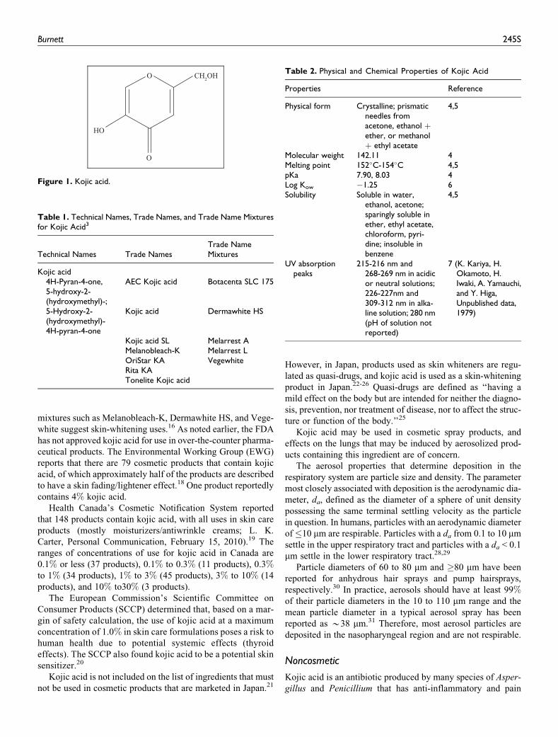
Kojic acid (CAS No 501-30-4) is the heterocyclic compound
that conforms to the structure depicted in Figure 1. Technical
names, traced names, and trade mixture names for this ingredi-
ent are listed in Table 1.3
Physical and chemical properties of kojic acid are described in
Table 2. UV absorption appears to vary as a function of the pH.
According to a review article by Beelik, the enolic hydroxyl
group at C5 gives kojic acid its weakly acidic property and
allows it to form salts with a number of metals.2
Kojic acid is naturally produced as a secondary metabolite
in the following Aspergillus strains: Aalbus, A alliaceus, A
awamori, A arachidicola, A bombycis, A caelatus, A candidus,
A clavatus, A effusus, A flavus, A fumigatus, A giganteus, A
glaucus, A gymnosardae, A leporis, A luteovirescens, A
lutescens, A minisclerotigenes, A nidulans, A nomius, A para-
siticus, A parvisclerotigenus, A pseudotamarii, A tamarii, and
A wentii.2,9 It is also the secondary metabolite of several strains
of Penicillium and Acetobacter fungi and several species of
acetic acid bacilli.2,10,11
Kojic acid can be detected with chromatographic or electro-
phoretic techniques.9,10,12-14
Use
Cosmetic
According to information supplied to the FDA by industry as
part of the Voluntary Cosmetic Registration Program (VCRP),
kojic acid is used in a total of 16 products. In a survey of current
use concentrations conducted by the Personal Care Products
Council, kojic acid is used at concentrations ranging from
0.1% to 2%, with the maximum concentration used in face and
neck creams, lotions, and powders.15 The available data on uses
and use concentration as a function of product type are pre-
sented in Table 3.
Gottschalck and Bailey described the current use of kojic
acid as an antioxidant; however, trade names and trade name
Corresponding Author:
Christina L. Burnett, Cosmetic Ingredient Review, 1101 17th Street, NW, Suite
412, Washington, DC 20036, USA.
Email: [email protected]
International Journal of Toxicology29(Supplement 4) 244S-273Sª The Author(s) 2010Reprints and permission:sagepub.com/journalsPermissions.navDOI: 10.1177/1091581810385956http://ijt.sagepub.com
mixtures such as Melanobleach-K, Dermawhite HS, and Vege-
white suggest skin-whitening uses.16 As noted earlier, the FDA
has not approved kojic acid for use in over-the-counter pharma-
ceutical products. The Environmental Working Group (EWG)
reports that there are 79 cosmetic products that contain kojic
acid, of which approximately half of the products are described
to have a skin fading/lightener effect.18 One product reportedly
contains 4% kojic acid.
Health Canada’s Cosmetic Notification System reported
that 148 products contain kojic acid, with all uses in skin care
products (mostly moisturizers/antiwrinkle creams; L. K.
Carter, Personal Communication, February 15, 2010).19 The
ranges of concentrations of use for kojic acid in Canada are
0.1% or less (37 products), 0.1% to 0.3% (11 products), 0.3%to 1% (34 products), 1% to 3% (45 products), 3% to 10% (14
products), and 10% to30% (3 products).
The European Commission’s Scientific Committee on
Consumer Products (SCCP) determined that, based on a mar-
gin of safety calculation, the use of kojic acid at a maximum
concentration of 1.0% in skin care formulations poses a risk to
human health due to potential systemic effects (thyroid
effects). The SCCP also found kojic acid to be a potential skin
sensitizer.20
Kojic acid is not included on the list of ingredients that must
not be used in cosmetic products that are marketed in Japan.21
However, in Japan, products used as skin whiteners are regu-
lated as quasi-drugs, and kojic acid is used as a skin-whitening
product in Japan.22-26 Quasi-drugs are defined as ‘‘having a
mild effect on the body but are intended for neither the diagno-
sis, prevention, nor treatment of disease, nor to affect the struc-
ture or function of the body.’’25
Kojic acid may be used in cosmetic spray products, and
effects on the lungs that may be induced by aerosolized prod-
ucts containing this ingredient are of concern.
The aerosol properties that determine deposition in the
respiratory system are particle size and density. The parameter
most closely associated with deposition is the aerodynamic dia-
meter, da, defined as the diameter of a sphere of unit density
possessing the same terminal settling velocity as the particle
in question. In humans, particles with an aerodynamic diameter
of�10 mm are respirable. Particles with a da from 0.1 to 10 mm
settle in the upper respiratory tract and particles with a da < 0.1
mm settle in the lower respiratory tract.28,29
Particle diameters of 60 to 80 mm and �80 mm have been
reported for anhydrous hair sprays and pump hairsprays,
respectively.30 In practice, aerosols should have at least 99%of their particle diameters in the 10 to 110 mm range and the
mean particle diameter in a typical aerosol spray has been
reported as *38 mm.31 Therefore, most aerosol particles are
deposited in the nasopharyngeal region and are not respirable.
Noncosmetic
Kojic acid is an antibiotic produced by many species of Asper-
gillus and Penicillium that has anti-inflammatory and pain
Table 2. Physical and Chemical Properties of Kojic Acid
Properties Reference
Physical form Crystalline; prismaticneedles fromacetone, ethanol þether, or methanolþ ethyl acetate
4,5
Molecular weight 142.11 4Melting point 152�C-154�C 4,5pKa 7.90, 8.03 4Log Kow �1.25 6Solubility Soluble in water,
ethanol, acetone;sparingly soluble inether, ethyl acetate,chloroform, pyri-dine; insoluble inbenzene
4,5
UV absorptionpeaks
215-216 nm and268-269 nm in acidicor neutral solutions;226-227nm and309-312 nm in alka-line solution; 280 nm(pH of solution notreported)
7 (K. Kariya, H.Okamoto, H.Iwaki, A. Yamauchi,and Y. Higa,Unpublished data,1979)
Table 1. Technical Names, Trade Names, and Trade Name Mixturesfor Kojic Acid3
Technical Names Trade NamesTrade NameMixtures
Kojic acid4H-Pyran-4-one,5-hydroxy-2-(hydroxymethyl)-;
AEC Kojic acid Botacenta SLC 175
5-Hydroxy-2-(hydroxymethyl)-4H-pyran-4-one
Kojic acid Dermawhite HS
Kojic acid SL Melarrest AMelanobleach-K Melarrest LOriStar KA VegewhiteRita KATonelite Kojic acid
O
HO
O
CH2OH
Figure 1. Kojic acid.
Burnett 245S
relief properties, with skin whitening activity reportedly caused
by the inhibition of tyrosinase.32
According to The Merck Index,4 kojic acid is used in maltol
and ethyl maltol synthesis and in flavor-enhancing additives in
food.
Uses for kojic acid in Hawley’s Condensed Chemical Dic-
tionary include chemical intermediate, metal chelation, and
insecticidal, antifungal, and antimicrobial agents.5
Kojic acid was reported to be used in a number of Japanese
foods, including soybean paste, soy sauce, sake, and mirin.33
Additional uses in foods include use as an antioxidant, a preser-
vative, a food additive to inhibit tyrosinase, an inhibitor of
nitrosopyrrolidine formation in fried food, and a reddening
agent in unripe strawberries.
General Biology
Absorption, Distribution, Metabolism, Excretion
Sansho Seiyaku Co, Ltd described a 1978 rat absorption, distri-
bution, metabolism, and excretion study (oral, subcutaneous,
and dermal routes) that was revised in 2001.34,35 [14C] Kojic
acid was biosynthesized by adding [14C-U] glucose into a
cultured broth of Aspergillus candidus, extracting with ethyl
acetate, and purifying by recrystallization. The purity of the
radiolabeled kojic acid was 99.9%. [14C] Kojic acid was admi-
nistered to groups of 3 male JCL-Wistar rats at a dose of 10
mCi/100 g body weight via a single oral, subcutaneous, or der-
mal administration. An additional group of rats received the
same subcutaneous dose over a period of 7 days. Blood samples
were collected from the tail tip 0.5, 1, 3, 6, 24, and 48 hours
after administration for the rats that received a single dose.
Urine and feces were collected from metabolic cages, and bile
samples were collected from cannulation in the common bile
duct at 0 to 10 minutes, 30 minutes to 1 hour, 1 to 3 hour, 3
to 6 hour, and 6 to 24 hour. In rats that received repeated doses
of kojic acid, blood, urine, and feces samples were collected at
24 hours after each administration. Enterohepatic circulation
was studied by connecting a cannula from a bile duct of a
treated rat to an untreated rat’s duodenum, from which the bile
samples were collected. At the end of the experiment, the rats
were killed 30 minutes, 1, 3, 6, 24, 48, or 72 hours after treat-
ment and tissues were collected and cut into sections for auto-
radiograph examination.
Radiolabel from the single oral exposure was found in the
intestine within 3 hours and in the cecum within 6 hours after
administration. The radioactivity was distributed in tissues and
organs very rapidly and maximum values were reached within
30 minutes of administration. Very high levels of radiolabel
were measured in the liver, kidneys, and pancreas, and high
levels were measured in the lungs, heart, and spleen. In the
blood, radioactivity decreased to 20.63% and 25.05% of total
radioactivity at 30 minutes and 1 hour, respectively, and
decreased to background levels within 24 hours. The amount
of 14C in the bile within 24 hours was approximately 0.5
mCi/10 mCi administered dose. No radioactivity was detected
in the bile samples from the enterohepatic circulation study.
Approximately 70% of the administered radioactivity was
excreted in the urine within 48 hours, while excretion in the
feces over the same time period was only 0.82%.
Distribution of the radiolabel in the tissues and organs fol-
lowing a single subcutaneous exposure was slightly slower than
that following the oral exposure. Distribution of radiolabel
after a single dermal exposure was further slowed. High levels
of radiolabel were measured in the kidney and liver 30 minutes
and 1 hour after subcutaneous exposure, while no remarkable
radioactivity was detected in the liver following dermal expo-
sure. In the blood, radioactivity was 13.29% and 21.67% at 30
minutes and 1 hour, respectively, following subcutaneous
exposure and 5% at 30 minutes following dermal exposure.
The amount of 14C in the bile within 24 hours was approxi-
mately 0.76 mCi/10 mCi and 0.5 mCi/10 mCi for the subcuta-
neous exposure and dermal exposure, respectively. No
radioactivity was measured in the bile samples from the enter-
ohepatic circulation study after either exposure type. Approxi-
mately 50% and 56% of the subcutaneous and dermal
administered radioactivity, respectively, were excreted in the
urine within 48 hours. Excretion in the feces over the same time
period was 2.62% and 1.58% of the administered subcutaneous
Table 3. Cosmetic Product Uses and Concentrations for Kojic Acid
Product Category
2009 Uses(Total Numberof Products inCategory)14,15
2008 Concentrations ofUse (Personal CareProducts Council,Unpublished Data,2010), %
Kojic acidBath products
Soaps anddetergents
1 (1665) –
Other bathproducts
1 (234) –
Eye makeupEye lotions �(254) 0.1-1
Skin care productsSkin cleansing
creams, lotions,liquids, and pads
2 (1446) –
Face and neckcreams, lotions,powders, and
sprays
2 (1583) 2a
Body and handcreams, lotions,powders, andsprays
�(1744) 1a
Moisturizers 2 (2508) –Skin fresheners 2 (259) –Other skinproducts
6 (1308) –
Total uses/ranges forKojic acid
16 0.1-2
a Concentrations of use reported for kojic acid in this category were not inspray products.
246S International Journal of Toxicology 29(Supplement 4)
and dermal doses, respectively. Recovery of radiolabel in
expired air in the rats administered a single subcutaneous dose
within 5 hours was 1.4%.
In the repeated subcutaneous dosed rats, radiolabel in blood
and urine samples increased until the fourth dosing and reached
an equilibrium state thereafter. Distribution of the radiolabel
was measured 10 minutes, 1, 6, 24, and 48 hours after the last
treatment. When compared to the single dose rats, radioactivity
was several times higher in all organs and tissues in the
repeated dose rats, especially in the intestinal tract 1-hour mea-
surement and in the pancreas and adipose tissues.
For all portions of the study, the major metabolites in the
urine and bile were glucuronide (6.4%-39.6% of total radioac-
tivity) and sulfate conjugates of kojic acid (35.6%-93.7% of
total radioactivity). Unmetabolized kojic acid was also
detected in the urine.34,35
The transfer of [14C] kojic acid (subcutaneous injection) to
fetuses and milk in pregnant JCL-Wistar rats was also investi-
gated.34,35 Groups of 2 pregnant rats received 10 mCi/100 g
body weight [14C] kojic acid subcutaneously on day 11 or 20
of gestation. Ten minutes, 30 minutes, or 3 hours after treat-
ment, fetuses were surgically extracted and prepared for auto-
radiograms, and the fluids, excreta, and tissues from the dams
were evaluated for radioactivity content as described above for
the male rats. For the milk transfer study, groups of 3 nursing
dams received 10 mCi/100 g body weight [14C] kojic acid sub-
cutaneously on day 3 of lactation. The stomachs of nursing
pups were extracted at 30 minutes, 1 hour, or 3 hours after treat-
ment of the dams to determine the radiolabel concentration in
milk.
In pregnant rats, the radioactivity was distributed rapidly in
tissues and organs. Very high values were observed in the kid-
ney and high values were observed in the liver, pancreas,
spleen, salivary gland, lungs, and kidney immediately after
administration. Radioactivity was also detected in the uterus,
placenta, amniotic fluid, and the fetus 30 minutes after treat-
ment. Fetal distribution of radiolabel was similar to that in the
adults, with high amounts detected in the liver and gastrointest-
inal tract. In nursing pups, radioactivity was detected in the sto-
mach wall and stomach content, with about 0.02% detected
3 hours after treatment. It was concluded that the radiolabel from
kojic acid was transported freely to the fetus, uterus and other
reproductive organs, and secreted into milk in this rat study.34,35
Dermal Penetration
In an in vitro percutaneous absorption and distribution study,36
[14C] kojic acid at 1.045% (w/w) in a formulation was applied
to human dermatomed skin. The integrity of the skin was tested
by measuring transepidermal water loss (TEWL) prior to test
material application. The formulation was applied at 2 mg/
cm2 (20.61 + 1.68 mgeq/cm2 of [14C] kojic acid) on the skin
surface. After 16 hours, the formulation was washed from the
skin surface with sodium lauryl ether sulfate and distilled
water. Liquid scintillation was employed to determine percuta-
neous absorption. Total recovery of the radiolabeled kojic acid
was 96.41% + 4.82% of the applied dose, with 75.55% +9.30% of the applied dose (15.52 + 1.43 mgeq/cm2) in skin
excess, 3.65% + 2.22% of the applied dose (0.76 + 0.48
mgeq/cm2) in the stratum corneum, 9.17% + 4.31% of the
applied dose (1.93 + 1.07 mgeq/cm2) in the epidermis and der-
mis, and 7.81% + 6.79% of the applied dose (1.65 + 1.49
mgeq/cm2) in the receptor fluid. The total absorbed amount of
[14C]-kojic acid was 16.98% + 10.28% of the applied dose
(3.58 + 2.38 mgeq/cm2).
In another study by Sansho Seiyaku Co, Ltd,37 the in vivo
percutaneous absorption of kojic acid was evaluated in human
volunteers. The study was open and uncontrolled. Six healthy
postmenopausal Japanese women received a single 500 mg
application of a cream formulation containing 1% kojic acid.
The test material was applied to the entire surface of the facial
skin (left and right cheeks). The participants were examined the
day before, immediately before, and 24 hours after application
and samples were collected for hematology, blood chemistry,
urinalysis, and immune serological tests. The amount of test
material in plasma was measured before application and at
0.5, 1, 1.5, 3, 6, 12, and 24 hours after application.
Kojic acid was detected in the plasma of all the participants
at one or more blood collection times. All the concentrations in
plasma were only slightly above the quantitation limit of 1 ng/
mL. The mean Cmax was 1.54 ng/mL and the mean AUC0-24 h
was 19.4 h�ng/mL. There were no adverse effects observed in
the participants. It was concluded that the potential dermal
transfer of kojic acid into the blood was very low.37
Based on the pharmacokinetic studies in rats and in vitro
percutaneous absorption values in human skin, a review by
Nohynek et al calculated a systemic exposure dose (SED) range
of 0.03 to 0.06 mg/kg per d in humans following a topical appli-
cation.38 This SED range was based on an application area of
the hands and face (400 and 590 cm2, respectively), a maxi-
mum application rate of 1.0 g of 1.0% kojic acid cream at 1
mg/cm2 (total application of 10 mg kojic acid/d), and percuta-
neous absorption of 17% of the applied dose (3.6 mg/cm2) in
humans.
Tyrosinase Inhibition
Cabanes et al stated that kojic acid is a slow-binding inhibitor
of catecholase activity of frog tyrosinase in a nonclassical man-
ner.39 In a study of several mammalian melanocyte tyrosinase
inhibitors, kojic acid was considered a potent free enzyme inhi-
bitor with an IC50 (50% inhibition concentration of tyrosinase
activity) value of 6.2 + 2 mg/mL.40 In this study, however,
Kojic acid did not reduce pigmentation in mammalian cells.
Melanocyte toxicity IC50 was >200 mg/mL, which indicated
that kojic acid was not considered cytotoxic.
Kojic acid was a reference sample in a study of the tyrosi-
nase activity of a nitrogen analog of stilbene.7 The IC50 value
of kojic acid was 275.6 mmol/L (39.17 mg/mL). In the same
study, kojic acid was a positive control for the evaluation of
superoxide dismutase-like (SOD-like) activity and melanin
production in the stilbene analog. Kojic acid inhibited 18.8%
Burnett 247S
and 21.9% SOD-like activity at concentrations of 10 (1.42 mg/
mL) and 50 mmol/L (7.11 mg/mL), respectively. Kojic acid did
not show inhibitory effects on melanin production at 10 (1.42
mg/mL) and 100 mmol/L (14.2 mg/mL) in cultured ‘‘melan-a’’
cells.
Kojic acid was a positive control in a study of the inhibitory
effects of oxyresveratrol and hydroxystilbene compounds on
mushroom and murine melanoma B-16 tyrosinase.41 At 100
mmol/L (14.2 mg/mL), kojic acid had a 76.7% + 1.1% inhibi-
tory effect on mushroom tyrosinase and a 43.0% + 2.5% inhi-
bitory effect on murine tyrosinase. The IC50 values of kojic
acid were 40.1 mmol/L (5.83 mg/mL) and >100 mmol/L (14.2
mg/mL) for mushroom and murine tyrosinases, respectively.
Mushroom tyrosinase inhibitory effects were dose-dependent.
Kojic acid was a competitive inhibitor of mushroom tyrosinase
in the kinetic portion of the study. In comparison, the IC50 val-
ues of oxyresveratrol were 1.2 mmol/L (0.29 mg/mL) in mush-
room tyrosinase and 52.7 mmol/L (12.9 mg/mL) in murine
tyrosinase. The percentage inhibition for 100 mmol/L (24.4
mg/mL) of this compound was 97.3% + 1.6% in mushroom
tyrosinase and 63.3% + 2.3% in murine tyrosinase.
Additional studies where kojic acid had been used as a pos-
itive control in mushroom tyrosinase inhibition studies have
been identified.42-45
Animal Toxicity
Acute Oral Toxicity
Kynoch and Lloyd46 reported the effects of acute doses of kojic
acid in fasted CFLP mice. The mice were divided into groups
of 2 males and 2 females and received 1, 4, or 16 g/kg kojic
acid in a 40% w/v suspension with 0.5% methylcellulose by
oral intubation. Dose volumes ranged from 10 to 40 mL/kg
body weight. The control group received 40 mL/kg of the vehi-
cle alone. Clinical signs of toxicity and mortalities were
recorded during the 14-day observation period. Mice that died
during the observation period and those that survived through
day 14 were necropsied. Preliminary findings indicated the
LD50 to be between 4 and 16 g/kg body weight. In order to pin-
point a more precise LD50, dosing was extended to groups of
5 male and 5 female mice. The groups received 4, 6.4, 10, or
16 g/kg kojic acid.
Clinical signs observed shortly after dosing included
lethargy, piloerection, hunched posture, ataxia, and depressed
respiratory rate. Mice treated with 6.4 g/kg body weight
also were observed gasping. One male and 2 females in the
4 g/kg, 4 males, and 3 females in the 6.4 g/kg, and all the males
and females in the 10 and 16 g/kg dose groups died within 1 to
3 hours of dosing. Necropsy of these animals revealed conges-
tion of the lungs and pallor of the liver, kidneys, and spleen.
Survivors completely recovered by day 4. Body weight gains
in females of the 4 g/kg dose group were slightly decreased
during the first week of observation but were comparable to
controls by the second week. No abnormalities were observed
in the surviving mice at necropsy. No clinical signs or deaths
were observed in the control group. The authors calculated the
LD50 of kojic acid in mice to be 5.1 g/kg body weight (95%confidence limits ¼ 3.9-6.7 g/kg body weight).46
A similar acute oral study of kojic acid was performed by
Kynoch and Lloyd47 using fasted CFY rats. The preliminary
LD50 was determined to be between 1 and 4 g/kg body weight.
To more precisely determine the LD50, the dose groups were
expanded to 5 males and 5 females and received 1, 1.6, 2.5,
or 4 g/kg body weight kojic acid in a 40% w/v suspension of
1.0% methylcellulose via oral intubation.
Lethargy, piloerection, ataxia, depressed respiratory rate,
and loss of righting reflex were observed shortly after treat-
ment. Rats treated with doses above 1 g/kg also had increased
salivation and body tremors. Increased lacrimation and diuresis
were observed in the 1.6 g/kg dose group and convulsions prior
to death were observed in the 2.5 and 4 g/kg dose groups. Two
males and 1 female in the 1.6 g/kg dose group and all of the
males and females in the 2.5 and 4 g/kg dose groups died within
3 to 67 hours after dosing. Necropsy of these rats revealed con-
gestion in the lungs with no specific cause of death evident. Opa-
city of the right eye was observed in 1 female in the 4 g/kg dose
group. Recovery of the survivors was complete within 7 days.
Body weight increases were slightly decreased in the 1.6 g/kg
dose group for the first week of observation but were comparable
to controls by the second week. No abnormalities were observed
in the surviving rats at necropsy. No clinical signs or deaths were
observed in the control group. The authors calculated the LD50
of kojic acid in rats to be 1.8 g/kg body weight (95% confidence
limits ¼ 1.5-2.0 g/kg body weight).
The acute oral toxicity of kojic acid was evaluated by
Manciaux48 in 6-week-old Wistar rats. The test material, pre-
pared in 0.5% methylcellulose, was administered at a dose of
2000 mg/kg (volume 10 mL/kg) by gavage to a group of 5 male
and 5 female fasted rats. Another group of 5 males and
5 females received the vehicle alone. Clinical signs, mortality,
and body weight gain were checked for 14 days following the
single administration. At the end of the observation period, the
animals were necropsied.
All animals in the treatment group were observed with seda-
tion or hypoactivity, dyspnea, and lateral recumbency on day 1.
One female rat was found dead 6 hours after treatment. The
remaining animals fully recovered on day 2. No clinical signs
or deaths were observed in the control group. Body weight gain
in the surviving rats was similar to the control group. No
abnormalities were observed at necropsy. It was concluded that
the oral LD50 of kojic acid was greater than 2 g/kg in rats.48
Acute Subcutaneous Toxicity
The effects of acute subcutaneous doses of kojic acid in CFLP
mice were studied.49 Preliminary findings indicated the LD50
to be between 4 and 16 g/kg body weight. In order to pinpoint
a more precise LD50, dosing was extended to groups of 5 male
and 5 female mice. The groups received 0, 1.6, 2.5, 4, 6.4, 10,
or 16 g/kg kojic acid as a 40% w/v suspension with 0.5%
248S International Journal of Toxicology 29(Supplement 4)
methylcellulose by injection. Clinical signs of toxicity and
mortalities were recorded during a 14-day observation period.
Hemorrhage at the injection site was observed immediately
after dosing in all mice receiving kojic acid. Clinical signs
observed shortly after dosing included lethargy, piloerection,
depressed respiratory rate, gasping, abnormal body carriage
(hunched posture), and ataxia. Mice treated with 2.5 g/kg body
weight also had coarse body tremors. In male mice, none from
the 1.6 g/kg dose group, 3 from the 4 g/kg dose groups, and all
in the remaining dose groups died. In female mice, 2 from the
1.6 g/kg dose group, 3 from the 2.5 dose groups, 4 in the 4, 6.4,
and 10 g/kg dose groups, and all in the 16 g/kg dose groups
died. Death occurred within 1 to 4 h after dosing. Necropsy
of these animals revealed the presence of dose material in sub-
cutaneous tissues near the injection site, pulmonary hemor-
rhage, and pallor of the liver. Opacities in the eyes were
observed in 1 mouse each of the 1.6 g/kg and 10 g/kg dose
groups. Survivors completely recovered by day 4. No abnorm-
alities were observed in the surviving mice at necropsy. No
clinical signs or deaths were observed in the control group. The
authors calculated the LD50 of kojic acid in mice to be 2.7 g/kg
body weight (95% confidence limits ¼ 1.9-3.9 g/kg body
weight.49
A similar acute subcutaneous study of kojic acid was done
using CFY rats.50 The preliminary LD50 was determined to
be between 4 and 16 g/kg body weight. To more precisely
determine the LD50, the dose groups were expanded to 5 males
and 5 females and received 1, 1.6, 2.5, 4, 6.4, or 10 g/kg body
weight kojic acid in a 40% w/v suspension of 1.0% methylcel-
lulose via injection.
Lethargy, piloerection, abnormal body carriage (hunched
posture), diuresis, and depressed respiratory rate were observed
shortly after treatment. Ataxia and convulsions accompanied
these signs in rats in the 2.5 g/kg dose groups and above. Rats
treated with 6.4 g/kg and above also had tremors. A total of 4
males and 3 females in the 2.5 g/kg dose group, 4 males and 4
females in the 4 g/kg dose group, all males and females in the
6.4 g/kg dose group, and all males and 3 females in the 10 g/kg
dose group died within 2 to 21 hours after dosing. Necropsy of
these rats revealed hemorrhage of the subcutaneous tissue at
the injection site, pulmonary hemorrhage, and pallor of the
liver. Opacity of one or both eyes was observed in about half
of the mortalities. Recovery of the survivors was complete
within 6 days. Body weight increases were slightly depressed
in surviving males in the 2.5 and 4.0 g/kg dose groups and in
the surviving female in the 4.0 g/kg dose group for the first
week of observation but were comparable to controls by the
second week. No abnormalities were observed in the surviving
rats at necropsy. No clinical signs or deaths were observed in
the control group.
An additional group of 5 male and 5 female rats were treated
subcutaneously with 4 g/kg kojic acid to further investigate the
opacities. Lenticular opacities were observed in both eyes of 2
male rats and drying and clouding of the cornea were observed
in 5 rats along with swelling of the cornea in 1 male and 1
female rat. This last effect obscured observation of the lens
in 2 rats. One male rat died before the reading 2.5 hours after
dosing. The authors determined that these opacities were not
inconsistent with those of acute reversible lens opacities that
have been ascribed to changes in the osmolarity of the aqueous
humor. The authors calculated the LD50 of kojic acid in rats to
be 2.6 g/kg body weight (95% confidence limits¼ 2.0-3.2 g/kg
body weight.50
Acute Intraperitoneal Toxicity
The effects of acute intraperitoneal injections of kojic acid in
CFLP mice were studied.7 Preliminary findings indicated the
LD50 to be between 1 and 4 g/kg body weight. To pinpoint a
more precise LD50, dosing was extended to groups of 5 male
and 5 female mice. The groups received 0, 1.6, 2.5, 4, 6.4, or
10 g/kg kojic acid in a 40% w/v suspension with 0.5% methyl-
cellulose by injection. Clinical signs of toxicity and mortalities
were recorded during a 14-day observation period.
Clinical signs observed shortly after dosing included
lethargy, piloerection, depressed respiratory rate, and ataxia.
Mice treated with 2.5 g/kg body weight were observed gasping.
Three male and 2 female mice from the 1.6 g/kg dose group and
all mice in the 4, 6.4, and 10 g/kg dose groups died. Death
occurred within 1 to 3 hours after dosing. Necropsy of these
animals revealed the pallor of the liver and kidneys, pulmonary
hemorrhage, and injection of the blood vessels of the abdom-
inal viscera. Survivors completely recovered within 2 days of
dosing. Body weight gains were comparable to controls. No
abnormalities were observed in the surviving mice at necropsy.
No clinical signs or deaths were observed in the control group.
The authors calculated the LD50 of kojic acid in mice to be 2.6
g/kg body weight (95% confidence limits ¼ 2.2-3.0 g/kg body
weight.51
A similar acute intraperitoneal study of kojic acid was done
using CFY rats.52 The preliminary LD50 was determined to be
between 1 and 4 g/kg body weight. To more precisely deter-
mine the LD50, the dose groups were expanded to 5 males and
5 females and received 1, 1.6, 2.5, or 4 g/kg body weight kojic
acid in a 40% w/v suspension of 1.0% methylcellulose via
intraperitoneal injection.
Lethargy, piloerection, abnormal body carriage (hunched
posture), ataxia, and depressed respiratory rate were observed
shortly after treatment. Coarse body tremors and convulsions
were observed in rats in the 1 g/kg dose group. Rats treated
with doses above 1 g/kg also had increased salivation, diuresis,
gasping, coarse body tremors, and convulsions prior to death.
One female rat in the 1 g/kg dose group had slight paralysis
of the hind limbs on day 3 that was still apparent at study ter-
mination. No deaths occurred in any of the males or females in
the 1 or 1.6 g/kg dose groups. All of the males and 3 females
each in the 2.5 and 4.0 g/kg dose group died between 1 and
19 hours post dosing. Necropsy of these rats revealed conges-
tion, pulmonary hemorrhage, pallor of the liver, and injection
of the blood vessels of the abnormal viscera. Opacities of one
or both eyes were observed in 7 of the 24 mortalities. Recovery
of the survivors was complete within 5 days. Body weight
Burnett 249S
increases were slightly decreased in the male 1.6 g/kg dose
group for the first week of observation but were comparable
to controls by the second week. No abnormalities were
observed in the surviving rats at necropsy. No clinical signs
or deaths were observed in the control group. The authors cal-
culated the LD50 of kojic acid in rats to be 2.4 g/kg body weight
(95% confidence limits ¼ 2.0-3.0 g/kg body weight.52
Acute Dermal Toxicity
The acute dermal toxicity of 100% kojic acid was evaluated in
8-week-old Wistar rats.53 The test material, in its original
powdered form, was applied to clipped skin on a gauze pad
(premoistened with 2 mL of purified water) at a dose of
2000 mg/kg to a group of 5 male and 5 female rats. Another
group of 5 males and 5 females were patched with just 2 mL
of purified water. The patches were applied for 24 hours and any
residual test material was removed with a moistened gauze pad.
Clinical signs and mortality were observed daily for 14 days, and
body weight gains were checked on days 1, 8, and 15. At the end
of the observation period, the animals were necropsied.
No deaths or clinical signs or cutaneous reactions were
observed during the study in either the test or control animals.
Body weight gains were slightly decreased between day 1 and
day 8 in 1/5 treated males and 3/5 treated females, when com-
pared to control animals. No abnormalities were observed at
necropsy. It was concluded that the dermal LD50 of kojic acid
is greater than 2000 mg/kg in rats.53
Short-Term Oral Toxicity
In a preliminary study for an in vivo genotoxicity study, male
mice received oral doses of kojic acid ranging from 0 to 2000
mg/kg for 5 days. The LD50 from the preliminary study was
calculated to be 1031.2 mg/kg per d kojic acid.54,55
Short-Term Dermal Toxicity
The dermal toxicity potential of kojic acid was evaluated in a 4-
week study in 104 Wistar Hannover rats.56 The rats were ran-
domly allocated to 3 treatment groups and 1 control group,
which received 100, 300, 1000 mg/kg per d kojic acid, or the
vehicle, 0.5% aqueous methyl cellulose solution (w/w), respec-
tively. The high-dose group and the control group consisted of
16 male and 16 female rats each, while the remaining groups
consisted of 10 male and 10 female rats each. The extra rats
in the high-dose and control groups were kept for a 2-week
treatment-free observation period. The rats received the treat-
ment or the control solutions daily to clipped dorsal skin. The
animals were checked daily for mortality and clinical signs of
toxicity. Body weights and food consumption were measured
once a week. Complete hematology and blood chemistry inves-
tigations and urinalysis were performed at the end of the treat-
ment period in the first 10 males and females of the high-dose
and control groups and in all of the remaining animals in the
other treatment groups. White blood cell and lymphocyte
counts were made in the reserved 6 males and 6 females of the
high-dose and control groups. All animals were killed at the
end of the treatment and treatment-free periods. Select organs
were weighed and a complete gross examination was per-
formed in all animals. Microscopic examinations were per-
formed on select tissues from the high-dose and control groups.
No deaths occurred and no relevant clinical signs were
observed during the treatment or treatment-free periods. Body
weight gains and food consumption were comparable to the
control group. Decreased lymphocyte counts were observed
at the end of the treatment period in both males and females
in the 300 and 1000 mg/kg per d dose groups. This effect had
partially reversed at the end of the treatment-free period in
males of the high-dose group. No treatment-related changes
were observed in blood chemistry parameters or urinalysis.
At necropsy, decreased absolute and relative spleen weights
were observed in the high-dose females, but there were no
treatment-related findings during the gross or microscopic
examinations in any dose group. The study concluded that the
no observable effect level (NOEL) was 100 mg/kg per d,
although the author noted that observed changes in lympho-
cytes and white blood cell counts in the higher dose groups
were minimal to mild in severity and the toxicological signifi-
cance of this finding was uncertain.56
Subchronic Oral Toxicity. In a subchronic study,8 male SD strain
rats received daily oral (by stomach tube) doses of 0, 0.25, 0.5,
1.0, 2.0, or 3.0 g/kg kojic acid suspended in 1.0% carboxy-
methylcellulose for 13 weeks. The dose groups included 20 rats
each. The administration period was followed by a 4-week
recovery period. During treatment, the rats were weighed and
observed for clinical signs of toxicity and mortality daily. Feed
and water intake were measured weekly. Rats from each group
were killed at 4, 13, and 17 weeks (5, 10, and 5 rats at each time
period, respectively) for necropsy, hematological and serobio-
chemical examinations, and urinalysis. Animals with lowest
weight gain in each treated group (except control) were
selected for removal at each time point. In dose groups where
the mortality exceeded the number of animals scheduled for
termination, no animals were removed.
Rats that received 0.5 g/kg or more of kojic acid had dysba-
sia 20 to 30 minutes after treatment and developed a strong
sedation followed by sleep. Animals in the 1.0, 2.0, and 3.0
g/kg dose groups during treatment bled from the eyes, and
exhibited ablepsy, exophthalmos, hematuria, epistaxis, and
vomiting. All animals in the 3.0 g/kg dose group died by week
3, while 11 animals in the 2.0 g/kg, 1 animal in the 1.0 g/kg, and
2 animals in the 0.5 g/kg died during the course of the study
period. No clinical signs of toxicity or mortalities were
observed in the 0.25 g/kg or control groups. Body weight gains
were significantly decreased in the 0.5, 1.0, and 2.0 g/kg dose
groups during treatment but became comparable to controls
during the recovery period. No significant changes in feed or
water intake were observed when compared to the control
group. No significant changes were observed with regard to
hematology or urinalysis in any treatment group when
250S International Journal of Toxicology 29(Supplement 4)
compared to controls. When compared to control serum chem-
istry values, serum glutamic-oxaloacetic transaminase (SGOT)
enzyme activity was increased in the 1.0 and 2.0 g/kg dose
groups and glutamate and calcium levels were decreased in the
2.0 g/kg dose group. Necropsies of animals that died during the
course of the study found pulmonary hemorrhage, congestion
of the stomach and intestine, adrenal gland hypertrophy, ocular
hemorrhage and opacity, and evidence of vomiting and clonic
or tonic spasm. Pyoid substance was noted in the lung with par-
tial sclerosis of pulmonary tissue in the rats from the 2.0 and 3.0
g/kg dose group. Necropsy at scheduled termination showed
similar findings in a dose-dependent manner. At 13-week
necropsy, weights of liver, kidneys, and testes increased in the
1.0 and 2.0 g/kg dose groups and the adrenal gland weights
were increased in the 2.0 g/kg dose group. At 17-week
necropsy, increases in testicular and thymic weights were noted
in the higher dose groups. The observations of normalization
during the recovery period suggested to the researchers that
kojic acid and its metabolites were rapidly excreted and that
toxicity occurred in a dose-dependent manner.8
In a 26-week toxicity study,57 male SD strain rats received
daily oral gavage doses of 0, 125, 250, 500, or 1000 mg/kg
kojic acid in 1% carboxymethylcellulose. The dose groups con-
sisted of 20 rats each except for the 125 mg/kg dose group. In
each group that contained 20 rats, 10 rats were used for a 5-
week recovery test following the treatment phase. Clinical
signs of toxicity and mortality were observed daily and body
weight, feed consumption, and water intake were measured
twice a week for the first 13 weeks and then once a week for
the remainder of the treatment phase. Urinalysis and hematol-
ogy and biochemistry tests were performed prior to necropsy at
study end. Tissues and organs were examined and weighed at
necropsy.
No deaths were observed in any group. Rats in the 250 mg/
kg dose group showed excitation followed by sedation, and
some rats in the 500 mg/kg and 1000 mg/kg had these clinical
signs accompanied by transient exophthalmos and salivation
that disappeared 2 to 3 hours after the dosing. Rats that received
250 mg/kg or more of kojic acid had significant suppression of
body weight gain when compared to the control group. Body
weight gains seemed to recover during the 5-week nontreat-
ment phase. Decreases in urine volume were observed in the
500 and 1000 mg/kg dose groups, with a decrease in the urinary
pH also occurring in the 1000 mg/kg dose group. The 1000 mg/
kg dose group also had a slight decrease in erythrocyte counts
and decrease of hematocrit value and hemoglobin concentra-
tion. Increases of SGOT and glutamic-pyruvic transaminase
(GPT) activities were observed in dose groups receiving 250,
500, and 1000 mg/kg. The 500 and 1000 mg/kg dose groups
had increased alkaline phosphatase (ALP) activity, and slight
increases in total cholesterol, bilirubin, and calcium were
observed in the 1000 mg/kg dose group. During the nontreat-
ment phase, the changes in urinalysis, hematology, and bio-
chemistry were not observed. At necropsy, the absolute and
relative weights of the adrenal glands were increased in the
dose groups receiving 500 and 1000 mg/kg kojic acid;
however, the absolute weights of the adrenal glands in the
recovery groups were almost the same as that for the control
group. In the 1000 mg/kg dose group, 2 rats had vacuolation
of anterior cells of the pituitary gland, but the researchers of
this study could not be certain this effect was treatment-related.
No other treatment-related effects in the tissues were observed.
It was concluded that the NOEL of kojic acid in this experiment
was 125 mg/kg per d.57
Chronic Toxicity
Studies of chronic exposures have been summarized in the Car-
cinogenicity section of this safety assessment.
Ocular and Dermal Irritation
A 3% aqueous solution of kojic acid was tested for ocular irri-
tation potential in rabbits (strain not reported).58 In a prelimi-
nary study, 0.05 mL of the kojic acid solution was instilled in
the right eye of 3 rabbits. The eyes were not rinsed. The rabbit
eyes were observed at 1, 3, 6, and 48 hours posttreatment. No
changes were observed. For the main study, the left eye of 5
rabbits was instilled with 0.05 mL of the kojic acid solution and
not rinsed. The eyes were examined at 0.5, 1, 6, 24, 48, and 72
hours and 1 week posttreatment. Slight redness was observed
only in 1 rabbit 0.5 hours after treatment. No other effects were
observed. To determine the accuracy of this study, another
laboratory performed a similar test in 4 Angola rabbits using
the same sample of kojic acid.59 Mild transient hyperemia was
observed in 2 of the rabbits. No other effects were observed. A
positive control, 3% Thesit Desitin in distilled water, yielded a
24-hour integrated edema value of 19, which was within the
normal response range (15-30). A supplemental study of the
3% kojic acid solution in 1 eye of 9 Angola rabbits found no
specific response and/or inflammatory response up to 72
hours.59
In a dermal irritation study,60 0.5 g of kojic acid was mixed
with 0.5 mL distilled water and applied to clipped, abraded, and
intact skin of 6 albino rabbits with gauze patches. The patches
were removed after 24 hours and the skin was evaluated for
reactions for a period of 72 hours. None of the animals had any
observable skin responses. The primary irritation index (PII)
was calculated to be 0 and kojic acid was not considered an irri-
tant to rabbit skin.
Kojic acid at 1% and 3% was evaluated for primary skin irri-
tation in a total of 12 male Japan white rabbits.61 A solution of
10% sodium lauryl sulfate (SLS) was used as a positive control.
The cream base at 0.25 g, the 1% or 3% kojic acid cream, or 0.1
mL of SLS were applied to clipped, abraded, and intact skin
(patch sites were 2 cm2 each). The patches were open. After
4 hours, the sites were wiped with warm water and assessed for
reactions after 4, 28, 48, and 72 hours. Erythema was observed
2 to 4 hours after application of both 1% and 3% kojic acid. A
score of 1 to 2 was apparent on almost all animals after 24
hours. Erythema gradually faded after 48 hours, with a few
sites exhibiting local and very slight erythema after 72 hours.
Burnett 251S
No significant difference was observed between abraded and
intact skin. No eschar formation or edema was observed in the
cream base or kojic acid patches. The PII were 0.78, 0.93, 0.85,
and 3.70 for the cream base, 1% kojic acid, 3% kojic acid, and
SLS, respectively. In this study, 1% and 3% kojic acid was a
mild skin irritant with a PII of no more than 1.
Dermal Sensitization
The potential of kojic acid to induce delayed contact hypersen-
sitivity was evaluated in albino Dunkin-Hartley guinea pigs.62
The control group and the treatment group consisted of 5 males
and 5 females and 10 males and 10 females, respectively. The
animals of the treatment group received 3 topical applications
(0.5 mL) of 30% kojic acid (w/w) in corn oil on the shaved
anterior flank on days 1, 8, and 15 of a 2-week induction phase.
The application sites were occluded for 6 hours after each treat-
ment. The animals in the control group received the 0.5 mL
corn oil vehicle alone on application sites, which were also
occluded. Following a 14-day rest period both groups of ani-
mals received a topical application of 30% kojic acid (w/w)
in corn oil to the posterior right flank. The left flank was treated
with only the corn oil and served as a negative control. Both
application sites were occluded for 6 hours. The skin was eval-
uated for reactions 24 and 48 hours after patch removal. The
animals were killed at the end of the study for skin sampling
of the challenge application sites in all control animals and in
animals that had cutaneous reactions in the treated group.
No clinical signs or deaths were observed during the study.
In the induction phase, very slight or well-defined skin reac-
tions were observed in a few of the animals that received kojic
acid. Following the challenge phase, no cutaneous reactions
were observed in the control group, while very slight erythema
occurred in 1 animal and well-defined erythema was observed
in another animal in the treatment group at the 24- and 48-hour
readings. The latter animal had slight edema at the 48-hour
observation. It was concluded that kojic acid should not be
classified as sensitizing to the skin.62
Dermal Depigmentation
The depigmenting effects of kojic acid along with 5 other sub-
stances, including phenylhydroquinone and hydroquinone,
were studied in a black guinea pig study.63 Kojic acid at 0.1
mL was applied at concentrations of 1% and 4% (w/v) in a
1:4 mixture of dimethyl sulfoxide (DMSO) and ethanol to the
shaved dorsal area (4 � 4 cm or 4� 3 cm) of 4 JY-4 black gui-
nea pigs. The vehicle alone was also tested. The test substance
was applied once a day, 6 days a week, for 5 successive weeks.
After the application period had ended, the animals were killed
and skin samples were prepared for examination. The depig-
mentation action was evaluated by macroscopic observation
and spectrophotometric colorimetry. Optical and electron
microscopy of epidermal melanocytes were also performed for
morphological examination. The mechanism for which skin
whitening occurs was also investigated by measuring oxygen
consumption and the relation of free radicals to melanin
synthesizing enzyme tyrosinase.
The skin whitening action of kojic acid was very weak when
compared to phenylhydroquinone: the results of the macro-
scopic evaluation of phenylhydroquinone at 1% and 4% were
‘‘þ’’ and ‘‘þþ,’’ respectively, while these results were ‘‘�’’
and ‘‘þ*+’’ in 1% and 4% kojic acid, respectively. The 4%kojic acid test group, however, showed no statistically signifi-
cant difference from the vehicle group in the colorimetric
value. A white substance that was thought to be crystals of the
applied kojic acid may have been causing the whitening rather
than an actual depigmenting action. With repeated application,
the white substance on the skin surface of the 4% kojic acid
group turned light brown. There was no difference in the mel-
anocyte count nor were there any morphological differences
between the kojic acid groups and the vehicle group. The num-
ber of melanocytes in the 1% and 4% kojic acid groups was
comparable to the vehicle group. Kojic acid did not show oxy-
gen consumption and free radical production, which indicated
melanocytes were not damaged. The authors concluded that
kojic acid showed almost no depigmenting action in black gui-
nea pigs.63
Phototoxicity
The effect of UV light on skin treated with kojic acid was eval-
uated using 10 albino Dunkin-Hartley guinea pigs.64 Kojic acid
(5% w/v; pH not reported) in absolute alcohol (0.5 mL) were
applied on 2 sites on clipped dorsal thoracic skin. One site was
occluded while the other site was left unoccluded. The guinea
pigs were irradiated with UV light (from 5 18 inch long Black-
lite tubes of 15 W each; wavelengths not reported) at a distance
of 6 inches from the dorsal skin for 30 minutes. After the UV
exposure, the patch was removed and both sites were assessed
for erythema and edema. The procedure was repeated daily for
5 consecutive days and the skin was assessed prior to each re-
exposure. On days 3, 4, and 5, the unoccluded site was cleaned
with absolute alcohol after the UV exposure to remove a resi-
dual brown stain. On these days, the sites were scored 30 min-
utes after cleaning. No dermal reactions were observed at any
of the occluded sites. Slight erythema was observed in 3 guinea
pigs on isolated occasions on days 1, 2, and 3. An additional
guinea pig developed erythema on day 3 that persisted to day
4. No reactions were observed in the remaining animals. It was
concluded that kojic acid may produce slight skin reactions
after UV irradiation in guinea pigs.
The photohypersensitization potential of 5% w/v kojic acid
in absolute alcohol was studied in albino guinea pigs.65 The test
material (0.2 mL) was applied to the shaved dorsal neck region
of 10 animals daily for 5 consecutive days. A control site on the
mid-dorsal region was treated daily with 0.2 mL absolute alco-
hol. After each induction exposure, the animals were irradiated
with UV light (from 5 18 inch long Blacklite tubes of 15 W
each; peak wavelength *350 nm) held 12 inches away from
the skin for 15 minutes and observed for the presence of
erythema. After a 10-day rest period, a challenge application
252S International Journal of Toxicology 29(Supplement 4)
of 1% w/v kojic acid in absolute alcohol was made to the induc-
tion sites on the neck region. The mid-dorsal region was again
treated with absolute alcohol. The sites were exposed to UV
irradiation for 15 minutes and then observed for the presence
of erythema at 0, 24, 48, and 72 hours.
No dermal reactions were observed during the first and sec-
ond induction exposures. Slight erythema was observed in 8 of
the 10 animals at the third, fourth, or fifth induction exposures.
No other dermal reactions were observed in any of the control
animals during induction. During the challenge, no dermal
reactions were observed in the test or control animals. The
study concluded that kojic acid did not induce delayed contact
photohypersensitization.65
The phototoxicity of 1% and 3% kojic acid in cream was
evaluated in 3 groups of 10 male Hartley albino guinea pigs.66
The positive control in this study was 10% anthracene ointment
with white petrolatum. The groups of animals received either
0.25 g of 1% kojic acid cream: distilled water (1:5), 0.25 g of
3% kojic acid cream: distilled water (1:5), or the positive con-
trol on the right shaved dorsal thoracic region on patch sites 2
cm2. The left dorsal regions of all animals served as vehicle
controls. Half of the sites were irradiated with an irradiation
device comprising 10 Blacklite lamps at a distance of 10 cm
from the skin surface for 38 minutes. To keep the light to no
more than 320 nm, a 3-mm thick glass filter was placed
between the lamps and the animals. Nonirradiated sites were
covered with a filter, aluminum foil, and tape. The irradiation
treatments were repeated daily for 5 consecutive days. Reac-
tions were evaluated 24 hours after irradiation. No dermal
reactions were observed following irradiation in the 1% or
3% kojic acid groups. The positive controls yielded the
expected results. It was concluded that kojic acid was not
phototoxic.
Reproductive and Developmental Effects
The effect of kojic acid on fertility and pregnancy in
CRL:COBS CD(SD)BR rats was studied.67 Doses of 0, 25,
150, and 900 mg/kg per d were orally administered in methyl
cellulose vehicle to groups of 20 rats of each gender. Male rats
at least 6 weeks in age were treated daily for 9 weeks prior to
mating and through mating in order for the effects of kojic acid
on spermatogenesis to be observed. Sexually mature females
were treated daily for 2 weeks prior to mating and through day
7 of gestation and were killed on day 20 of gestation.
The 900 mg/kg per d dose group had a transient increase in
activity followed by lethargy accompanied by prone posture,
lacrimation, dyspnea, unsteadiness, and catalepsy. This group
also exhibited slight aggressiveness, increased salivation, and
brown discoloration of saliva, urine, and coats. The 150 mg/
kg per d dose group had slightly increased activity and saliva-
tion. One death in a 25 mg/kg per d dose group female was
unrelated to treatment. No treatment-related effects were
observed in the 25 mg/kg per d dose group. Body weight gains
of both genders in the 900 mg/kg per d dose group were
decreased and feed consumption of males at week 9 was
significantly decreased. No body weight changes or feed and
water consumption effects were observed in the 25 and 150
mg/kg per d dose groups.
Slight delayed mating was observed in the 900 mg/kg per d
dose group and lower values of mean litter size and number of
implantations per litter were observed in this group when com-
pared to the control group. No other mating performance or preg-
nancy rate effects were observed in the other treatment groups.
There were nonsignificant differences in respect to lower corpora
lutea count and higher preimplantation loss in the 900 mg/kg per
d dose group, which resulted in nonsignificant lower values for
litter weights. No treatment-related effects were observed in any
treatment group with regard to postimplantation loss, mean fetal
weight, or embryonic or fetal development.67
In another study, pregnant New Zealand white rabbits
received 0, 20, 100, or 500 mg/kg per d kojic acid in 1%methylcellulose through gavage on days 6 through 18 of gesta-
tion.68 There were 13 rabbits in each dose group. The animals
were observed daily for clinical signs of toxicity and mortality,
and body weights were recorded. All animals were killed on
day 29 of gestation, and litter parameters were measured and
fetuses were examined for abnormalities.
The rabbits in the 500 mg/kg dose group had marginally
lower body weight gains throughout treatment. Post-dosing
reactions from day 12 of gestation included mydriasis,
lethargy, and tachypnea. No effects on body weights were
observed in the remaining dose groups when compared to con-
trol values. A sporadic occurrence of post-dosing reactions was
observed in the 20 and 100 mg/kg dose groups but was not con-
sidered significant. No treatment-related effects on litter size,
postimplantation loss, litter and mean fetal weights, or embryo-
nic and fetal development were observed.68
The effect of oral administration of kojic acid on reproduc-
tion and development was studied on pregnant ddy-SLC
mice.69 Groups of 35 mice received 0, 25, 150, or 900 mg/kg
per d kojic acid in 1% methylcellulose by gavage on days 6
through 15 of gestation. Clinical signs of toxicity and mortality
were observed daily. On day 18 of gestation, 2/3 of the mice
underwent Cesarean section to observe toxicity and teratogeni-
city in the fetuses. The remaining mice were allowed to deliver
their litters naturally. From these litters, 4 male and female
newborn mice per litter were chosen on day 4 after birth and
2 male and female pups per litter were chosen at weaning to
observe growth and reproduction ability. The remaining weanl-
ings underwent skeletal examination.
The maternal mice in the 900 mg/kg dose group exhibited
mild calmness and ataxia, and in some cases, coma and dys-
pnea. In this dose group, there were no treatment-related body
weight changes, feed consumption, water intake, course of
gestation, or findings in delivery or lactation. Body weight
gains in the 25 mg/kg maternal mice were significantly greater
than the control values. An increase in body weight gain was
also observed in the 150 mg/kg group, but it was not signifi-
cant. No abnormal effects were observed in the 25 and 150
mg/kg maternal mice, but dams in the 900 mg/kg dose group
had decreased heart weights compared to the controls.
Burnett 253S
No significant effects of treatment were noted in the 25 and
150 mg/kg dose groups, including numbers of corpus luteum
verum, implantations, living fetuses, resorbed and dead
embryos, survival rate, body weight, weight of placenta, or
gender ratio. A slight but significant decrease in body weights
of male fetuses in the 900 mg/kg dose group was observed.
Male and female fetuses of this dose group also had slight but
significant retardation of ossification. A significant dose-
dependent decrease in the number of fetuses with ossified cal-
caneus was observed in the 150 and 900 mg/kg dose group
fetuses. Hypoplasia of the lung and heart was observed in
5.1%, 4.8%, and 7.6% of the 25, 150, and 900 mg/kg dose
group fetuses. A slight increase in body weights was observed
at birth in the 25 mg/kg dose group pups. Pups in the 900 mg/kg
dose group had significantly increased kidney weights at 3
weeks of age. No other effects were observed in the fetuses or
weanlings in any dose group. F1 dams from the 900 mg/kg dose
group had significantly decreased heart weights on day 18 of
gestation while 13-week males of the 25 and 900 mg/kg dose
groups had decreased adrenal and prostate glands, respectively.
No other abnormalities were observed in the reproduction of the
F1 mice or in the development of the F2 fetuses. The no observa-
ble adverse effect level (NOAEL) for maternal toxicity and
embryotoxicity in this study was 150 mg/kg per d.69
The effect of kojic acid was investigated on pregnant
Slc:ddy mice and F1 offspring.70 The pregnant mice received
once daily oral doses of 0, 30, 160, and 800 mg/kg on days
15 of gestation to day 21 postpartum. All dams were allowed
spontaneous delivery of the pups and the second generation
of mice were subjected to postnatal observations, with litter
size adjusted to 4 males and 4 females per litter analyzed on
day 4 postpartum and 2 males and 2 females per litter analyzed
at weaning for growth and reproductive ability. The remaining
weanlings were subjected to skeletal examination.
Dams in the 800 mg/kg per d dose group showed signs of
calmness and ventral posture from days 15 of gestation to
weaning. A significant decrease in feed consumption and water
intake at the terminal stage of gestation accompanied by a sig-
nificant decrease in body weight also were observed with this
dose group. A significant decrease in body weights was also
noted during the lactation period in the 800 mg/kg dose group,
although no abnormalities were observed in lactation behavior.
Gestation duration was significantly prolonged in this dose
group. Significant decreases in the absolute and relative organ
weights were observed in the kidney of the 160 mg/kg dose
group, the thymus, and the spleen (absolute only) of the 800
mg/kg dose group, and the liver of both the 160 and 800 mg/
kg dose groups. No significant adverse effects were noted in the
dams in the 30 mg/kg dose group.
The number of live female pups at birth and total number of
live pups were significantly decreased in the 800 mg/kg dose
group when compared to the control values. One dam in this
dose group had an entire stillborn litter. No other abnormal lit-
ter parameters, including numbers of implantations, total new-
borns, perinatal mortality, live male pups, gender ratio, or body
weights of pups were observed at any dose level. There were no
treatment-related effects on skeletal formation or motor
responses in the F1 mice. A significant decrease in body weight
gain was observed in female weanlings of the 800 mg/kg dose
group. Three-week-old F1 mice had decreased relative organ
weights in the liver (160 and 800 mg/kg dose groups), the brain,
the kidney, and the adrenals (160 mg/kg dose group), and the tes-
tis (30 mg/kg dose group). Vaginal opening was delayed in the
30 and 160 mg/kg dose groups, and incisor eruption was retarded
significantly and dose-dependently in the 160 and 800 mg/kg
dose groups. No other developmental or reproductive abnormal-
ities were observed in the F1 mice and no changes were noted in
the F2 offspring. This study concluded that kojic acid was not
teratogenic or a reproductive toxicant in the F2 mice.70
The effect of oral administration of kojic acid, as well as 2
mycotoxins, on pregnant albino rats was studied.71 The rats
were divided into 4 groups, with 1 group of 7 receiving the
vehicle (0.1 mL propylene glycol) and 1 group of 8 receiving
50 mg/d kojic acid dissolved in glycol on days 1 to 5 post coi-
tum. The remaining 2 groups received either aflatoxin B1 or
patulin. The rats were laparotomized on day 8 of pregnancy
to examine corpora lutea and implantation sites. Litter sizes
were recorded at term as well as teratogenic defects, death of
young, and behavior of the dams.
The rats that were given kojic acid had significant decreases
in implantation sites and loss of viability 2 to 3 days after litter-
ing, when compared to the control group. A significant decrease
in litter size was also observed in the females given kojic acid.
No teratogenic effects were observed in any treatment groups;
however, mortality of litter was significant in the kojic acid
group. Mothers of these litters had cannibalistic behavior 2 days
after delivery. In the kojic acid group, 1 rat died before litter
delivery, and 2 other rats had acute nasal and mouth infections.
Significant decreases in the number of implantations occurred,
but no decline in the number of corpora lutea were observed. The
authors concluded that kojic acid causes an anti-implantation
effect, an abortifacient effect, and litter death in albino rats,
which is mainly due to maternal toxicity.71
The potential of kojic acid to cause toxic effects on fertility
and cannibalistic behavior was evaluated in another study of
mycotoxins.72 Eight male Sprague Dawley rats with proven
fertility received oral doses of 50 mg/d kojic acid in propylene
glycol for 21 days. A control group of 7 rats received propylene
glycol alone for the same time period. Fertility performance
was studied during days 16 through 21 of treatment when each
male was caged separately with 2 females of proven fertility. In
rats with confirmed pregnancies, a laparotomy was performed
on day 8 of gestation to examine and record the number of cor-
pora lutea and implantation sites in addition to litter size, tera-
togenic effects, and number of live and dead fetuses. The dams
were observed for changes in behavior. The male rats were
killed on day 22 and were necropsied. The fructose content
in the coagulating gland and acid phosphatase activity in the
ventral prostate was examined. Spermatozoa were collected
from the caput, corpus, and cauda epididymis, and vas deferens
and studied microscopically, and their number, morphology,
and mortality were recorded.
254S International Journal of Toxicology 29(Supplement 4)
Body weights were significantly decreased in males
exposed to kojic acid and in females with which they were
mated. Weights of the testis and epididymis in the males were
also significantly decreased when compared to the control
group. There were no treatment-related effects on the fructose
content of the coagulating gland, acid phosphatase activity, or
on spermatogenesis or sperm parameters. Of the 8 males
treated with kojic acid, 6 bred successfully with a total of 8
females, as compared to 6 of the 7 control males. Implantations
and litter sizes were significantly decreased in the treated
group. Also noted was a loss of viability among the litter on the
second or third day after delivery. Dams mated to males treated
with kojic acid started to eat their litter 2 days after delivery;
this was thought to be due to a disturbance in the chemical
interaction of the mothers with the litters as there was no nutri-
tional deficiency observed in the control group. The authors
concluded that kojic acid caused anti-implantation and canni-
balistic effects in females mated with treated males and
decreased litter viability.72
The potential of kojic acid to cause toxic effects on embryo-
nic and fetal development was studied in mated female Wistar
Han rats.73 Three groups of 6 female rats (10 weeks old)
received kojic acid at doses of 100, 300, or 1000 mg/kg per d
via oral gavage on days 6 through 17 of pregnancy. An addi-
tional group of 6 mated females received the 0.5% methylcel-
lulose vehicle alone as the control. Clinical signs of toxicity,
including evidence of abortion/resorption and mortality, were
checked daily. Feed consumption and body weight gain were
recorded on days 2, 6, 9, 12, 15, 18, and 20 post coitum. The
rats were killed on day 20 of pregnancy and fetuses were
removed. The dams were examined macroscopically and num-
ber of corpora lutea, implantation sites, early and late resorp-
tions, and dead and live fetuses were recorded. The fetuses
were weighed, sexed, and submitted for external examination.
In the dams, no clinical signs of toxicity, abortions/resorp-
tions, or death were observed at any dose level. Body weight
gains in the 300 and 1000 mg/kg dose groups were slightly
lower than the control group on the first 3 days of treatment.
The body weight gains of the 100 mg/kg dose group were sim-
ilar to that of the control. Feed consumption in all dose groups
was similar to the control group. No abnormal macroscopic
findings were observed at any dose level, and there were no
treatment-related effects on litter parameters nor external mal-
formations or anomalies in fetuses in any dose group. The study
concluded that aside from slight and transient maternal body
weight decreases in the 300 and 1000 mg/kg dose groups, kojic
acid caused no signs of maternal toxicity or fetal developmen-
tal effects in this study.73
Genotoxicity
Bacterial Assays
An Ames assay was performed on several 1,2-dicarbonyl com-
pounds, including kojic acid, utilizing Salmonella typhimurium
strains TA 98 and TA 100.74 Kojic acid concentrations were 10
to 10 000 mg/plate, with and without S9 metabolic activation.
Solvent controls were water or DMSO and positive controls
were quercetin, sterigmatocystin, and benzo[a]pyrene. A
dose-dependent increase in revertant colonies was observed
in strain TA 100, but not in TA 98, with or without S9. The
authors concluded that kojic acid was mutagenic in TA 100.
The mutagenic potential of kojic acid was studied in an Ames
test using S typhimurium strains TA 98, TA 100, TA 1535, and
TA 1537, with and without S9 metabolic activation.54,75 The test
concentrations were 500, 1000, 2000, or 4000 mg/plate. The pos-
itive controls were N-ethyl-N’-nitro-N-nitroguanidine (ENNG),
furylfuramide (AF2), 9-aminoacridine, and 2-aminoanthracene.
In the presence and absence of S9, dose-dependent increases in
the number of mutant colonies were observed at doses of 1000
or 2000 mg/plate and above in all but the TA 1537 strain. The
positive controls yielded expected results. Kojic acid was found
to be a weak mutagen in this Ames test.
The mutagenic potential of kojic acid was studied in an
Ames assay using S typhimurium strains TA 98 and TA 100,
with and without S9, at concentrations ranging from 100 to
6000 mg/plate.76 The negative control was the solvent, distilled
water, and the positive controls were 2-aminofluorene (both
strains with S9), methylmethane sulfonate (TA 100 without
S9), and 2-nitrofluorene (TA 98 without S9). In TA 98, kojic
acid was toxic at 1000 mg/plate and above without S9 and
mutagenic at concentrations of 100 mg/plate and above without
S9 and at 2000 mg/plate and above with S9. Mutagenicity was
observed in the TA 100 at concentrations of 1000 mg/plate and
above without S9 and at 2000 mg/plate and above with S9.
Kojic acid was mutagenic in TA 98 and TA 100 in this Ames
assay.
The mutagenicity of kojic acid was studied in S typhimurium
strain TA 100, with and without S9.77 To rule out the possibil-
ity that mutagenicity observed in earlier studies was due to
contaminants in kojic acid samples, the researchers purified 3
samples of kojic acid (reagent, food additive, and cosmetic
lots) by high-performance liquid chromatography (HPLC) and
tested the resulting fractions. In the mutation assay, kojic acid
was tested at 500, 1000, and 1500 mg/plate. Positive controls
were 4-nitroquinoline 1-oxide (without S9) and ben-
zo[a]pyrene (with S9) and these yielded expected results. The
3 samples of kojic acid were found to have similar mutagenic
activities, before and after separation by HPLC and with and
without S9, in a linear dose-dependent manner.
The mutagenicity of kojic acid was studied in an Ames test
using S typhimurium strains TA 98, TA 100, TA 102, TA 1535,
and TA 1537, with and without S9.78 Doses of kojic acid per
plate ranged from 0 to 5000 mg (diluted in distilled water). The
positive controls for assays with S9 were 2-anthramine (for TA
98, TA 100, TA 1535, and TA 1537) and benzo[a]pyrene (for
TA 102), and the positive controls for assays without S9 were
sodium azide (for TA 100 and TA 1535), 9-aminoacridine (for
TA 1537), 2-nitrofluorene (for TA 98), and mitomycin C (for
TA 102). The positive controls yielded expected results. Kojic
acid induced mutagenic activity in all 5 Salmonella strains,
with and without metabolic activation.
Burnett 255S
The potential of kojic acid to induce gene mutation was
studied in S typhimurium strains TA 98, TA 100, TA 1535, and
TA 1537 and in Escherichia coli strain WP2 uvrA using the
reverse mutation assay.79 The assay was performed with and
without S9 metabolic activation, with the concentrations 0,
33, 100, 333, 1000, 2500, and 5000 mg/plate kojic acid (in
DMSO). Positive controls for assays without metabolic activa-
tion were sodium azide (in TA 100 and TA 1535), 4-nitro-o-
phenylenediamine (in TA 98 and TA 1537), and methyl
methane sulfonate (in WP2 uvrA). The positive control in
assays with metabolic activation was 2-aminoanthracene in all
strains and species. In the first experiment, toxicity was
observed in TA 1537 at 5000 mg/plate, with and without S9.
In both experiments, a dose-dependent increase in revertant
colony numbers was observed at higher concentrations in all
strains treated with kojic acid, except in TA 1537, with and
without S9. Positive controls yielded expected results. It was
concluded that kojic acid induced gene mutations (through base
pair changes and frame shifts) in S typhimurium strains TA 98,
TA 100, TA 1535 and E. coli strain WP2 uvrA.
In another reverse mutation assay,80 S typhimurium strains
TA 98 and TA 100 received kojic acid at either concentrations
ranging from 0 to 5000 mg/plate with S9 or 0 to 1000 mg/plate
without S9. The solvent, DMSO, proved to be toxic to TA 98
without S9 and was replaced with deionized water. The posi-
tive control for both strains with S9, for TA 98 without S9, and
for TA 100 without S9 were 2-aminoanthracene, 4-nitro-o-phe-
nylenediamine, and sodium azide, respectively. ‘‘Erratic toxic
effects’’ were observed in the first experiment; results for treat-
ment with S9 only were reported. In both experiments, toxic
effects were observed without S9 at concentrations of 333
mg/plate and greater in TA 98 and at concentrations of 100
mg/plate and greater in TA 100. No significant or reproducible
increases in revertant colony numbers were observed in either
test strain at any dose level, with or without S9. The positive
controls yielded expected results. It was concluded that kojic
acid was nonmutagenic to S typhimurium strains TA 98 and
TA 100 in this assay.
Mammalian Cell Assays
The potential for kojic acid to induce sister chromatid
exchanges (SCEs) in Chinese hamster ovary (CHO) cells was
studied.76 The cells were incubated for 2 hours with kojic acid
(with and without S9 metabolic activation) at concentrations of
3, 4.5, or 6 mg/mL, washed, and incubated for another 24 hours
in fresh medium containing 5-bromodeoxyuridine. Cells were
also incubated in the negative control, M-199 culture medium,
or the positive controls, methylmethane sulfonate (without S9)
and cyclophosphamide (with S9). Colchicine was added for the
last 3 hours of culture. Cells were fixed and stained. At least 30
metaphases were scored for each dose per duplicate flask.
Cytotoxicity was tested in M-199 culture medium at con-
centrations of kojic acid ranging from 1.5 to 12 mg/mL. Kojic
acid was cytotoxic at concentrations of 9 mg/mL and above.
The TC50 (50% toxic concentration) was 10.86 + 3.86 mg/
mL based on loss of cellular proteins.
A dose-related and significant increase in SCE in CHO cells
was observed after exposure with kojic acid, with and without
metabolic activation. However, binding of kojic acid to consti-
tuents of the S9 mix may have resulted in reduction of SCE fre-
quency in the groups that was treated with metabolic activation.
The positive controls yielded expected results. It was con-
cluded that kojic acid was genotoxic in this SCE study.
In the same study, the potential of kojic acid to induce chro-
mosomal aberrations in CHO cells was studied.76 The investiga-
tion was similar in methodology as the SCE study above except
that the slides were stained with 4% Giemsa and that at least 100
metaphases were scored for each dose. Positive controls in this
study were triethylenemelamine (without S9) and cyclophospha-
mide (with S9). A dose-related and significant increase in the
percentage of aberrant CHO cells was observed after exposure
with kojic acid, with and without metabolic activation. Except
for ring aberrations, all categories of chromosomal aberrations
increased with increased doses of kojic acid without S9. The
authors concluded that kojic acid was clastogenic in this study.
Kojic acid was studied for cell mutation in mouse
lymphoma L5178Y TKþ/� cells at the hprt locus.81 After a
range-finding test to measure cytotoxicity, 2 independent
experiments were performed. The concentrations for both
experiments ranged from 300 to 1421 mg/mL, with and without
S9 metabolic activation. The doses were selected to determine
viability and 6-thioguanine resistance 7 days after treatment.
Relative survival at the highest concentration was 79% with
S9 and 95% without S9, respectively, in the first experiment,
and 81% with S9 and 92% without S9 in the second experi-
ment. The vehicle control was purified water. The positive
controls were benzo[a]pyrene with S9 and 4-nitroquinoline
1-oxide without S9. A small, statistically significant increase
in mutation frequency was observed at 300 mg/mL with S9 in
the second experiment. There was no evidence of a dose-
related response, however, and no other statistically significant
increases in mutation frequency were observed at any dose
level tested with or without S9 in either experiment. The con-
trols yielded expected results. It was concluded that kojic acid
was not mutagenic in the cell mutation assay.
The mutagenic activity of kojic acid was evaluated in gua-
nidine-resistant Chinese hamster V79 cells.55,82 The cells were
assayed without S9 at concentrations of 0, 30, 100, 300, 1000,
or 3000 mg/mL kojic acid in culture medium. The positive con-
trol was ethyl methanesulfonate (EMS). Cells were treated for
16 hours and then washed and successively cultured at 2-day
intervals for 3 times. Cells were then plated for a culture period
of 12 days with 10 mg/mL 6-thioguanine. No significant
increase in mutation rate was observed at any dose level and
there was no statistically significant difference between the
treatment and the solvent control groups. The positive control
produced expected results. In this study, kojic acid was not
mutagenic in Chinese hamster V79 cells.
The potential of kojic acid to induce structural chromosome
aberrations was assessed in vitro using V79 cells of Chinese
256S International Journal of Toxicology 29(Supplement 4)
hamsters.83 A range-finding experiment was used to determine
the concentrations of the test material to be evaluated with and
without S9 metabolic activation in 2 independent experiments.
Toxic effects were observed only in the absence of S9. In
experiment 1, the concentrations of kojic acid tested were
355, 710, or 1420 mg/mL, with and without S9, and in experi-
ment 2, the concentrations tested with S9 were 355, 710, or
1420 mg/mL and those without S9 were 250, 500, or 1000
mg/mL. Each experiment had 2 parallel cultures. The culture
medium and deionized water served as the negative and solvent
controls while EMS (without S9) and cyclophosphamide (with
S9) were the positive controls. The treatment period for experi-
ment 1 was 4 hours with a 14-hour recovery in both the pres-
ence and absence of S9, while the treatment periods in
experiment 2 were 4 hours with a 24-hour recovery in the pres-
ence of S9 and 18 or 28 hours with no recovery in the absence
of S9. Cytogenetic analysis for chromosome aberrations was
performed on 100 metaphases/culture.
In the range-finding assay, no toxicity occurred at any con-
centration after 4 hours, with or without S9, but toxic effects
were observed at concentrations of 710 mg/mL and higher with-
out S9. In experiment 1, no toxic effects were observed in cul-
tures tested with S9, but a dose-dependent reduction in cell
numbers were observed in both experiments 1 and 2 without
S9 and with S9 in experiment 2. The number of cells did not
fall below 50% of the solvent control, however. Weak clasto-
genic effects were observed in experiment 2 with number of
cells with aberrations increased significantly after 18 hours
(250 and 1000 mg/mL) and 28 hours (1000 mg/mL). No preci-
pitation and no relevant influence of kojic acid on pH value or
osmolarity were observed. No biologically relevant increase in
polyploid cells was observed when compared to the controls.
The positive controls yielded the expected results. It was con-
cluded that in the absence of S9 metabolic activation and after
18 or 28 hours exposures, kojic acid was a weak clastogen,
although the effects observed may be related to cytotoxicity.83
In Vivo Mammalian Tests
The genotoxic potential of kojic acid was evaluated using a
micronucleus test.84 The main study was preceded by range-
finding studies. NMRI mice received 500, 750, 1000, or 2000
mg/kg body weight kojic acid. The test material was adminis-
tered by a single intraperitoneal injection in 1% carboxyl
methyl cellulose (CMC) at a volume of 10 mL/kg body weight.
In the main study, mice received 187.5, 375, or 750 mg/kg body
weight of the test material. Each treatment group consisted of 5
males and 5 females. There were also vehicle (1% CMC) and
positive (cyclophosphamide) control groups. Mice in all dose
groups were killed at 24 hours; an additional 750 mg/kg dose
group was killed at 48 hours (the high-dose groups had 6 males
and 6 females, each). Bone marrow was sampled upon death in
all mice. Two thousand polychromatic erythrocytes (PCEs) per
animal were studied for the presence of micronuclei. Normo-
chromatic erythrocytes (NCEs) were also studied for micronu-
clei. The PCE/NCE ratio was measured in 2000 erythrocytes.
In the range-finding studies, deaths occurred within 1 hour
of dosing in the 2000 mg/kg dose group. Toxic effects in the
other dose groups included reduced spontaneous activity,
abdominal position, eyelid closure, and apathy. In the main
study, the 750 mg/kg dose group was also observed with the
aforementioned clinical signs of toxicity. The mean number
of NCEs was not increased after treatment with kojic acid when
compared to vehicle control values, indicating that kojic acid
was not cytotoxic in the bone marrow. In all dose groups, the
number of micronucleated PCE was not statistically increased
when compared to the vehicle control group. The positive con-
trol group yielded expected results. It was concluded that kojic
acid was not genotoxic in this micronucleus assay.84
The genotoxic potential of kojic acid was studied in another
micronucleus test using male ddY mice.85 The main study was
preceded by a range finding study in which groups of 2 mice
received a single intraperitoneal injection of 125, 250, 500,
1000, 2000, or 4000 mg/kg body weight kojic acid in 0.9%physiological saline in a dose volume of 10 mL/kg. In the main
study, groups of 6 mice received either 2 or 5 intraperitoneal
injections at 24-hour intervals. The doses for the ‘‘2-repeated
dose’’ mice were 125, 250, 500, or 1000 mg/kg body weight
kojic acid, and the doses for the ‘‘5-repeated dose’’ mice were
125, 250, or 500 mg/kg body weight kojic acid. There were also
vehicle (0.9% physiological saline) and positive (mitomycin C)
control groups. All mice were killed 6 hours after the final dos-
ing. Bone marrow was sampled upon death in all mice. One
thousand PCEs per animal were studied for the presence of
micronuclei. A single dose of 1000 mg/kg body weight kojic
acid killed 5 of the 6 mice. In the surviving mouse of that dose
group, no micronucleus was observed in the 1000 PCEs. The
number of micronucleated PCEs was not increased in the
125, 250, or 500 mg/kg dose groups for the 2-day or 5-day
exposures when compared to the vehicle control group. The
positive control group yielded expected results. Kojic acid was
not genotoxic in bone marrow cells of mice.
In a micronucleus assay, male ddY mice (3 and 9 weeks old)
and male F344 rats (9 weeks old) in groups of 4 received 0, 500,
or 1000 mg/kg kojic acid by gastric intubation.77 Groups of 3
rodents received the positive control compounds, diethylnitro-
samine or cyclophosphamide. At 24 hours after treatment, two-
thirds partial hepatectomies were performed on the 9-week-old
animals. After 4 days, all animals were killed and the livers
were prepared for analysis. In the 3-week-old mice, partial
hepatectomies were not performed and livers were removed for
analysis at 72, 96, or 120 hours after treatment. The number of
micronucleated hepatocytes among 1000 hepatocytes was
recorded for each animal. Mean values of micronucleated
hepatocytes in the 9-week-old mice were increased dose depen-
dently. At 1000 mg/kg, the value was significantly increased
over the negative control. No increases were observed in the
rats or in the 3-week-old mice. Positive controls yielded
expected results. The authors concluded that while genotoxi-
city was observed in the mouse liver following kojic acid expo-
sure, it was not proved that this genotoxicity is involved in
hepatic tumor development in mice.
Burnett 257S
A dominant lethal test of kojic acid in 1% sodium carboxy-
methylcellulose was conducted on groups of 30 BDF1 mice.54,55
Male mice received 0, 350, or 700 mg/kg kojic acid by oral
gavage. At the end of the dosing period, each male mouse was
mated with a single female. Mating continued for 56 days, with
the male mating with an unmated female every 4 days. Thirteen
days after mating, the females were killed, necropsied, and num-
ber of successful pregnancy, corpora lutea, implantations, and
live and dead fetuses were recorded. The number of pregnant
females in the treated groups was comparable to the negative
control. Postimplantation losses were slight but decreased in a
statistically significant manner in the 700 mg/kg per d dose group
during mating days 37 to 40. No other induced dominant lethality
was observed in either concentration. The positive control, 7,12-
dimethylbenz(a)anthracene, induced the expected dominant
lethal response. It was concluded that kojic acid did not induce
dominant lethality in this test.54,55
An unscheduled DNA synthesis study of 100% kojic acid
was conducted on Wistar HanIbm male rats.86 The rats
received a single oral gavage dose of 150 or 1500 mg/kg body
weight of the test material. Each dose group included 4 rats, 3
of which were processed for the assay. A vehicle control group
received 10 mL/kg body weight deionized water and a positive
control group received 10 mg/kg body weight 2-acetylamino-
fluorene. At 2- and 16-hour postadministration, primary hepa-
tocytes were isolated from the rats and incubated with tritiated
methyl thymidine for 4 hours and then incubated overnight in
medium containing unlabelled thymidine before processing for
autoradiography.
The viability of the hepatocytes was not substantially
affected in any dose group for either treatment period.
Enhanced mean nuclear and cytoplasmic grain counts in addi-
tion to slight shifts of the percentage distribution of nuclear
grain counts to higher values at the 2- and 16-hour treatment
interval after dosing with 1500 mg/kg kojic acid were
observed. The net grain values of all dose groups, however,
were consistently negative and comparable to the vehicle con-
trol. The positive controls yielded expected results. This study
concluded that kojic acid did not induce DNA damage leading
to unscheduled DNA synthesis in rat hepatocytes and, thus, was
not genotoxic to rats.86
Kojic acid (100.6% pure) was tested in an in vivo Comet
assay in male Wistar rats.87 Groups of 5 males received 2 oral
doses of 0, 1000, or 2000 mg/kg body weight kojic acid in a
0.5% aqueous solution of cremophor. The 2 doses were 21
hours apart. The positive control was EMS (300 mg/kg body
weight in a single oral dose). The animals were killed 24 hours
after the last treatment (3 hours for positive controls) and the
stomach, colon, and liver were examined. Slides were prepared
with nuclei isolated from homogenized tissue samples for
the Comet assay. Electrophoresis was performed in an ice bath
for 40 minutes (30 minutes for stomach cells) at 25 V and at
300 mA.
During a pilot study for this assay, rats in both dose groups
had roughened fur, strongly semianesthetized state, and
strongly reduced motility. In the main study, rats in the 2000
mg/kg dose group showed signs of toxicity (no details pro-
vided). No treatment-related cytotoxicity was observed in the
liver, stomach, or colon cells after isolation. No biologically
significant increases in mean Comet tail length were observed
in the cells from rats treated with kojic acid, but such increases
occurred as expected in the positive controls. Kojic acid was
considered not genotoxic in this Comet assay of rat liver, sto-
mach, and colon cells.87
DNA adduct formation from kojic acid exposure was inves-
tigated in male F344/DuCrj rats.88,89 Rats in groups of 3
received 100.3% kojic acid in the diet at concentrations of
0%, 0.5%, or 2.0% for 7 or 28 days. The positive control,
2-acetylaminofluorene, was administered by gavage once at
16 hours before necropsy. The rats were observed daily for
clinical signs of toxicity and weighed weekly. The animals
were killed 1 day after the last treatment, and organs were
examined and livers weighed. The 32P-postlabeling method
was utilized in determining the DNA adducts. Chromatography
was performed using 3 solvent systems for kojic acid analysis
in order to determine unknown DNA adducts.
No treatment-related clinical signs of toxicity were observed
during the treatment period and no abnormalities were
observed during gross pathology. Rats in the 2% kojic acid
treatment group had significantly decreased body weights after
the day 7 treatment when compared to the control group. This
treatment group also had slightly decreased food consumption.
Liver weights in all treatment groups were comparable to the
control group. An unclear autoradiograph pattern was observed
in 2 of the solvent systems for the 2.0% treatment groups. A
second experiment was performed and these results could not
be reproduced. No distinct spots of DNA adducts were detected
for the control or 0.5% treatment group. The positive control
yielded expected spots of DNA adducts on the autoradiogram.
It was concluded in this study that kojic acid has no potential to
form DNA adducts in rat liver.88,89
The formation of DNA adducts and 8-hydroxydeoxyguano-
sine (8-OHdG) in rat thyroids was studied in rat thyroids after
exposure to kojic acid.24 Groups of 20 male F344 rats received
food with either 0% or 2% kojic acid for 1 or 2 weeks. After the
designated treatment period, the thyroids were removed from
the rats and the DNA was extracted. Twenty thyroid lobes per
animal from 10 animals per group were combined and 2 sam-
ples were achieved for the DNA adduct investigation; 6 lobes
from 3 rats were combined as one sample for the 8-OHdG
investigation; a total of 5 and 6 samples were created from the
control animals for the 1 and 2 week exposures, respectively.32P-postlabeling analysis with HPLC coupled to an electroche-
mical detector was utilized in determining the DNA adducts
and 8-OHdG. No spots indicating DNA adduct formation were
detected in the thyroids of rats fed the diet containing 2% kojic
acid for 2 weeks. The 8-OHdG values were slightly reduced at
1 week after administration of 2% kojic acid and became sig-
nificantly decreased after 2 weeks when compared to the con-
trols. The authors of this study concluded that kojic acid has no
potential to form DNA adducts or 8-OHdG in rat thyroid.
Genotoxicity studies are summarized in Table 4.
258S International Journal of Toxicology 29(Supplement 4)
Photogenotoxicity
The potential of 100% pure kojic acid to induce gene mutations
in E coli strain WP2 during irradiation was investigated by
Wollny.90 The concentrations of kojic acid (dissolved in
DMSO) for each experiment were 33, 100, 333, 1000, 2500,
or 5000 mg/plate. The positive control was 8-methoxypsoralen
(MOP) and the negative control was the solvent. Irradiation
was performed with a metal halogenide light source. The UV
doses were 10 mJ/cm2 UVA and 0.5 mJ/cm2 UVB and the
duration was 10 seconds.
No relevant toxic effects were observed. In the first experi-
ment, the 2500 mg/plate concentration had an increase in rever-
tant colonies slightly exceeding the threshold when compared
to the solvent control. The threshold was exceeded in the
2500 and 5000 mg/plate concentrations in the second experi-
ment. However, irradiation did not further increase the number
of revertant colonies when compared to the corresponding
treated but nonirradiated controls. The positive control yielded
expected results. The author concluded that irradiation had no
influence on the mutagenic potential of kojic acid.79
A photo-reverse mutation assay of kojic acid in S typhimur-
ium strains TA 98 (concentration ranges 0-2500 mg/mL) and
TA 102 (concentration ranges 0-5000 mg/mL) and in E coli
strain WP2/pKM101 (concentration ranges 0-5000 mg/mL) was
done.89 The bacteria were tested with the plate method with or
without UV irradiation and in the absence of metabolic activa-
tion. Positive controls were mitomycin C or AF2 (without irra-
diation) and MOP or chlorpromazine hydrochloride (with
irradiation). Revertant colonies were twice the negative control
in TA 102 at 5000 mg/mL and in WP2/pKM101 at 2000 mg/mL
and higher with UV irradiation. A dose-dependent response
was observed. An increase of revertant colonies was also
observed in UV irradiation groups as compared to groups with-
out irradiation. An increase of more than twice that of the neg-
ative control was not observed in the TA 98 strain, with or
without irradiation. The positive controls yielded expected
results. The authors concluded that kojic acid was a weak
photo-mutagen.
The potential of kojic acid to produce chromosome aberra-
tions in Chinese hamster lung cells following UV irradiation
was studied.89 The cells were exposed to 0.35, 0.70, or 1.4
mg/mL kojic acid with and without light irradiation. The sol-
vent control group was treated with DMSO and the positive
control groups were treated with either N-methyl-N’-nitro-N-
nitrosoguanidine ([MNNG] without irradiation) or MOP (with
irradiation). A nontreated control group that received light irra-
diation was also prepared. No statistically significant increase
of cells with structural chromosome aberrations or polyploidy
cells was observed at any dose level without UV irradiation.
Statistically significant increases of cells with structural chro-
mosome aberrations (1.4 mg/mL dose) and polyploidy (0.70
and 1.4 mg/mL doses) were observed. It was concluded that
kojic acid was a weak photo-mutagen.
A micronucleus study on male HR-1 mice to determine the
photomutagenticity of kojic acid was also done.89 The backs of
the mice were treated with a cream containing 1.0% or 3.0%kojic acid or a positive control solution containing MOP dis-
solved in acetone:olive oil (2 groups of 3 mice for each sub-
stance plus an additional 2 groups of 3 mice that received a
control cream that did not contain kojic acid). The materials
were applied at 24-hour intervals and 1 group of mice from
each treatment type was exposed to UVA irradiation. At 48
hours after the second irradiation, epidermal cells of mouse
skin were prepared for micronucleus examination.
After the first irradiation, the skin of the mice treated with
kojic acid became brown in tone. No clinical signs of toxicity
of mortality were observed in any of the dose groups. Micronu-
cleated cells in the kojic acid-treated groups, with or without
UV irradiation, were comparable to the control values. Positive
control values yielded expected results both with and without
UV irradiation. It was concluded that kojic acid did not produce
micronuclei in mouse epidermal cells, in the presence or
absence of UV irradiation.89
Carcinogenicity
International Agency for Research on Cancer (IARC) deter-
mined that kojic acid is ‘‘not classifiable as to its carcinogeni-
city to humans (Group 3)’’.91
A 78 week carcinogenicity study of kojic acid in mice was
done.92 Male and female B6C3F1 mice were fed diets contain-
ing 0%, 0.16%, 0.4%, or 1% kojic acid. The mice were
observed daily for clinical signs of toxicity and mortality, while
body weight and feed consumption were measured once a week
for 13 weeks and then once every 4 weeks. The mice were
killed and necropsied at the end of the treatment period.
A few deaths occurred in both male and female mice during
the course of the study, but these occurrences were comparable
with the control group. The cumulative survival rates were 92%and 100% for male and female mice, respectively. Gross exter-
nal examination discovered preputial gland swelling, Harderian
gland enlargement, and palpable masses in the fermoral subcu-
tis in the treated male and control groups, but the authors deter-
mined that these findings were not related to kojic acid
exposure. A slight decrease in body weight gain was observed
in both males and females in the 1% dose group, starting at
week 3 in males and week 11 in females. Slight body weight
gain decreases were also noted in the 0.4% females and
0.16% males but were considered insignificant by the research-
ers due to the briefness of the occurrence and the fact that the
opposite gender in each dose did not have similar results. There
were no significant differences in feed consumption between
the treated groups and the controls.
Females in the 0.16% dose groups and higher and males in
the 0.4% dose groups and higher had a significant increase in
both the absolute and relative thyroid weights. Statistically sig-
nificant, but very slight (less than 1%) and nondose-dependent
increases or decreases in absolute organ weights were observed
in the prostate glands, adrenal glands of males and females,
lungs of males, salivary glands of males, and kidneys of
Burnett 259S
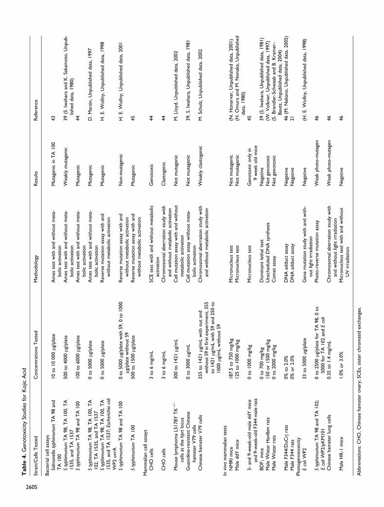
Tab
le4.
Gen
oto
xic
ity
Studie
sfo
rK
ojic
Aci
d
Stra
in/C
ells
Tes
ted
Conce
ntr
atio
ns
Tes
ted
Met
hodolo
gyR
esults
Ref
eren
ce
Bac
teri
alce
llas
says
Salm
onel
laty
phim
uriu
mT
A98
and
TA
100
10
to10
000mg
/pla
teA
mes
test
with
and
without
met
a-bolic
activa
tion
Muta
genic
inT
A100
43
Sty
phim
uriu
mT
A98,T
A100,T
A1535,an
dT
A1537
500
to4000mg
/pla
teA
mes
test
with
and
without
met
a-bolic
activa
tion
Wea
kly
muta
genic
39
(S.Iw
ahar
aan
dK
.Sa
kam
oto
,U
npub-
lished
dat
a,1980)
Sty
phim
uriu
mT
A98
and
TA
100
100
to6000mg
/pla
teA
mes
test
with
and
without
met
a-bolic
activa
tion
Muta
genic
44
Sty
phim
uriu
mT
A98,T
A100,T
A102,T
A1535,an
dT
A1537
0to
5000mg
/pla
teA
mes
test
with
and
without
met
a-bolic
activa
tion
Muta
genic
D.M
arzi
n,U
npublis
hed
dat
a,1997
Sty
phim
uriu
mT
A98,T
A100,T
A1535,a
nd
TA
1537;E
sche
rich
iaco
liW
P2
uvr
A
0to
5000mg
/pla
teR
ever
sem
uta
tion
assa
yw
ith
and
without
met
abolic
activa
tion
Muta
genic
H.E.W
olln
y,U
npublis
hed
dat
a,1998
Sty
phim
uriu
mT
A98
and
TA
100
0to
5000mg
/pla
tew
ith
S9,0
to1000
mg/p
late
without
S9R
ever
sem
uta
tion
assa
yw
ith
and
without
met
abolic
activa
tion
Non-m
uta
genic
H.E.W
olln
y,U
npublis
hed
dat
a,2001
Sty
phim
uriu
mT
A100
500
to1500mg
/pla
teR
ever
sem
uta
tion
assa
yw
ith
and
without
met
abolic
activa
tion
Muta
genic
45
Mam
mal
ian
cell
assa
ysC
HO
cells
3to
6m
g/m
LSC
Ete
stw
ith
and
without
met
abolic
activa
tion
Gen
oto
xic
44
CH
Oce
lls3
to6
mg/
mL
Chro
moso
mal
aber
ration
study
with
and
without
met
abolic
activa
tion
Cla
stoge
nic
44
Mouse
lym
phom
aL5
178Y
TKþ
/�
cells
atth
ehp
rtlo
cus
300
to1421mg
/mL
Cel
lmuta
tion
assa
yw
ith
and
without
met
abolic
activa
tion
Not
muta
genic
M.Ll
oyd
,U
npublis
hed
dat
a,2002
Guan
idin
e-re
sist
ant
Chin
ese
ham
ster
V79
cells
0to
3000mg
/mL
Cel
lm
uta
tion
assa
yw
ithout
met
a-bolic
activa
tion
Not
muta
genic
39,S.
Iwah
ara,
Unpublis
hed
dat
a,1981
Chin
ese
ham
ster
V79
cells
355
to1421mg
/mL
with
out
and
withoutS9
infir
stex
per
imen
t,355
to1421mg
/mL
with
S9an
d250
to1000mg
/mL
without
S9
Chro
moso
mal
aber
ration
study
with
and
without
met
abolic
activa
tion
Wea
kly
clas
toge
nic
M.Sc
hulz
,U
npublis
hed
dat
a,2002
Invi
vom
amm
alia
nte
sts
NM
RIm
ice
187.5
to750
mg/
kgM
icro
nucl
eus
test
Not
muta
genic
(N.H
onar
var,
Unpublis
hed
dat
a,2001)
Mal
eddY
mic
e125
to1000
mg/
kgM
icro
nucl
eus
test
Not
muta
genic
(H.O
mura
and
M.N
onak
a,U
npublis
hed
dat
a,1980)
3-
and
9-w
eek-
old
mal
eddY
mic
ean
d9-w
eek-
old
F344
mal
era
ts0
to1000
mg/
kgM
icro
nucl
eus
test
Gen
oto
xic
only
in9
wee
kold
mic
e45
BD
F 1m
ice
0to
700
mg/
kgD
om
inan
tle
thal
test
Neg
ativ
e39
(S.Iw
ahar
a,U
npublis
hed
dat
a,1981)
Mal
eW
ista
rH
anIb
mra
ts150
or
1500
mg/
kgU
nsc
hed
ule
dD
NA
synth
esis
Not
genoto
xic
(W.V
olk
ner
,U
npublis
hed
dat
a,1997)
Mal
eW
ista
rra
ts0
to2000
mg/
kgC
om
etas
say
Not
genoto
xic
(S.B
rendle
r-Sc
hw
aab
and
B.K
ram
er-
Bau
tz,U
npublis
hed
dat
a,2004)
Mal
eF3
44/D
uC
rjra
ts0%
to2.0
%D
NA
adduct
assa
yN
egat
ive
46
(M.N
akan
o,U
npublis
hed
dat
a,2005)
Mal
eF3
44
rats
0%
or
2.0
%D
NA
adduct
assa
yN
egat
ive
21
Photo
genoto
xic
ity
Eco
liW
P2
33
to5000mg
/pla
teG
ene
muta
tion
study
with
and
with-
out
light
irra
dia
tion
Neg
ativ
e(H
.E.W
olln
y,U
npublis
hed
dat
a,1998)
Sty
phim
uriu
mT
A98
and
TA
102;
Eco
liW
P2/p
KM
101
0to
2500mg
/pla
tefo
rT
A98,0
to5000
for
TA
102
and
Eco
liPhoto
-rev
erse
muta
tion
assa
yW
eak
photo
-muta
gen
46
Chin
ese
ham
ster
lung
cells
0.3
5to
1.4
mg/
mL
Chro
moso
mal
aber
ration
study
with
and
without
light
irra
dia
tion
Wea
kphoto
-muta
gen
46
Mal
eH
R-1
mic
e1.0
%or
3.0
%M
icro
nucl
eus
test
with
and
without
UV
irra
dia
tion
Neg
ativ
e46
Abbre
viat
ions:
CH
O,C
hin
ese
ham
ster
ova
ry;SC
Es,
sist
erch
rom
atid
exch
ange
s.
260S
males and females. At necropsy, hepatic adenomas and heman-
giomas, pulmonary adenomas, malignant lymphomas, leuke-
mia, or pituitary adenomas were observed. These tumor
incidences did not differ between the kojic acid treatment
groups and the control group. Likewise, nodular hyperplasia
in the liver, adrenal subcapsular spindle cell hyperplasia, and
uterus cystic endometrial hyperplasia did not occur at signifi-
cantly differing rates in the treatment groups versus the control
groups. The researchers concluded that kojic acid was not
tumorigenic to mice in this 78-week study.92
The tumorigenic potential of kojic acid was evaluated, using
heterozygous p53-deficient CBA, p53(þ/�), mice and wild
type littermates, p53(þ/þ).22 The mice were fed diet contain-
ing 0%, 1.5%, or 3.0% kojic acid for 26 weeks. The mice were
observed daily for clinical signs of toxicity and were weighed
weekly. All surviving mice were killed after blood sampling for
hormone assays and necropsied. Livers and thyroid glands
were removed and weighed. These organs along with the pitui-
tary, spleen, lungs, and other organs and tissues with macro-
scopic lesions were fixed for histopathological examination.
Additionally, tissue sections were immunohistochemically
stained for proliferating cell nuclear antigen (PCNA). Five
thousand hepatocellular nuclei in normal background parench-
yma in each mouse were counted for PCNA determination.
One wild type male from the 3.0% dose group was found
dead at week 13. Both p53(þ/�) and p53(þ/þ) mice of the
3.0% dose group had decreased body weight gains compared
to controls. Absolute thyroid gland weights were significantly
(P < .01) increased in a dose-related fashion by 209% and
444% in the 1.5% and 3.0% kojic acid dose groups, respec-
tively, in p53(þ/�) mice and by 140% and 374% in
p53(þ/þ) mice. Absolute and relative liver weights in the kojic
acid-treated groups had somewhat higher values in both types
of mice when compared to controls but was not significant
except for the relative weight in the 3.0% p53(þ/þ) mice.
Diffuse hypertrophy and hyperplasia of thyroid follicular
epithelial cells were observed along with decreased serum thyr-
oxine (T4) levels in both p53(þ/�) and p53(þ/þ) mice treated
with kojic acid. No thyroid tumors were observed, however. In
the liver, the incidence of altered hepatocellular foci was sig-
nificantly increased at 1.5% and 3.0% in p53(þ/�) and at
1.5% in p53(þ/þ) mice. The authors concluded that there is
tumorigenic potential of kojic acid in the liver but not in the
thyroid follicular epithelial cells in CBA mice. The genotoxic
potential of kojic acid on hepatocellular tumor development
could not be ruled out.22
The above study was repeated using male CBA mice that
received 0%, 0.5%, 1%, or 2% kojic acid in their diet for 26
weeks.25 Incidences of hepatocellular adenomas were 5%,
17%, 10%, and 21%, respectively. Incidences of hepatocellular
foci in these dose groups were 15%, 39%, 45%, and 47%,
respectively, with a statistically significant difference (P <
.05) only between the control group and the 2% dose group.
Male F344 rats were used in a 55-week toxicity dietary
study of kojic acid.93 The 7-week-old rats were divided into
groups of 20 and received 0%, 0.5%, or 2.0% kojic acid
(equivalent to 0, 227, or 968 mg/kg body weight/d, respec-
tively). One week prior to treatment, rats received a single sub-
cutaneous injection of 5 mL/kg saline. The rats were observed
daily for clinical signs of toxicity and were weighed regularly.
Feed consumption was recorded weekly. At the end of treat-
ment, surviving rats were killed after blood sampling and
necropsied. Major organs and tissues were weighed and/or
fixed for histopathological examination. Additionally, liver
sections were studied immunohistochemically for glutathione
S-transferase-placental form (GST-P), PCNA, and single-
strand DNA (ssDNA).
No mortality or obvious clinical signs of toxicity were
observed during the treatment period. Body weight gains were
decreased in the 2.0% group from week 6 until treatment end,
when compared to the controls. No significant changes in feed
consumption were observed. In both the 0.5% and 2.0% treat-
ment groups, red blood cell counts and hematocrit values were
decreased. Significant increases or a tendency for increase
were observed in aspartate aminotransferase (AST), alanine
aminotransferase (ALT), ALP, g-glutamyl transpeptidase (g-
GTP), blood urea nitrogen (BUN), and sodium values in both
the 0.5% and 2.0% dose groups. In the 2.0% group, total pro-
tein, total bilirubin, and total cholesterol values were signifi-
cantly increased and the albumin/globulin ratio was decreased.
Absolute and relative spleen and thyroid gland weights were
increased or had a tendency for increase in both the 0.5% and
2.0% dose groups. In the 2.0% dose group, absolute and rela-
tive weights of heart, lungs, liver, adrenal glands, testes, and
relative weights of brain and kidneys were significantly
increased. Single-cell necrosis of hepatocytes and proliferation
of small bile ducts or ductules were recorded in animals from
both treatment groups, with the incidence of the proliferation
of bile ducts significantly increased in the 2.0% dose group. All
2.0% dose group animals had diffuse hepatocellular hypertro-
phy and/or vacuolization and formation of microgranulomas
containing crystals and/or brown pigment; the incidence of the
granulomas was significantly increased. Areas of GST-P-posi-
tive foci were significantly increased in the liver of the 2.0%dose group. Incidences of hyaline casts and basophilic tubules
were also significantly increased in the 2.0% dose group. Dif-
fuse follicular cell hyperplasia was noted in the thyroid glands
in both treatment groups, with focal follicular cell hyperplasia,
and adenomas and/or carcinomas observed in the 2.0% group.
The 2.0% dose group also had increased hypertrophy of corti-
cal cells in zona fasciculate in the adrenal glands. The study
concluded that the NOAEL of kojic acid was below 0.5%(227 mg/kg body weight/d).93
Carcinogenicity studies are summarized in Table 5.
Tumor Promotion
The carcinogenesis-modifying action of kojic acid in rat liver
using a 2-stage model with initiation by diisopropanolnitrosa-
mine (DHPN) was investigated.94 Sixty male F344 rats
received either a single subcutaneous injection of 2000 mg/
kg DHPN or the vehicle and then were fed a diet containing
Burnett 261S
0%, 0.125%, 0.5%, or 2% kojic acid for 20 weeks. At the end of
the treatment period, the rats were killed and necropsied. The
liver was removed, weighed, and prepared for paraffin section-
ing. H&E staining and immunostaining to GST-P and PCNA
were performed and the sections were investigated histopatho-
logically and cell-kinetically.
Rats treated with 2% kojic acid with DHPN initiation had
significantly increased (P < .01) relative liver weights. Histo-
pathology revealed an increased incidence of microgranuloma
and vacuolation of centrilobular hepatocytes. The number and
area of GST-P-positive foci per unit area of the liver in the
DHPN and 2% kojic acid group were 22.30 foci and 3745
mm2, respectively, which was a significant increase (P < .01)
when compared the 8.48 foci and 531 mm2 in the group treated
with only DHPN. The incidence of GST-P-positive foci and the
percentage of PCNA-positive cells were more prominent in
animals with marked vacuolation of hepatocytes. In the group
treated with 2% kojic acid without DHPN, the number and area
of GST-P-positive foci were 1.39 foci and 109.5 mm2, respec-
tively, which was also a significant increase when compared
to the control group values of 0.40 foci and 9.7 mm2. No treat-
ment-related effects were observed in the rats treated with 0.5%kojic acid or lower, with or without DHPN. The researchers
concluded that kojic acid has a carcinogenesis-promoting
action in the rat liver and may be carcinogenic without
promotion.94
Further study on the tumor promotion potential of kojic acid
was done.95 Groups of 20 male F344 rats received 0%, 0.5%, or
2% kojic acid in feed for 20 weeks without DHPN initiation. At
the end of the treatment period, the rats were killed and necrop-
sied, and the livers were studied in the same manner as
described above. Dose-related increases in absolute and rela-
tive liver weights were observed in both kojic acid treatment
groups. Numbers and areas of GST-P-positive foci were signif-
icantly increased (P < .01) in the 2% kojic acid group when
compared to the control group. Increased incidences of micro-
granuloma and vacuolation of hepatocytes were observed in the
2% kojic acid treatment group. PCNA expression was signifi-
cantly increased (P < .05) in the 2% kojic acid dose group when
compared to the control group, with PCNA-positive hepato-
cytes mainly localized around the vacuolated and granuloma-
tous regions.
The authors also performed a medium-term liver bioassay of
kojic acid in groups of 25 F344 male rats at concentrations of
0%, 0.125%, 0.5%, or 2% to determine kojic acid’s promoting
influence.95 Two weeks prior to the start of the 6-week dietary
exposure of kojic acid, the rats received a single intraperitoneal
injection of 200 mg/kg N-diethylnitrosamine (DEN). At week
3, the rats were subjected to a two-third partial hepatectomy.
At the end of the treatment period, the rats were killed and
livers were prepared for analysis as above. A dose-related
decrease in body weight gains and an increase in relative liver
weights were observed, with statistical significance (P < .01) in
the 2% dose group. Significant increases (P < .01) in number
and areas of GST-P-positive foci were observed in the 2% dose
group when compared to the control group. The authors
concluded that kojic acid at 2% was tumor-promoting and had
weak hepatocarcinogenic potential. The authors further opined
that the enhanced replication of hepatocytes related to toxic
changes may have been involved as an underlying mechanism.
Tumor Initiation
A study on the tumor-initiating potential of kojic acid in mouse
liver was performed using male ICR mice.23 The mice received
a diet containing 0% or 3% kojic acid for 4 weeks, followed by
distilled water containing 0 or 500 ppm phenobarbital (PB) for
14 weeks. Two weeks after the treatment with PB, a two-third
partial hepatectomy was performed on all mice. At the end of
the study, all mice were killed and liver slices were performed
to evaluate g-glutamyltransferase-positive foci as preneoplastic
foci markers in the liver as well as PCNA.
No treatment-related deaths were observed and there were
no significant changes in feed consumption or body weights
during the course of the study. No proliferative lesions were
observed in any dose groups during microscopic examinations.
There were no differences in the number of g-glutamyltransfer-
ase-positive cells between the kojic acid and distilled water and
the kojic acid þ PB groups. Significant increases in the label-
ing index of PCNA were observed in the control þ PB and
kojic acid þ PB dose groups as compared to the control þ dis-
tilled water group (1.28 + 1.93); however, no significant dif-
ference in the positivity of PCNA was observed between the
controlþ PB and the kojic acid þ PB groups. The authors con-
cluded that kojic acid has no tumor-initiating activity in mouse
liver.23 In reviewing this report, however, the SCCP concluded
that the kojic acid effect on proliferation of liver cells cannot be
excluded since kojic acid þ distilled water PCNA values were
increased compared to basal diet þ distilled water.20
The initiation potential of kojic acid (99.5% pure) in rat liver
was examined in a 2-part study.26
In the first experiment, groups of 5 male F344 rats were fed a
diet containing 0% or 2% kojic acid for 3, 7, or 28 days. All rats
were injected with 100 mg/kg body weight bromodeoxyuridine
(BrdU) intraperitoneally once a day for the last 2 days of expo-
sure and 2 hours prior to termination. Livers were removed and
weighed at necropsy and slices were prepared for BrdU immu-
nostaining. Labeling indices (LIs) were calculated as percentages
of cells positive for BrdU incorporation divided by the total num-
ber of cells counted. In addition, 8-oxodeoxyguanosine (8-
OxodG) was measured in nuclear DNA to examine the formation
of oxidative DNA adduct by HPLC-ECD detection.
On day 28 of the experiment, body weight gains in the 2%kojic acid group were significantly decreased compared to the
control group. In the 2% kojic acid dose group, absolute liver
weights were significantly increased on day 7 but decreased
on day 28. Relative liver weights were significantly increased
at all time points. The LI values of hepatocytes of the 2% dose
group were significantly increased as compared to the controls
on days 3 and 7. All 8-OxodG levels in the liver DNA in the 2%dose group were slightly higher than the control values but
were not statistically significant.
262S International Journal of Toxicology 29(Supplement 4)
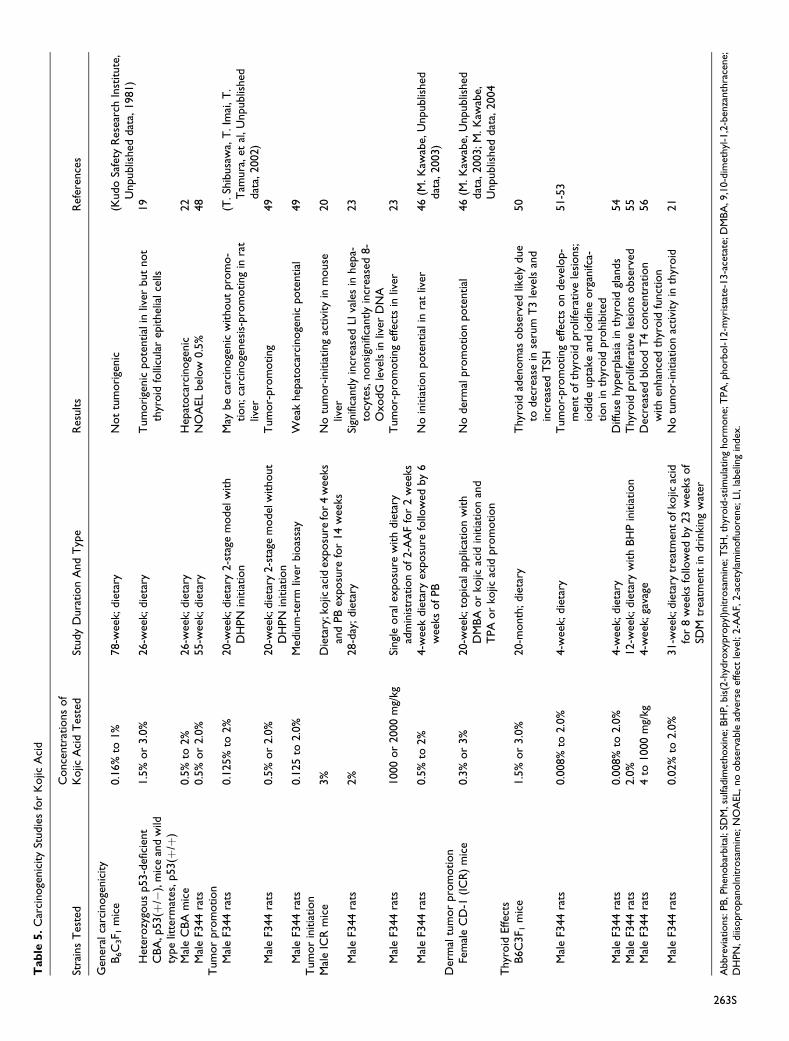
Tab
le5.
Car
cinoge
nic
ity
Studie
sfo
rK
ojic
Aci
d
Stra
ins
Tes
ted
Conce
ntr
atio
ns
of
Kojic
Aci
dT
este
dSt
udy
Dura
tion
And
Typ
eR
esults
Ref
eren
ces
Gen
eral
carc
inoge
nic
ity
B6C
3F 1
mic
e0.1
6%
to1%
78-w
eek;
die
tary
Not
tum
ori
genic
(Kudo
Safe
tyR
esea
rch
Inst
itute
,U
npublis
hed
dat
a,1981)
Het
erozy
gous
p53-d
efic
ient
CB
A,p
53(þ
/�),
mic
ean
dw
ildty
pe
litte
rmat
es,p53(þ
/þ)
1.5
%or
3.0
%26-w
eek;
die
tary
Tum
ori
genic
pote
ntial
inliv
erbut
not
thyr
oid
folli
cula
rep
ithel
ialce
lls19
Mal
eC
BA
mic
e0.5
%to
2%
26-w
eek;
die
tary
Hep
atoca
rcin
oge
nic
22
Mal
eF3
44
rats
0.5
%or
2.0
%55-w
eek;
die
tary
NO
AEL
bel
ow
0.5
%48
Tum
or
pro
motion
Mal
eF3
44
rats
0.1
25%
to2%
20-w
eek;
die
tary
2-s
tage
model
with
DH
PN
initia
tion
May
be
carc
inoge
nic
without
pro
mo-
tion;ca
rcin
oge
nes
is-p
rom
oting
inra
tliv
er
(T.Sh
ibusa
wa,
T.Im
ai,T
.T
amura
,et
al,U
npublis
hed
dat
a,2002)
Mal
eF3
44
rats
0.5
%or
2.0
%20-w
eek;
die
tary
2-s
tage
model
without
DH
PN
initia
tion
Tum
or-
pro
moting
49
Mal
eF3
44
rats
0.1
25
to2.0
%M
ediu
m-t
erm
liver
bio
assa
yW
eak
hep
atoca
rcin
oge
nic
pote
ntial
49
Tum
or
initia
tion
Mal
eIC
Rm
ice
3%
Die
tary
;kojic
acid
exposu
refo
r4
wee
ksan
dPB
exposu
refo
r14
wee
ksN
otu
mor-
initia
ting
activi
tyin
mouse
liver
20
Mal
eF3
44
rats
2%
28-d
ay;die
tary
Sign
ifica
ntly
incr
ease
dLI
vale
sin
hep
a-to
cyte
s,nonsi
gnifi
cantly
incr
ease
d8-
OxodG
leve
lsin
liver
DN
A
23
Mal
eF3
44
rats
1000
or
2000
mg/
kgSi
ngl
eora
lex
posu
rew
ith
die
tary
adm
inis
trat
ion
of2-A
AF
for
2w
eeks
Tum
or-
pro
moting
effe
cts
inliv
er23
Mal
eF3
44
rats
0.5
%to
2%
4-w
eek
die
tary
exposu
refo
llow
edby
6w
eeks
ofPB
No
initia
tion
pote
ntial
inra
tliv
er46
(M.K
awab
e,U
npublis
hed
dat
a,2003)
Der
mal
tum
or
pro
motion
Fem
ale
CD
-1(I
CR
)m
ice
0.3
%or
3%
20-w
eek;
topic
alap
plic
atio
nw
ith
DM
BA
or
kojic
acid
initia
tion
and
TPA
or
kojic
acid
pro
motion
No
der
mal
pro
motion
pote
ntial
46
(M.K
awab
e,U
npublis
hed
dat
a,2003;M
.K
awab
e,U
npublis
hed
dat
a,2004
Thyr
oid
Effec
tsB
6C
3F 1
mic
e1.5
%or
3.0
%20-m
onth
;die
tary
Thyr
oid
aden
om
asobse
rved
likel
ydue
todec
reas
ein
seru
mT
3le
vels
and
incr
ease
dT
SH
50
Mal
eF3
44
rats
0.0
08%
to2.0
%4-w
eek;
die
tary
Tum
or-
pro
moting
effe
cts
on
dev
elop-
men
tofth
yroid
pro
lifer
ativ
ele
sions;
iodid
eupta
kean
dio
din
eorg
anifc
a-tion
inth
yroid
pro
hib
ited
51-5
3
Mal
eF3
44
rats
0.0
08%
to2.0
%4-w
eek;
die
tary
Diff
use
hyp
erpla
sia
inth
yroid
glan
ds
54
Mal
eF3
44
rats
2.0
%12-w
eek;
die
tary
with
BH
Pin
itia
tion
Thyr
oid
pro
lifer
ativ
ele
sions
obse
rved
55
Mal
eF3
44
rats
4to
1000
mg/
kg4-w
eek;
gava
geD
ecre
ased
blo
od
T4
conce
ntr
atio
nw
ith
enhan
ced
thyr
oid
funct
ion
56
Mal
eF3
44
rats
0.0
2%
to2.0
%31-w
eek;
die
tary
trea
tmen
tofk
ojic
acid
for
8w
eeks
follo
wed
by
23
wee
ksof
SDM
trea
tmen
tin
dri
nki
ng
wat
er
No
tum
or-
initia
tion
activi
tyin
thyr
oid
21
Abbre
viat
ions:
PB
,Phen
obar
bital
;SD
M,s
ulfa
dim
ethoxin
e;B
HP,b
is(2
-hyd
roxyp
ropyl
)nitro
sam
ine;
TSH
,thyr
oid
-stim
ula
ting
horm
one;
TPA
,phorb
ol-12-m
yris
tate
-13-a
ceta
te;D
MB
A,9
,10-d
imet
hyl
-1,2
-ben
zanth
race
ne;
DH
PN
,diis
opro
pan
oln
itro
sam
ine;
NO
AEL,
no
obse
rvab
lead
vers
eef
fect
leve
l;2-A
AF,
2-a
cety
lam
inoflu
ore
ne;
LI,la
bel
ing
index
.
263S
In the second experiment of this study, 30 male F344 rats
were subjected to a two-third partial hepatectomy on day 0.
At 12-hour postsurgery, the rats were treated once orally with
carboxymethylcellulose vehicle (8 rats), 1000 mg/kg kojic acid
(12 rats), or 2000 mg/kg kojic acid (10 rats) at a dose volume of
10 mL/kg body weight. The rats were then fed basal diet for 2
weeks and then diet containing 0.015% 2-acetylaminofluorene
(2-AAF) for another 2 weeks. At 3 weeks post kojic acid
administration, rats received a single 0.8 mL/kg body weight
dose of carbon ttetrachloride (CCl4). Surviving rats were killed
at the end of week 5 and slices of all liver lobes were stained
immunohistochemically for GST-P. The mean area and num-
ber of GST-P-positive foci per unit area of all liver sections
were calculated. During the course of the experiment, 1 rat in
the control group died. Slight decreases were observed in the
mean area and numbers of GST-P positive foci, but these dif-
ferences were not statistically significant.
The researchers of this second experimental study con-
cluded that kojic acid has neither liver initiation activity nor the
capability of 8-OxodG formation; however, the findings sug-
gest that kojic acid has liver tumor-promoting effects.26
The initiation potential of kojic acid (100.3% pure) in a liver
carcinogenesis bioassay was performed on F344 male rats.89,96
In one portion of the study, groups of 15 rats received 0%,
0.5%, 1%, or 2% kojic acid or the positive control 2-AAF at
concentrations of 0.01% or 0.001% in their feed for 4 weeks.
After the treatment period, all rats received basal diet for 1
week, and then a diet containing 0.5% phenobarbital sodium
salt (SPB) for 6 weeks. In another portion of the study, groups
of 9 rats received 0% or 2% kojic acid or 0.01% or 0.001% 2-
AAF in feed for 4 weeks, and then all rats received basal diet
for 7 weeks. At 6 weeks after the beginning of the study, all ani-
mals from both portions of the study underwent a two-third par-
tial hepatectomy. Rats were checked twice daily for clinical
signs of toxicity and mortality. Body weights were measured
weekly and daily feed consumption and intake of kojic acid,
2-AAF, and SPB were calculated. All surviving rats were killed
at study end, and organs were examined macroscopically. Liver
weights were recorded and sections from 3 liver lobes were
stained immunohistochemically for GST-P.
No treatment-related effects or deaths were observed during
the study. Rats that received 2% kojic acid in both portions of
the study had significant decreases in body weights during
initiation period of the study, but body weights returned to con-
trol levels during the SPB or basal diet treatments. Decreases in
feed consumption during the initiation period occurred in the
1.0% and 2.0% kojic acid groups, but increases in feed con-
sumption during the SPB or basal diet treatment were marked
with increases in body weight change. No treatment-related
differences were observed in final body or liver weights, with
or without SPB. Numbers of GST-P-positive foci in kojic
acid-treated groups were similar to the control values, with or
without SPB. No other treatment-related effects were observed.
In the positive control groups, the numbers of GST-P-positive
foci were statistically significantly increased in the 0.01% 2-
AAF groups, with and without SPB. This study concluded that
kojic acid did not possess initiation potential in the rat
liver.89,96
Dermal Tumor Promotion
A skin carcinogenesis bioassay to determine the promotion
potential of kojic acid (reported as 100.3% pure) in a cream for-
mulation was performed using female CD-1 (ICR) mice.89,96,97
The positive initiator control was 9,10-dimethyl-1,2-benzan-
thracene (DMBA) and the positive promoter control was phor-
bol-12-myristate-13-acetate (TPA). Groups of 10 or 15 mice
were treated in the following manner: DMBA þ vehicle,
DMBA þ 0.3% kojic acid, DMBA þ 3% kojic acid, DMBA
þ TPA, acetone þ 0.3% kojic acid, acetone þ 3% kojic acid,
vehicle þ TPA, or 3% kojic acid þ TPA. The control or test
substances were applied to the shaved backs of the mice (4
cm2). The mice receiving DMBA or acetone were treated once
at the beginning of the experiment while the mice treated with
vehicle þ TPA or 3% kojic acid þ TPA received 50 mg of the
test substances daily for 1 week. A week after the study com-
mencement, the treatment groups with DMBA or acetone
received 50 mg of the test substances 5 times weekly for 19
weeks. The remaining groups received TPA twice weekly for
19 weeks 1 or 2 weeks after study commencement. Animals
were checked for clinical signs of toxicity and mortality once
daily and for skin nodules once weekly. All surviving animals
were killed after the completion of the promoter treatment and
examined macroscopically. A histological examination of the
skin was performed and liver weights were recorded.
No treatment-related mortalities were observed. Body
weight gain was significantly decreased in week 2 or weeks
3 and 4 in the DMBA þ 0.3% kojic acid and acetone þ 3%dose groups, respectively. Squamous cell papilloma was
observed in 1 mouse from the DMBA þ 3% kojic acid. The
positive control group, DMBA þ TPA, had significantly
increased body weight gain (starting at week 3) and absolute
and relative liver weights. The positive control group also had
skin nodules, which were revealed to be squamous cell hyper-
plasia, squamous cell papilloma, or squamous cell carcinoma at
necropsy. It was concluded that kojic acid did not possess pro-
motion potential for skin carcinogenesis.89,96
Thyroid Effects
The tumorigenicity of kojic acid was studied in a 20-month
study in B6C3F1 mice.98 Groups of 65 male and female mice
received 0%, 1.5%, or 3.0% kojic acid in feed for 20 months.
Subgroups of 5 animals were killed at 6 and 12 months after the
beginning of treatment. Serum was collected for hormone
assessment at 6, 12, and 20 months from 5 animals in each
treatment group. Another subgroup of 10 to 14 animals in each
treatment group was switched to normal diet at month 19. At
the end of the treatment period, all surviving animals were
killed and necropsied, with major organs and tissues weighed
and fixed for histopathological examination.
264S International Journal of Toxicology 29(Supplement 4)
Survival rates in mice in the treatment groups were compa-
rable with the control groups during the course of the adminis-
tration period. Thyroid weights were increased significantly in
the kojic acid-treated groups of both genders, especially in the
male groups; there were no significant differences in other
major organ or tissue weights or hematological values or serum
biochemical parameters in any of the treatment groups. Inci-
dences of thyroid gland hyperplasia and follicular adenomas
were significantly increased in all treatment groups. In mice
that received normal feed 30 days prior to termination, inci-
dences of thyroid gland adenomas were significantly
decreased, although average thyroid weights were unchanged.
The serum-free triiodothyronine (T3) levels in the 3.0% dose
groups of both genders were significantly lower than the con-
trol at month 6, while the thyroid-stimulating hormone (TSH)
levels were increased. The decreases in the free T3 levels con-
tinued at the later measurements, but changes in the TSH levels
disappeared. It was concluded that chronic high doses of kojic
acid induces thyroid adenomas in male and female B6C3F1
mice. The authors proposed that the likely mechanism is the
decrease in serum T3 levels and increased TSH.98
A study was performed to determine the mechanisms of
serum thyroid hormone reduction and thyroid tumor-promotion
effects of kojic acid exposure in rats.99 Groups of 8 male F344
rats received basal diet containing 0%, 0.008%, 0.03%,
0.125%, 0.5%, or 2.0% kojic acid for 4 weeks (doses equivalent
to 0, 5.85, 23.8, 95.3, 393.6, and 1387.3 mg/kg body weight/d).
At the end of treatment, blood was collected from 5 rats per
group for hormone assays. The remaining animals were
injected intraperitoneally with 0.4 mL of 0.1 mol/L Na125I in
saline 24 hours before they were killed. Measurement of 125I
uptake was taken and the thyroid was examined for
organification.
No significant changes in body weights were observed in the
treated rats when compared to the control rats. Absolute and
relative thyroid gland weights were increased in all groups
treated with kojic acid in a dose-dependent manner, with signif-
icant increases occurring at 0.5% or more. The relative pitui-
tary gland weights were significantly increased in the 2.0%kojic acid group and relative liver weights were significantly
greater in all kojic acid groups except the 0.125% group. These
last two observations were not dose-dependent or associated
with significant changes in absolute weights, and thus were not
biologically relevant. A statistically significant decrease in
serum T3 and T4 levels was observed in the 2.0% kojic acid
group when compared with the control group. The serum TSH
in the 2% kojic acid group was significantly increased when
compared to the controls. There were no other significant dif-
ferences in these parameters in the other dose groups. Thyroid125I uptake was significantly decreased in a dose-dependent
manner starting at 0.03% kojic acid. A significant reduction
in organic formation of iodine was observed in the 2.0% kojic
acid group.
Histopathologic examination revealed decreased colloid in
the thyroid follicles and follicular cell hyperthrophy in the thyr-
oid in high incidences in groups that received 0.03% kojic acid
or more. All rats in the 2.0% kojic acid group had thyroid cap-
sular fibrosis. In a quantitative morphometric analysis, the ratio
of the area of follicular epithelial cells to the area of colloids
was significantly increased in the 0.03% kojic acid dose group
and higher. In this rat study, kojic acid inhibited iodide uptake
and iodine organification in the thyroid, with tumor-promoting
effects on the development of thyroid proliferative lesions.
These effects were likely secondary to prolonged serum TSH
stimulation resulting from negative-feedback through the pitui-
tary�thyroid axis.99 Additional studies found similar
results.100,101
The mechanism of tumorigenesis in the thyroid from expo-
sure to kojic acid was examined in a 3-part study.102
In the first experiment, groups of 9 male F344 rats received
0%, 0.008%, 0.03%, 0.125%, 0.5%, or 2.0% kojic acid in their
diets for 4 weeks. Twenty-four hours prior to experiment end,
4 rats in each dose group received 0.2 mL/100 g body weight
Na125I at 0.1 mol/L in saline. Rats were killed and the thyroid
glands were weighed and examined for 125I uptake. The remain-
ing 5 animals were killed on the same day. Thyroid gland
weights were increased in a dose-dependent manner in rats
receiving 0.125% or more kojic acid in diet, with the thyroid
gland weights from the 2.0% dose group 9 times that of the con-
trols. 125I uptake into the thyroid gland was more sensitive to
kojic acid treatment, with significant suppression at 0.03%.
Organic 125I formation was interrupted only in the 2.0% dose
group. Serum T3, T4, and TSH levels were affected only at 2.0%.
In the second experiment, male and female F344 rats were
divided into 8 and 4 groups, respectively, with each group con-
sisting of 8 animals. The groups received diet containing 0% or
2.0% kojic acid. Male groups were killed at weeks 1, 2, 3, and 4
and female groups were killed at weeks 2 and 4. Half of the rats
were studied for 125I uptake and the other half for hormonal and
histopathological examination. In males, thyroid gland weights
increased linearly from 11 to 98 mg in the 4 weeks of treatment
with 2.0% kojic acid. A less prominent, but still significant,
increase in thyroid gland weights was observed in females,
from 7.5 to 40 mg. The suppression of 125I uptake was also time
dependent and in males, the decrease started at 1 week after
kojic acid treatment and reached about 2% of control values
by week 3, with organic 125I formation significantly decreased
by 50% compared to the controls. These effects were not as sig-
nificant in females, with only 20% suppression of 125I uptake at
week 4. Serum T3 and T4 levels were decreased to minimum
levels after 2 weeks of kojic acid treatment, but recovered
thereafter although at lower than control values in both gen-
ders. Serum TSH started to increase at week 1 and reached a
maximum at weeks 2 and 3.
For the final experiment in this study, 6 groups of 8 male
F344 rats received 0% and 2.0% kojic acid in diet for 4 weeks.
At the end of the treatment, kojic acid was replaced with basal
diet for 0, 6, 12, 24, or 48 hours. The groups were killed and
examined as in the first 2 experiments, except that 125I was
injected 12 hours before death. The organic 125I formation
returned to normal limits after 6 hours and 125I uptake per unit
of thyroid weight increased to 70% of the control values within
Burnett 265S
24 hours. Serum T3 and T4 were 47% and 34% of the control
values after 4 weeks of the kojic acid diet. The levels increased
to normal limits within 48 hours after return to basal diet and
high levels of TSH decreased to normal within 24 hours.
The histopathological investigation on thyroid glands in
these 3 experiments found a diffuse type of hyperplasia caused
by the kojic acid diet. After 2 weeks of returning to basal diet,
normal thyroid follicular structure was apparent in enlarged
thyroid glands. The authors of this study suggest that the pro-
liferative effect of kojic acid on the thyroid is not related to a
genotoxic pathway.102
In a study to determine whether kojic acid causes a promot-
ing effect on thyroid carcinogenesis, male F344 rats were initi-
ated with N-bis(2-hydroxypropyl)nitrosamine (BHP) with a
single subcutaneous injection (2800 mg/kg).103 The dose
groups included 10 rats each. One week later, the rats received
basal diet containing 0% or 2% kojic acid for 12 weeks. An
additional group of 8 rats received no BHP initiation or kojic
acid and were fed basal diet for 13 weeks. Half of the rats were
killed at week 4 and the remainder after the last week of expo-
sure. In the second experiment of the same study, another 2
groups of 10 rats not initiated with BHP received diet contain-
ing 0% or 2% kojic acid for 20 weeks. Again, half of the rats
were killed at week 4 and the remainder after week 20. Body
weights were recorded and blood samples for hormone analysis
were taken before death in all animals.
Body weights were decreased in the rats that received kojic
acid at both week 4 and 12. Rats in both experiments exposed
to kojic acid also had increased absolute and relative thyroid
weights up to 25-fold greater than the control group, as well
as increased relative liver weights at each time point. Absolute
liver weights were significantly increased in rats exposed to
kojic acid for 20 weeks. Serum T3 and T4 levels were signifi-
cantly decreased (approximately one half to one third the values
of the BHP alone group) and serum TSH was significantly
increased (13-19 times higher than the BHP alone group) in the
BHPþ kojic acid group at both time periods. Similar changes in
other serum thyroid-related hormones were observed in the 2%kojic acid alone group at week 4 but not at week 20.
At week 4, 4 of the 5 rats in the BHPþ kojic acid group had
focal thyroid follicular hyperplasias, while 3 of the 5 rats had
focal thyroid follicular adenomas. These lesions were observed
in all rats in the BHP þ kojic acid group by week 12. Rats that
only received kojic acid had marked diffuse hypertrophy of fol-
licular epithelial cells at week 4 and 20. The BHP alone and the
untreated control groups had no changes in thyroid-related hor-
mone levels or histopathological lesions. There were no signif-
icant intergroup changes of the liver T4-uridine diphosphate
glucuronosyltransferase (UDP-GT) activity. The authors con-
cluded that kojic acid induced thyroid proliferative lesions due
to continuous serum TSH stimulation through the negative
feedback mechanism of the pituitary�thyroid axis, with
decreases of T3 and T4 caused by a mechanism independent
of T4-UDP-GT activity.103
In a study on the effect of kojic acid on thyroid function, 24
groups of 10 male F344/Du Crj rats received 0, 4, 15, 62.5, 250,
or 1000 mg/kg kojic acid daily for 4 weeks.104 Kojic acid was
suspended in 0.5% carboxymethylcellulose and administered at
a dosing volume of 5 mL/kg via gavage. At the end of each
treatment week, a group of rats from each dose group were
killed and necropsied (1 group of rats were necropsied prior
to test material administration).
No abnormalities were observed in rats in the 0 to 250 mg/
kg dose groups during treatment. Several rats in the 1000 mg/
kg dose group had transient and slight decreases in motility 30
minutes to 1 hour after dosing on day 18 to 28 of treatment.
Body weights and feed consumption in the 1000 mg/kg dose
group were significantly inhibited when compared to the con-
trol group. The absolute and relative weights of the thyroid
glands were nearly comparable to the control in the 4 to 250
mg/kg dose groups throughout the treatment period. Absolute
and relative weights of the thyroid gland in the 1000 mg/kg
dose groups were 1.2-fold and 1.3-fold greater than the control
group, respectively. Serum T3 concentration in the 250 mg/kg
dose group had a significant decrease only at week 1 when
compared to the control group, but the other dose groups
showed no significant differences compared to the control at
week 2 to 4. The serum T4 concentration in the 1000 mg/kg
dose group was significantly decreased at week 4, but no
dosage of kojic acid affected the serum TSH concentration sig-
nificantly. The 1000 mg/kg dose group had hypertrophy of
epithelial cells in the thyroid gland at week 1 to 4; this was not
observed in the 250 mg/kg dose group.
In this study, the uptake of iodine and iodination were deter-
mined prior to the beginning of treatment and at week 1, 2, 3,
and 4 of treatment in 5 animals in each dose group. The rats
received 125I-NaI intraperitoneally 24 hours after the last treat-
ment at the end of each week and blood was collected to mea-
sure radioactivity 24 hours after each administration of the
radiolabel. Animals were killed and thyroid glands were
excised and homogenized for radioactivity measurement.
Radioactive iodine uptake in the 4 to 250 mg/kg dose groups
was comparable to the control group at week 1 to 4. In the
1000 mg/kg dose group, the iodine uptake was about 2-fold
greater than the control group in week 1; the uptake in this
group continued to be constant and high through week 4. The
TCA-precipitable radioactive iodine in the thyroid gland was
also increased in the 1000 mg/kg dose group.
This study also determined the absorption of radioactive
kojic acid in male Wistar rats dosed with a single-oral dose
of 10 mCi/100 g body weight 14C-U-kojic acid. Blood was col-
lected 10 and 30 minutes and 1, 3, 6, and 24 hours after admin-
istration and radioactivity was measured with liquid
scintillation. The absorption of kojic acid was rapid as mani-
fested by the Tmax of blood concentration of radioactivity,
which was as short as 1.0 + 0.0 hours and the t1/2 was 4.8
+ 0.3 hours. Blood concentrations of radioactivity had nearly
disappeared by 24 hours after treatment. The authors con-
cluded that kojic acid may decrease blood T4 concentration
and that thyroid function may be enhanced compensatorily;
however, the toxic effect observed on the thyroid gland from
the 1000 mg/kg dose group may depend on a fast decrease
266S International Journal of Toxicology 29(Supplement 4)
following a transient increase of concentration of kojic acid in
the blood.104
The potential thyroid gland tumor initiation activity of kojic
acid was evaluated in a 2-part study on rats.24 Groups of 20
male F344 rats received a diet containing 0%, 0.02%, 0.2%,
or 2% kojic acid for 8 weeks that was followed by treatment
with 0.1% sulfadimethoxine (SDM) in drinking water for 23
weeks. A 13-week recovery period followed the SDM treat-
ment. Controls included a group that received 4 subcutaneous
injections of BHP during the initiation period followed by an
administration of 0.1% SDM, a group that received diet con-
taining 2% kojic acid for the initial 8 weeks alone, a group that
received 2% kojic acid for the entire 31 weeks, and a group that
received only basal diet. Body weights were measured weekly.
At the end of 31 weeks of experimenting, blood was drawn for
hormone analysis. Half of the rats in each group were killed
prior to the recovery and the remaining rats were killed after.
All rats were necropsied. Thyroid glands from the animals were
weighed, fixed, and underwent histopathological examination.
During the treatment and recovery periods, deaths from tra-
cheal obstruction from extremely hypertrophied thyroids were
observed in the BHP control group (5 in total), the 31-week
administration of kojic acid control group (3 in total), the 8-
week kojic acid control group (1 in total), and the 2% kojic acid
þ SDM treatment group (1 in total). Significant suppression of
body weight gains was observed in the BHP and 31-week kojic
acid control groups during administration that continued until
the end of the recovery period in the 31-week kojic acid con-
trol. All treated groups had significantly increased absolute and
relative thyroid gland weights when compared to the untreated
(basal diet) control group at the end of the administration
period. These values, however, were decreased at the end of the
recovery period, except in the BHP control group. When com-
pared to the untreated controls, serum T3 levels in the 0% kojic
acid þ SDM, 2% kojic acid þ SDM, and BHP control group
were significantly decreased at the end of the administration
period, as were the serum T4 levels in all treatment groups
except the 8-week kojic acid control. The serum T3 and T4 lev-
els in the 8-week kojic acid control were significantly increased
compared to the untreated controls. Dose-dependent significant
increases in the serum TSH levels occurred in all treatment
groups, except the 8-week kojic acid control. These increases
were also dependent on treatment duration in the groups that
received kojic acid.
Thyroid carcinomas and adenomas were observed in all rats
of the BHP control group while no histopathological lesions
were observed in the untreated control group. One adenoma
was observed in the 31-week kojic acid control group, but no
other carcinomas or adenomas were observed in the remaining
treatment groups. At the end of administration, focal follicular
cell hyperplasias were significantly higher in rats in the 2%kojic acid þ SDM, BHP control, and 31-week kojic acid con-
trol groups. This effect was observed in the latter 2 groups until
the end of the recovery period. The mean percentage of PCNA-
positive cells to 150 to 700 follicular cells counted per prolif-
erative lesion was significantly increased in the BHP control
and the 31-week kojic acid control group. The authors con-
cluded that kojic acid had no tumor-initiation activity in the
thyroid and observed thyroid tumorigenic activity in earlier
studies was likely attributable to nongenotoxic mechanisms.24
In this safety assessment, the only thyroid carcinogenesis
data available are those pertaining to rodents. A review by
Capen reported that rodent thyroid glands, especially in male
rats, have greater sensitivity to chemical substances and phy-
siologic perturbations than human thyroid glands.105 This dif-
ference is attributed to several factors, including shorter
plasma half-life of T4 in rodents and differences in transport
and binding of proteins for thyroid hormones. Capen concluded
that induction of neoplasia in humans from prolonged stimula-
tion of the human thyroid by TSH would occur only in excep-
tional circumstances. In contrast, a review by Hill et al stated
that the US Environmental Protection Agency (EPA) follows
the position that chemically induced rodent thyroid tumors are
presumed to be relevant to humans and that when interspecies
information is lacking, the default is to assume comparable car-
cinogenic sensitivity in rodents and humans.106 The SCCP
noted that while thyroid tumor induction due to tumor-promot-
ing effect from hormonal disruption occurs in rodents, the
effect of kojic acid on human thyroid glands does not pose a
significant carcinogenic risk.20
Clinical Assessment of Safety
Case Studies
A 30-year-old woman that developed hyperpigmentation fol-
lowing sclerotherapy for varicose veins was prescribed a cream
containing 3% kojic acid, 10% urea, 2% hydroquinone, 4% lac-
tic acid, 74% witch hazel, 5% castor oil, 1% citric acid, 1% cel-
lulose, and 10% propylene glycol.107 After 4 months of use, she
saw no improvement of the hyperpigmentation and was pre-
scribed another medication (a mixture of melilotus, alpha bisa-
bolol, Ginko biloba extract, and ascorbic acid) to use along
with the cream. A few weeks later, the patient presented with
eczematous eruption on and around the hyperpigmentation.
Patch tests with the Grupo Espanol de Investigacion Dermatitis
de Contacto (GEIDC) series were negative, while a patch test
of the entire cream was þþ after 4 days. The individual com-
ponents of the cream were tested, including kojic acid aqueous
solutions of 0.1%, 0.5%, 1%, and 5%. All kojic acid patches
were positive after 2 and 4 days, with aþþ reaction to concen-
trations of 1% and 5%. Patch tests of the other components
were negative. Twenty controls tested with the same kojic acid
concentrations were negative.
In another case study, a 54-year-old woman with actinic len-
tigines on her arms and forearms developed pigmented contact
dermatitis on her arms.108 The patient admitted to using a com-
pound with a formulation similar to the one described above
containing 3% kojic acid for 5 years. One year before presenta-
tion, she noticed progressive, asymptomatic erythematous and
hyperpigmented areas on her arms but continued applying the
skin lightening compound. Biopsy showed pigmentary
Burnett 267S
incontinence, melanophagia, and moderate lymphohistiocytic
infiltrate without a spongiotic epidermis. Patch tests with
GEIDC series, disperse dyes, and photopatch tests were nega-
tive. Patch tests with 1% aqueous kojic acid and the compound
‘‘as is’’ were negative on day 2, but hyperpigmentation was
present at both sites on day 4 and 7. These lesions persisted for
1 month. Twenty controls tested with the same compound and
1% aqueous kojic acid were negative.
Clinical Testing and Therapeutic Use
A human repeat insult patch test (HRIPT) of the potential of
kojic acid to induce primary or cumulative irritation and/or
allergic contact sensitization was conducted using 54 partici-
pants.109 The participants received applications of a cream
product containing 1% kojic acid. Induction applications were
made to the same, previously untreated site on the back 3 times
per week for 3 successive weeks. An amount sufficient to cover
the contact surface of kojic acid was applied to a 3=4 inch square
absorbent pad portion of an adhesive dressing. The test sites
were occluded. The patches were removed after 24 hours. Fol-
lowing the 2-week nontreatment period, the challenge applica-
tion was applied to a previously untreated site for 24 hours, and
the site was scored 24 and 72 hours after patch removal. No
responses were observed during either the induction or chal-
lenge tests.
In another HRIPT study, the potential of a formula contain-
ing 2% kojic acid to induce sensitization was evaluated using
218 participants. The induction phase consisted of 9 consecu-
tive applications of 0.2 mg of the test material. The test mate-
rial was applied on a 2 cm� 2 cm Webril pad, and the test sites
were semioccluded. The patches were removed after 24 hours,
and the test sites were evaluated after 48 or 72 hours. After a 2-
week rest period, the participants received challenge applica-
tions on previously untreated sites for 24 hours, and the test
sites were evaluated after 48 or 72 hours. During the induction
phase, 11 minimal or doubtful (‘‘?’’) responses and 4 definite
erythema (‘‘þ’’) responses were observed. Only one minimal
or doubtful response was observed at 48 hours but was resolved
at 72 hours. The study concluded that there was no evidence of
sensitization in a formula containing 2% kojic acid.
Of the 220 female patients patch tested for suspected cos-
metic-related contact dermatitis, 5 reacted to kojic acid as well
as products they owned that contained 1% kojic acid.110 Reac-
tions to 1% and 5% kojic acid in these patients wereþ andþþ.
The 5 patients had developed facial dermatitis within 1 to 12
months of using kojic acid-containing cosmetic products. The
remaining 215 patients in the patch test group, including 3 that
had previous exposures to the kojic acid, did not have any reac-
tions to kojic acid.
The effectiveness of hydroquinone and kojic acid (concen-
tration of 2%) formulations with glycolic acid for the treatment
of melasma in 39 patients was compared.111 The formulations
were applied on each half of the face once daily (increasing to
twice daily if well tolerated) for a month. Burning and desqua-
mation were reported in all patients, with the kojic acid
formulation being more irritating of the 2 formulations tested.
None of the patients discontinued treatment, however.
The effectiveness of a gel containing 2% kojic acid, 10%glycolic acid, and 2% hydroquinone to treat melasma was
determined in a 12-week study of 40 Chinese women.112 One
half of each woman’s face was treated with the test gel and the
other half was treated with a gel that did not contain kojic acid.
All patients experienced redness, stinging, and mild exfoliation
on both halves of the face, with symptoms settling by the third
week of the study. Three patients had to withdraw from the
study due to these side effects.
Prignano et al32 described the use of kojic acid in treat-
ment for melasma (cloasma). Kojic acid is normally used
in 1% preparations for this skin condition at a frequency
of 2 times daily for 2 months. A side effect of this treatment
is contact allergy.
Summary
Kojic acid is used as an antioxidant in cosmetics and is derived
from several fungal species.
The FDA reports that kojic acid is used in a total of 16 prod-
ucts. In an industry survey of current use concentrations, kojic
acid is used at concentrations ranging from 0.1% to 2%. Health
Canada and the EWG report 148 and 93 uses, respectively, with
the uses in Canada reported as high as 10% to 30%. Kojic acid
may be used in cosmetic spray products, but the particle sizes
produced by such products are not respirable.
The European Commission’s SCCP determined that, based
on a margin of safety calculation, the use of kojic acid at
1.0% in skin care formulations poses a risk to human health due
to potential systemic effects. The SCCP also found that kojic
acid is a potential skin sensitizer. Kojic acid is not included
on the list of ingredients that must not be used in cosmetic prod-
ucts that are marketed in Japan.
Noncosmetic uses reported for kojic acid include therapeu-
tic uses for melasma, antioxidant and preservative in foods,
antibiotic, chemical intermediate, metal chelate, pesticide, and
antimicrobial agents.
In rats, kojic acid is rapidly absorbed and distributed to all
organs in oral treatments. Kojic acid is not as rapidly absorbed
or distributed in subcutaneous treatments, is slowly absorbed in
dermal treatments, and can be transferred at low levels to milk.
Kojic acid is mainly excreted in the urine; metabolites are sul-
fate and glucuronide conjugates of kojic acid.
Absorption of kojic acid through human dermatomed skin
resulted in 17% of the applied dose being absorbed. A study of
percutaneous absorption of kojic acid in human volunteers
found the potential for dermal transfer into the blood to be
very low. Based on application of a 1% kojic acid cream to the
hands and face and percutaneous absorption of applied dose in
human skin, a SED range of 0.03 to 0.06 mg/kg per d was
calculated.
Because of its well-documented ability to inhibit tyrosinase
activity, kojic acid has been used in numerous studies as a pos-
itive control.
268S International Journal of Toxicology 29(Supplement 4)
In acute mouse studies with kojic acid, oral, subcutaneous,
and intraperitoneal LD50 values were 5.1, 2.7, and 2.6 g/kg
body weight, respectively. In rats, the LD50 values were greater
than 2 g/kg body weight in oral and dermal studies, and 2.6 and
2.4 g/kg body weight in subcutaneous and intraperitoneal stud-
ies, respectively.
A short-term dermal study in rats found that exposure to
kojic acid lowered lymphocyte counts at doses of 300 and
1000 mg/kg per d and decreased absolute and relative spleen
weights at 1000 mg/kg per d. The NOEL for this study was
100 mg/kg per d.
The subchronic oral toxicity study in male rats concluded
with a NOEL for kojic acid of 125 mg/kg per d. Rats that
received 250 mg/kg or more of kojic acid had significant sup-
pression of body weight gain when compared to the control
group. The 1000 mg/kg dose group also had a slight decrease
in erythrocyte counts and decreases of hematocrit value and
hemoglobin concentration. Increases of GOT and GPT were
observed in dose groups receiving 250, 500, and 1000 mg/
kg. The 500 and 1000 mg/kg dose groups had increased ALP,
and slight increases of total cholesterol, bilirubin, and calcium
were observed in the 1000 mg/kg dose group. At necropsy, the
absolute and relative weights of the adrenal glands were
increased in the dose groups receiving 500 and 1000 mg/kg
kojic acid.
Kojic acid was not an ocular irritant but was a mild dermal
irritant in rabbits. In guinea pigs, this ingredient was not a der-
mal sensitizer but did produce slight skin reactions with UV
light exposure in acidic conditions in human repeat insult patch
tests, 1% and 2% kojic acid was not sensitizing. A study of 1%and 4% kojic acid in black guinea saw almost no skin-whiten-
ing effects.
Several studies of kojic acid, with doses tested up to 900 mg/
kg per d in rodents, found the substance was not a reproductive
or developmental toxicant.
Kojic acid was genotoxic in several bacterial assays, but the
results in mammalian cell assays were mixed. In vivo mamma-
lian tests of kojic acid were negative for genotoxicity. Kojic
acid was a weak photo-mutagen in a photo-reverse mutation
assay and a chromosomal aberration study with light
irradiation.
International Agency for Research on Cancer has concluded
that kojic acid is a group 3 carcinogen—not classifiable to
human carcinogenicity. Several studies on mice and rat liver
found kojic acid to have carcinogenesis-promoting potential
but not an initiation potential. Kojic acid did not possess initia-
tion or promotion potential for skin carcinogenesis in mice.
Studies on the effect of kojic acid on rodent thyroids found the
chemical inhibits iodine uptake and organification in the thyr-
oid, which causes a proliferative effect.
Thyroid proliferative responses in rodent systems may be
due to such factors as shorter plasma half-life of T4 in rodents
and differences in transport and binding of protein for thyroid
hormones that do not occur in humans.
Case studies of contact dermatitis have been reported in
patients that have used cosmetic products or medicinal creams
containing 1% kojic acid. Kojic acid is reportedly used to treat
melasma. An efficacy study in Chinese women reported that
the patients experienced redness, itchiness, and exfoliation,
although these results were also observed on skin that was not
treated with kojic acid. Another therapeutic study reported that
the side effect of the treatment of melasma with 1% kojic acid
was contact allergy.
Discussion
Because kojic acid is not a toxicant in acute, chronic, reproduc-
tive, and genotoxicity studies, the Cosmetic Ingredient Review
(CIR) Expert Panel considered that these data posed no safety
issues. The Panel did note that some animal data suggest tumor
promotion and weak carcinogenicity. Kojic acid, however, is
slowly absorbed into the circulation from human skin, and
likely would not reach the systemic level at which these effects
were seen. The available human sensitization data support the
safety of kojic acid at a concentration of 2% in leave-on cos-
metics, suggesting that a limit of 2% might be appropriate. A
depigmentation study of kojic acid in black guinea pigs, how-
ever, found that skin whitening was statistically significantly at
a concentration of 4%. In the same study, a kojic acid concen-
tration of 1% did not result skin whitening that was different
from the vehicle control. Kojic acid did not appear to damage
melanocytes, and the skin-whitening effect at 4% likely is
attributed to tyrosinase inhibition. While reversible, the Panel
considers tyrosinase inhibition to be an adverse effect with a
NOEL of 1%. Therefore, the Expert Panel finds that kojic acid
should only be used up to a concentration of 1% in cosmetic
products.
The Panel recognizes that the EWG on its Web site and
Health Canada in its product database have reported uses of
kojic acid at concentrations greater than 1%. Because these
data may include over-the-counter drug uses, it was not possi-
ble to determine the extent to which cosmetic products were
being sold with concentrations greater than 1%, the limit estab-
lished by the Panel.
The CIR Expert Panel noted the large number of studies on
the effects of kojic acid on rodent thyroid glands. The weight of
evidence indicates differing factors, such as shorter plasma
half-life of T4 in rodents and differences in transport and bind-
ing of protein for thyroid hormones between rodents and
humans, allow the rodent thyroid system to be more likely to
have a proliferative response to physical or chemical stimula-
tion attributable to an indirect effect on thyroid hormone synth-
esis and secretion rather than a genotoxic mechanism.
Recognizing that the rodent thyroid gland is sensitive to chem-
ical substances and physiologic perturbations in ways different
from that in humans, the Expert Panel concluded that kojic acid
would not pose significant risk to human thyroid glands at the
levels used in cosmetic products.
The potential adverse effects of inhaled aerosols depend on
the specific chemical species, the concentration, and the dura-
tion of the exposure and their site of deposition within the
respiratory system. In practice, aerosols should have at least
Burnett 269S
99% of their particle diameters in the 10 to 110 mm range and
the mean particle diameter in a typical aerosol spray has been
reported as *38 mm. Particles with an aerodynamic diameter
of �10mm are respirable. In the absence of inhalation toxicity
data, the Expert Panel determined that kojic acid can be used
safely in cosmetic spray products, because the product particle
size is not respirable.
Conclusion
The CIR Expert Panel concluded that kojic acid is safe for use
in cosmetic products up to a concentration of 1%.
Author’s Note
The 2010 Cosmetic Ingredient Review Expert Panel members are:
Chairman, Wilma F. Bergfeld, MD, FACP; Donald V. Belsito, MD;
Ronald A. Hill, PhD; Curtis D. Klaassen, PhD; Daniel C. Liebler,
PhD; James G. Marks Jr, MD, Ronald C. Shank, PhD; Thomas J.
Slaga, PhD; and Paul W. Snyder, DVM, PhD.
The CIR Director is F. Alan Andersen, PhD. This report was pre-
pared by Christina L. Burnett, CIR Scientific Analyst/Writer.
Unpublished sources cited in this report are available from the
Director, Cosmetic Ingredient Review, 1101 17th St., Suite 412,
Washington, DC 20036, USA.
Conflict of Interest
The author’s declared no potential conflict of interest relevant to this
article was reported. F. Alan Andersen and Christina L. Burnett are
employed by the Cosmetic Ingredient Review.
Funding
The author(s) disclosed receipt of the following financial support for
the research and/or authorship of this article: The Cosmetic Ingredient
Review Program is financially supported by the Personal Care Prod-
ucts Council.
References
1. Draelos ZD. Skin lightening preparations and the hydroquinone
controversy. Dermatol Ther. 2007;20(5):308-313.
2. Beelik A. Kojic acid. Adv Carbohydr Chem. 1956;11:145-183.
3. Gottschalck TE, Bailey JE. International Cosmetic Ingredient Dic-
tionary and Handbook. 12th ed. Washington, DC: CTFA; 2008.
4. O’Neil MJ. The Merck Index. 14th ed. Whitehouse Station, NJ:
Merck Research Laboratories; 2006:920-920.
5. Lewis RJ. Hawley’s Condensed Chemical Dictionary. 13th ed.
New York, NY: John Wiley; 1997.
6. SRC Inc. KowWin (LogKow) Log P Calculation of 4H-Pyran-4-
one, 5-hydroxy-2-(hydroxymethyl)-. 2010. http://www.srcinc.com/
what-we-do/databaseforms.aspx?id¼385. Accessed February 15,
2010.
7. Choi SY, Kim S, Kim H, et al. (4-Methoxy-benzylidene)-(3-meth-
oxy-phenyl)-amine, a nitrogen analog of stilbene as a potent inhi-
bitor of melanin production. Chem Pharm Bull. 2002;50(4):
450-452.
8. Kariya, K., Okamoto, H., Iwaki, H., Yamauchi, A., and Higa, Y.
Subacute toxicity test of kojic acid - 4 and 13-week consecutive
oral administration. Unpublished data. 1979.
9. Pildain MB, Frisvad JC, Vaamonde G, Cabral D, Varga J,
Samson RA. Two novel aflatoxin-producing Aspergillus species
from Argentinian peanuts. Int J Syst Evol Microbiol. 2008;58(pt
3):725-735.
10. Masse MO, Duvallet V, Borremans M, Goeyens L. Indentifica-
tion and quantitative analysis of kojic acid and arbutine in
skin-whitening cosmetics. Int J Cosmet Sci. 2001;23(4):
219-232.
11. Agha N, Betina V, Kutkova M, Nemec P. Kojic acid from Peni-
cillium simlicissimum (Oud.) Thom. Biologia. 1968;23(6):
466-467.
12. Owens RG, Welty RE, Lucas GB. Gas chromatogrphaic analysis
of the mycotoxins kojic acid, terreic acid, and terrein. Anal Bio-
chem. 1970;35(1):249-258.
13. Lin YH, Yang YH, Wu SM. Experimental design and capillary
electrophoresis for simultaneous analysis of arbutin, kojic acid
and hydroquinone in cosmetics. J Pharm Biomed Anal. 2007;
44(1):279-282.
14. Lin CH, Wu HL, Huang YL. Combining high-performance liquid
chromatography with on-line microdialysis sampling for the
simultaneous determination of ascorbyl glucoside, kojic acid, and
niacinamide in bleaching cosmetics. Anal Chim Acta. 2007;
581(1):102-107.
15. Personal Care Products Council. Current use concentrations -
Kojic Acid. Unpublished data. 2010.
16. Food and Drug Administration (FDA). Frequency of use of Kojic
Acid in cosmetic ingredients. FDA database. 2009.
17. Food and Drug Administration (FDA). Frequency of use of cos-
metic ingredients. FDA database. 2009.
18. Environmental Working Group. Skin deep: cosmetic safety
database. 2010. http://www.cosmeticsdatabase.com/index.php.
Accessed April 9, 2010.
19. Carter, LK. Personal Communication - Cosmetic Ingredient
Review Query on Kojic Acid. 2-15-2010.
20. Scientific Committee on Consumer Products (SCCP). Scientific
Committee on Consumer Products opinion on kojic acid. Eur-
opean Commission; 2008:79 pages. http://ec.europa.eu/health/
ph_risk/committees/04_sccp/docs/sccp_o_148.pdf. Accessed
March 13, 2009.
21. Ministry of Health Labor and Welfare. MHW Ordinance No. 331,
Appendix 1. List of ingredients that cosmetics shall not contain;
2005.
22. Takizawa T, Mitsumori K, Tamura T, et al. Hepatocellular tumor
induction in heterozygous p53-deficient CBA mice by a 26-week
dietary administration of Kojic Acid. Toxicol Sci. 2003;73(2):
287-293.
23. Moto M, Mori T, Okamura M, Kashida Y, Mitsumori K. Absence
of liver tumor-initiating activity of Kojic Acid in mice. Arch Tox-
icol. 2006;80(5):299-304.
24. Tamura T, Mitsumori K, Totsuka Y, et al. Absence of in vivo gen-
otoxic potential and tumor initiation activity of kojic acid in the
rat thyroid. Toxicology. 2006;222(3):213-224.
25. Watanabe T, Mori T, Okamura M, Kashida Y, Mitsumori K.
Induction of hepatocellular proliferative lesions in CBA mice
by a 26-week dietary administration of kojic acid. J Toxicol
Pathol. 2005;18(3):159-165.
270S International Journal of Toxicology 29(Supplement 4)
26. Watanabe T, Mori T, Kitamura Y, et al. Lack of initiating activity
of kojic acid on hepatocarcinogenesis in F344 rats. J Toxicol
Pathol. 2005;18(2):79-84.
27. Ministry of Health Labor and Welfare. MHW Ordinance No. 332,
Ingredients of quasi-drugs. Products to be used directly on the
body; 2000.
28. James AC, Stahlhofen W, Rudolf G, et al. Annexe D. Deposition
of inhaled particles. Ann ICRP. 1994;24(1-3):231-232.
29. Oberdorster G, Oberdorster E, Oberdorster J. Nanotoxicology: an
emerging discipline evolving from studies of ultrafine particles.
Environ Health Perspect. 2005;113(7):823-839.
30. Bower D. 1999. Unpublished information on hair spray particle
sizes provided at the September 9, 1999 CIR Expert Panel meeting.
31. Johnson MA. The influence of particle size. Spray Technology
and Marketing 2004;November:24-27.
32. Prignano F, Ortonne G, Buggiani G, Lotti T. Therapeutical
approaches in melasma. Dermatologic Clinics. 2007;25(3):
337-342.
33. Burdock GA, Soni MG, Carabin IG. Evaluation of health aspects
of kojic acid in food. Regul Toxicol Pharmacol. 2001;33(1):
80-101.
34. Laboratory for Biological Science, Co. Ltd. Absorption, distribu-
tion, metabolism, and excretion of kojic acid - study on the
absorption, distribution, metabolism, and excretion of 14C-kojic
acid in rats. Unpublished data. 1978. 93 pages.
35. Sansho Seiyaku Co., Ltd. Absorption, distribution, metabolism,
and excretion (ADME) of kojic acid in the rat - revision of
2001. Unpublished data. 2001. 52 pages.
36. Cadene, A. In vitro percutaneous absorption of [14C] kojic acid in
human dermatomed skin. ADME Bioanalsis study number ERO/
ACK/02001. Unpublished data. 2002. 23 pages.
37. Sansho Seiyaku Co., Ltd. Percutaneous absorption study of kojic
acid in humans. Study report KACO406. Unpublished data. 2005.
44 pages.
38. Nohynek GJ, Kirkland D, Marzin D, Toutain H, Leclerc-
Ribaud C, Jinnai H. An assessment of the genotoxicity and human
health risk of topical use of kojic acid [5-hydroxy-2-(hydroxy-
methyl)-4H-pyran-4-one. Food Chem Toxicol. 2004;42(1):
93-105.
39. Cabanes J, Chazarra S, Garcia-Carmona F. Kojic acid, a cos-
metic skin whitening agent, is a slow-binding inhibitor of cate-
cholase activity in tyrosinase. J Pharm Pharmacol. 1994;46(12):
982-985.
40. Curto EV, Kwong C, Hermersdorfer H, et al. Inhibitors of mam-
malian melanocyte tyrosinase: In vivo comparisons of alkyl esters
of gentisic acid with other putative inhibitors. Biochem Pharma-
col. 1999;57(6):663-672.
41. Kim YM, Yun J, Lee CK, Lee H, Min KR, Kim Y. Oxyresveratrol
and hydroxystilbene compounds: inhibitory effect on tyrosinase
and mechanism of action. J Biol Chem. 2002;277(18):
16340-16344.
42. Sasaki K, Yoshizaki F. Nobiletin as a tyrosinase inhibitor from the
peel of citrus fruit. Biol Pharm Bull. 2002;25(6):806-808.
43. Ishikawa M, Kawase I, Ishii F. Combination of amino acids
reduces pigmentation in B16F0 melanoma cells. Biol Pharm Bull.
2007;30(4):677-681.
44. Donsing P, Limpeanchob N, Viyoch J. Evalution of the effect
of Thai breadfruit’s heartwood extract on melanogenesis-
inhibitory and antioxidation activities. Int J Cosmet Sci. 2008;
59(1):41-58.
45. Abdel-Halim OB, Marzouk AM, Mothana R, Awadh N. A new
tyrosinase inhibitor from Crinum yemense as potential treatment
for hyperpigmentation. Pharmazie. 2008;63(5):405-407.
46. Kynoch, S. R. and Lloyd, G. K. Acute oral toxicity to mice of
kojic acid. Huntingdon Research Centre study number 8086/D4/
77. Unpublished data. 1977. 1-6.
47. Kynoch, S. R. and Lloyd, G. K. Acute oral toxicity to rats of kojic
acid. Huntingdon Research Centre study number 8084/D1/77.
Unpublished data. 1977. 1-6.
48. Manciaux, X. Acute oral toxicity in rats. CIT stdy number 16692
TAR. Unpublished data. 1998. 1-23.
49. Kynoch, S. R. and Lloyd, G. K. Acute subcutaneous toxicity to
mice of kojic acid. Huntingdon Research Centre study number
8087/D5/77. Unpublished data. 1977. 1-6.
50. Kynoch, S. R. and Lloyd, G. K. Acute subcutaneous toxicity to
rats of kojic acid. Huntingdon Research Centre study number
8083/D2/77. Unpublished data. 1977. 1-7.
51. Kynoch, S. R. and Lloyd, G. K. Acute intraperitoneal toxicity to
mice of kojic acid. Huntingdon Research Centre study number
8088/D6/77. Unpublished data. 1977. 1-5.
52. Kynoch, S. R. and Lloyd, G. K. Acute intraperitoneal toxicity to
rats of kojic acid. Huntingdon Research Centre study number
8085/D3/77. Unpublished data. 1977. 1-6.
53. Manciaux, X. Acute dermal toxicity in rats. CIT study number
16693 TAR. Unpublished data. 1998. 1-23.
54. Shibuya T, Murota T, Sakamoto K, Iwahara S, Ikeno M. Muta-
genicity and dominant lethal test of kojic acid—Ames test, for-
ward mutation test in cultured Chinese hamster cells and
dominant lethal test in mice. J Toxicol Sci. 1982;7(4):255-262.
55. Iwahara, S. Mutagenicity study on kojic acid - dominant lethal test
in mice. Hatano Reseach Institute study number 56-234-2.
Unpublished data. 1981.
56. Roger, R. Four-week toxicity study by cutaneous route in rats fol-
lowed by a two-week treatment-free period. CIT study number
17002 TSR. Unpublished data. 1999.
57. Kyudo Co., Ltd. Tests on chronic toxicity of kojic acid and its
recovery: chronic toxicity test of kojic acid and its recovery in
male rats. Unpublished data. 1980. 30 pages.
58. Shino, T. Irritation test of kojic acid aqueous solutions against the
eye mucosa of rabbits. Unpublished data. 1978. 1-5.
59. Suzuki, M., Yoshimura, M, and Kojso, I. Eye mucosa irritation
test of kojic acid. Pola Chemical Research Institute. Unpublished
data. 1978. 1-3.
60. Kynoch, S. R. and Liggett, M. P. Irritant effects of kojic acid on
rabbit skin. Huntingdon Research Centre study number 9556/
14D/78. Unpublished data. 1978. 1-5.
61. Sato, K., Katsuta, T., Inoue, H., Fujisaki, Y, and Ide, H. Kyudo
Co., Ltd. Evaluation of the primary skin irritation of kojic acid
on rabbit skin. Unpublished data. 1982.
62. Manciaux, X. Skin sensitization test in guinea pigs (Buehler test:
3 applications). CIT study number 16696 TSG. Unpublished data.
1998. 1-31.
Burnett 271S
63. Tayama K. Experimental pathology study on the depigmenting
action of phenylhydroquinone and mechanisms underlying mela-
nocyte damage (English translation). J Saitama Med Univ. 2002;
29(1):1-20.
64. Elliott, P. H and Seaber, J. A. Effect of ultra-violet light on skin
treated wit kojic acid in the albino guinea-pig. Huntingdon Research
Centre study number 9933/D28/78. Unpublished data. 1978.
65. Elliott, P. H and Seaber, J. A. Screening test for delayed contact
photohypersensitivity with kojic acid in the albino guinea-pig.
Huntingdon Research Centre study number 9931/D16/78. Unpub-
lished data. 1978. 1-7.
66. Sato, K., Katsuta, T., Inoue, H., Fujisaki, Y, and Ide, H. Kyudo
Co., Ltd. Evaluation of the phototoxicity of kojic acid in albino
guinea-pigs. Unpublished data. 1982.
67. Palmer, A. K., Edwards, J. A., and Clark, R. Effect of kojic acid
on fertility and early pregnancy of the rat. Huntingdon Research
Centre study no. SSO/21/79429. Unpublished data. 1979.
68. Palmer, A. K., Edwards, J. A., and Clark, R. Effect of kojic acid
on pregrancy of the New Zealand White rabbit. Huntingdon
Research Centre study number SS0/22/79320. Unpublished data.
1979. 55 pages.
69. Environmental Health Bioresearch Center Co., Ltd. Teratogeni-
city study of kojic acid: effect of kojic acid on reproduction of
mice - study of administration during the fetal organogenic
period. Unpublished data. 1980. 72 pages.
70. Environmental Health Bioresearch Center Co., Ltd. Kojic acid
reproduction studies: a study in mice treated during the perinatal
and lactation periods. Unpublished data. 1983. 97 pages.
71. Choudhary DN, Sahay GR, Singh JN. Effect of some mycotoxins
on reproduction in pregnant albino rats. J Food Sci Technol. 1992;
29(4):264-265.
72. Choudhary DN, Sahay GR, Singh JN. Antifertility and cannibalis-
tic properties of some mycotoxins in albino rats. J Food Sci Tech-
nol. 1994;31(6):497-499.
73. Richard, J. Preliminary study for effects on embryo-fetal develop-
ment by oral administration (gavage) in rats. CIT study number
16699 RSR. Unpublished data. 1998. 107 pages.
74. Bjeldanes LF, Chew H. Mutagenicity of 1,2-dicarbonyl com-
pounds: maltol, kojic acid, diacetyl and related substances. Mutat
Res. 1979;67(4):367-371.
75. Iwahara, S. and Sakamoto, K. Mutation induction test of kojic
acid - Ames test. Hatano Research Institute study number 54-
079, 54-119. Unpublished data. 1980.
76. Wei CI, Huang TS, Fernando SY, Chung KT. Mutagenicity stud-
ies of kojic acid. Toxicol Lett. 1991;59(1-3):213-220.
77. Ishikawa S, Sasaki YF, Kawaguchi S, Mochizuki M, Nagao M.
Characterization of genotoxicity of kojic acid by mutagenicity
in Salmonella and micronucleus induction in rodent liver. Genes
Environ. 2006;28(1):31-37.
78. Marzin, D. Mutagenicity test on bacteria (Salmonella typhimur-
ium his-) using B.N. Ames technique with 53 758. Institut Pasteur
de Lille report number IPL-R 970118/53 758/. Unpublished data.
1997.
79. Wollny, H. E. Salmonella typhimurium and Escherichia coli
reverse muation assay with kojic acid. RCC-CCR project number
612701. Unpublished data. 1998.
80. Wollny, H. E. Salmonella typhimurium reverse muation assay
with kojic acid. RCC-CCR project number 696301. Unpublished
data. 2001.
81. Lloyd, M. Kojic acid: Mutation at the hprt locus of L5178Y
mouse lymphoma cells using the microtitre fluctuation tech-
nique. Covance report number 413/45/D6173. Unpublished
data. 2002.
82. Iwahara, S. Mutagenicity study on kojic acid - mutation induction
test in Chinese hamster V79 cultured cells. Hatano Research Insti-
tute study number 56-234-1. Unpublished data. 1981.
83. Schulz, M. In vitro chromosome aberration test in Chinese ham-
ster V79 cells with kojic acid. RCC-CCR project number 696302.
Unpublished data. 2002.
84. Honarvar, N. Micronucleus assay in bone marrow cells of the
mouse with kojic acid. RCC-CCR project number 696303.
Unpublished data. 2001.
85. Omura, H. and Nonaka, M. A study on mutagenic activity of kojic
acid - micronucleus tests. Unpublished data. 1980.
86. Volkner, W. In vivo/In vitro unscheduled DNA synthesis in rat
hepatocytes with acide kojique. RCC-CCR project number
593300. Unpublished data. 1997.
87. Brendler-Schwaab, S. and Kramer-Bautz, B. Kojic acid: comet
assay in vivo in male rat liver, stomach, and colon. Bayer Health-
Care AG report number AT01715. Unpublished data. 2004.
88. Nakano, M. DNA adducts formation study of kojic acid in the
livers of rats. Sumika Technoservice Corp. study number
STS0439. Unpublished data. 2005.
89. Higa Y, Kawabe M, Nabae K, et al. Kojic acid-absence of
tumor-initiating activity in rat liver, and of carcinogenic and
photo-genotoxic potential in mouse skin. J Toxicol Sci. 2007;
32(2):143-159.
90. Wollny, H. E. Escherichia coli photo mutagenicity assay with
kojic acid. RCC-CCR project number 612702. Unpublished data.
1998.
91. International Agency for Research on Cancer (IARC). IARC
Monographs on the Evaluation of Carcinogenic Risks to Humans:
Some Thyrotropic Agents. 79th ed. Lyon, France: IARC; 2001:
607-618.
92. Kudo Safety Research Institute. A carcinogenicity study in mice
fed on a diet containing kojic acid for 78 weeks. Unpublished
data. 1981.
93. Ota Y, Imai T, Onose J, Takami S, Cho Y-M, Hirose M,
Nishikawa A. A 55-week chronic toxicity study of dietary admi-
nistered kojic acid (KA) in male F344 rats. J Toxicol Sci. 2009;
34(3):305-313.
94. Shibusawa, T., Imai, T., Tamura, T., Ueda, M., Onodera, H.,
Yasuhara, K., Takagi, H., Mitsumori, K., and Hirose, M. Carcino-
genesis-modifying action of kojic acid in the rat liver. Unpub-
lished data. 2002.
95. Takizawa T, Imai T, Onose J, et al. Enhancement of hepatocarci-
nogenesis by kojic acid in rat two-stage models after initiation
with N-bis(2-hydroxypropyl)nitrosamine or N-diethylnitrosa-
mine. Toxicol Sci. 2004;81(1):43-49.
96. Kawabe, M. Rat medium-term liver carcinogenesis bioassy of
kojic acid for initiation potential. Daiyu-kai Institute of Medical
Science study number 0305. Unpublished data. 2003.
272S International Journal of Toxicology 29(Supplement 4)
97. Kawabe, M. Mouse medium-term skin carcinogenesis bioassay of
kojic acid. Daiyu-kai Institute of Medical Science study number
0306. Unpublished data. 2004.
98. Fujimoto N, Watanabe H, Nakatani T, Roy G, Ito A. Induction
of thyroid tumours in (C57BL/6N x C3H/N)F1 mice by oral admin-
istration of kojic acid. Food Chem Toxicol. 1998;36(8):697-703.
99. Tamura T, Mitsumori K, Onodera H, et al. Inhibition of thyroid
iodine uptake and organification in rats treated with Kojic Acid.
Toxicol Sci. 1999;47(2):170-175.
100. Tamura T, Mitsumori K, Onodera H, et al. Time course observa-
tion of thyroid proliferative lesions and serum levels of related
hormones in rats treated with kojic acid after DHPN initation.
J Toxicol Sci. 1999;24(3):145-155.
101. Tamura T, Mitsumori K, Onodera H, et al. Dose-threshold for
thyroid tumor-promoting effects of orally administered kojic
acid in rats after initiation with N-bis(2-hydroxypropyl)nitrosa-
mine. J Toxicol Sci. 2001;26(2):85-94.
102. Fujimoto N, Onodera H, Mitsumori K, Tamura T, Maruyama S,
Ito A. Changes in thyroid function during development of thyr-
oid hyperplasia induced by kojic acid in F344 rats. Carcinogen-
esis. 1999;20(8):1567-1571.
103. Mitsumori K, Onodera H, Takahashi M, et al. Promoting effects of
kojic acid due to serum TSH elevation resulting from reduced
serum thyroid hormone levels on development of thyroid prolifera-
tive lesions in rats initiated with N-bis(2-hydroxypropyl)-
nitrosamine. Carcinogenesis. 1999;20(1):173-176.
104. Higa Y, Ohkubo A, Kitajima S, Hatori A, Kariya K. Studies on
thyroid function in rats subjected to repeated oral administration
with kojic acid. J Toxicol Sci. 2000;25(3):167-175.
105. Capen CC. Mechanistic data and risk assessment of selected
toxic end points of the thyroid gland. Toxicol Pathol. 1997;
25(1):39-48.
106. Hill RN, Crisp TM, Hurley PM, Rosenthal SL, Singh DV. Risk
assessment of thyroid follicular cell tumors. Environ Health Per-
spect. 1998;106(8):447-458.
107. Serra-Baldrick E, Tribo MJ, Camarasa JG. Allergic contact der-
matitis from kojic acid. Contact Dermatitis. 1998;39:86-87.
108. Garcia-Gavin J, Gonzalez-Vilas D, Fernandez-Redondo V,
Toribio J. Pigmented contact dermatitis due to kojic acid. A
paradoxical side effect of a skin lightener. Contact Dermatitis.
2010;62(1):63-64.
109. Consumer Product Testing Co. Repeated insult patch test of a
cream containing 1% kojic acid. Experiment reference number
C06-0308.01. Unpublished data. 2006.
110. Nakagawa M, Kawai K, Kawai K. Contact allergy to kojic acid
in skin care products. Contact Dermatitis. 1995;32(1):9-13.
111. Garcia A, Fulton JE. The combination of glycolic acid and
hydroquinone or kojic acid for the treatment of melasma and
related conditions. Dermatol Surg. 1996;22(5):443-447.
112. Lim JT. Treatment of melasma using kojic acid ina gel contain-
ing hydroquinone and glycolic acid. Dermatol Surg. 1999;25(4):
282-284.
Burnett 273S