Embed Size (px)
Citation preview

[Frontiers in Bioscience 4, d9-21, January 1, 1999]
9
AMINOGLYCOSIDE PHOSPHOTRANSFERASES: PROTEINS, STRUCTURE, AND MECHANISM
Gerard D. Wright and Paul R. Thompson
Department of Biochemistry, McMaster University, 1200 Main Street West, Hamilton, Ontario, Canada L8N 3Z5
TABLE OF CONTENTS
1. Abstract2. Introduction3. Aminoglycoside kinases (APHs)
3.1. Nomenclature3.2. APH(3’)
3.2.1. APH(3’) from clinical isolates3.2.2. APH(3’) from antibiotic producing organisms
3.3. Other Aminoglycoside Kinases3.3.1.APH(2”)3.3.2.APH(3”) and APH(6)3.3.3.APH(9)3.3.4. APH(4)
3.3.5. APH(7”) 3.3.6. Other aminoglycoside and miscellaneous phosphotransferases 3.3.7. Mycobacterial APHs 3.4. Phylogenetic relationship between APHs 4. APH(3’)-IIIa Structure and Mechanism 5. Inhibition of APHs 6. Evolution of APHs 7. Acknowledgments8. References
1. ABSTRACT
Aminoglycoside antibiotics constitute an importantclass of clinically useful drugs which are imperiled by theemergence of resistant organisms. Aminoglycoside resistancein the clinics is primarily due to the presence of modifyingenzymes which N-acetylate, O-adenylate or O-phosphorylatethe antibiotics. The latter family of enzymes are termed theaminoglycoside phosphotransferases or kinases and are thesubject of this review. There are seven classes ofaminoglycoside phosphotransferases (APH(3’), APH(2”),APH(3”), APH(6), APH(9), APH(4), APH(7”)) and manyisozymes in each class, and although there is very little overallgeneral sequence homology among these enzymes, certainsignature residues and sequences are common. The recentdetermination of the three-dimensional structure of the broadspectrum aminoglycoside kinase APH(3’)-IIIa complexed withthe product ADP, in addition to mechanistic and mutagenicstudies on this and related enzymes, has added a great deal toour understanding of this class of antibiotic resistance enzyme.In particular, the revelation of structural and mechanisticsimilarities between APHs and Ser/Thr and Tyr kinases has setthe stage for future inhibition studies which could proveimportant in reversing aminoglycoside resistance.
2. INTRODUCTION
The aminoglycoside/aminoclyclitol antibioticsinclude many clinically important drugs such asgentamicin, amikacin, tobramycin, and streptomycin whichfind extensive use in the treatment of infections caused bymany bacteria. These antibiotics constitute a large familyof amino-compounds which exhibit broad antibacterial and
antiprotozoal activity and have found clinical use sincetheir discovery in the mid-1940s. The aminoglycosidestarget the bacterial ribosome, and in particular footprint tothe 16S rRNA where they are thought to interfere withtranslation, often resulting in incorrect reading of themRNA, which results in a variety of downstream effects.Unlike other antibiotics which interfere with bacterialtranslation such as tetracycline and chloramphenicol, mostaminoglycosides are bactericidal rather than bacteriostatic.This property makes aminoglycosides highly desirable anti-infective agents.
One of the more significant consequencesassociated with translational errors caused by manyaminoglycosides is membrane damage (1). This results in abreach of membrane integrity and a disruption of iongradients which precipitates cell death. Aminoglycosideswhich bind to the ribosome but do not bring aboutmistranslation, such as hygromycin or spectinomycin, donot result in membrane damage and as a consequence arebacteriostatic (2).
Aminoglycosides all contain a six-memberedaminocyclitol ring (a cyclohexane group to which areattached amino and hydroxyl groups) and carbohydratemoieties, many of which are aminosugars. Thusaminoglycosides are water soluble, basic in nature, andgenerally positively charged at physiological pH. Theaminocyclitol ring is generally derived from glucose-6-phosphate either through the synthesis of myo-inositolfollowed by oxidation and transamination to give scyllo-
Aminoglycoside kinases
10
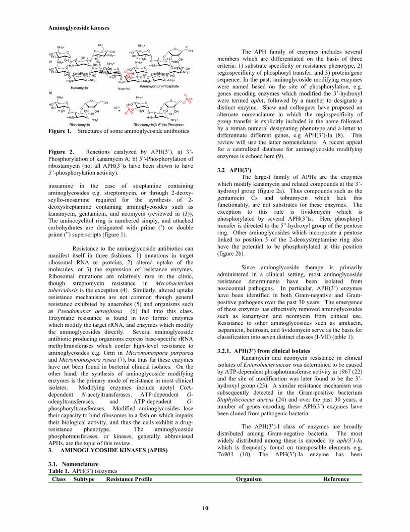
Figure 1. Structures of some aminoglycoside antibiotics
Figure 2. Reactions catalyzed by APH(3’). a) 3’-Phosphorylation of kanamycin A; b) 5”-Phosphorylation ofribostamycin (not all APH(3’)s have been shown to have5”-phosphorylation activity).
inosamine in the case of streptamine containingaminoglycosides e.g. streptomycin, or through 2-deoxy-scyllo-inosamine required for the synthesis of 2-deoxystreptamine containing aminoglycosides such askanamycin, gentamicin, and neomycin (reviewed in (3)).The aminocyclitol ring is numbered simply, and attachedcarbohydrates are designated with prime (’) or doubleprime (”) superscripts (figure 1).
Resistance to the aminoglycoside antibiotics canmanifest itself in three fashions: 1) mutations in targetribosomal RNA or proteins, 2) altered uptake of themolecules, or 3) the expression of resistance enzymes.Ribosomal mutations are relatively rare in the clinic,though streptomycin resistance in Mycobacteriumtuberculosis is the exception (4). Similarly, altered uptakeresistance mechanisms are not common though generalresistance exhibited by anaerobes (5) and organisms suchas Pseudomonas aeruginosa (6) fall into this class.Enzymatic resistance is found in two forms: enzymeswhich modify the target rRNA, and enzymes which modifythe aminoglycosides directly. Several aminoglycosideantibiotic producing organisms express base-specific rRNAmethyltransferases which confer high-level resistance toaminoglycosides e.g. Grm in Micromonospora purpureaand Micromonospora rosea (7), but thus far these enzymeshave not been found in bacterial clinical isolates. On theother hand, the synthesis of aminoglycoside modifyingenzymes is the primary mode of resistance in most clinicalisolates. Modifying enzymes include acetyl CoA-dependent N-acetyltransferases, ATP-dependent O-adenyltransferases, and ATP-dependent O-phosphoryltransferases. Modified aminoglycosides losetheir capacity to bind ribosomes in a fashion which impairstheir biological activity, and thus the cells exhibit a drug-resistance phenotype. The aminoglycosidephosphotransferases, or kinases, generally abbreviatedAPHs, are the topic of this review.3. AMINOGLYCOSIDE KINASES (APHS)
3.1. Nomenclature
The APH family of enzymes includes severalmembers which are differentiated on the basis of threecriteria: 1) substrate specificity or resistance phenotype, 2)regiospecificity of phosphoryl transfer, and 3) protein/genesequence. In the past, aminoglycoside modifying enzymeswere named based on the site of phosphorylation, e.g.genes encoding enzymes which modified the 3’-hydroxylwere termed aphA, followed by a number to designate adistinct enzyme. Shaw and colleagues have proposed analternate nomenclature in which the regiospecificity ofgroup transfer is explicitly included in the name followedby a roman numeral designating phenotype and a letter todifferentiate different genes, e.g APH(3’)-Ia (8). Thisreview will use the latter nomenclature. A recent appealfor a centralized database for aminoglycoside modifyingenzymes is echoed here (9).
3.2 APH(3’)The largest family of APHs are the enzymes
which modify kanamycin and related compounds at the 3’-hydroxyl group (figure 2a). Thus compounds such as thegentamicin Cs and tobramycin which lack thisfunctionality, are not substrates for these enzymes. Theexception to this rule is lividomycin which isphosphorylated by several APH(3’)s. Here phosphoryltransfer is directed to the 5”-hydroxyl group of the pentosering. Other aminoglycosides which incorporate a pentoselinked to position 5 of the 2-deoxystreptamine ring alsohave the potential to be phosphorylated at this position(figure 2b).
Since aminoglycoside therapy is primarilyadministered in a clinical setting, most aminoglycosideresistance determinants have been isolated fromnosocomial pathogens. In particular, APH(3’) enzymeshave been identified in both Gram-negative and Gram-positive pathogens over the past 30 years. The emergenceof these enzymes has effectively removed aminoglycosidessuch as kanamycin and neomycin from clinical use.Resistance to other aminoglycosides such as amikacin,isepamicin, butirosin, and lividomycin serve as the basis forclassification into seven distinct classes (I-VII) (table 1).
3.2.1. APH(3’) from clinical isolatesKanamycin and neomycin resistance in clinical
isolates of Enterobacteriaceae was determined to be causedby ATP-dependent phosphotransferase activity in 1967 (22)and the site of modification was later found to be the 3’-hydroxyl group (23). A similar resistance mechanism wassubsequently detected in the Gram-positive bacteriumStaphylococcus aureus (24) and over the past 30 years, anumber of genes encoding these APH(3’) enzymes havebeen cloned from pathogenic bacteria.
The APH(3’)-I class of enzymes are broadlydistributed among Gram-negative bacteria. The mostwidely distributed among these is encoded by aph(3’)-Iawhich is frequently found on transposable elements e.g.Tn903 (10). The APH(3’)-Ia enzyme has been
Table 1. APH(3’) isozymesClass Subtype Resistance Profile Organism Reference
HO OHO
OOH
NH3+O
OOH
+H3N NH3+
HONH3+
OH
HO
HO O
O+H3N
NH3+
O
+H3NNH3+
O
OH
OH
OHHO
HOO
HO
O+H3N
NH3+
OHOH
+H3N NH3+
3'
Kanamycin A
HO
Ribostamycin
Neamine
4 6 2" 3' 4 5
5"
3' 4 6
HO OHO
OOH
NH3+
O
OOH
+H3N NH3+HO
NH3+OH
HO
OPO--O
OHO O
OOH
NH3+
O
OOH
+H3N NH3+HO
NH3+OH
HO
ATP
Kanamycin Kanamycin(3')-Phosphate
a)
b)
HO O
O+H3N
NH3+
O
+H3N NH3+
O
OH
OH
OHHO
HO
Ribostamycin
HO O
O+H3N
NH3+
O
+H3N NH3+
O
OHOH
HO
Ribostamycin(3',5")bis-Phosphate
OP
O-O-
O
-OP
O-O
OATP

Aminoglycoside kinases
12
I a Kan, Neo, Paro, Rib, Liv, Gent B Escherichia coli (10)b E. coli (11)c Klebsiella pneumonia (12)
II a Kan, Neo, Paro, Rib, But, Gent B E. coli (13)III Kan, Amk, Isep, Neo, Paro, Rib, Liv, But, Gent B Enterococcus and
Staphylococcus(14, 15)
IV a Kan, Neo, Paro, Rib, But Bacillus cirulans (16)V a Kan Neo, Paro, Rib Streptomyces fradiae (17)
b Streptomyces ribosidificus (18)c Micromonospora chalcea (19)
VI a Kan, Neo, Paro, Rib, But, Gent B Acinetobacter baumani (20)VII a Kan, Amk, Isep, Neo, Paro, Rib, Liv, But, Gent B Campylobacter jejuni (21)
Kan, kanamycin; Amk, amikacin; Isep, isepamicin; Neo, neomycin; Paro, paromomycin; Rib, ribostamycin; Liv, lividomycin;But, butirosin; Gent B, gentamicin B.
overexpressed in E. coli, purified and enzymaticallycharacterized by the group of S. Mobashery (25). Theenzyme has a monomer molecular mass of 31 kDa, afeature common to most APH(3’)s. In a fashion analogousto APH(3’)-IIIa (vide infra), APH(3’)-Ia can be isolated asa DTT sensitive dimer, indicative of intermoleculardisulfide bonds. Steady state kinetic analysis demonstrateda broad aminoglycoside substrate specificity as predictedby the resistance phenotype with kcat/Km between 106 and108 M-1 s-1 (25), values approaching the diffusion limit forsmall molecules in solution, thus APH(3’)-Ia is a highlyevolved catalyst.
Two other APH(3’)-I isozymes have recentlybeen described. The first, designated APH(3’)-St, ispresent on the Salmonella typhimurium plasmid NTP16and encoded by transposon derived sequences and isvirtually identical (>95%) to APH(3’)-Ia (26). The second,which we have designated APH(3’)-Id based on itshomology to other type I APH(3’)s, is most closely relatedto APH(3’)-Ib and is encoded on the bacterial,incompatibility group Q, plasmid pIE693 (27).
The APH(3’)-II class of enzymes find frequentuse as a tool in molecular biology. In particular, the neogene derived from Tn5 (13) is a general antibioticresistance marker in wide use for both prokaryotic(kanamycin and neomycin resistance) and eukaryotic(geneticin resistance) studies. The corresponding enzyme,APH(3’)-IIa, has been overexpressed in E. coli, purifiedand characterized (28). The roles of specific amino acidresidues in APH(3’)-IIa has been studied by site directedmutagenesis, in particular His188 which is invariant in allAPHs, was determined to be important by virtue of anincrease in aminoglycoside antibiotic minimal inhibitoryconcentration (MIC) (29, 30) and decrease in enzymeactivity (30). Mutation of Tyr218 to Ser, Asp or Pheresulted in a change in aminoglycoside recognition but notof ATP (31), and Arg211 to His, Lys, and Pro mutationswere determined to alter ATP binding (32). The specificroles for these amino acids can now be inferred using thethree-dimensional structure of APH(3’)-IIIa.
Interestingly, a chromosomally encodedAPH(3’)-IIb has recently been described in Pseudomonas
aeruginosa (33). APH(3’)-IIb is highly homologous toAPH(3’)-IIa (approximately 52% identity) and itschromosomal location in P. aeruginosa may contribute tothe low level resistance to aminoglycoside antibioticsintrinsically associated with Pseudomonas strains.
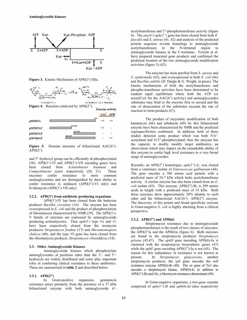
The gene encoding APH(3’)-IIIa has been clonedfrom Enterococcus faecalis (15) and Staphylococcusaureus (14). In E. faecalis, the gene is located on themulti-resistance plasmid pJH1 along with streptomycin andmacrolide resistance determinants. The gene has also beenfound on plasmid pIP1433 in the Gram-negative organismCamplylobacter coli (34). APH(3’)-IIIa has beenoverexpressed in E. coli, purified and characterized (35).Like APH(3’)-Ia, the enzyme can be isolated as a monomeror a kinetically indistinguishable disulfide bridged dimer.The enzyme exhibits a very broad aminoglycoside substraterange with specificity constants (kcat/Km) generally on theorder of 106 M-1s-1. Steady state kinetic analysis of the enzymerevealed a special case of an ordered BiBi mechanism termedTheorell-Chance with ATP binding first followed by theaminoglycoside (figure 3) (36). Release of the phosphorylatedaminoglycoside then precedes release of ADP. The specific caseof a Theorell-Chance mechanism indicates that under steadystate conditions, the chemical conversion of the ternary complex:(ATP•Aminoglycoside•Enz↔ADP•Phospho-aminoglycoside•Enz), does not contribute to the observed maximal rate, kcat.Generally, this implies that second product release is rate-limiting, in this case ADP. The kinetic mechanism wasvalidated and rate-limiting release of ADP demonstratedthrough a series of experiments including solvent isotopeeffects, ATPgammaS thio effect and viscosity effects (37).
In addition, the regiospecificity of phospho-transfer by APH(3’)-IIIa was definitively established bypurification of phosphorylated aminoglycosides followedby detailed analysis by a variety of NMR and mass spectraltechniques (35, 38). Based on these experiments, it wasdetermined that the enzyme phosphorylates 4,6-disubstituted-2-deoxystreptamine aminoglycosides such askanamycin and amikacin exclusively at the 3’-hydroxyl.On the other hand, 4,5-disubstituted-2-deoxystreptaminesuch as lividomycin which lacks a 3’-hydroxyl aresubstrates, but phosphorylation occurs at the 5”-hydroxylgroup of the pentose ring. Aminoglycosides with both a 3’-
Aminoglycoside kinases
13
Figure 3. Kinetic Mechanism of APH(3’)-IIIa.
Figure 4. Reaction catalyzed by APH(2”).
Figure 5. Domain structure of bifunctional AAC(6’)-APH(2”).
and 5”-hydroxyl group can be efficiently di-phosphorylated(38). APH(3’)-VI and APH(3’)-VII encoding genes havebeen cloned from Acinetobacter baumani andCampylobacter jejuni respectively (20, 21). Theseenzymes confer resistance to most commonaminoglycosides and are distinguished by their ability toconfer resistance to amikacin (APH(3’)-VI only) andlividomycin (APH(3’)-VII only).
3.2.2. APH(3’) from antibiotic producing organismsAPH(3’)-IV has been cloned from the butirosin
producer Bacillus circulans (16). The enzyme has beenoverexpressed in E. coli and the product of phosphorylationof ribostamycin characterized by NMR (39). The APH(3’)-V family of enzymes are expressed by aminoglycosideproducing actinomycetes. Thus aph(3’) type Va and Vchave been respectively cloned from the neomycinproducers Streptomyces fradiae (17) and Micromonosporachalcea (40), and the type Vb gene has been cloned fromthe ribostamycin producer, Streptomyces ribosidificus (18).
3.3. Other Aminoglycoside KinasesAminoglycoside kinases which phosphorylate
aminoglycosides at positions other than the 3’- and 5”-hydroxyls are widely distributed and some play importantroles in conferring clinical resistance to these antibiotics.These are summarized in table 2 and described below.
3.3.1. APH(2”)In Gram-positive organisms, gentamicin
resistance arises primarily from the presence of a 57 kDabifunctional enzyme with both aminoglycoside 6’-
acetyltransferase and 2”-phosphotransferase activity (figure4). The aac(6’)-aph(2”) gene has been cloned from both E.faecalis and S. aureus (41, 42) and analysis of the predictedprotein sequence reveals homology to aminoglycosideacetyltransferases in the N-terminal region toaminoglycoside kinases in the C-terminus. Ferretti at al.have prepared truncated gene products and confirmed thepredicted location of the two aminoglycoside modificationactivities (figure 5) (42).
The enzyme has been purified from S. aureus andS. epidermidis (65), and overexpressed in both E. coli (66)and Bacillus subtilis (D. Daigle & G. Wright, in press). Thekinetic mechanisms of both the acetyltransferase andphospho-transferase activities have been determined to berandom rapid equilibrium where both the ATP (oracetylCoA for the AAC(6’) activity) and aminoglycosidessubstrates may bind to the enzyme first or second and therate of dissociation of the substrates exceeds the rate ofreaction to form products (67).
The product of enzymatic modification of bothkanamycin (66) and arbekacin (68) by this bifunctionalenzyme have been characterized by NMR and the predictedregiospecificities confirmed. In addition, both of thesestudies detected some product which was both N-6’-acetylated and O-2”-phosphorylated, thus the enzyme hasthe capacity to doubly modify target antibiotics, anobservation which may impact on the remarkable ability ofthis enzyme to confer high level resistance to a very broadrange of aminoglycosides.
Recently, an APH(2”) homologue, aph(2”)-Ic, was clonedfrom a veterinary isolate of Enterococcus gallinarum (44).The gene encodes a 306 amino acid protein with apredicted mass of 34.7 kDa which lacks acetyltransferaseactivity. A similar enzyme has also been cloned from an E.coli isolate (43). This enzyme, APH(2”)-Ib, is 299 aminoacids in length with a predicted mass of 33 kDa. Boththese enzymes show approximately 20% identity to eachother and the bifunctional AAC(6’)- APH(2”) enzyme.The discovery of this potent and broad specificity enzymein Gram-negative E. coli is highly alarming from a clinicalperspective.
3.3.2. APH(3”) and APH(6)Streptomycin resistance due to aminoglycoside
phosphotransferases is the result of two classes of enzymes,the APH(3”)s and the APH(6)s (figure 6). Both enzymesare found in the streptomycin producer Streptomycesgriseus (45,47). The aphD gene encoding APH(6)-Ia isclustered with the streptomycin biosynthetic genes (47)while the aphE gene encoding APH(3”)-Ia is not (45). Thereason for this redundancy in resistance is not known atpresent. In Streptomyces glaucescens, anotherstreptomycin producer, the sph gene encodes the selfresistance enzyme APH(6)-Ib (48). The str gene of Tn5 alsoencodes a streptomycin kinase, APH(6)-Ic in addition toAPH(3’)-IIa and ble, a bleomycin resistance determinant (49).
In Gram-negative organisms, a two-gene cassettecomprised of aph(3”)-Ib and aph(6)-Id (also respectively

Aminoglycoside kinases
14
Table 2. Other APHsEnzyme (gene) Resistance Profile Organism ReferenceAAC(6’)-APH(2”)-Ia Kan, Tobr, Neo, Liv, Gent C Staphylococci and Enterococci (41, 42)APH(2”)-Ib Kan, Tobr, Neo, Liv, Gent C Escherichia coli (43)APH(2”)-Ic Kan, Tobr, Neo, Liv, Gent C Enterococcus gallinarum (44)APH(3”)-Ia (aphE) Strep Streptomyces griseus (45)APH(3”)-Ib (strA) Strep E. coli (46)APH(6)-Ia (aphD) Strep Streptomyces griseus (47)APH(6)-Ib (sph) Strep Streptomyces glaucescens (48)APH(6)-Ic (str) Strep E. coli (49)APH(6)-Id (strB) Strep E. coli (46)APH(9)-Ia Spect Legionella pneumophila (50)APH(9)-Ib (spcN) Spect Streptomyces flavopersicus (51)APH(4)-Ia Hygr E. coli (52)APH(4)-Ib (glpA) Hygr, Glyphosate Pseudomonas pseudomallei (53)APH(7”)-Ia Hygr Streptomyces hygroscopicus (54)APH(3')-VSr Kan, Neo Streptomyces rimosus (55)Hydroxyurea kinase (hur) hydroxyurea Streptomyces aureofaciens (56)APH-Ll (orf8) unknown Lactococcus lactis subsp. lactis (57)APH-STRN (strN) unknown S. griseus (58)MPH-I (mphA, mphk) erythromycin E. coli (59, 60)MPH-II (mphB ) erythromycin E. coli (61)Viomycin kinase (vph) viomycin Streptomyces vinaceus (62)Capreomycin kinase (cph) capreomycin Streptomyces capreolus (63)MtPH-I unknown Mycobacterium tuberculosis (64)MtPH-II unknown M. tuberculosis (64)MtPH-III unknown M. tuberculosis (64)Kan, kanamycin; Tobr, tobramycin; Neo, neomycin; Liv, lividomycin; Gent C, gentamicin C complex; Strep, streptomycin;Spect, spectinomycin; Hygr, hygromycin.
known as strA and strB) is located on broad host rangeplasmids e.g RSF1010 (46). A recent survey ofenvironmental isolates has shown that the strA-strB genesare widely distributed in the environment (69).
3.3.3. APH(9)Recently, two genes encoding spectinomycin
kinases have been cloned from Legionella pneumophila(50) and the spectinomycin producer Streptomycesflavopersicus (51). The L. pneumophila enzyme has beenoverexpressed in E. coli, characterized, and the product ofspectinomycin phosphorylation determined to beexclusively spectinomycin-9-phosphate by NMR methods(90). Based on these results, we propose that the enzymebe classified as APH(9)-Ia (figure 7).
The S. flavopersicus gene product has also beenshown to phosphorylate spectinomycin, though theregiospecificity of phosphoryl transfer has not beenestablished. As phylogenetic analysis (see section 3.4)reveals that this enzyme and APH(9)-Ia cluster together andgiven the demonstrated site of phospho-transfer in the latterenzyme, we predict that these enzymes will sharespecificities and thus the S. flavopersicus enzyme should bedesignated APH(9)-Ib.
3.3.4. APH(4)Resistance to the aminoglycoside hygromycin is
a useful genetic marker for a number of molecularbiological experiments in both prokaryotes and eukaryotes.The APH(4)-Ia gene has been cloned from E. coli (52), andthe regiospecificity of phosphorylation determined by
NMR (figure 8a) (70). One other hygromycinphosphotransferase has been identified in Pseudomonaspseudomallei, designated here APH(4’)-Ib. This APH(4)-Ia homologue confers tolerance to the herbicide glyphosate(N-phosphonomethylglycine) in E. coli presumably byformation of the acylphosphate which is necessary formetabolism of this inhibitor of 5-enoylpyruvylshikimate-3-phosphate synthase (figure 8b) (53).
3.3.5. APH(7”)Hygromycin resistance in the producing
organism Streptomyces hygroscopicus is conferred by akinase which has been demonstrated to phosphorylate theantibiotic at position 7” (figure 9) (71). The gene has beencloned (54) and the enzyme purified and characterized froman E. coli construct (72, 73).
3.3.6. Other aminoglycoside and miscellaneousphosphotransferases
The sequencing of various genes clusters as wellas bacterial genomes have resulted in the identification ofseveral new genes which either have been confirmed to benovel APHs, or which show significant homology toaminoglycoside kinases. Specifically, kinases which showthe signature catalytic sequence HGD(X)4N, where X is anyamino acid, (see section 4 for discussion of the importance ofthis peptide sequence) have been included here.
An APH(3')-VSr has been cloned fromStreptomyces rimosus (55) and while it mediates resistanceto kanamycin and neomycin, little else is known except that

Aminoglycoside kinases
15
Figure 6. Reactions catalyzed by APH(3”) and APH(6).
Figure 7. Reaction catalyzed by APH(9).
Figure 8. Reaction catalyzed by APH(4).
Figure 9. Reaction catalyzed by APH(7”).
Figure 10. Reactions catalyzed by APH homologues.
APH(3')-VSr shows little homology to other type V3'APHs. In fact it is most closely related to the hygromycinphosphotransferase APH(7")-Ia (see section 3.3.5).
The gene, hur, encoding a hydroxyurea resistanceelement has been cloned from the chlortetracyclineproducer Streptomyces aureofaciens (figure 10a) (56).Expression of the hur gene product in E. coli confersresistance to hydroxyurea, a synthetic inhibitor of DNAsynthesis. Hydroxyurea kinase (HK) is a 340 amino acidprotein which shows approximately 50% identity toAPH(6)-Ia. Despite this significant homology tostreptomycin kinase, HK does not confer resistance toeither streptomycin (≤10 microg/mL) or neomycin,kanamycin and spectinomycin.
A gene encoding an APH-like enzyme (orf8), hasbeen found within the His operon of Lactococcus lactissubsp. lactis. The function of this gene with regard to Hisbiosynthesis is not known at present. The predicted protein,APH-Ll, while weakly homologous to APH(3’) enzymes (13-21% similarity), does not confer resistance to kanamycin,tobramycin, butirosin, lividomycin, neomycin, dibekacin,amikacin, streptomycin and spectinomycin but does contain theexpected catalytic sequence HGDYCLPN (57).
APH-STRN is a 35.6 kDa aminoglycosidephosphotransferase cloned from the streptomycin producerStreptomyces griseus (58). The regiospecificity of thisenzyme has not been established, but APH-STRN is mostclosely related to the APH(9)s. It is thought that StrN maybe involved in the control of streptomycin biosynthesis.
Erythromycin resistance in E. coli can beconferred by two APH-like genes that encode the macrolidephosphotransferase proteins MPH(2')-I and MPH(2')-II(figure 10b) (59-61). These proteins are somewhatdifferent from APHs in that they incorporate a HGD(X)8Dsequence rather than the APH signature HGD(X)4N motif,and thus the general similarity in terms of structure andmechanism remains to be demonstrated. MPH(2')-I is a301 amino acid E. coli protein that in combination withMRX, a protein of unknown function, confers high levelresistance to erythromycin. MPH(2')-II is a 302 amino acidE. coli protein that is 42 % identical to MPH(2')-I. TheMPHs are most homologous to the 2"-aminoglycosidephosphotransferases, particularly in their C-termini.
Also related to the 2"-aminoglycosidephosphotransferases are two kinases which modify thecyclic peptide antibiotic viomycin. The vph gene from theviomycin producer Streptomyces vinaceus encodes a kinase(62), which O-phosphorylates viomycin and the relatedcapreomycins IA and IIA (figure 10c). A related kinasedesignated CK, cloned from Streptomyces capreolus, has asimilar substrate profile but preferentially phosphorylatescapreomycin IA over viomycin (63).
3.3.7. Mycobacterial APHsThree mycobacterial protein sequences,
designated here MtPH-I, MtPH-II, and MtPH-III, wereidentified based on their homology to the known
Aminoglycoside kinases
16
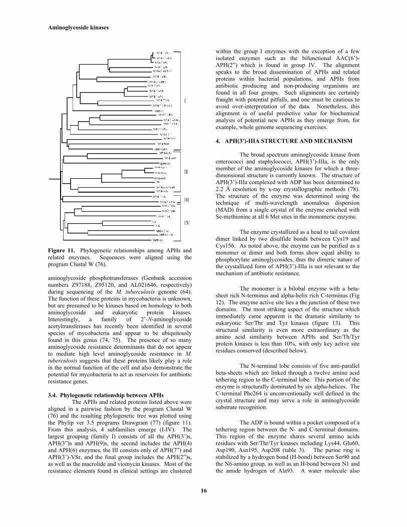
Figure 11. Phylogenetic relationships among APHs andrelated enzymes. Sequences were aligned using theprogram Clustal W (76).
aminoglycoside phosphotransferases (Genbank accessionnumbers Z97188, Z95120, and AL021646, respectively)during sequencing of the M. tuberculosis genome (64).The function of these proteins in mycobacteria is unknown,but are presumed to be kinases based on homology to bothaminoglycoside and eukaryotic protein kinases.Interestingly, a family of 2’-N-aminoglycosideacetyltransferases has recently been identified in severalspecies of mycobacteria and appear to be ubiquitouslyfound in this genus (74, 75). The presence of so manyaminoglycoside resistance determinants that do not appearto mediate high level aminoglycoside resistance in M.tuberculosis suggests that these proteins likely play a rolein the normal function of the cell and also demonstrate thepotential for mycobacteria to act as reservoirs for antibioticresistance genes.
3.4. Phylogenetic relationship between APHsThe APHs and related proteins listed above were
aligned in a pairwise fashion by the program Clustal W(76) and the resulting phylogenetic tree was plotted usingthe Phylip ver 3.5 programs Drawgram (77) (figure 11).From this analysis, 4 subfamilies emerge (I-IV). Thelargest grouping (family I) consists of all the APH(3’)s,APH(3”)s and APH(9)s, the second includes the APH(4)and APH(6) enzymes, the III consists only of APH(7”) andAPH(3’)-VSr, and the final group includes the APH(2”)s,as well as the macrolide and viomycin kinases. Most of theresistance elements found in clinical settings are clustered
within the group I enzymes with the exception of a fewisolated enzymes such as the bifunctional AAC(6’)-APH(2”) which is found in group IV. The alignmentspeaks to the broad dissemination of APHs and relatedproteins within bacterial populations, and APHs fromantibiotic producing and non-producing organisms arefound in all four groups. Such alignments are certainlyfraught with potential pitfalls, and one must be cautious toavoid over-interpretation of the data. Nonetheless, thisalignment is of useful predictive value for biochemicalanalysis of potential new APHs as they emerge from, forexample, whole genome sequencing exercises.
4. APH(3’)-IIIA STRUCTURE AND MECHANISM
The broad spectrum aminoglycoside kinase fromenterococci and staphylococci, APH(3’)-IIIa, is the onlymember of the aminoglycoside kinases for which a three-dimensional structure is currently known. The structure ofAPH(3’)-IIIa complexed with ADP has been determined to2.2 Å resolution by x-ray crystallographic methods (78).The structure of the enzyme was determined using thetechnique of multi-wavelength anomalous dispersion(MAD) from a single crystal of the enzyme enriched withSe-methionine at all 6 Met sites in the monomeric enzyme.
The enzyme crystallized as a head to tail covalentdimer linked by two disulfide bonds between Cys19 andCys156. As noted above, the enzyme can be purified as amonomer or dimer and both forms show equal ability tophosphorylate aminoglycosides, thus the dimeric nature ofthe crystallized form of APH(3’)-IIIa is not relevant to themechanism of antibiotic resistance.
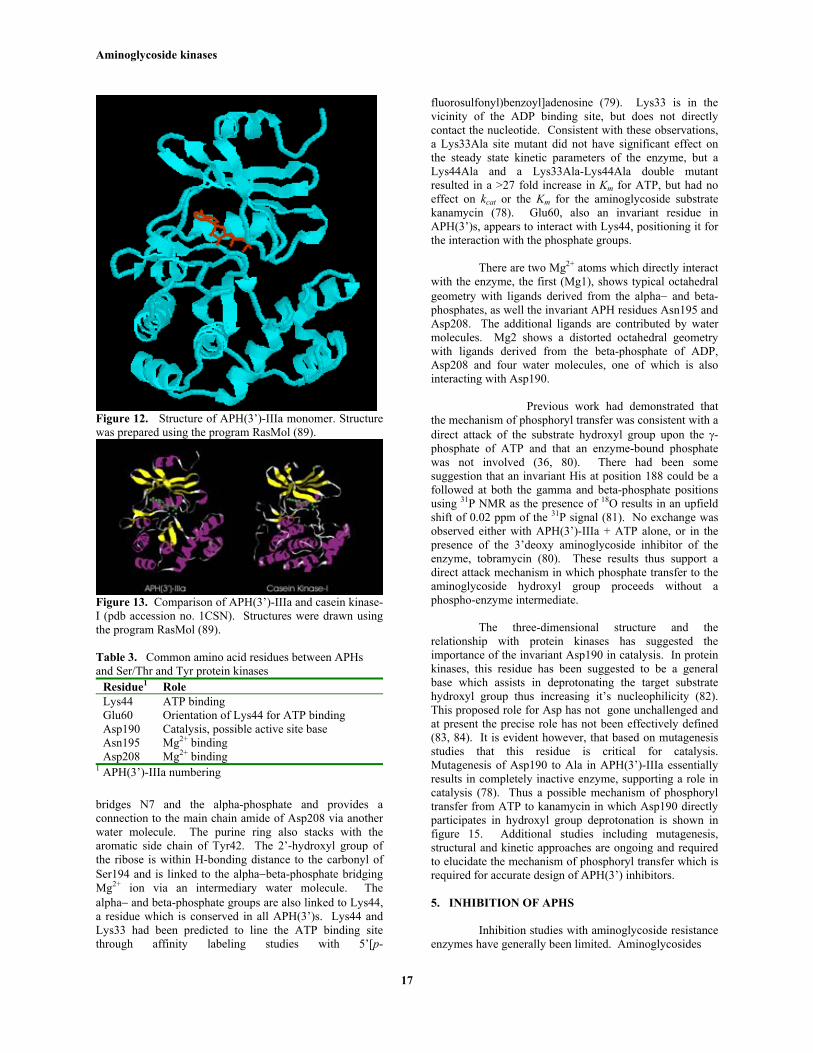
The monomer is a bilobal enzyme with a beta-sheet rich N-terminus and alpha-helix rich C-terminus (Fig12). The enzyme active site lies a the junction of these twodomains. The most striking aspect of the structure whichimmediately came apparent is the dramatic similarity toeukaryotic Ser/Thr and Tyr kinases (figure 13). Thisstructural similarity is even more extraordinary as theamino acid similarity between APHs and Ser/Th/Tyrprotein kinases is less than 10%, with only key active siteresidues conserved (described below).
The N-terminal lobe consists of five anti-parallelbeta-sheets which are linked through a twelve amino acidtethering region to the C-terminal lobe. This portion of theenzyme is structurally dominated by six alpha-helices. TheC-terminal Phe264 is unconventionally well defined in thecrystal structure and may serve a role in aminoglycosidesubstrate recognition.
The ADP is bound within a pocket composed of atethering region between the N- and C-terminal domains.This region of the enzyme shares several amino acidsresidues with Ser/Thr/Tyr kinases including Lys44, Glu60,Asp190, Asn195, Asp208 (table 3). The purine ring isstabilized by a hydrogen bond (H-bond) between Ser90 andthe N6-amino group, as well as an H-bond between N1 andthe amide hydrogen of Ala93. A water molecule also
Aminoglycoside kinases
17
Figure 12. Structure of APH(3’)-IIIa monomer. Structurewas prepared using the program RasMol (89).
Figure 13. Comparison of APH(3’)-IIIa and casein kinase-I (pdb accession no. 1CSN). Structures were drawn usingthe program RasMol (89).
Table 3. Common amino acid residues between APHsand Ser/Thr and Tyr protein kinases
Residue1 RoleLys44 ATP bindingGlu60 Orientation of Lys44 for ATP bindingAsp190 Catalysis, possible active site baseAsn195 Mg2+ bindingAsp208 Mg2+ binding
1 APH(3’)-IIIa numbering
bridges N7 and the alpha-phosphate and provides aconnection to the main chain amide of Asp208 via anotherwater molecule. The purine ring also stacks with thearomatic side chain of Tyr42. The 2’-hydroxyl group ofthe ribose is within H-bonding distance to the carbonyl ofSer194 and is linked to the alpha−beta-phosphate bridgingMg2+ ion via an intermediary water molecule. Thealpha− and beta-phosphate groups are also linked to Lys44,a residue which is conserved in all APH(3’)s. Lys44 andLys33 had been predicted to line the ATP binding sitethrough affinity labeling studies with 5’[p-
fluorosulfonyl)benzoyl]adenosine (79). Lys33 is in thevicinity of the ADP binding site, but does not directlycontact the nucleotide. Consistent with these observations,a Lys33Ala site mutant did not have significant effect onthe steady state kinetic parameters of the enzyme, but aLys44Ala and a Lys33Ala-Lys44Ala double mutantresulted in a >27 fold increase in Km for ATP, but had noeffect on kcat or the Km for the aminoglycoside substratekanamycin (78). Glu60, also an invariant residue inAPH(3’)s, appears to interact with Lys44, positioning it forthe interaction with the phosphate groups.
There are two Mg2+ atoms which directly interactwith the enzyme, the first (Mg1), shows typical octahedralgeometry with ligands derived from the alpha− and beta-phosphates, as well the invariant APH residues Asn195 andAsp208. The additional ligands are contributed by watermolecules. Mg2 shows a distorted octahedral geometrywith ligands derived from the beta-phosphate of ADP,Asp208 and four water molecules, one of which is alsointeracting with Asp190.
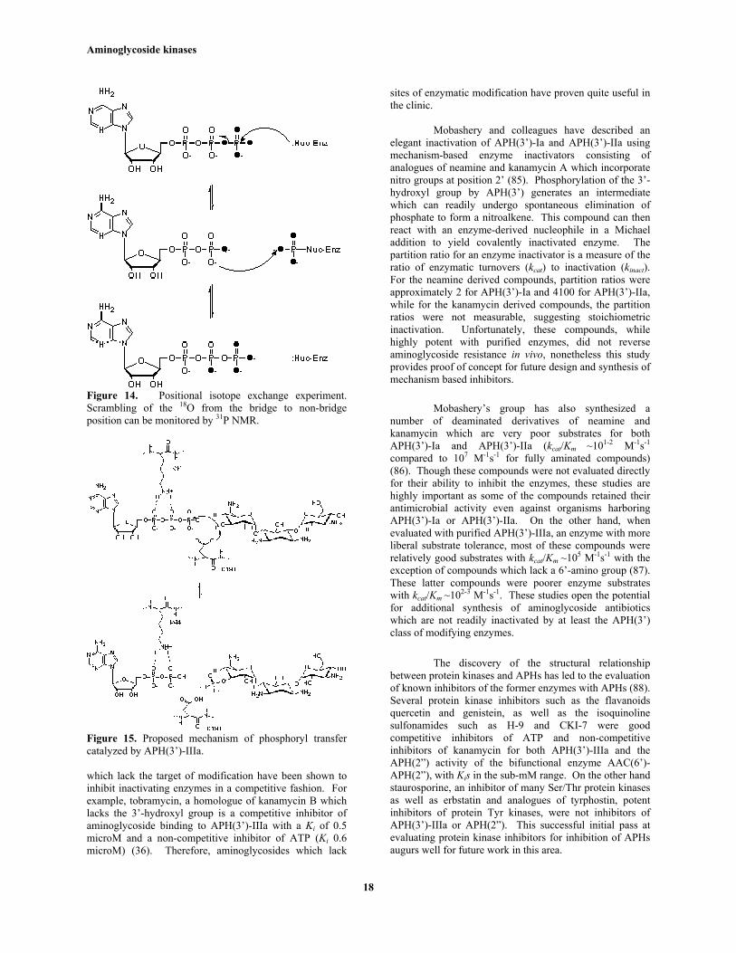
Previous work had demonstrated thatthe mechanism of phosphoryl transfer was consistent with adirect attack of the substrate hydroxyl group upon the γ-phosphate of ATP and that an enzyme-bound phosphatewas not involved (36, 80). There had been somesuggestion that an invariant His at position 188 could be afollowed at both the gamma and beta-phosphate positionsusing 31P NMR as the presence of 18O results in an upfieldshift of 0.02 ppm of the 31P signal (81). No exchange wasobserved either with APH(3’)-IIIa + ATP alone, or in thepresence of the 3’deoxy aminoglycoside inhibitor of theenzyme, tobramycin (80). These results thus support adirect attack mechanism in which phosphate transfer to theaminoglycoside hydroxyl group proceeds without aphospho-enzyme intermediate.
The three-dimensional structure and therelationship with protein kinases has suggested theimportance of the invariant Asp190 in catalysis. In proteinkinases, this residue has been suggested to be a generalbase which assists in deprotonating the target substratehydroxyl group thus increasing it’s nucleophilicity (82).This proposed role for Asp has not gone unchallenged andat present the precise role has not been effectively defined(83, 84). It is evident however, that based on mutagenesisstudies that this residue is critical for catalysis.Mutagenesis of Asp190 to Ala in APH(3’)-IIIa essentiallyresults in completely inactive enzyme, supporting a role incatalysis (78). Thus a possible mechanism of phosphoryltransfer from ATP to kanamycin in which Asp190 directlyparticipates in hydroxyl group deprotonation is shown infigure 15. Additional studies including mutagenesis,structural and kinetic approaches are ongoing and requiredto elucidate the mechanism of phosphoryl transfer which isrequired for accurate design of APH(3’) inhibitors.
5. INHIBITION OF APHS
Inhibition studies with aminoglycoside resistanceenzymes have generally been limited. Aminoglycosides
Aminoglycoside kinases
18
Figure 14. Positional isotope exchange experiment.Scrambling of the 18O from the bridge to non-bridgeposition can be monitored by 31P NMR.
Figure 15. Proposed mechanism of phosphoryl transfercatalyzed by APH(3’)-IIIa.
which lack the target of modification have been shown toinhibit inactivating enzymes in a competitive fashion. Forexample, tobramycin, a homologue of kanamycin B whichlacks the 3’-hydroxyl group is a competitive inhibitor ofaminoglycoside binding to APH(3’)-IIIa with a Ki of 0.5microM and a non-competitive inhibitor of ATP (Ki 0.6microM) (36). Therefore, aminoglycosides which lack
sites of enzymatic modification have proven quite useful inthe clinic.
Mobashery and colleagues have described anelegant inactivation of APH(3’)-Ia and APH(3’)-IIa usingmechanism-based enzyme inactivators consisting ofanalogues of neamine and kanamycin A which incorporatenitro groups at position 2’ (85). Phosphorylation of the 3’-hydroxyl group by APH(3’) generates an intermediatewhich can readily undergo spontaneous elimination ofphosphate to form a nitroalkene. This compound can thenreact with an enzyme-derived nucleophile in a Michaeladdition to yield covalently inactivated enzyme. Thepartition ratio for an enzyme inactivator is a measure of theratio of enzymatic turnovers (kcat) to inactivation (kinact).For the neamine derived compounds, partition ratios wereapproximately 2 for APH(3’)-Ia and 4100 for APH(3’)-IIa,while for the kanamycin derived compounds, the partitionratios were not measurable, suggesting stoichiometricinactivation. Unfortunately, these compounds, whilehighly potent with purified enzymes, did not reverseaminoglycoside resistance in vivo, nonetheless this studyprovides proof of concept for future design and synthesis ofmechanism based inhibitors.
Mobashery’s group has also synthesized anumber of deaminated derivatives of neamine andkanamycin which are very poor substrates for bothAPH(3’)-Ia and APH(3’)-IIa (kcat/Km ~101-2 M-1s-1
compared to 107 M-1s-1 for fully aminated compounds)(86). Though these compounds were not evaluated directlyfor their ability to inhibit the enzymes, these studies arehighly important as some of the compounds retained theirantimicrobial activity even against organisms harboringAPH(3’)-Ia or APH(3’)-IIa. On the other hand, whenevaluated with purified APH(3’)-IIIa, an enzyme with moreliberal substrate tolerance, most of these compounds wererelatively good substrates with kcat/Km ~105 M-1s-1 with theexception of compounds which lack a 6’-amino group (87).These latter compounds were poorer enzyme substrateswith kcat/Km ~102-3 M-1s-1. These studies open the potentialfor additional synthesis of aminoglycoside antibioticswhich are not readily inactivated by at least the APH(3’)class of modifying enzymes.
The discovery of the structural relationshipbetween protein kinases and APHs has led to the evaluationof known inhibitors of the former enzymes with APHs (88).Several protein kinase inhibitors such as the flavanoidsquercetin and genistein, as well as the isoquinolinesulfonamides such as H-9 and CKI-7 were goodcompetitive inhibitors of ATP and non-competitiveinhibitors of kanamycin for both APH(3’)-IIIa and theAPH(2”) activity of the bifunctional enzyme AAC(6’)-APH(2”), with Kis in the sub-mM range. On the other handstaurosporine, an inhibitor of many Ser/Thr protein kinasesas well as erbstatin and analogues of tyrphostin, potentinhibitors of protein Tyr kinases, were not inhibitors ofAPH(3’)-IIIa or APH(2”). This successful initial pass atevaluating protein kinase inhibitors for inhibition of APHsaugurs well for future work in this area.
Aminoglycoside kinases
19
6. EVOLUTION OF APHS
Aminoglycoside resistance determinants musthave either been preexisting or co-evolved withaminoglycoside biosynthesis in antibiotic producingorganisms. The three-dimensional structure of APH(3’)-IIIa has presented the possibility that protein kinases andAPHs share a common evolutionary origin. Furthermore,the fact that protein kinases and APHs share similarcatalytic strategies also supports such a link. We haverecently demonstrated that APHs under certain conditionscan indeed act as Ser protein kinases (91).Aminoglycosides are produced primarily by actinomycetesor bacilli. In recent years, members of both of thesefamilies of organisms have been shown to encodeeukaryotic-like Ser/Thr kinases. The coexistence of bothAPHs and protein kinases in antibiotic producingorganisms places both these genes in the same context andprovides a suggestive link between them. It is therefore notunreasonable to suggest that APHs and protein kinasesevolved from a common ancestor despite the low overallamino acid sequence homology.
In addition, it is clear that based on the sequencesof other antibiotic detoxifying enzymes such as viomycinand hydroxyurea kinases and their similarity to APHs (andthus protein kinases), that the kinase fold and catalyticmechanism (inferred) is one which exhibits broad generalapplication in biology. The sequencing of whole bacterialgenomes has already provided a wealth of new data onpotential new aminoglycoside resistance proteins e.g. inmycobacteria, and APHs in particular. Current work onestablishing the role(s) of some of these cryptic genes inbacterial metabolism will shed light not only on theevolution of the genes currently found in the clinics, butalso will be of value in the prediction of the emergence ofnew resistance determinants in the future.
7. ACKNOWLEDGMENTS
Work described in this review from ourlaboratory was funded through grants from the MedicalResearch Council of Canada. P.R.T. is the recipient of agraduate scholarship from the Natural Sciences andEngineering Research Council of Canada.
8. REFERENCES
1. B. D. Davis, L. L. Chen & P. C. Tai: Misread proteincreates membrane channels: an essential step in thebactericidal action of aminoglycosides. Proc. Natl. Acad.Sci. U S A 83, 6164-6168 (1986)2. E. P. Bakker: Aminoglycoside and aminocyclitolantibiotics: hygromycin B is an atypical bactericidalcompound that exerts effects on cells of Escherichia colicharacteristics for bacteriostatic aminocyclitols. J. Gen.Microbiol. 138, 563-569 (1992)3. W. Piepersberg: Molecular Biology. Biochemistry, andfermentation of aminoglycoside antibiotics. In:Biotechnology of industrial antibiotics. Eds: Strohl W.,Marcel Dekker, New York, 81-163 (1997)
4. J. M. Musser: Antimicrobial agent resistance inmycobacteria: molecular genetic insights. Clin. Microbiol.Rev. 8, 496-514 (1995)5. D. Schlessinger: Failure of aminoglycoside antibioticsto kill anaerobic, low-pH, and resistant cultures. Clin.Microbiol. Rev. 1, 54-59 (1988)6. M. L. Young, M. Bains, A. Bell & R. E. Hancock: Roleof Pseudomonas aeruginosa outer membrane protein OprHin polymyxin and gentamicin resistance: isolation of anOprH-deficient mutant by gene replacement techniques.Antimicrob. Agents Chemother. 36, 2566-2568 (1992)7. G. H. Kelemen, E. Cundliffe & I. Financsek: Cloningand characterization of gentamicin-resistance genes fromMicromonospora purpurea and Micromonospora rosea.Gene 98, 53-60 (1991)8. K. J. Shaw, P. N. Rather, R. S. Hare & G. H. Miller:Molecular genetics of aminoglycoside resistance genes andfamilial relationships of the aminoglycoside-modifyingenzymes. Microbiol. Rev. 57, 138-163 (1993)9. R. Vanhoof, E. Nannecart-Pokorni & J. Content:Nomenclature of genes encoding aminoglycoside-modifying enzymes. Antimicrob. Agents Chemother. 42,483 (1998)10. A. Oka, H. Sugisaki & M. Takanami: Nucleotidesequence of the kanamycin resistance transposon Tn903. J.Mol. Biol. 147, 217-226 (1981)11. W. Pansegrau, L. Miele, R. Lurz & E. Lanka:Nucleotide sequence of the kanamycin resistancedeterminant of plasmid RP4: Homology to otheraminoglycoside 3'-phosphotransferases. Plasmid 18, 193-204 (1987)12. K.-Y. Lee, J. D. Hopkins &M. Syvanen: Evolvedneomycin phosphotransferase from an isolate of Klebsiellapneumonia. Mol. Microbiol. 5, 2039-2046 (1991)13. E. Beck, G. Ludwig, E. A. Auerswald, B. Reiss & H.Schaller: Nucleotide sequence and exact localization of theneomycin phosphotransferase gene from transposon Tn5.Gene 19, 327-336 (1982)14. G. S. Gray & W. M. Fitch: Evolution of antibioticresistance genes: The DNA sequence of a kanamycinresistance gene from Staphylococcus aureus. Mol. Biol.Evol. 1, 57-66 (1983)15. P. Trieu-Cuot & P. Courvalin: Nucleotide sequence ofthe Streptococcus faecalis plasmid gene encoding the 3'5"-aminoglycoside phosphotransferase type III. Gene 23, 331-341 (1983)16. C. J. Herbert, M. Sarwar, S. S. Ner, G. I.G. & M.Akhtar: Sequence and interspecies transfer of anaminoglycoside phosphotransferase gene (APH) of Bacilluscirculans. Self-defense mechanism in antibiotic-producingorganisms. Biochem. J. 233, 383-393 (1986)17. C. J. Thompson & G. S. Gray: Nucleotide sequence ofa streptomycete aminoglycoside phosphotransferase geneand its relationship to phosphotransferases encoded byresistance plasmids. Proc. Natl. Acad. Sci. U S A 80, 5190-5194 (1983)18. S. Hoshiko, C. Nojiri, K. Matsunaga, K. Katsumata, E.Satoh & K. Nagaoka: Nucleotide sequence of theribostamycin phosphotransferase gene and of its controlregion in Streptomyces ribosidificus. Gene 68, 285-296(1988)
Aminoglycoside kinases
20
19. D. Salauze, J.-A. Perez-Gonzalez, W. Piepersberg & J.Davies: Characterization of aminoglycosideacetyltransferase-encoding genes of neomycin-producingMicromonospora chalcea and Streptomyces fradiae. Gene101, 143-148 (1991)20. P. Martin, E. Jullien & P. Courvalin: Nucleotidesequence of Acinetobacter baumannii aphA-6 gene:evolutionary and functional implications of sequencehomologies with nucleotide-binding proteins, kinases andother aminoglycoside-modifying enzymes. Mol. Microbiol.2, 615-625 (1988)21. F. C. Tenover, T. Gilbert & P. O'Hara: Nucleotidesequence of a novel kanamycin resistance gene, aphA-7,from Campylobacter jejuni and comparison to otherkanamycin phosphotransferase genes. Plasmid 22, 52-58(1988)22. H. Umezawa, M. Okanishi, S. Kondo, K. Hamana, R.Utahara, K. Maeda & S. Mitsuhashi: Phosphorylativeinactivation of aminoglycoside antibiotics by Escherichiacoli carrying R factor. Science 157, 1559-1561 (1967)23. S. Kondo, M. Okanishi, R. Utahara, K. Maeda & M.Okanishi: Isolation of kanamycin and paromamineinactivated by E. coli carrying R factor. J. Antibiot. 21, 22-29 (1968)24. O. Doi, M. Miyamoto, N. Tanaka & H. Umezawa:Inactivation and phosphorylation of kanamycin by drug-resistant Staphylococcus aureus. Appl. Microbiol. 16, 1282-1284 (1968)25. J. J. Siregar, K. Miroshnikov & S. Mobashery:Purification, characterization, and investigation of themechanism of aminoglycoside 3'-phosphotransferase TypeIa. Biochemistry 34, 12681-12688 (1995)26. P. M. Cannon & P. Strike: Complete nucleotidesequence and gene organization of plasmid NTP16.Plasmid 27, 220-230 (1992)27. E. Tietze & J. Brevet: Nucleotide sequence of thebacterial Streptothricin resistance gene Sat 3. Biochim.Biophys. Acta. 1263, 176-178 (1995)28. J. J. Siregar, S. A. Lerner & S. Mobashery:Purification and characterization of aminoglycoside 3'-phosphotransferase Type IIa and kinetic comparison with anew mutant enzyme. Antimicrob. Agents Chemother. 38,641-647 (1994)29. J. Blázquez, J. Davies & F. Moreno: Mutations in theaphA-2 gene of transposon Tn5 mapping within the regionshighly conserved in aminoglycoside-phosphotransferasesstrongly reduce aminoglycoside resistance. Mol. Microbiol.5, 1511-1518 (1991)30. S. Kocabiyik & M. H. Perlin: Site-specific mutationsof conserved C-terminal residues in aminoglycoside 3'-phosphotransferase II: Phenotypic and structural analysis ofmutant enzymes. Biochem. Biophys. Res. Commun. 185,925-931 (1992)31. S. Kocabiyik & M. H. Perlin: Altered substratespecificity by substitutions at Tyr218 in bacterialaminoglycoside 3'-phosphotransferase. FEMS Microbiol.Lett. 93, 199-202 (1992)32. S. Kocabiyik & M. H. Perlin: Amino acidsubstitutions within the analogous nucleotide binding loop(P-loop) of aminoglycoside 3'-phosphotransferase-II. Int. J.Biochem. 26, 61-66 (1994)
33. H. Hachler, Santarnam, P., & Kayser, F.H.: Sequenceand characterization of a novel chromosomalaminoglycoside phosphotransferase gene aph(3')-IIb inPseudomonas aeruginosa. Antimicrob. Agents Chemother.40, 1254-1256 (1996)34. S. S. Taylor, D. R. Knighton, J. Zheng, L. F. Ten Eyck& J. M. Sowadski: Structural framework for the proteinkinase family. Annu. Rev. Cell Biol. 8, 429-462 (1992)35. G. A. McKay, P. R. Thompson & G. D. Wright:Broad spectrum aminoglycoside phosphotransferase typeIII from Enterococcus: Overexpression, purification, andsubstrate specificity. Biochemistry 33, 6936-6944 (1994)36. G. A. McKay & G. D. Wright: Kinetic mechanism ofaminoglycoside phosphotransferase type IIIa: Evidence fora Theorell-Chance mechanism. J. Biol. Chem. 270, 24686-24692 (1995)37. G. A. McKay & G.D. Wright : Catalytic mechanismof enterococcal kanamycin kinase (APH(3')-IIIa):Viscosity, thio, and solvent isotope effects support aTheorell-Chance mechanism. Biochemistry 35, 8680-8685(1996)38. P. R. Thompson, D. W. Hughes & G. D. Wright:Regiospecificity of aminoglycoside phosphotransferasefrom Enterococci and Staphylococci (APH(3')-IIIa).Biochemistry 35, 8686-8695 (1996)39. M. Sarwar & M. Akhtar: Cloning of aminoglycosidephosphotransferase (APH) gene from antibiotic-producingstrain of Bacillus circulans into a high-expression vector,pKK223-3. Purification, properties and location of theenzyme. Biochem. J. 268, 671-677 (1990)40. D. Salauze & J. Davies: Isolation and characterizationof an aminoglycoside phosphotransferase from neomycin-producing Micromonospora chalcea: Comparison with thatof Streptomyces fradiae and other producers of 4,6-disubstituted 3-deoxystreptamine antibiotics. J. Antibiot.44, 1432-1443 (1991)41. D. A. Rouch, M. E. Byrne, Y. C. Kong & R. A.Skurray: The aacA-aphD gentamicin and kanamycinresistance determinant of Tn4001 from Staphylococcusaureus: Expression and nucleotide sequence analysis. J.Gen. Microbiol. 133, 3039-3052 (1987)42. J. J. Ferretti, K. S. Gilmore & P. Courvalin:Nucleotide sequence analysis of the gene specifying thebifunctional 6'-aminoglycoside acetyltransferase 2"-aminoglycoside phosphotransferase enzyme inStreptococcus faecalis and identification and cloning ofgene regions specifying the two activities. J. Bacteriol. 167,631-638 (1986)43. K. J. Shaw: Personal communication. (1997)44. J. W. Chow, M. J. Zervos, S. A. Lerner, L. A. Thal, S.M. Donabedian, D. D. Jaworski, S. Tsai, K. J. Shaw & D.B. Clewell: A novel gentamicin resistance gene inEnterococcus. Antimicrob. Agents Chemother. 41, 511-514(1997)45. P. Heinzel, O. Werbitzky, J. Distler & W. Piepersberg:A second streptomycin resistance gene from Streptomycesgriseus codes for streptomycin-3"-phosphotransferase.Relationships between antibiotic and protein kinases. Arch.Microbiol. 150, 184-192 (1988)46. P. Scholz, V. Haring, B. Wittmann-Liebold, K.Ashman, M. Bagdasarian & E. Scherzinger: Complete
Aminoglycoside kinases
21
nucleotide sequence and gene organization of the broad-host-range plasmid RSF1010. Gene 75, 271-288 (1989)47. J. Distler, C. Bräun, A. Ebert & W. Piepersberg: Genecluster for streptomycin biosynthesis in Streptomycesgriseus: analysis of a central region including the majorresistance gene. Mol. Gen. Genet. 208, 204-210 (1987)48. M. Vögtli & R. Hütter: Characterisation of thehydroxystreptomycin phosphotransferase gene (sph) ofStreptomyces glaucescens: nucleotide sequence andpromoter analysis. Mol. Gen. Genet. 208, 195-203 (1987)49. P. Mazodier, P. Cossart, E. Giraud & F. Gasser:Completion of the nucleotide sequence of the central regionof Tn5 confirms the presence of three resistance genes.Nucleic Acids Res. 13, 195-205 (1985)50. T. M. Suter, V. K. Viswanathan & N. P. Cianciotto:Isolation of a gene encoding a novel spectinomycinphosphotransferase from Legionella pneumophila.Antimicrob. Agents Chemother. 41, 1385-1388 (1997)51. D. Lyutzkanova, J. Distler & J. Altenbuchner: Aspectinomycin resistance determinant from thespectinomycin producer Streptomyces flavopersicus.Microbiology 143, 2135-2143 (1997)52. L. Gritz & J. Davies: Plasmid-encoded hygromycin Bresistance: the sequence of hygromycin Bphosphotransferase gene and its expression in Escherichiacoli and Saccharomyces cerevisiae. Gene 25, 179-188(1983)53. A. Peñaloza-Vazquez, G. L. Mena, L. Herrera-Estrella& A. M. Bailey: Cloning and sequencing of genes involvedin glyphosphate utilization by Pseudomonas pseudomallei.Appl. Environ. Microbiol. 161, 538-543 (1995)54. M. Zalacain, A. Gonzalez, M. C. Guerrero, R. J.Mattaliano, F. Malpartida & A. Jimenez: Nucleotidesequence of the hygromycin B phosphotransferase genefrom Streptomyces hygroscopicus. Nucleic Acids Res. 14,1565-1581 (1986)55. K. E. Akopiants & V. N. Danilenko: Instability of thegenome and 'silent' genes in Actinomycetes. DirectSubmission (1995)56. J. Kormanec, M. Farkasovsky, L. Potuchkova & S.Godar: A gene (hur) fromStreptomyces aureofaciensconferring resistance to hydroxyurea is related to genesencoding streptomycin phosphotransferases. Gene 114,133-137 (1992)57. C. Delorme, S. D. Ehrlich & P. Renault: Histidinebiosynthesis genes in Lactococcus lactis subsp. lactis. J.Bacteriol. 174, 6571-6579 (1992)58. K. Pissowotzki, K. Mansouri & W. Piepersberg:Genetics of streptomycin production in Streptomycesgriseus: molecular structure and putative function of genesstrELMB2N. Mol. Gen. Genet. 231, 113-123 (1991)59. N. Noguchi, A. Emura, H. Matsuyama, K. O'Hara, M.Sasatsu & M. Kono: Nucleotide sequence andcharacterization of erythromycin resistance determinantthat encodes macrolide 2'-phosphotransferase-I inEscherichia coli. Antimicrob. Agents Chemother. 39, 2359-2363 (1995)60. S. Kim & E. Choi: Nucleotide sequence, expressionand transcriptional analysis of the Escherichia coli mphKgene encoding a macrolide phosphotransferase K. Mol.Cells 6, 153-160 (1996)
61. N. Noguchi, J. Katayama & K. O'Hara: Cloning andnucleotide sequence of the mphB gene for macrolide 2'-phosphotransferase-II in Escherichia coli. FEMSMicrobiol. Lett. 144, 197-202 (1996)62. M. J. Bibb, M. J. Bibb, J. M. Ward & S. N. Cohen:Nucleotide sequences encoding and promoting expressionof three antibiotic resistance genes indigenous toStreptomyces. Mol. Gen. Genet. 199, 26-36 (1985)63. A. S. Thiara & E. Cundliffe: Analysis of twocapreomycin resistance determinants from Streptomycescapreolus and characterization of the action of theirproducts. Gene 167, 121-126 (1995)64. W. J. Philipp, S. Poulet, K. Eiglmeier, L. Pascopella,V. Balasubramanian, B. Heym, S. Bergh, B. R. Bloom, W.R. J. Jacobs & S. T. Cole: An integrated map of thegenome of the tubercle bacillus, Mycobacteriumtuberculosis H37Rv, and comparison with Mycobacteriumleprae. Proc. Natl. Acad. Sci. USA 93, 3132-3137 (1996)65. K. Ubukata, N. Yamashita, A. Gotoh & M. Konno:Purification and characterization of aminoglycoside-modifying enzymes from Staphylococcus aureus andStaphylococcus epidermidis. Antimicrob. AgentsChemother. 25, 754-759 (1984)66. E. Azucena, I. Grapsas & S. Mobashery: Properties ofa bifunctional bacterial antibiotic resistance enzyme thatcatalyzes ATP-dependent 2"-phosphorylation and acetyl-CoA-dependent 6'-acetylation of aminoglycosides. J. Am.Chem. Soc. 119, 2317-2318 (1997)67. A. Martel, M. Masson, N. Moreau & F. L. Goffic:Kinetic studies of aminoglycoside acetyltransferase andphosphotransferase from Staphylococcus aureus RPAL.Eur. J. Biochem. 133, 515-521 (1983)68. S. Kondo, A. Tamura, S. Gomi, Y. Ikeda, T. Takeuchi& S. Mitsuhashi: Structures of enzymatically modifiedproducts of arbekacin by methicillin-resistantStaphylococcus aureus. J. Antibiot. 46, 310-315 (1993)69. G. W. Sundin &C. L. Bender: Dissemination of thestrA-strB streptomycin-resistance genes among commensaland pathogenic bacteria from humans, animals, and plants.Mol. Ecol. 5, 133-143 (1996)70. R. N. Rao, N. E. Allen, J. N. J. Hobbs, W. E. J. Alborn,H. A. Kirst & J. W. Paschal: Genetic and enzymatic basisof hygromycin B resistance in Escherichia coli.Antimicrob. Agents Chemother. 24, 689-695 (1983)71. J. M. Pardo, F. Malpartida, M. Rico & A. Jimenez:Biochemical basis of resistance to hygromycin B inStreptomyces hygroscopicus--the producing organism. J.Gen. Microbiol. 131, 1289-1298 (1985)72. M. Zalacain, F. Malpartida, D. Pulido & A. Jimenez:Cloning and expression in Escherichia coli of ahygromycin B phosphotransferase gene from Streptomyceshygroscopicus. Eur. J. Biochem. 162, 413-418 (1987)73. M. Zalacain, J. M. Pardo & A. Jimenez: Purificationand characterization of a hygromycin B phosphotransferasefrom Streptomyces hygroscopicus. Eur. J. Biochem. 162,419-422 (1987)74. J. A. Aínsa, C. Martin, B. Gicquel & R. Gomez-Lus:Characterization of the chromosomal aminoglycoside 2'-N-acetyltransferase gene from Mycobacterium fortuitum.Antimicrob. Agents Chemother. 40, 2350-2355 (1996)75. J. A. Aínsa, E. Pérez, V. Pelicic, F. X. Berthet, B.Gicquel & C. Martín: Aminoglycoside 2'-N-
Aminoglycoside kinases
22
acetyltransferase genes are universally present inmycobacteria: characterization of the aac(2')-Ic gene fromMycobacterium tuberculosis and the aac(2')-Id gene fromMycobacterium smegmatis. Mol. Microbiol. 24, 431-441(1997)76. J. D. Thompson, D. G. Higgins & T. J. Gibson:CLUSTAL W: improving the sensitivity of progressivemultiple sequence alignment through sequence weighting,position-specific gap penalties and weight matrix choice.Nucl. Acids Res. 22, 4673-4680 (1994)77. J. Felsenstein: PHYLIP (Phylogeny InferencePackage) version 3.5c. (1993)78. W. C. Hon, G. A. McKay, P. R. Thompson, R. M.Sweet, D. S. C. Yang, G. D. Wright & A. M. Berghuis:Structure of an enzyme required for aminoglycosideresistance reveals homology to eukariotic protein kinases.Cell 89, 887-895 (1997)79. G. A. McKay, R. A. Robinson, W. S. Lane & G. D.Wright: Active-site labeling of an aminoglycosideantibiotic phosphotransferase (APH(3')-IIIa). Biochemistry33, 14115-14120 (1994)80. P. R. Thompson, D. W. Hughes & G. D. Wright:Mechanism of aminoglycoside 3'-phosphotransferase typeIIIa:His188 is not a phosphate-accepting residue. Chem.Biol. 3, 747-755 (1996)81. J. J. Villafranca: Positional isotope exchange usingphosphorus-31 nuclear magnetic resonance. Methods.Enzymol. 177, 390-403 (1989)82. Madhusudan, E. A. Trafny, N.-H. Xuong, J. A. Adams,L. F. Ten Eyck, S. S. Taylor & J. M. Sowadski: cAMP-dependent protein kinase: Crystallographic insights intosubstrate recognition and phosphotransfer. Prot. Sci. 3,176-187 (1994)83. P. A. Cole, M. R. Grace, R. S. Phillips, P. Burn & C.T. Walsh: The role of the catalytic base in the proteintyrosine kinase Csk. J. Biol. Chem. 270, 22105-22108(1995)84. J. Zhou & J. A. Adams: Is there a catalytic base in theactive site of cAMP-dependent protein kinase?Biochemistry 36, 2977-84 (1997)85. J. Roestamadji, I. Grapsas & S. Mobashery:Mechanism-based inactivation of bacterial aminoglycoside3'-phosphotransferases. J. Am. Chem. Soc. 117, 80-84(1995)86. J. Roestamadji, I. Grapsas & S. Mobashery: Loss ofindividual electrostatic interactions betweenaminoglycoside antibiotics and resistance enzymes as aneffective means to overcoming bacterial drug resistance. J.Am. Chem. Soc. 117, 11060-11069 (1995)87. G. A. McKay, J. Roestamadji, S. Mobashery & G. D.Wright: Recognition of aminoglycoside antibiotics byenterococcal-staphylococcal aminoglycoside 3'-phosphotransferase type IIIa: Role of substrate aminogroups. Antimicrob. Agents Chemother. 40, 2648-2650(1996)88. D. M. Daigle, G. A. McKay & G. D. Wright:Inhibition of aminoglycoside antibiotic resistance enzymesby protein kinase inhibitors. J. Biol. Chem. 272, 24755-24758 (1997)89. R. Sayle & E. J. Milner-White: RasMol: Biomoleculargraphics for all. Trends Biochem. Sci. 20, 374 (1995)
90. P.R. Thompson, D.W. Hughes, N.P. Cianciotto & G.D.Wright: Spectinomycin Kinase from Legionellapneomophila. Characterization of substrate specificity andidentification of catalytically important residues. J. Biol.Chem. 273, 14788-14795 (1998)91. D.M. Daigle, G.A. McKay, P.R. Thompson & G.D.Wright: Aminoglycoside antibiotic phosphotransferasesare also serine protein kinases. Chem. & Biol. 6, 11-18(1998)
Key Words: Aminoglycoside, Antibiotic, Resistance,Phosphoryltransfer, Inhibition, Kinase, EnzymeMechanism
Send correspondence to: Dr. G.D. Wright, Department ofBiochemistry, McMaster University, 1200 Main StreetWest, Hamilton, Ontario, Canada L8N 3Z5, Tel: (905)-525-9140, Ext. 22943, Fax: (905)-522-9033, E-mail:[email protected]
Received 3/31/98 Accepted 6/2/98
![[Frontiers in Bioscience 4, d713-730, October 15, 1999 ... · [Frontiers in Bioscience 4, d713-730, October 15, 1999] 713 POTENTIAL REGULATION OF CARTILAGE METABOLISM IN OSTEOARTHRITIS](https://img.pdfslide.net/doc/110x75/5e539caa773adc12c06e031c/frontiers-in-bioscience-4-d713-730-october-15-1999-frontiers-in-bioscience.jpg)
![[Frontiers in Bioscience 14, 5291-5338, June 1, 2009 ... [Frontiers in Bioscience 14, 5291-5338, June 1, 2009] 5291 Neurobiology of depression, fibromyalgia and neuropathic pain Vladimir](https://img.pdfslide.net/doc/110x75/5f4d827d68593756d475cb0a/frontiers-in-bioscience-14-5291-5338-june-1-2009-frontiers-in-bioscience.jpg)
![[Frontiers in Bioscience 8, e172-189, January 1, 2003 ... Front Biosci 2003.pdf · [Frontiers in Bioscience 8, e172-189, January 1, 2003] 172 ACUTE VERSUS CHRONIC FUNCTIONAL ASPECTS](https://img.pdfslide.net/doc/110x75/5f1aacf81b888b2bd7546847/frontiers-in-bioscience-8-e172-189-january-1-2003-front-biosci-2003pdf.jpg)


![[Frontiers in Bioscience 14, 1337-1361, January 1, 2009] … [Frontiers in Bioscience 14, 1337-1361, January 1, 2009] 1337 Biochemical properties of plasminogen activator inhibitor-1](https://img.pdfslide.net/doc/110x75/5f31d3bd17e984560260dc0a/frontiers-in-bioscience-14-1337-1361-january-1-2009-frontiers-in-bioscience.jpg)
![[Frontiers in Bioscience 13, 2757-2773, January 1, 2008] … · 2009-03-16 · [Frontiers in Bioscience 13, 2757-2773, January 1, 2008] 2757 Fundamental principles and applications](https://img.pdfslide.net/doc/110x75/5f46768ef970013bc94661bf/frontiers-in-bioscience-13-2757-2773-january-1-2008-2009-03-16-frontiers.jpg)
![[Frontiers in Bioscience 10, 1946-1960, May 1, 2005 ...library.ibp.ac.cn/html/slwj/000232319800077.pdf · [Frontiers in Bioscience 10, 1946-1960, May 1, 2005] 1946 APPLICATION OF](https://img.pdfslide.net/doc/110x75/5ac1962e7f8b9a1c768cf131/frontiers-in-bioscience-10-1946-1960-may-1-2005-frontiers-in-bioscience.jpg)
![[Frontiers in Bioscience 5212-5240, May 1, 2008] Origins ... · [Frontiers in Bioscience 5212-5240, May 1, 2008] 5212 Origins and evolution of modern biochemistry: insights from genomes](https://img.pdfslide.net/doc/110x75/5ecfff36d3ee0724a0699851/frontiers-in-bioscience-5212-5240-may-1-2008-origins-frontiers-in-bioscience.jpg)
![[Frontiers in Bioscience E3, 788-800, January 1, 2011 ...€¦ · [Frontiers in Bioscience E3, 788-800, January 1, 2011] 788 Dental pulp and dentin tissue engineering and regeneration:](https://img.pdfslide.net/doc/110x75/5f3dc5968fe42175d60d313f/frontiers-in-bioscience-e3-788-800-january-1-2011-frontiers-in-bioscience.jpg)
![[Frontiers in Bioscience 5212-5240, May 1, 2008] Origins and ......[Frontiers in Bioscience 5212-5240, May 1, 2008] 5212 Origins and evolution of modern biochemistry: insights from](https://img.pdfslide.net/doc/110x75/5f49005b20d86c7fe45afa3c/frontiers-in-bioscience-5212-5240-may-1-2008-origins-and-frontiers.jpg)
![[Frontiers in Bioscience, 3, d208-236, February 15, 1998 ...[Frontiers in Bioscience, 3, d208-236, February 15, 1998] 208 CELL-CELL COMMUNICATION IN CARCINOGENESIS James E. Trosko1,Randall](https://img.pdfslide.net/doc/110x75/5e7a41b6970d6f051943d6e3/frontiers-in-bioscience-3-d208-236-february-15-1998-frontiers-in-bioscience.jpg)
![[Frontiers in Bioscience 16, 1663-1674, January 1, 2011 ... › a1a2 › 2bf5cf072987... · [Frontiers in Bioscience 16, 1663-1674, January 1, 2011] 1663 Inflammatory markers and](https://img.pdfslide.net/doc/110x75/5f03c4f17e708231d40aae1b/frontiers-in-bioscience-16-1663-1674-january-1-2011-a-a1a2-a-2bf5cf072987.jpg)
![[Frontiers in Bioscience 8, e94-109, January 1, 2003] …...[Frontiers in Bioscience 8, e94-109, January 1, 2003] 94 CHAGAS’ HEART DISEASE: CLINICAL-PATHOLOGICAL CORRELATION Marcos](https://img.pdfslide.net/doc/110x75/5fddb75a5b2d67635b578662/frontiers-in-bioscience-8-e94-109-january-1-2003-frontiers-in-bioscience.jpg)
![[Frontiers in Bioscience E4, 2085-2100, January 1, 2012 ... · [Frontiers in Bioscience E4, 2085-2100, January 1, 2012] 2085 Histology of epiphyseal cartilage calcification and endochondral](https://img.pdfslide.net/doc/110x75/5e8f2217c77359741c35360b/frontiers-in-bioscience-e4-2085-2100-january-1-2012-frontiers-in-bioscience.jpg)

![[Frontiers in Bioscience 17, 1108-1119, January 1, …...[Frontiers in Bioscience 17, 1108-1119, January 1, 2012] 1108 Histamine in two component system-mediated bacterial signaling](https://img.pdfslide.net/doc/110x75/5f0567197e708231d412c98b/frontiers-in-bioscience-17-1108-1119-january-1-frontiers-in-bioscience.jpg)
![[Frontiers in Bioscience 11, 2179-2192, September 1, 2006]](https://img.pdfslide.net/doc/110x75/62356e1e67d47524e43cfd5f/frontiers-in-bioscience-11-2179-2192-september-1-2006.jpg)

![[Frontiers In Bioscience, Elite, 11, 109-120, Jan 1, 2019]](https://img.pdfslide.net/doc/110x75/616a66ee11a7b741a3521a58/frontiers-in-bioscience-elite-11-109-120-jan-1-2019.jpg)

![[Frontiers in Bioscience, Landmark, 26, 478-495, Jan 1, 2021]](https://img.pdfslide.net/doc/110x75/619139c2adb37e26e40b75b4/frontiers-in-bioscience-landmark-26-478-495-jan-1-2021.jpg)

![[Frontiers in Bioscience 13, 7096-7114, May 1, 2008] Early ......[Frontiers in Bioscience 13, 7096-7114, May 1, 2008] 7096 Early signals after stretch leading to cardiac hypertrophy](https://img.pdfslide.net/doc/110x75/5ff8ec139b3a430a6641433a/frontiers-in-bioscience-13-7096-7114-may-1-2008-early-frontiers-in.jpg)