Embed Size (px)
Citation preview

CORRECTION
A novel method to study contact inhibition of locomotion usingmicropatterned substratesElena Scarpa, Alice Roycroft, Eric Theveneau, Emmanuel Terriac, Matthieu Piel and Roberto Mayor
There was an error published in Biol. Open 2, 901-906.
In Fig. 4D, the y-axis scale was incorrect; the correct Fig. 4 is shown below. There are no changes to the figure legend, which is accurate.This error does not affect the conclusions of the paper.
The authors apologise to the readers for any confusion that this error might have caused.
Fig. 4. 1D-substrates increase the probability of successfulcollision and provide a simpler readout of CIL. (a) Diagramshowing the possible outcomes of cell–cell collisions onfibronectin lines. (b) Examples showing the different outcomesdepicted in panel A. When CIL occurs it leads to completerepolarization of the direction of migration with cells moving awayfrom each other (ai,bi). Alternatively, cells can fail to dissociateafter contact and adhere to each other (aii,bii). Finally, cells maynot react to their physical contact and walk past each otherfollowing their original path of migration (aiii,biii). (c) Percentage ofcell–cell collisions in which both colliding cells repolarize uponcontact in 2D or 1D cultures (*P<0.05). (d) Percentages of NCcells displaying CIL, adhesion or Walk-Past behavior in controlconditions or upon Wnt/PCP inhibition (DEP+) or Rho Kinaseinhibition (Y-27632). *P<0.05; **P<0.01. (e) Distance betweennuclei of colliding cells 30 minutes after initial contact in controlconditions or upon DEP+ or Y-27632 treatment (**P<0.01).Scale bar: 10 μm (b).
This is an Open Access article distributed under the terms of the Creative Commons Attribution License (http://creativecommons.org/licenses/by/3.0), which permits unrestricted use, distribution andreproduction in any medium provided that the original work is properly attributed.
1553
© 2016. Published by The Company of Biologists Ltd | Biology Open (2016) 5, 1553 doi:10.1242/bio.020917
BiologyOpen
by guest on May 25, 2018http://bio.biologists.org/Downloaded from

A novel method to study contact inhibition oflocomotion using micropatterned substrates
Elena Scarpa1, Alice Roycroft1, Eric Theveneau1, Emmanuel Terriac2, Matthieu Piel2 and Roberto Mayor1,*1Department of Cell and Developmental Biology, University College London, Gower Street, London WC1E 6BT, UK2Institut Curie, CNRS UMR144, 26 rue d’Ulm, 75248 Paris Cedex 05, France
*Author for correspondence ([email protected])
Biology Open 2, 901–906doi: 10.1242/bio.20135504Received 14th May 2013Accepted 7th June 2013
SummaryThe concept of contact inhibition of locomotion (CIL) describes
the ability of a cell to change the direction of its movement
after contact with another cell. It has been shown to be
responsible for physiological and developmental processes such
as wound healing, macrophage dispersion and neural crest cell
migration; whereas its loss facilitates cancer cell invasion and
metastatic dissemination. Different assays have been developed
to analyze CIL in tissue culture models. However, these
methods have several caveats. Collisions happen at low
frequency between freely migrating cells and the orientation
of the cells at the time of contact is not predictable. Moreover,
the computational analysis required by these assays is often
complicated and it retains a certain degree of discretion. Here,
we show that confinement of neural crest cell migration on a
single dimension by using a micropatterned substrate allows
standardized and predictable cell–cell collision. CIL can thus
easily be quantified by direct measurement of simple cellular
parameters such as the distance between nuclei after collision.
We tested some of the signaling pathways previously identified
as involved in CIL, such as small GTPases and non-canonical
Wnt signaling, using this new method for CIL analysis. The
restricted directionality of migration of cells in lines is a
powerful strategy to obtain higher predictability and higher
efficiency of the CIL response upon cell–cell collisions.
� 2013. Published by The Company of Biologists Ltd. This is an
Open Access article distributed under the terms of the Creative
Commons Attribution License (http://creativecommons.org/
licenses/by/3.0), which permits unrestricted use, distribution
and reproduction in any medium provided that the original
work is properly attributed.
Key words: Contact inhibition of locomotion, Neural crest,
Micropatterned fibronectin substrates
IntroductionMore than five decades ago, Abercrombie and Heaysman found
that the direction of migration of fibroblasts cultured in vitro was
affected by their interaction with other cells (Abercrombie and
Heaysman, 1953). The process was named contact inhibition of
locomotion (CIL) and it was proposed as the main force driving
wound healing of epithelia (Abercrombie, 1979; Abercrombie
and Ambrose, 1962). CIL is defined as the ability of a cell to
change the direction of its movement after contact with another
cell. It consists of a stereotyped sequence of steps: (i) cell–cell
contact, (ii) inhibition of membrane protrusions at the site of
contact, (iii) repolarization through generation of a new
protrusion away from the site of cell contact and (iv) migration
in the direction of the new protrusion (Mayor and Carmona-
Fontaine, 2010). The potential importance of this idea became
immediately apparent when it was observed that malignant
mesenchymal cells showed a reduced CIL response, being able to
invade fibroblast cultures in what was compared to invasive
metastasis (Abercrombie, 1979; Abercrombie and Ambrose,
1962; Abercrombie and Heaysman, 1954a). More recently,
Eph-Ephrin signaling was shown to be important to regulate
the invasiveness of prostate cancer cells towards stromal
fibroblast via an inhibition of the CIL response in the
malignant cells (Astin et al., 2010). Furthermore, the
fundamental relevance of CIL in guiding complex migratory
phenomena during embryonic development has been
demonstrated in vivo for neural crest (NC) cells and
macrophages (Carmona-Fontaine et al., 2008; Stramer et al.,
2010).
CIL prevents the formation of protrusions between cells.
Therefore, when cells are at high cell density only the cells with a
free edge can produce lamellipodia whereas cells surrounded by
other cells can only generate smaller transient protrusions. As a
consequence of this behavior, cells exhibiting CIL do not crawl
over their neighbours leading to monolayer formation in groups
and to scattering in single cells. Furthermore, when two cell
clusters exhibiting CIL-like behavior are juxtaposed, they will
tend to remain separated rather than invading each other
(Carmona-Fontaine et al., 2008).
Since its discovery in 1953, several assays have been
developed to identify, analyze and quantify CIL as a biological
phenomenon. The initial observations made by Abercrombie and
Heaysman were obtained by analyzing the cell behavior in the
area between two embryonic chick heart explants: where the two
explants encounter, the fibroblasts do not clump on top of each
other. Instead, they halt their migration or disperse elsewhere
(Abercrombie and Heaysman, 1954b). A similar strategy to
analyze CIL behavior among group of cells has been developed
for cultured Xenopus neural crest cell explants (Carmona-
Fontaine et al., 2008). In invasion assays, two differently
Research Article 901
Bio
logy
Open
by guest on May 25, 2018http://bio.biologists.org/Downloaded from

labeled pieces of NC tissue are plated adjacent to each other
(Fig. 1a). Over time, the explants will tend to spread and form a
monolayer thereby contacting each other. When two cell
populations show reciprocal CIL they collapse their protrusions
at the sites of cell–cell contact therefore remaining separated. If
at least one of the explants fails to display CIL, it will invade the
other tissue thus leading to an extensive overlap of the two
populations (Fig. 1b). Invasion assays proved useful to
functionally identify molecules involved in CIL signaling
(Carmona-Fontaine et al., 2008; Theveneau et al., 2010).
However, they require labeling each explant with differential
markers, the use of whole tissue explants and are imaged at low
magnification, thus not allowing fine dissection of the CIL
phenomenon at the cellular level.
Therefore, several assays using dissociated cultured cells have
been devised. In a simple collision occurring on a 2D substrate
where cells can freely move, it is possible to measure cell
velocity before and after collisions (Fig. 1c). CIL leads to an
arrest of migration followed by a change in velocity and a
consequent acceleration when cells move away from each other
(Abercrombie and Heaysman, 1953). However, variation of
velocity over time can occur by chance and to conclude about
CIL, such changes must occur upon cell–cell collisions. Thus, the
angle between the direction of migration before and after contact,
which represent the repolarization owing to CIL, has to be
measured as well (Fig. 1d). Statistical analysis of the distribution
of the angles formed by the position of a cell before and after
contact demonstrates that these changes are not stochastic but are
strongly biased in the opposite direction to the collision
(Carmona-Fontaine et al., 2008).
Another means through which CIL is quantified (Astin et al.,
2010; Paddock and Dunn, 1986) is by comparing contact
acceleration indices (Cx) of free-moving cells and colliding
cells. Cells are tracked before and after collision, and analysis of
vectors is used to indicate how a cell’s migration path deviates
from a straight line after collision (Fig. 1e,f). This deviation is
represented as a contact acceleration index (Cx) which represents
the difference between how far the cell has progressed in the
direction of migration and how far it would have gone had there
been no collision.
Finally, as CIL induces formation of new protrusions away
from the cell–cell contact, it can be quantified by plotting the
distribution of persisting versus de novo protrusions after contact
(Fig. 1g,h) (Kadir et al., 2011).
However, there are limitations to these measurements when
culturing cells on 2D substrates. Cells can collide at any
incoming angle. In fact, Abercrombie and colleagues showed
that a stronger response to the contact is observed with leading
edge to leading edge (head-to-head) collisions compared to a
leading edge to cell body (head-to-side) (Abercrombie and
Fig. 1. Methods to analyze contact inhibition of locomotion in tissues and
cells cultured on 2D and 1D-substrates. (a) Invasion assay: two differentiallylabeled tissue explants are placed in close proximity. (b) If the tissues undergoCIL a sharp boundary is established between the two cell populations. Inabsence of CIL the tissues invade each other. This amount of overlap can be
measured and used as a quantification of CIL. (c) 2D-collision assay: a cellcolliding with another one deviates from its trajectory if exhibit CIL. (d) Theacceleration vector a and its associated angle h can be calculated via celltracking. (e,f) Analysis of CIL via calculation of the acceleration index. Cxrepresents the difference between how far the cell has progressed in thedirection of migration and how far it would have gone had there been nocollision. (g) Protrusion plot analysis of CIL. New leading edges formed away
from contact upon collision are plotted in green, existing leading edges that arenot affected by the contact are plotted in red. (h) Upon CIL, cells preferentiallyform new leading edge away from contact while collapsing pre-existingprotrusions at the site of contact. This results in a polarized plot with mostprotrusions located away from the contact. In absence of CIL the plots aresymmetrical indicating that the cells have not responded to the contact. (i) 1D-
collision assay. Cells are confined to migrate on a straight line. Colliding cellscan only make head-to-head collisions and, when undergoing CIL, cells areforced to change direction at a 180˚angle to move away from each other. Thus,this method abolishes the need to measure the angle between the originaldirection and the new direction after repolarization. The acceleration a can becalculated via cell tracking.
Contact inhibition of locomotion 902
Bio
logy
Open
by guest on May 25, 2018http://bio.biologists.org/Downloaded from

Dunn, 1975). In addition, cells do not instantly separate upon
collision and can rotate while in contact, making the
measurement of angle difficult. Analysis of Cx is based on
projection of the velocity vectors which get rid of the angles all
together. Since cells colliding at various angles can show great
differences in their response critical information is lost in this
analysis. Protrusion plots are a fine description of the change of
cell polarity after contact but they do not take into account any
motility features of the colliding cells, such as velocity or
acceleration before and after contact, nor the angles. Finally,
because the cells are randomly migrating in a 2D environment,
the frequency of cell–cell collisions events is low and the
predictability of the collisions is scarce, making the collision
assays in dissociated cells inefficient. Moreover, the
computational analysis that follows image acquisition required
by these assays can be complicated and often requires an
intermediate step of cell tracking (as for assays shown in
Fig. 1c–f), and the extraction of indirect parameters
(acceleration, Cx acceleration index) to ascertain CIL.
Here, we develop a novel 1D collision assay (Fig. 1i). Cell
migration is confined on straight fibronectin lines obtained by
microcontact printing-based micropatterning. Restriction of their
movements on a single dimension forces cells to collide head-to-
head. Collisions are followed by a complete repolarization and
migration at a fixed 180˚ angle, therefore standardizing the CIL
response by eliminating the variability associated to the
redirection angle.
Results and DiscussionA new way to look at contact inhibition of locomotion: 1D
collision assays on micropatterned substrates
Micropatterning allows the control of cell adhesion geometry on
a surface, and proved an inspiring technique for several questions
in cell biology. It allowed important biological findings, in the
fields of apoptosis (Chen et al., 1997), control of cell–cell
architecture (Thery et al., 2006a), cell internal organization
(Thery et al., 2006b), and division axis (Thery et al., 2005). Here,
we adapted a micropatterning strategy (Thery and Piel, 2009) to
standardize the analysis of CIL. Xenopus neural crest cells are
cultured on 22-mm wide straight fibronectin lines. The width of
the lane was chosen according to the size of neural crest cells, to
avoid major effects on cell motility and cell polarity as described
for narrower lanes (Doyle et al., 2009). The migratory features of
Xenopus NCCs under 2D (Fig. 2a,b; supplementary material
Movie 1) and 1D (Fig. 2c,d; supplementary material Movie 1)
culture conditions were compared. Non-colliding single cells
were tracked over time and their overall behavior analyzed. We
found the speed (Fig. 2e) and directionality (Fig. 2f) to be
slightly reduced on 1D-substrates. This might be due to the
constraint imposed by the 1D-culture. While on a 2D-substrate
cells can move in any direction; on a line, cells are forced either
to keep walking forward or to completely repolarize. In addition,
the repolarization process is not instantaneous. Thus, space
constraint on 1D-culture is likely to account for the observed
reduction of both velocity and directionality.
Fig. 2. Overall dynamics of neural crest cell migration is not affected by 1D-cultures. (a) Still photograph from a time-lapse movie showing dissociated NC cellsmigrating in all directions on a 2D fibronectin substrate. (b) Cell tracking analysis show that NC cells migrate randomly (4 representative batches have beenoverlapped). (c) NC cell migration on 1D fibronectin lines. (d) Cell tracks reveal spatial restrictions imposed by the pattern (4 representative batches have beenoverlapped). (e) Average velocity on 2D and 1D matrices (***P,0.001). (f) Directionality of migration on 2D and 1D cultures. (g) Average time single migrating
cells spend on ‘‘run’’ or ‘‘tumble’’ mode in 2D and 1D conditions. (h–m) Image subtraction analysis on single migrating cells in 2D (h–j) and 1D (k–m) cell cultures.Protrusions and retractions are color-coded in red and green respectively. (n) Average protrusive area for 2D and 1D substrates. (o) Average retraction area on 2D or1D cultures. All error bars represent the standard error of the mean. Scale bars: 20 mm.
Contact inhibition of locomotion 903
Bio
logy
Open
by guest on May 25, 2018http://bio.biologists.org/Downloaded from
Migration of individual NCC cells has been characterized by
an alternation between two phases: run and tumble (Theveneau et
al., 2010). ‘‘Run’’ corresponds to a phase of directional migration,
while ‘‘tumble’’ is a reorientation phase characterized by collapse
of protrusions and by a series of small, randomly oriented
movements, with no net migration. Alternation between run and
tumbling phases reflects the intrinsic tendency of NCCs to
change direction. Importantly, this cycle of run and tumble
phases, measured as described (Theveneau et al., 2010), is not
altered by the 1D-substrates (Fig. 2g). To further characterize the
migratory behavior of single cells in 1D-cultures, we performed
image subtraction analysis that allows to distinguish and to
quantify the protrusive activity at the leading edge and the
trailing edge retraction of a migrating cell (Fig. 2h–m). We
measured the average membrane protrusion area (Fig. 2n)
and the average trailing edge retracting areas (Fig. 2o), which
are not affected by spatial restriction of migration on 1D lines.
Overall, our comparative analysis of NC cell migration on 2D
versus 1D-cultures does not reveal any major differences in cell
behavior and migratory abilities.
Analysis of collisions in 1D-cultures
We then compared collisions occurring on 2D (Fig. 3a,b) and
1D-substrates (Fig. 3c,d). First, we asked whether the frequencyof collision would improve by plating cells on a 1D-substrate.
Frequency of collisions increases proportionally to the cell
density in an exponential manner in both 2D and 1D-cultures but
no significant difference was observed (Fig. 3e). We then looked
at the time two cells spend in contact and how the distance
between them evolves over time during CIL, using the cell nucleias references. Importantly, the average time cells spend in contact
with each other is unchanged (Fig. 3f), suggesting that CIL
occurs with comparable dynamics on 2D and 1D-cultures. During
collision, the distance between the nuclei drops at the time point
of physical contact and remains constant as long as the cells stay
together (Fig. 3g). The distance then linearly increases as the
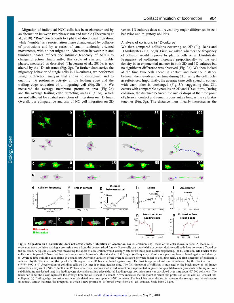
Fig. 3. Migration on 1D-substrates does not affect contact inhibition of locomotion. (a) 2D collision. (b) Tracks of the cells shown in panel A. Both cellsrepolarize upon collision making a protrusion away from the contact (third frame). Since cells can rotate while in contact their overall path does not seem affected bythe collision. A typical CIL analysis measuring the angle of acceleration would wrongly categorize these cells as non-responding. (c) 1D collision. (d) Tracks of the
cells shown in panel C. Note that both cells move away from each other at a sharp 180˚angle. (e) Frequency of collisions per time frame plotted against cell density.(f) Average time colliding cells spend in contact. (g) Over time variation of the average distance between nuclei of colliding cells. The first timepoint of collision isindicated by the black arrow. (h) Speed of colliding cells on 1D lines is plotted against time. The first timepoint of collision is indicated by the black arrow(***P,0.001). (i) Acceleration of colliding cells on 1D lines is plotted against time. The first timepoint of collision is indicated by the black arrow. (j–m) Imagesubtraction analysis of a NC–NC collision. Protrusive activity is represented in red, retraction is represented in green. For quantitative analysis, each colliding cell wassubdivided (green dashed line) in a leading edge side and a trailing edge side. (n) Leading edge protrusion area was calculated over time upon NC–NC collisions. The
black bar under the x-axis represent the average time the cells spent in contact. Arrow indicates the timepoint at which the protrusion at the cell–cell contact sitecollapses. (o) Trailing edge protrusion area was calculated over time upon NC–NC collisions. The black bar under the x-axis represent the average time the cells spentin contact. Arrow indicates the timepoint at which a new protrusion is formed away from cell–cell contact. Scale bars: 20 mm.
Contact inhibition of locomotion 904
Bio
logy
Open
by guest on May 25, 2018http://bio.biologists.org/Downloaded from

cells move away. We empirically determined that 30 minutes
after collision most cells have migrated away from each other anddistance between cells at this time point can thus be used as aneasy readout of CIL.
Data extracted from cell tracking analysis of cells on 1D-
substrates revealed a similar behavior upon CIL to thosepreviously described for cells in 2D-conditions (Carmona-Fontaine et al., 2008). A drop in cell speed at the time of
contact (Fig. 3h, arrow) is followed by a sudden increase(Fig. 3h). This can be shown as a deceleration upon contact(Fig. 3i, arrow) followed by an acceleration (Fig. 3i).
Importantly, this acceleration is coupled with a repolarizationin the opposite direction. This can be assessed by monitoringprotrusion/retraction dynamics over time. To do so, growing andretracting regions of the cells are color-coded by image
subtraction (Fig. 3j–m; see Materials and Methods for details).Before collision, the cell extends a protrusion at its leading edge(Fig. 3j, red) oriented towards the other cell. Upon collision
(Fig. 3k), the protrusion collapses, and a new protrusion isextended on the former trailing edge of the cell (Fig. 3l). Thisrepresents a complete switch of cell polarity. The cells eventually
separate (Fig. 3m). The average protrusion and retraction areaswere measured over time (Fig. 3n,o). Interestingly, the switch ofpolarity occurs immediately after the two cells make contact. The
initial protrusion collapses at the time of contact (Fig. 3n, arrow)and a new protrusion is created at the opposite side of the cell(Fig. 3o, arrow).
To summarize, CIL occurs with similar frequency and
comparable timing in 2D and 1D-cultures. Critically, theduration of the cell–cell contact, the dynamics of cellprotrusion leading to repolarization and its consequences on
cell velocity and acceleration are preserved on 1D-substrates.
Standardization and validation of a unique parameter forassaying CIL
In a 1D collision assay, cell migration is restricted on straightlines. When cells enter in contact with one another there are threemain outcomes. If cells exhibit a clear CIL response, cell–cellcontact results in full repolarization and migration of the two
cells away from each other (Fig. 4ai,bi; supplementary materialMovie 2). If the cells do not exhibit CIL towards each other theymay not dissociate the contact established upon collision and
remain together (Fig. 4aii,bii; supplementary material Movie 2)or may be unaffected by their physical interaction and walk pasteach other (Fig. 4aiii,biii; supplementary material Movie 2).
Therefore, spatial confinement of cell migration simplifiesthe distinction between a CIL and a non-CIL response. We thenanalyzed whether the quality of the collisions would be improvedby the 1D culture conditions. In 2D, we defined a collision
‘‘successful’’ as an event where both cells efficiently repolarizedupon contact with one another and moved away. Importantly,we observed that the percentage of successful collisions
was significantly increased on 1D substrates (Fig. 4c). Earlyobservations by Abercrombie and Dunn on chick heart fibroblastindicated that the cells undergo a stronger repolarization response
when they enter in contact with each other in a head-to-headfashion when both leading edges make a frontal contact(Abercrombie and Dunn, 1975). Since cells are forced to interact
head-to-head when cultured on 1D-substrates, this might accountthe observed improvement of the efficiency of CIL on 1D versus2D-cultures.
The micropatterning technique here described allows both
higher predictability and higher efficiency of the CIL response in
cell–cell collisions. Because all of the functional assays available
so far to assess CIL require more than one quantitation step and
very often include cell-tracking, we aimed at identifying a
cellular parameter whose direct measurement could provide an
easy readout of CIL. The distance between the nuclei of two
colliding cells (Fig. 3k) appeared as an interesting candidate
parameter, as it is maintained relatively constant if the cells
establish a contact but increases linearly when two cells
repolarize and move away. Time-lapse movies are still needed
to be able to distinguish cells that are far away due to CIL or
walk-past behavior but importantly cell tracking is not necessary.
To validate this method we tested some of the signaling
pathways that have been described to be involved in CIL of
neural crest cells (Carmona-Fontaine et al., 2008). We blocked
CIL by interfering with Wnt/PCP and Rho signaling using a
Fig. 4. 1D-substrates increase the probability of successful collision and
provide a simpler readout of CIL. (a) Diagram showing the possible
outcomes of cell–cell collisions on fibronectin lines. (b) Examples showing thedifferent outcomes depicted in panel A. When CIL occurs it leads to completerepolarization of the direction of migration with cells moving away from eachother (ai,bi). Alternatively, cells can fail to dissociate after contact and adhereto each other (aii,bii). Finally, cells may not react to their physical contact andwalk past each other following their original path of migration (aiii,biii).(c) Percentage of cell–cell collisions in which both colliding cells repolarize
upon contact in 2D or 1D cultures (*P,0.05). (d) Percentages of NC cellsdisplaying CIL, adhesion or Walk-Past behavior in control conditions or uponWnt/PCP inhibition (DEP+) or Rho Kinase inhibition (Y-27632). *P,0.05;**P,0.01. (e) Distance between nuclei of colliding cells 30 minutes afterinitial contact in control conditions or upon DEP+ or Y-27632 treatment(**P,0.01). Scale bar: 10 mm (b).
Contact inhibition of locomotion 905
Bio
logy
Open
by guest on May 25, 2018http://bio.biologists.org/Downloaded from
dominant-negative of Dishevelled (DshDEP+) (Carmona-
Fontaine et al., 2008) and the ROCK inhibitor Y-27632(Carmona-Fontaine et al., 2008; Kadir et al., 2011). Collisionbetween control cells, DshDEP+ expressing cells or Y-27632
treated cells were analyzed qualitatively (Fig. 4d) and thedistance between nuclei was measured in parallel (Fig. 4e). Asignificant reduction in the CIL response was observed for both
treatments. Importantly, the loss of CIL was associated with asignificant reduction of the distance between nuclei of collidingcells 30 minutes after the initial contact (Fig. 4e) confirming thatthe distance between cell nuclei at a given time point after
collision is a good readout of CIL.Here we show how a micropatterning technique can facilitate
the analysis of a phenomenon of significant biological relevance
such as CIL. Confinement provided by one-dimensional culturesfacilitates the identification of cell–cell collisions by increasingtheir predictability. It improves the efficiency of CIL by forcing
the cells to undergo head-to-head collisions and eliminatinghead-to-side collisions. Moreover, by preventing randommovement and rotation during collision, 1D-cultures abolish the
time-consuming steps of monitoring angles of acceleration andperforming cell tracking. Furthermore, it simplifies the analysisof changes in polarity by image subtraction. Finally,simplification provided by cultures on fibronectin lines allows
detection of a functional CIL response by measurement of asimple and unique parameter such as the distance between twocell nuclei.
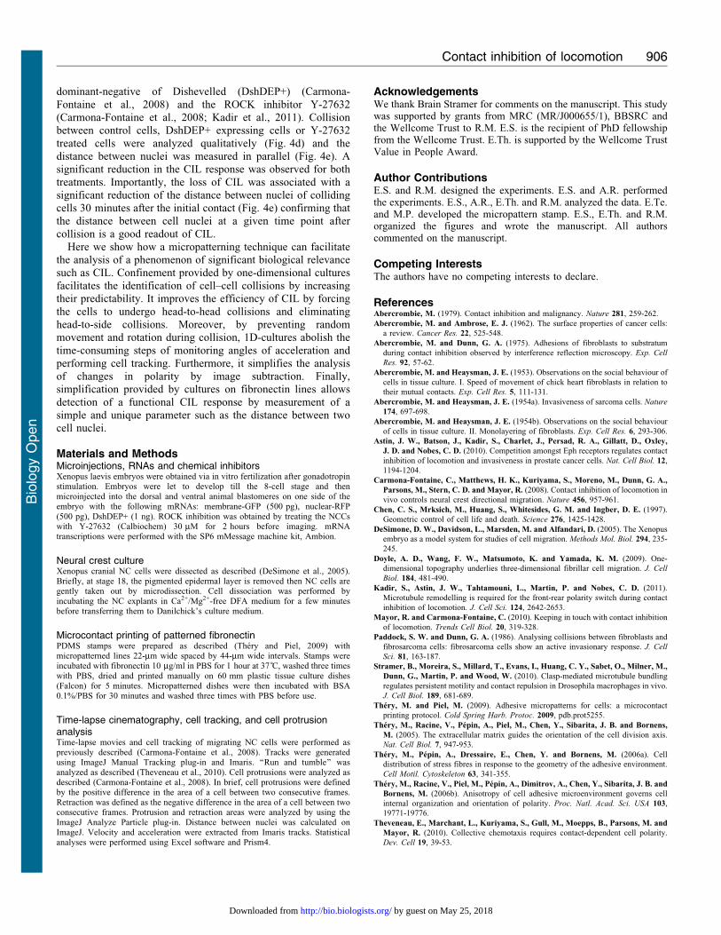
Materials and MethodsMicroinjections, RNAs and chemical inhibitorsXenopus laevis embryos were obtained via in vitro fertilization after gonadotropinstimulation. Embryos were let to develop till the 8-cell stage and thenmicroinjected into the dorsal and ventral animal blastomeres on one side of theembryo with the following mRNAs: membrane-GFP (500 pg), nuclear-RFP(500 pg), DshDEP+ (1 ng). ROCK inhibition was obtained by treating the NCCswith Y-27632 (Calbiochem) 30 mM for 2 hours before imaging. mRNAtranscriptions were performed with the SP6 mMessage machine kit, Ambion.
Neural crest cultureXenopus cranial NC cells were dissected as described (DeSimone et al., 2005).Briefly, at stage 18, the pigmented epidermal layer is removed then NC cells aregently taken out by microdissection. Cell dissociation was performed byincubating the NC explants in Ca2+/Mg2+-free DFA medium for a few minutesbefore transferring them to Danilchick’s culture medium.
Microcontact printing of patterned fibronectinPDMS stamps were prepared as described (Thery and Piel, 2009) withmicropatterned lines 22-mm wide spaced by 44-mm wide intervals. Stamps wereincubated with fibronectin 10 mg/ml in PBS for 1 hour at 37 C, washed three timeswith PBS, dried and printed manually on 60 mm plastic tissue culture dishes(Falcon) for 5 minutes. Micropatterned dishes were then incubated with BSA0.1%/PBS for 30 minutes and washed three times with PBS before use.
Time-lapse cinematography, cell tracking, and cell protrusionanalysisTime-lapse movies and cell tracking of migrating NC cells were performed aspreviously described (Carmona-Fontaine et al., 2008). Tracks were generatedusing ImageJ Manual Tracking plug-in and Imaris. ‘‘Run and tumble’’ wasanalyzed as described (Theveneau et al., 2010). Cell protrusions were analyzed asdescribed (Carmona-Fontaine et al., 2008). In brief, cell protrusions were definedby the positive difference in the area of a cell between two consecutive frames.Retraction was defined as the negative difference in the area of a cell between twoconsecutive frames. Protrusion and retraction areas were analyzed by using theImageJ Analyze Particle plug-in. Distance between nuclei was calculated onImageJ. Velocity and acceleration were extracted from Imaris tracks. Statisticalanalyses were performed using Excel software and Prism4.
AcknowledgementsWe thank Brain Stramer for comments on the manuscript. This studywas supported by grants from MRC (MR/J000655/1), BBSRC andthe Wellcome Trust to R.M. E.S. is the recipient of PhD fellowshipfrom the Wellcome Trust. E.Th. is supported by the Wellcome TrustValue in People Award.
Author ContributionsE.S. and R.M. designed the experiments. E.S. and A.R. performedthe experiments. E.S., A.R., E.Th. and R.M. analyzed the data. E.Te.and M.P. developed the micropattern stamp. E.S., E.Th. and R.M.organized the figures and wrote the manuscript. All authorscommented on the manuscript.
Competing InterestsThe authors have no competing interests to declare.
ReferencesAbercrombie, M. (1979). Contact inhibition and malignancy. Nature 281, 259-262.
Abercrombie, M. and Ambrose, E. J. (1962). The surface properties of cancer cells:
a review. Cancer Res. 22, 525-548.
Abercrombie, M. and Dunn, G. A. (1975). Adhesions of fibroblasts to substratum
during contact inhibition observed by interference reflection microscopy. Exp. Cell
Res. 92, 57-62.
Abercrombie, M. and Heaysman, J. E. (1953). Observations on the social behaviour of
cells in tissue culture. I. Speed of movement of chick heart fibroblasts in relation to
their mutual contacts. Exp. Cell Res. 5, 111-131.
Abercrombie, M. and Heaysman, J. E. (1954a). Invasiveness of sarcoma cells. Nature
174, 697-698.
Abercrombie, M. and Heaysman, J. E. (1954b). Observations on the social behaviour
of cells in tissue culture. II. Monolayering of fibroblasts. Exp. Cell Res. 6, 293-306.
Astin, J. W., Batson, J., Kadir, S., Charlet, J., Persad, R. A., Gillatt, D., Oxley,
J. D. and Nobes, C. D. (2010). Competition amongst Eph receptors regulates contact
inhibition of locomotion and invasiveness in prostate cancer cells. Nat. Cell Biol. 12,
1194-1204.
Carmona-Fontaine, C., Matthews, H. K., Kuriyama, S., Moreno, M., Dunn, G. A.,
Parsons, M., Stern, C. D. and Mayor, R. (2008). Contact inhibition of locomotion in
vivo controls neural crest directional migration. Nature 456, 957-961.
Chen, C. S., Mrksich, M., Huang, S., Whitesides, G. M. and Ingber, D. E. (1997).
Geometric control of cell life and death. Science 276, 1425-1428.
DeSimone, D. W., Davidson, L., Marsden, M. and Alfandari, D. (2005). The Xenopus
embryo as a model system for studies of cell migration. Methods Mol. Biol. 294, 235-
245.
Doyle, A. D., Wang, F. W., Matsumoto, K. and Yamada, K. M. (2009). One-
dimensional topography underlies three-dimensional fibrillar cell migration. J. Cell
Biol. 184, 481-490.
Kadir, S., Astin, J. W., Tahtamouni, L., Martin, P. and Nobes, C. D. (2011).
Microtubule remodelling is required for the front-rear polarity switch during contact
inhibition of locomotion. J. Cell Sci. 124, 2642-2653.
Mayor, R. and Carmona-Fontaine, C. (2010). Keeping in touch with contact inhibition
of locomotion. Trends Cell Biol. 20, 319-328.
Paddock, S. W. and Dunn, G. A. (1986). Analysing collisions between fibroblasts and
fibrosarcoma cells: fibrosarcoma cells show an active invasionary response. J. Cell
Sci. 81, 163-187.
Stramer, B., Moreira, S., Millard, T., Evans, I., Huang, C. Y., Sabet, O., Milner, M.,
Dunn, G., Martin, P. and Wood, W. (2010). Clasp-mediated microtubule bundling
regulates persistent motility and contact repulsion in Drosophila macrophages in vivo.
J. Cell Biol. 189, 681-689.
Thery, M. and Piel, M. (2009). Adhesive micropatterns for cells: a microcontact
printing protocol. Cold Spring Harb. Protoc. 2009, pdb.prot5255.
Thery, M., Racine, V., Pepin, A., Piel, M., Chen, Y., Sibarita, J. B. and Bornens,
M. (2005). The extracellular matrix guides the orientation of the cell division axis.
Nat. Cell Biol. 7, 947-953.
Thery, M., Pepin, A., Dressaire, E., Chen, Y. and Bornens, M. (2006a). Cell
distribution of stress fibres in response to the geometry of the adhesive environment.
Cell Motil. Cytoskeleton 63, 341-355.
Thery, M., Racine, V., Piel, M., Pepin, A., Dimitrov, A., Chen, Y., Sibarita, J. B. and
Bornens, M. (2006b). Anisotropy of cell adhesive microenvironment governs cell
internal organization and orientation of polarity. Proc. Natl. Acad. Sci. USA 103,
19771-19776.
Theveneau, E., Marchant, L., Kuriyama, S., Gull, M., Moepps, B., Parsons, M. and
Mayor, R. (2010). Collective chemotaxis requires contact-dependent cell polarity.
Dev. Cell 19, 39-53.
Contact inhibition of locomotion 906
Bio
logy
Open
by guest on May 25, 2018http://bio.biologists.org/Downloaded from


![Locomotion [2015]](https://img.pdfslide.net/doc/110x75/55d39c9ebb61ebfd268b46a2/locomotion-2015.jpg)


![Locomotion [2014]](https://img.pdfslide.net/doc/110x75/5564e3eed8b42ad3488b4e94/locomotion-2014.jpg)