Embed Size (px)
Citation preview

The Plant Cell, Vol. 14, 1691–1704, August 2002, www.plantcell.org © 2002 American Society of Plant Biologists
RESEARCH ARTICLE
Functional Rice Centromeres Are Marked by a Satellite Repeat and a Centromere-Specific Retrotransposon
Zhukuan Cheng,
a
Fenggao Dong,
a
Tim Langdon,
b
Shu Ouyang,
c
C. Robin Buell,
c
Minghong Gu,
d
Frederick R. Blattner,
e
and Jiming Jiang
a,1
a
Department of Horticulture, University of Wisconsin-Madison, Madison, Wisconsin 53706
b
Institute of Grassland and Environmental Research, Plas Gogerddan, Aberystwyth SY23 3EB, United Kingdom
c
The Institute for Genomic Research, 9712 Medical Center Drive, Rockville, Maryland 20850
d
Department of Agronomy, Yangzhou University, Yangzhou 225009, People’s Republic of China
e
Department of Genetics, University of Wisconsin-Madison, Madison, Wisconsin 53706
The centromere of eukaryotic chromosomes is essential for the faithful segregation and inheritance of genetic infor-mation. In the majority of eukaryotic species, centromeres are associated with highly repetitive DNA, and as a conse-quence, the boundary for a functional centromere is difficult to define. In this study, we demonstrate that the centers ofrice centromeres are occupied by a 155-bp satellite repeat, CentO, and a centromere-specific retrotransposon,
CRR
.The CentO satellite is located within the chromosomal regions to which the spindle fibers attach. CentO is quantita-tively variable among the 12 rice centromeres, ranging from 65 kb to 2 Mb, and is interrupted irregularly by
CRR
ele-ments. The break points of 14 rice centromere misdivision events were mapped to the middle of the CentO arrays, sug-gesting that the CentO satellite is located within the functional domain of rice centromeres. Our results demonstratethat the CentO satellite may be a key DNA element for rice centromere function.
INTRODUCTION
The centromere is the most characteristic landmark of chro-mosomes in higher eukaryotic species and appears cytolog-ically as a distinct primary constriction on condensedmetaphase chromosomes. The centromere is responsiblefor sister chromatid cohesion and is the site for kinetochoreassembly and spindle fiber attachment, allowing for thefaithful pairing and segregation of sister chromatids duringcell division. Determining the precise DNA boundary of acentromere has proven to be a difficult task. In the majority ofeukaryotic species, centromeres are embedded within mega-bases of highly repetitive DNA that cannot be sequencedprecisely by even the most tenacious genome-sequencingprojects (Arabidopsis Genome Initiative, 2000; InternationalHuman Genome Sequencing Consortium, 2001).
The budding yeast
Saccharomyces cerevisiae
is the moststriking exception to the rule of DNA sequence complexity ineukaryotic centromeres. Each of the “point” centromeres in
S
.
cerevisiae
consists of only
�
125 bp of unique sequence(Clarke, 1990, 1998). By contrast, the centromeres of mostother model eukaryotic species, including fission yeast(
Schizosaccharomyces pombe
),
Drosophila melanogaster
,and human, encompass long tracts of highly repetitive DNAsequences (for review, see Henikoff et al., 2001). Satellite re-peats often are the major DNA component of centromeres inhigher eukaryotic species (Csink and Henikoff, 1998). Thebest-characterized centromeric satellite DNA is the
�
satel-lite located in human centromeres.
The
�
satellite DNA is composed of arrays of an
�
171-bpmonomer repeat. The amount of the
�
satellite DNA in hu-man centromeres varies from
�
250 kb to
�
4 Mb (Wevrickand Willard, 1989; Oakey and Tyler-Smith, 1990). Human ar-tificial chromosomes have been assembled successfully us-ing either synthetic or cloned
�
satellite DNA as the cen-tromere component (Harrington et al., 1997; Ikeno et al.,1998; Henning et al., 1999), suggesting that a long stretch of
�
satellite DNA can act as a functional human centromere.The centromeres of the model plant Arabidopsis
havebeen defined genetically (Copenhaver et al., 1999) and con-tain various types of repetitive DNA elements, including ret-roelements, transposons, and telomere-like repeats (Richardset al., 1991; Thompson et al., 1996; Brandes et al., 1997; Haupt
1
To whom correspondence should be addressed. E-mail [email protected]; fax 608-262-4743.Article, publication date, and citation information can be found atwww.plantcell.org/cgi/doi/10.1105/tpc.003079.
Dow
nloaded from https://academ
ic.oup.com/plcell/article/14/8/1691/6009797 by guest on 27 Septem
ber 2021

1692 The Plant Cell
et al., 2001). The most abundant DNA element within func-tional Arabidopsis
centromeres is the pAL1 repeat, a 180-bpsatellite repeat family that constitutes 3% of the Arabidopsisgenome (Martinez-Zapater et al., 1986; Maluszynska andHeslop-Harrison, 1991; Round et al., 1997). Kumekawa etal. (2001) recently demonstrated that the amount of thepAL1 repeat in Arabidopsis centromeres was underesti-mated previously and that each Arabidopsis centromeremay contain several megabases of this satellite repeat.
The centromeres of supernumerary B chromosomes ofmaize have been well studied. A repetitive element specificto the B centromere was isolated (Alfenito and Birchler,1993). A functional role was proposed for this repeat, in partbecause of its strong homology with the knob repeat ofmaize. The maize knobs are cytologically distinct hetero-chromatin structures on meiotic pachytene chromosomesand can function as neocentromeres in certain geneticbackgrounds. Thus, the B centromere repeat may contrib-ute to centromere function through a mechanism similar tothat of the knob repeat. Kaszas and Birchler (1996) demon-strated that the B centromere repeat is present in signifi-cantly rearranged B centromeres. Transmission studies andmolecular characterization of rearranged B centromereseventually may define the critical component of a functionalB centromere (Kaszas and Birchler, 1998).
Several centromeric repetitive DNA elements have beenreported in rice (Dong et al., 1998; Nonomura and Kurata,1999). Sequence analysis revealed that most of these cen-tromeric DNA elements are derived from a Ty3/
gypsy
-classretrotransposon family that is specific to the centromeric re-gions of grass chromosomes (Miller et al., 1998a; Prestinget al., 1998; Langdon et al., 2000). However, the 155-bp sat-ellite repeat CentO, previously named RCS2 (Dong et al.,1998), is unique to rice and is located exclusively in rice cen-tromeres.
In this study, we conducted high-resolution cytologicalmapping of the CentO satellite repeat in the rice genome.The distribution and organization of the CentO satellite, to-gether with the centromere-specific retrotransposon
CRR
,in the 12 rice centromeres are illustrated using a combina-tion of cytological and molecular methods. Quantification ofthe CentO repeat in normal and telocentric rice chromo-somes revealed that the break points of centromere misdivi-sions always are located within the CentO loci, suggestingthat the CentO satellite is a key component of functional ricecentromeres.
RESULTS
The CentO Satellite Is Located in Cytologically Defined Rice Centromeres
The CentO satellite was identified originally in the clonepRCS2, which contains four tandemly arranged monomers
with units between 154 and 165 bp and maps to the centro-meric regions of somatic metaphase chromosomes of rice(Dong et al., 1998). Because of the low mapping resolutionusing somatic metaphase chromosomes, it is not known ifthe CentO satellite is associated with the primary constric-tions. However, meiotic pachytene chromosomes of rice are
�
10 times the length of somatic metaphase chromosomesand provide superior resolution for fluorescence in situ hy-bridization (FISH) mapping (Cheng et al., 2001b).
Pachytene FISH analysis using pRCS2 revealed a singleCentO locus on each of the 12 rice chromosomes (Figures1A to 1C). The pRCS2 FISH signals clearly were associatedwith the primary constriction in pachytene cells, in which themorphology of the chromosomes was well preserved afterthe FISH procedure. Noncentromeric signals were not ob-served on pachytene chromosomes, even under conditionsof low hybridization stringency (30% formamide at 37
�
C), in-dicating that the CentO satellite is highly specific to the cen-tromeres. On meiotic metaphase I chromosomes, the FISHsignals are located consistently on the tips (the most pole-ward positions) of the bivalent chromosomes (Figures 1D to1F), suggesting that the chromosomal regions containingthe CentO satellite are associated with the kinetochore pro-tein complex.
We also observed a unique FISH hybridization pattern insome mitotic metaphase cells. The centromeric regions ofthe chromosomes in these cells were stretched out (Figure1G), possibly as a result of the attachment of spindle fibersand the mechanical stretching of the spindle fibers imposedby the crushing in the cytological preparation. Interestingly,the FISH signals derived from the CentO satellite alwayscolocalized with the stretched centromeric regions (Figures1H and 1I). This unique FISH signal pattern indicates thatthe CentO satellite is located within the chromosomal re-gions that are the sites of kinetochore formation and spindlefiber attachment.
Complex Composition of the CentO Satellite
We previously identified a rice BAC clone (17p22) that is de-rived from a centromeric region of
Oryza
sativa
subsp
indica
var IR-BB21. Several repetitive DNA elements, including theCentO satellite sequence, were isolated from this BAC (Donget al., 1998). To obtain more sequence information from17p22, we conducted 10-fold redundant shotgun sequencingof an M13 library prepared from 17p22. A rigorous assemblyprocess (see Methods) coupled with fiber-FISH analysis re-sulted in a single contig of 62 kb (Figures 2A and 2B).
The 62-kb insert of 17p22 consists of two uninterruptedCentO arrays of 9 and 39.5 kb that are separated by a singleretrotransposon insertion and flanked on one side by a com-plex arrangement of additional retrotransposon sequences(Figure 2A). All CentO monomers are arranged in the sameorientation, but their organization and polymorphism aresurprisingly complex. There are two distinct subfamilies,
Dow
nloaded from https://academ
ic.oup.com/plcell/article/14/8/1691/6009797 by guest on 27 Septem
ber 2021

Functional Rice Centromeres 1693
Figure 1. Centromeric Localization of the CentO Satellite in Rice.
(A) to (C) A single CentO locus is detected on each of the 12 meiotic pachytene chromosomes prepared from var Nipponbare rice.(A) A complete pollen mother cell at the pachytene stage.(B) FISH signals derived from the CentO probe pRCS2. Note that the sizes and intensities of the FISH signals are significantly different amongthe 12 centromeres.(C) A merged image of the pachytene chromosomes and the FISH signals. The individual pachytene chromosomes are identified based on mor-phology. Bar � 10 �m.(D) to (F) Locations of the CentO satellite on meiotic metaphase I chromosomes prepared from var Wuyujing 8 rice.(D) A complete pollen mother cell at metaphase I.(E) FISH signals derived from the CentO probe pRCS2.(F) A merged image of the bivalent chromosomes and the FISH signals. Note that the signals are located on the stretched terminal regions onevery bivalent chromosome.(G) to (I) Locations of the CentO satellite on mitotic metaphase chromosomes prepared from var Nipponbare rice.(G) A complete somatic cell at metaphase.(H) FISH signals derived from the CentO probe pRCS2.(I) A merged image of the chromosomes and the FISH signals. Note that the signals are located on the stretched chromosomal regions. Bar �10 �m.
Dow
nloaded from https://academ
ic.oup.com/plcell/article/14/8/1691/6009797 by guest on 27 Septem
ber 2021
1694 The Plant Cell
with consensus lengths of 155 bp (252 copies) and 164 bp(61 copies), which differ principally as a result of a single 10-bpinsertion in the larger family (Figure 3). All 164-bp monomersare flanked on both sides by the 155-bp monomers.
Within each subfamily, there are many variants, consistingmainly of single base changes and/or single base insertions/deletions relative to the consensus. There are a limited num-ber of common polymorphisms, although all monomers are
�
90% identical to the relevant consensus. Few bases areconserved absolutely, and there are no simple universalconsensus motifs present. Two variants have internal dele-tions (11 and 35 bp), and the deletions are centered on thesame consensus position, yet otherwise these variants ap-pear unrelated by polymorphism at other positions (Figure3). The majority of variants occur only once, but some (in-cluding the 35-bp deletion) are repeated moderately (6 to 14copies). Repeated variants generally are dispersed widelythroughout the clone, and there are no simple tandem ar-rays of identical units.
Sequence Similarity between the CentO Satellite and the Maize Centromeric Repeat CentC
An initial search in GenBank using several monomers of theCentO satellite revealed partial sequence similarity with a156-bp satellite repeat, CentC, located in maize cen-tromeres (Ananiev et al., 1998). Alignment of the two satel-lite families confirms that they are most likely descended
from a common ancestor (Figure 3). Relative to the pub-lished CentC orientation (Ananiev et al., 1998), CentC ishighly divergent from CentO in the central region, where adeletion of 22 or 32 bp is present compared with the 155-and 164-bp subfamilies, respectively. However, CentO andCentC retain strong similarities at the 5
�
and 3
�
ends.The central CentC deletion appears to have been bal-
anced by a degenerate duplication of the terminal 25 bp(shown in Figure 3 as a 3
�
extension relative to CentO), suchthat the length of both satellite families remains similar, sug-gesting that this may have functional importance. Interest-ingly, the conserved 3
�
region is predicted to have signifi-cant potential to direct bending, based on the criteria of thebend.it server (http://www2.icgeb.trieste.it/~dna/bend_it.html;see Methods), whereas both deletions found in the CentOvariants have occurred at this point, suggesting that it maybe susceptible to breakage. Therefore, it is likely that struc-tural constraints play a role in the pattern of sequence con-servation observed between CentC and CentO.
Quantification of the CentO Satellite in Individual Rice Centromeres
The sizes and intensities of the FISH signals derived fromthe CentO satellite differ significantly on individual rice chro-mosomes (Figures 1B, 1E, and 1H), suggesting that thecopy number of the CentO repeat is highly variable amongthe 12 rice centromeres. We quantified the amount of the
Figure 2. Structure of Rice Centromeric BAC 17p22.
(A) The middle diagram shows that the insert of BAC 17p22 contains two blocks of the CentO satellite that are 9 and 39.5 kb, respectively, andthat are separated by two CRR-related DNA sequences. The top diagram shows the structure of the two CRR-related fragments. The 9.2-kbfragment contains three truncated or rearranged CRR elements. The 4.4-kb fragment contains a single CRR element with two LTRs. Each CRRelement is represented by a purple arrow. Open arrows indicate open reading frames; light blue arrows indicate LTRs; the dark blue arrow indi-cates the reverse transcriptase domain; red arrows indicate the integrase domain; the green arrow indicates the protease domain; yellow arrowsindicate the gag domain. The bottom diagram shows that the two CRR-related DNA fragments are covered by six plasmid clones: pRCE1,pRCE2, pRCH1, pRCH2, pRCH3, and pRCS1. These six plasmid clones were used as the CRR probe for FISH analysis.(B) A fiber-FISH image of a single 17p22 molecule visualized by three probes: BAC vector (blue), CRR probe (red), and CentO probe pRCS2(green). The sequence assembly of BAC 17p22 in (A) is confirmed by the fiber-FISH result.
Dow
nloaded from https://academ
ic.oup.com/plcell/article/14/8/1691/6009797 by guest on 27 Septem
ber 2021

Functional Rice Centromeres 1695
CentO satellite in each of the 12 rice centromeres in
O. sa-tiva
subsp
japonica
var Nipponbare and
O
.
sativa
subsp
in-dica
var Zhongxian 3037 (Table 1). The fluorescence intensi-ties of the FISH signals derived from the CentO satellitewere measured on rice pachytene chromosomes using IPLabsoftware.
The 10 best cells in which all 12 pachytene bivalents wereidentified unambiguously were selected for measurements.The intensity of the FISH signal derived from centromere 8was calibrated as 1, and the relative intensities of the signalsderived from other centromeres were calculated and aresummarized in Table 1. A BAC clone, RC8-1, was found tobe closely linked to the CentO locus of chromosome 8. UsingRC8-1 as a reference marker, we were able to determine thelength of the CentO locus of chromosome 8 (CentO-8) using
fiber-FISH (Figure 4A). The sizes of the CentO-8 in Nipponbareand Zhongxian 3037 were measured as 64 and 81 kb, re-spectively, by fiber-FISH analysis (Figure 4A, Table 1). Theamount of the CentO satellite in other centromeres was cal-culated based on relative fluorescence intensities comparedwith centromere 8 (Table 1).
We also identified DNA markers that are closely linked tothe CentO locus on chromosome 11 (CentO-11). Using astrategy similar to that demonstrated in Figure 4A, we mea-sured the sizes of the CentO-11 in Nipponbare and Zhongxian3037 to be 1.64 and 0.64 Mb, respectively, based on five fiber-FISH signals from each rice variety (data not shown). Thesemeasurements are similar to the estimated sizes of CentO-11(1.90 and 0.42 Mb) based on relative fluorescence intensi-ties (Table 1). These results demonstrated an appreciable
Figure 3. Sequence Similarity between Rice CentO and Maize CentC Centromeric Satellite Repeats.
Five CentC monomers (top) are compared with CentO sequences from the 17p22 or pRCS2 clones. CentC contains an internal deletion and adegenerate duplication (CentC numbering is as in the database entry; the degenerate duplications are marked by arrows above). CentC se-quences shown are from single, complete monomers, whereas CentO sequences represent single monomers followed by part (�24 bp) of theadjacent downstream monomer (boxed). The CentO variants displayed include two partial deletions and four members of the 164-bp subfamily.The region removed in the two partial deletions has the maximum predicted potential for bending within the CentO consensus. Sequence con-servation between the displayed repeats is indicated by background shading (black represents 100% conservation, dark gray represents atleast 80%, and light gray represents at least 60%).
Dow
nloaded from https://academ
ic.oup.com/plcell/article/14/8/1691/6009797 by guest on 27 Septem
ber 2021
1696 The Plant Cell
accuracy of the quantification method based on measure-ments of FISH signal intensities.
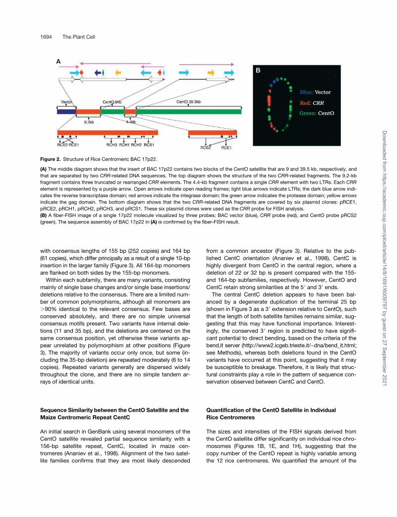
The total amounts of the CentO satellite are
�
7 and 5 Mb inNipponbare and Zhongxian 3037, respectively, and account for1.6 and 1.2% of the 430-Mb rice genome (Arumuganathanand Earle, 1991). The centromeres of chromosomes 1, 2, 3,4, 8, and 12 in the two rice varieties, which represent twodifferent subspecies, show similar amounts of the CentOsatellite. But the centromeres of the remaining chromo-somes in the two varieties differ significantly in their CentOcontents. For example, centromere 6 in Nipponbare andZhongxian 3037 contains
�
820 and 160 kb of the CentOsatellite, representing a fivefold difference.
The Break Points of Rice Centromere Misdivisions Are Located in the Middle of the CentO Arrays
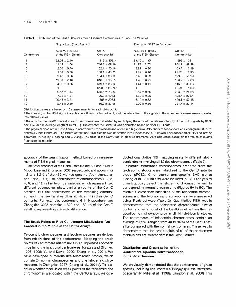
Telocentric chromosomes and isochromosomes are derivedfrom misdivisions of the centromeres. Mapping the breakpoints of centromere misdivisions is an important approachin defining the functional centromeres (Kaszas and Birchler,1996, 1998; Yu and Dawe, 2000; Zhang et al., 2001). Wehave developed numerous rice telotrisomic stocks, whichcontain 24 normal chromosomes and one telocentric chro-mosome, in Zhongxian 3037 (Cheng et al., 2001c). To dis-cover whether misdivision break points of the telocentric ricechromosomes are located within the CentO arrays, we con-
ducted quantitative FISH mapping using 14 different telotri-somic stocks involving all 12 rice chromosomes (Table 2).
Somatic metaphase chromosomes prepared from thetelotrisomic stocks were hybridized to the CentO satelliteprobe pRCS2. Chromosome arm–specific BAC clones(Cheng et al., 2001a) also were included in FISH analysis tounambiguously detect the telocentric chromosome and itscorresponding normal chromosome (Figures 5A to 5C). Therelative fluorescence intensities of the telocentric chromo-somes and the two normal chromosomes were measuredusing IPLab software (Table 2). Quantitative FISH resultsdemonstrated that the telocentric chromosomes alwayscontain a lower amount of the CentO satellite than their re-spective normal centromeres in all 14 telotrisomic stocks.The centromeres of telocentric chromosomes contain anaverage of 65% (ranging from 48 to 84%) of the CentO sat-ellite compared with the normal centromeres. These resultsdemonstrate that the break points of all of the centromeremisdivisions are located within the CentO arrays.
Distribution and Organization of the Centromere-Specific Retrotransposon in the Rice Genome
We previously demonstrated that the centromeres of grassspecies, including rice, contain a Ty3/
gypsy
-class retrotrans-poson family (Miller et al., 1998a; Langdon et al., 2000). This
Table 1.
Distribution of the CentO Satellite among Different Centromeres in Two Rice Varieties
Nipponbare (
japonica
rice) Zhongxian 3037 (
indica
rice)
CentromereRelative Intensityof the FISH Signal
a
CentO Content
b
(kb)Relative Intensityof the FISH Signal
a
CentO Content
b
(kb)
1 22.04
�
2.46 1,418
�
158.3 23.45
�
1.35 1,898
�
1092 11.14
�
1.06 716.6
�
68.19 11.17
�
0.72 904.1
�
58.283 2.83
�
0.78 182.1
�
50.18 2.27
�
0.20 183.7
�
16.194 1.68
�
0.70 108.1
�
45.03 1.22
�
0.16 98.75
�
12.955 2.40
�
0.56 154.4
�
36.02 7.40
�
0.63 599.0
�
50.996 12.69
�
2.46 816.3
�
158.3 1.93
�
0.21 156.2
�
17.007 4.96
�
0.56 319.1
�
36.02 1.44
�
0.11 116.6 � 8.9038 1 64.33 � 25.73c 1 80.94 � 11.33c 9 9.57 � 1.14 615.6 � 73.33 2.57 � 0.30 208.0 � 24.28
10 7.32 � 1.64 470.9 � 105.5 1.59 � 0.25 128.7 � 20.2411 29.48 � 3.21 1,896 � 206.5 5.19 � 0.62 420.1 � 50.1812 2.43 � 0.59 156.3 � 37.95 2.90 � 0.36 234.7 � 29.14
Distribution values are based on 10 measurements for each data point.a The intensity of the FISH signal in centromere 8 was calibrated as 1, and the intensities of the signals in the other centromeres were convertedinto relative values.b The error for the CentO content in each centromere was calculated by multiplying the error of the relative intensity of the FISH signals by 64.33or 80.94 kb (the average length of CentO-8). The error for the CentO-8 was calculated based on fiber-FISH data.c The physical sizes of the CentO array in centromere 8 were measured on 10 and 6 genomic DNA fibers of Nipponbare and Zhongxian 3037, re-spectively (see Figure 4A). The length of the fiber-FISH signals was converted into kilobases by 3.18 kb/�m (unpublished fiber-FISH calibrationparameter in rice by Z. Cheng and J. Jiang). The sizes of the CentO loci in other centromeres were calculated based on the values of relativefluorescence intensity.
Dow
nloaded from https://academ
ic.oup.com/plcell/article/14/8/1691/6009797 by guest on 27 Septem
ber 2021

Functional Rice Centromeres 1697
Figure 4. Fiber-FISH Analysis of Rice Centromeric DNA.
(A) BAC clone RC8-1 (red) is located �50 kb away from the CentO locus in rice chromosome 8 (CentO-8). Fiber-FISH signals derived fromCentO-8 (green) can be identified unambiguously and measured using RC8-1 as a reference marker. The top signal is from var Nipponbare rice;the bottom signal is from var Zhongxian 3037 rice. Note that the red signals within the CentO-8 loci were derived from the CRR sequences con-tained in BAC RC8-1. Bar � 10 �m.(B) Representative fiber-FISH signals demonstrate highly variable densities and nesting of the CRR elements (red signals) within CentO arrays(green signals). An �400-kb CentO signal (fiber i) contains no CRR insertion. Fiber ii (�200 kb) shows five insertions (arrowheads). Long CRRsignals (�20 kb) inserted into CentO satellite DNA are detected within fibers iii and iv. A CRR signal independent of CentO satellite DNA (fiber v)contains �100-kb CRR-related sequences. The long CRR signals most likely are derived from nested CRR elements. Bar � 10 �m.(C) A fiber-FISH signal consists of interspersed signals from CRR and CentO. The top fiber signal (green) is from CentO; the middle fiber signal(red) is from CRR; and the bottom fiber signal is a merged image. Bar � 10 �m.
Dow
nloaded from https://academ
ic.oup.com/plcell/article/14/8/1691/6009797 by guest on 27 Septem
ber 2021
1698 The Plant Cell
centromere-specific family in rice is referred to as CRR(Centromeric Retrotransposon of Rice). The insert of BAC17p22 contains two regions, 4.4 and 9.2 kb, respectively,that are derived from the CRR family (Figure 2A).
The 4.4-kb region represents an insertion of a nonautono-mous element into the CentO satellite array. This insertionhas occurred relatively recently, as judged by the lack of di-vergence of its long terminal repeat (LTR) sequences. Aswith other members of the nonautonomous subfamily ofCRR elements, an open reading frame that has homologywith the gag gene of full-length elements is fused to a read-ing frame of unknown function (Langdon et al., 2000). Nocanonical enzymatic motifs are present, but elements thatshare a similar structure to this 4.4-kb region are found innumerous rice DNA sequence entries in GenBank, consis-tent with their dispersal by recent retrotransposition. The5-bp duplication “signature” observed at the junctions of ret-rotransposed elements also is found at the terminus of the4.4-kb CRR element.
The 9.2-kb region is derived from three CRR elements,each of which is truncated differently (Figure 2A). The most5� element is a nonautonomous element whose upstreamsequences have been lost in cloning that has inserted intothe integrase region of a full-length element. The 5� LTR ofthe full-length element is recombined exactly with the 3�
LTR of a second full-length element, generating two tandemoverlapping elements. The second full-length element haslost almost all upstream sequences, apparently as a conse-quence of a deletion event that extended into the CentO ar-ray. This deletion may have occurred during propagation of
the BAC clone, because we found recently that many BACclones containing tandem repeats are not stable in Escheri-chia coli (Song et al., 2001).
To investigate the distribution of CRR elements in the ricegenome, we screened a Nipponbare BAC library (http://www.genome.clemson.edu/orders/Product.html) using the CentOsatellite pRCS2 probe and the pRCS1 probe derived fromthe integrase domain of CRR (Miller et al., 1998a). ProbespRCS2 and pRCS1 hybridized to 569 (1.50%) and 712(1.87%) of the 38,016 clones, respectively. A total of 157BACs hybridized to both probes. We expect that the CRRelements in these 157 BACs are either located adjacent toor inserted in the middle of the CentO arrays.
All of the CentO- and CRR-positive clones were used tosearch the BAC fingerprint database developed by ClemsonUniversity (http://www.genome.clemson.edu/projects/rice/fpc/). Among the 569 clones associated with the CentO re-peat, 267 clones are singletons and the other 302 clones aredistributed in 26 different contigs. Among the 712 clonesassociated with CRR, 604 clones are distributed on 70 con-tigs and 98 clones are singletons. These results suggest thatthe CentO satellite is organized into long arrays with fewdisruptions by other sequences, whereas the CRR elementsare dispersed in a wider range of chromosomal regions thanthe CentO satellite. In addition, many of the BACs contain-ing the CentO satellite may not produce distinct restrictionpatterns; thus, they appear as singletons.
To gain a cytological view of the distribution of the CRRelements, we conducted FISH analysis by pooling the sixplasmid clones derived from different parts of the CRR ele-ment as a single probe (Figure 2A; referred to as CRR probehereafter). FISH signals derived from the CRR probe werehighly enriched in the centromeric regions on both somaticmetaphase and meiotic pachytene chromosomes (Figure6A). However, cross-hybridization of the CRR probe to otherchromosomal regions was observed. The noncentromericsignals were not consistent among different cells and gener-ally were much weaker than the signals detected in the cen-tromeres. We could not determine whether such signalswere derived from noncentromeric members of the CRRfamily or from cross-hybridization with other retrotranspo-son sequences. Based on the size and intensity of the FISHsignals, the copy number of the CRR element varied signifi-cantly among the 12 rice centromeres. The CRR elementswere distributed in a much wider range of the centromericregions than the CentO satellite (Figures 6B to 6E), which isconsistent the BAC library screening results.
The FISH signals derived from the CRR probe were notdistributed uniformly in the majority of the centromeres. Inseveral centromeres, the signals from the CRR probe wereclearly enriched on both sides of the CentO locus (Figures6C to 6E). In some chromosomes, the CRR signal on oneside of the centromere was significantly stronger than thesignal on the other side of the centromere. The FISH resultsalso indicated that the majority of the CRR elements flankthe CentO arrays rather than insert into the CentO arrays.
Table 2. The Content of the CentO Satellite in Telocentric Rice Chromosomes
TelotrisomicStock
CentO Content in the Telocentric Chromosome (%)
2n•1S 60.97 � 5.362n•1L 65.01 � 7.482n•2S 57.80 � 3.292n•3L 47.81 � 4.412n•4S 56.39 � 3.102n•5L 70.91 � 4.302n•6S 84.41 � 4.832n•7S 57.09 � 3.822n•8L 77.47 � 4.822n•9S 52.17 � 7.442n•10S 67.57 � 3.312n•11L 66.87 � 4.212n•12S 69.65 � 4.502n•12L 76.25 � 4.67
CentO content data are based on five measurements for each datapoint and are presented as the percentage of the corresponding nor-mal chromosome.
Dow
nloaded from https://academ
ic.oup.com/plcell/article/14/8/1691/6009797 by guest on 27 Septem
ber 2021

Functional Rice Centromeres 1699
The CRR and CentO probes were used for FISH analysison extended DNA fibers prepared from Nipponbare. Inser-tions of the CRR elements into the CentO arrays were visu-alized (Figure 4B). The density of the CRR elements insertedin the CentO arrays varied significantly among different fiber-FISH signals (Figure 4B). We observed CentO arrays as longas 500 kb without disruptions by CRR elements (Figure 4B).Another interesting observation is the nesting of the CRR ele-ments in some centromeric regions. Most of the fiber-FISHsignals derived from the CRR probe consisted of two to fourfluorescent spots, representing �1 to 3 �m or 3 to 9 kb.However, contiguous CRR signals as long as 50 �m were ob-served. Such signals most likely were derived from nesting ofmultiple CRR elements (Figure 4B). The 9.2-kb CRR-relatedregion in BAC 17p22 (Figure 2A) may represent one suchnesting case. Although in the majority of the fiber-FISH im-
ages, the CRR signals appear to occupy separate domainsfrom the CentO signals, images with significant interspersingof the CRR and CentO signals were observed (Figure 4C).
DISCUSSION
DNA sequences associated with centromeric regions havebeen reported in numerous plant species (Alfenito and Birchler,1993; Harrison and Heslop-Harrison, 1995; Aragon-Alcaideet al., 1996; Jiang et al., 1996; Brandes et al., 1997; Miller etal., 1998b; Nagaki et al., 1998; Francki, 2001; Gindullis et al.,2001; Hudakova et al., 2001; Page et al., 2001; Saunders andHouben, 2001). However, large-scale sequencing and organi-zation studies of centromeric DNA have been documented in
Figure 5. Quantification of the CentO Satellite in the Centromere of Telocentric Chromosome 2S.
(A) A complete prometaphase cell of telotrisomic 2S.(B) FISH signals derived from the CentO satellite. The arrow indicates the signal on telocentric chromosome 2S. Arrowheads point to the signalson normal chromosome 2.(C) Merged signals of chromosomes and FISH signals derived from the CentO satellite (red) and BAC clone a0095P12 (green) specific to theshort arm of rice chromosome 2 (Cheng et al., 2001a). Note that the FISH signal derived from CentO in the telocentric chromosome is signifi-cantly weaker and smaller than those in normal chromosome 2.(D) Diagram of centromere misdivision. A division in the middle of the centromere results in two functional centromeres associated with the twotelocentric chromosomes.
Dow
nloaded from https://academ
ic.oup.com/plcell/article/14/8/1691/6009797 by guest on 27 Septem
ber 2021

1700 The Plant Cell
Figure 6. Distribution and Organization of the CentO Satellite and the Centromere-Specific Retrotransposon CRR in Rice Chromosomes.
(A) Localization of the CentO satellite (red) and the CRR elements (green) on mitotic prometaphase chromosomes (left) and meiotic pachytenechromosomes (right).(B) to (E) Images of individual rice centromeres of pachytene chromosomes hybridized with the CentO satellite (red) and the CRR probe (green)(B). The composite images are separated digitally into images of FISH signals derived from the CentO satellite (C), images of FISH signals de-rived from the CRR probe (D), and merged images of FISH signals from both probes (E).(F) Diagrams depicting the distribution of the CentO satellite (red) and CRR elements (yellow) in individual rice centromeres. The sizes of the redbars represent the relative amount of the CentO satellite (see Table 1). The density of the yellow lines represents the relative density of the CRRelements in different centromeres.
Dow
nloaded from https://academ
ic.oup.com/plcell/article/14/8/1691/6009797 by guest on 27 Septem
ber 2021

Functional Rice Centromeres 1701
only a few plant species. The most abundant DNA element inArabidopsis centromeres is the 180-bp satellite repeat pAL1.The pAL1 arrays may be interrupted by the 106B repeat, adiverged copy of the LTR of the Athila retrotransposon(Fransz et al., 2000), but complete Athila elements, the mostdominant retrotransposon family in Arabidopsis, are highlyenriched in the pericentromeric region rather than in func-tional centromeres (Fransz et al., 2000). Satellite repeatsand retrotransposons also are the major component of cen-tromeric DNA in barley (Hudakova et al., 2001) and Betaspecies (Gindullis et al., 2001).
In this study, we demonstrate that the centers of rice cen-tromeres are occupied by the CentO satellite repeat se-quence. Cytological mapping revealed that the CentO satel-lite is located within the chromosomal regions at which thekinetochore is formed and the spindle fibers are attached(Figures 1D to 1F and 1G to 1I). A Ty3/gypsy-class ret-rotransposon family, CRR, is colocalized with the CentOsatellite within rice centromeres. CRR is highly specific tothe centromeres, and its homologous sequences have beenfound in the centromeric regions of all grass chromosomes(Ananiev et al., 1998; Miller et al., 1998a; Presting et al.,1998; Langdon et al., 2000).
Despite its centromeric specificity, the copy number anddensity of CRR elements within and outside of the CentOarrays are highly variable among the 12 rice centromeres. Inaddition, a majority of the CRR elements flank the CentO ar-rays rather than insert into the CentO satellite sequences.Frequent and drastic nesting of the CRR element was de-tected (Figure 4B). It will be interesting to determine if thisretrotransposon has any direct or indirect role in rice cen-tromere function. Other retroelements that are not specificto the centromeres have been identified in rice centromericregions (Nonomura and Kurata, 2001).
The mechanism of centromere function is an intriguingpuzzle for biologists. The functional role of the centromere incell division, including both meiosis and mitosis, is highlyconserved in all eukaryotic species. Several proteins in-volved in centromeric function have been found to be con-served in highly divergent eukaryotic species, including S.cerevisiae, Caenorhabditis elegans, D. melanogaster, hu-man, and plants (for reviews, see Dobie et al., 1999; Yu etal., 2000). However, the centromeric DNA sequences areextremely variable even among closely related species.
Satellite repeats often are the major component of com-plex eukaryotic centromeres (Csink and Henikoff, 1998). Thecentromeric satellite repeats generally have a similar mono-mer length, ranging from 150 to 180 bp (for review, seeHenikoff et al., 2001), which is close to the range of nucleo-somal unit length. Henikoff et al. (2001) recently proposedthat adaptive modifications of the centromeric histone 3 inresponse to centromeric satellite expansion may be thedriving force of centromere evolution.
Although satellite repeats are the most common feature ofeukaryotic centromeres and have been proposed to be thekey centromeric DNA component in many species, their true
functional role is difficult to verify. Human � satellite is theonly centromeric satellite whose function has been demon-strated by artificial chromosome assays (Harrington et al.,1997; Ikeno et al., 1998; Henning et al., 1999; Schueler etal., 2001), and its in vivo association with centromeric his-tone H3 also has been demonstrated by immunoprecipita-tion (Vafa and Sullivan, 1997).
We have obtained several lines of evidence that indicatethat the CentO satellite is the key component of functionalrice centromeres. First, the CentO satellite is cytologicallylocated at the tip of the bivalent chromosomes at metaphaseI of meiosis, indicating that the CentO satellite is locatedwithin the chromosomal regions at which the kinetochoreforms and the spindle fibers attach. Second, sequencecomparison revealed that the CentO satellite repeats sharesignificant similarity with the maize centromeric satellite re-peat CentC. Both the localization of these two satellites andthe features that appear to have been conserved duringtheir divergence since a common origin support a functionalrole for these sequences. Third, and most importantly, wehave demonstrated that the break points of all 14 cen-tromere misdivision events are located in the middle of theCentO loci, suggesting that the CentO satellite occupies thecenter of functional rice centromeres.
The human and Arabidopsis genomes have been se-quenced using a clone-by-clone approach (Arabidopsis Ge-nome Initiative, 2000; International Human Genome Se-quencing Consortium, 2001). However, the centromeres ofthese chromosomes were all left as “gaps” in the sequenc-ing data. Thus, even the most rigorous clone-by-clone se-quencing approach has not yielded data on the completeDNA sequences of a centromere from either of these twospecies. There are arguments about whether sequencing ofcentromeres is necessary in genome projects, because it isknown that the centromeres contain mainly known satelliterepeats. However, in humans, only part of the � satellite ar-rays in the centromeres is involved in kinetochore assembly,because antibodies to kinetochore proteins localize to onlya portion of the � satellite DNA (Warburton et al., 1997) andnot all � satellite arrays can form artificial chromosomes(Ikeno et al., 1998). Characterization of the functional cen-tromere of human X chromosome by Schueler et al. (2001)demonstrated that a DNA sequencing effort, together withfunctional assays, represents a powerful approach not onlyto define functional centromeres but also to reveal the plas-ticity and evolution of eukaryotic centromeres. Schueler etal. (2001) showed that only a subset of the � satellite re-peats is responsible for the function of the X chromosome.
Rice provides an excellent model for functional and evolu-tionary studies of centromeres. Several rice centromeres con-tain only a limited amount of satellite repeat compared withhuman and Arabidopsis centromeres. It should be technicallyfeasible to construct BAC contigs that span the entire cen-tromeres of such chromosomes. The BAC contigs would pro-vide an unprecedented resource for centromere sequencing.
It has been demonstrated in several eukaryotic species
Dow
nloaded from https://academ
ic.oup.com/plcell/article/14/8/1691/6009797 by guest on 27 Septem
ber 2021

1702 The Plant Cell
that a centromeric motif would have to be reiterated overhundreds of kilobases to achieve the minimum size of fullyfunctional centromeres. In humans, a minimum of 100 kb of� satellite DNA is required to maintain the centromere func-tion of minichromosomes derived from the Y chromosome(Yang et al., 2000). The centromere of a Drosophila mini-chromosome is contained within a 420-kb region of centricrepetitive DNA. Deletion of DNA in this region leads to a pro-gressive reduction of transmission (Sun et al., 1997). A simi-lar minichromosome-based study in maize also demon-strated that 500 kb of centromeric repeat seems to be theminimum for fully functional B centromeres (Kaszas andBirchler, 1998). Therefore, it is surprising that some rice cen-tromeres contain 100 kb of centromeric satellite repeat. Itwould be of great interest to determine if 60 to 80 kb ofCentO is sufficient to act as a functional centromere in riceor if the DNA sequences flanking the CentO satellite are in-volved in centromere function.
METHODS
Materials
Rice (Oryza sativa subsp japonica var Nipponbare and Wuyujing 8 andO. sativa subsp indica var Zhongxian 3037) was used for cytologicalstudies. The telotrisomic stocks used in this study were developedfrom Zhongxian 3037 (Cheng et al., 2001c). All BAC clones used forfluorescence in situ hybridization (FISH) analysis were selected from aBAC library developed from Nipponbare by Clemson University (http://www.genome.clemson.edu/orders/Product.html). The rice chromo-some-specific BAC markers and rice centromeric DNA probes, includ-ing pRCS1, pRCS2, pRCH1, pRCH2, pRCH3, pRCE1, and pRCE2,were described previously (Dong et al., 1998; Cheng et al., 2001a).
FISH and Fiber-FISH
The preparation of somatic metaphase chromosomes and meioticpachytene chromosomes was as described (Cheng et al., 2001a).The FISH procedure applied to both mitotic and meiotic preparationswas essentially the same as the published protocols (Jiang et al.,1995). DNA probes were labeled with biotin-dUTP, digoxigenin-dUTP, or fluorescein isothiocyanate–dUTP (Boehringer Mannheim).Chromosomes were counterstained with propidium iodide or 4�,6-diamidino-2-phenylindole in Vectashield antifade solution (VectorLaboratories, Burlingame, CA). FISH procedures on genomic DNA fi-bers and BAC molecules were as described previously (Jackson etal., 1998, 1999). Hybridization signals in two-color fiber-FISH weredetected with a three-layer antibody detection system as described(Jackson et al., 1998). Signal detection in three-color fiber-FISH wasaccording to Hsieh et al. (2000).
Cytological Measurements and Analysis
All images were captured digitally using a SenSys charge-coupleddevice camera (Roper Scientific, Tucson, AZ) attached to an Olympus
BX60 epifluorescence microscope (Tokyo, Japan). The charge-cou-pled device camera was controlled using IPLab Spectrum version3.1 software (Signal Analytics, Vienna, VA) on a Macintosh computer.Gray-scale images were captured for each color channel and thenmerged. Measurements were made on the digital images of the FISHsignals within IPLab Spectrum software. For quantitative FISH anal-ysis, the intensity of 5 to 10 signals from the same chromosome wasmeasured, and the numerical data were analyzed in Microsoft Excel98 using the data analysis package. Only the mitotic metaphase cellsor meiotic pachytene cells in which all 12 chromosomes or all tar-geted chromosomes could be identified unambiguously were se-lected for quantitative FISH analysis.
DNA Sequencing and Analysis
An M13 library was constructed from nebulized, size-fractionatedDNA prepared from BAC 17p22. DNA templates were purified fromrandom library clones, and sequences were collected using dye-ter-minator-labeled fluorescent cycle sequencing Prism reagents andABI377 automated sequencers (Applied Biosystems, Foster City,CA). Sequences were assembled into contigs with the Seqman IIprogram (DNASTAR, Madison, WI), and clones were selected for se-quencing from the opposite end to fill coverage, resolve ambiguities,and close gaps (Burland et al., 1993). Final coverage was �10-fold.
Unique sequences (vector and nonsatellite) were readily assem-bled as described previously (Blattner et al., 1997). The assembly ofhighly repetitive sequences presented a challenge. Because thehighly repetitive satellite sequences led to misassembly under theusual conditions, they were reassembled under higher stringencyand using clone coverage information (dual-end sequence pairs de-rived from the same clone of �1 to 2.5 kb). Each initial clonal regionwas microedited to resolve ambiguities and then used as a longersequence for another round of assembly. This rigorous recursive as-sembly process was repeated until all collected data were added tothe project.
Sequence alignments were refined manually and displayed usingGeneDoc (http://www.psc.edu/biomed/genedoc). DNA bending pre-dictions were made using the bend.it server (http://www2.icgeb.trieste.it/~dna/bend_it.html). This server predicts DNA curvaturefrom DNA sequences, calculated as a vector sum of dinucleotide ge-ometries (roll, tilt, and twist angles) using the BEND algorithm ofGodsell and Dickerson and expressed as degrees per helical turn(Munteanu et al., 1998).
Upon request, all novel materials described in this article will bemade available in a timely manner for noncommercial research pur-poses. No restrictions or conditions will be placed on the use of anymaterials described in this article that would limit their use for non-commercial research purposes.
Accession Number
The GenBank accession number for BAC clone 17p22 is AY101510.
ACKNOWLEDGMENTS
This research was supported by a grant from the Consortium forPlant Biotechnology Research, Novartis Seeds, and Dow Agro-
Dow
nloaded from https://academ
ic.oup.com/plcell/article/14/8/1691/6009797 by guest on 27 Septem
ber 2021

Functional Rice Centromeres 1703
Sciences, by Department of Energy Grant DE-FG02-01ER15266,and by funds from the Graduate School of the University of Wis-consin-Madison to J.J. This work also was partially supported byChinese Project 973 Grant G1999011601 to Z.C. and M.G. Fundingsupport to C.R.B. included U.S. Department of Agriculture Grant 99-35317-8275, National Science Foundation Grant DBI998282, andDepartment of Energy Grants DE-FG02-99ER20357 and DE-FG01-01ER15265.
Received March 13, 2002; accepted May 6, 2002.
REFERENCES
Alfenito, M.R., and Birchler, J.A. (1993). Molecular characterization ofa maize B chromosome centric sequence. Genetics 135, 589–597.
Ananiev, E.V., Phillips, R.L., and Rines, H.W. (1998). Chromo-some-specific molecular organization of maize (Zea mays L.) cen-tromeric regions. Proc. Natl. Acad. Sci. USA 95, 13073–13078.
Arabidopsis Genome Initiative. (2000). Analysis of the genomesequence of the flowering plant Arabidopsis thaliana. Nature 408,796–815.
Aragon-Alcaide, L., Miller, T., Schwarzacher, T., Reader, S., andMoore, G. (1996). A cereal centromeric sequence. Chromosoma105, 261–268.
Arumuganathan, K., and Earle, E.D. (1991). Nuclear DNA contentof some important plant species. Plant Mol. Biol. Rep. 9, 208–218.
Blattner, F.R., et al. (1997). The complete sequence of Escherichiacoli K-12. Science 277, 1453–1474.
Brandes, A., Thompson, H., Dean, C., and Heslop-Harrison, J.S.(1997). Multiple repetitive DNA sequences in the paracentricregions of Arabidopsis thaliana L. Chromosome Res. 5, 238–246.
Burland, V., Daniels, D.L., Plunkett, G., III, and Blattner, F.R.(1993). Genome sequencing on both strands: The Janus strategy.Nucleic Acids Res. 21, 3385–3390.
Cheng, Z.K., Buell, C.R., Wing, R.A., Gu, M.H., and Jiang, J.(2001a). Toward a cytological characterization of the rice genome.Genome Res. 11, 2133–2141.
Cheng, Z.K., Presting, G.G., Buell, C.R., Wing, R.A., and Jiang, J.(2001b). High resolution pachytene chromosome mapping of bac-terial artificial chromosomes anchored by genetic markers revealsthe centromere location and the distribution of genetic recombi-nation along chromosome 10 of rice. Genetics 157, 1749–1757.
Cheng, Z.K., Yan, H., Yu, H., Tang, S., Jiang, J., Gu, M.H., andZhu, L. (2001c). Development and applications of a complete setof rice telotrisomics. Genetics 157, 361–368.
Clarke, L. (1990). Centromeres of budding and fission yeasts.Trends Genet. 6, 150–154.
Clarke, L. (1998). Centromeres: Proteins, protein complexes, andrepeated domains at centromeres of simple eukaryotes. Curr.Opin. Genet. Dev. 8, 212–218.
Copenhaver, G.P., et al. (1999). Genetic definition and sequenceanalysis of Arabidopsis centromeres. Science 286, 2468–2474.
Csink, A.K., and Henikoff, S. (1998). Something from nothing: Theevolution and utility of satellite repeats. Trends Genet. 14, 200–204.
Dobie, K.W., Hari, K.L., Maggert, K.A., and Karpen, G.H. (1999).Centromere proteins and chromosome inheritance: A complexaffair. Curr. Opin. Genet. Dev. 9, 206–217.
Dong, F., Miller, J.T., Jackson, S.A., Wang, G.-L., Ronald, P.C.,and Jiang, J. (1998). Rice (Oryza sativa) centromeric regions con-sist of complex DNA. Proc. Natl. Acad. Sci. USA 95, 8135–8140.
Francki, M.G. (2001). Identification of Bilby, a diverged centromericTy1-copia retrotransposon family from cereal rye (Secale cerealeL.). Genome 44, 266–274.
Fransz, P.F., Armstrong, A., de Jong, J.H., Parnell, L.D., vanDrunen, C., Dean, C., Zabel, P., Bisseling, T., and Jones, G.H.(2000). Integrated cytogenetic map of chromosome arm 4S of A.thaliana: Structural organization of heterochromatic knob andcentromere region. Cell 100, 367–376.
Gindullis, F., Desel, C., Galasso, I., and Schmidt, T. (2001). Thelarge-scale organization of the centromeric region in Beta spe-cies. Genome Res. 11, 253–265.
Harrington, J.J., Bokkelen, G.V., Mays, R.W., Gustashaw, K., andWillard, H.F. (1997). Formation of de novo centromeres and con-struction of first-generation human artificial microchromosomes.Nat. Genet. 15, 345–355.
Harrison, G.E., and Heslop-Harrison, J.S. (1995). Centromericrepetitive DNA in the genus Brassica. Theor. Appl. Genet. 90,157–165.
Haupt, W., Fischer, T.C., Winderl, S., Fransz, P., and Torres-Ruiz,R.A. (2001). The CENTROMERE1 (CEN1) region of Arabidopsisthaliana: Architecture and functional impact of chromatin. Plant J.27, 285–296.
Henikoff, S., Ahmad, K., and Malik, H.S. (2001). The centromerecomplex: Stable inheritance with rapidly evolving DNA. Science293, 1098–1102.
Henning, K.A., Novotny, E.A., Compton, S.T., Guan, X.-Y., Liu,P.P., and Ashlock, M.A. (1999). Human artificial chromosomesgenerated by modification of a yeast artificial chromosome con-taining both human alpha satellite and single-copy DNAsequences. Proc. Natl. Acad. Sci. USA 96, 592–597.
Hsieh, H.B., Wang, M., Lersch, R.A., Kim, U.-J., and Weier, H.-U.G.(2000). Rational design of landmark probes for quantitative DNAfiber mapping (ODFM). Nucleic Acids Res. 28, e30.
Hudakova, S., Michalek, W., Presting, G.G., ten Hoopen, R., dosSantos, K., Jasencakova, Z., and Schubert, I. (2001). Sequenceorganization of barley centromeres. Nucleic Acids Res. 24, 5029–5035.
Ikeno, M., Grimes, B., Okazaki, T., Nakano, M., Saitoh, K.,Hoshino, H., McGill, N.I., Cooke, H., and Masumoto, H. (1998).Construction of YAC-based mammalian artificial chromosomes.Nat. Biotechnol. 16, 431–439.
International Human Genome Sequencing Consortium. (2001).Initial sequencing and analysis of the human genome. Nature 409,860–921.
Jackson, S.A., Dong, F., and Jiang, J. (1999). Digital mapping ofbacterial artificial chromosomes by fluorescence in situ hybridiza-tion. Plant J. 17, 581–587.
Jackson, S.A., Wang, M.L., Goodman, H.M., and Jiang, J. (1998).Application of Fiber-FISH in genome analysis of Arabidopsisthaliana. Genome 41, 566–572.
Jiang, J., Gill, B.S., Wang, G.-L., Ronald, P.C., and Ward, D.C.(1995). Metaphase and interphase fluorescence in situ hybridiza-tion mapping of the rice genome with bacterial artificial chromo-somes. Proc. Natl. Acad. Sci. USA 92, 4487–4491.
Jiang, J., Nasuda, S., Dong, F., Scherrer, C.W., Woo, S.S., Wing,R.A., Gill, B.S., and Ward, D.C. (1996). A conserved repetitiveDNA element located in the centromeres of cereal chromosomes.Proc. Natl. Acad. Sci. USA 93, 14210–14213.
Dow
nloaded from https://academ
ic.oup.com/plcell/article/14/8/1691/6009797 by guest on 27 Septem
ber 2021

1704 The Plant Cell
Kaszas, E., and Birchler, J.A. (1996). Misdivision analysis ofcentromere structure in maize. EMBO J. 15, 5246–5255.
Kaszas, E., and Birchler, J.A. (1998). Meiotic transmission ratescorrelate with physical features of rearranged centromeres inmaize. Genetics 150, 1683–1692.
Kumekawa, N., Hosouchi, T., Tsuruoka, H., and Kotani, H. (2001).The size and sequence organization of the centromeric region ofArabidopsis thaliana chromosome 4. DNA Res. 8, 285–290.
Langdon, T., Seago, C., Mende, M., Leggett, M., Thomas, H.,Forster, J.W., Thomas, H., Jones, R.N., and Jenkins, G. (2000).Retrotransposon evolution in diverse plant genomes. Genetics156, 313–325.
Maluszynska, J., and Heslop-Harrison, J.S. (1991). Localization oftandemly repeated DNA sequences in Arabidopsis thaliana. PlantJ. 1, 159–166.
Martinez-Zapater, J.M., Estelle, M.A., and Somerville, C.R.(1986). A high repeated DNA sequence in Arabidopsis thaliana.Mol. Gen. Genet. 204, 417–423.
Miller, J.T., Dong, F., Jackson, S.A., Song, J., and Jiang, J.(1998a). Retrotransposon-related DNA sequences in the cen-tromeres of grass chromosomes. Genetics 150, 1615–1623.
Miller, J.T., Jackson, S.A., Nasuda, S., Gill, B.S., Wing, R.A., andJiang, J. (1998b). Cloning and characterization of a centromere-specific repetitive DNA element from Sorghum bicolor. Theor.Appl. Genet. 96, 832–839.
Munteanu, M.G., Vlahovicek, K., Parthasaraty, S., Simon, I., andPongor, S. (1998). Rod models of DNA: Sequence-dependentanisotropic elastic modelling of local bending phenomena. TrendsBiochem. Sci. 23, 341–346.
Nagaki, K., Tsujimoto, H., and Sasakuma, T. (1998). A novelrepetitive sequence of sugar cane, SCEN family, locating on cen-tromeric regions. Chromosome Res. 6, 295–302.
Nonomura, K.I., and Kurata, N. (1999). Organization of the 1.9-kbrepeat unit RCE1 in the centromeric region of rice chromosomes.Mol. Gen. Genet. 261, 1–10.
Nonomura, K.I., and Kurata, N. (2001). The centromere composi-tion of multiple repetitive sequences on rice chromosome 5. Chro-mosoma 110, 284–291.
Oakey, R., and Tyler-Smith, C. (1990). Y chromosome DNA haplo-typing suggests that most European and Asian men aredescended from one of two males. Genomics 7, 325–330.
Page, B.T., Wanous, M.K., and Birchler, J.A. (2001). Characteriza-tion of a maize chromosome 4 centromeric sequence: Evidencefor an evolutionary relationship with the B chromosome cen-tromere. Genetics 159, 291–302.
Presting, G.G., Malysheva, L., Fuchs, J., and Schubert, I. (1998).A Ty3/gypsy retrotransposon-like sequence localizes to the cen-tromeric regions of cereal chromosomes. Plant J. 16, 721–728.
Richards, E.J., Goodman, H.M., and Ausubel, F.M. (1991). Thecentromere region of Arabidopsis thaliana chromosome 1 con-tains telomere-similar sequences. Nucleic Acids Res. 19, 3351–3357.
Round, E.K., Flowers, S.K., and Richards, E.J. (1997). Arabidopsisthaliana centromere regions: Genetic map positions and repetitiveDNA structure. Genome Res. 7, 1045–1053.
Saunders, V.A., and Houben, A. (2001). The pericentromeric het-erochromatin of the grass Zingeria biebersteiniana (2n�4) iscomposed of Zbcen1-type tandem repeats that are intermingledwith accumulated dispersedly organized sequences. Genome44, 955–961.
Schueler, M.G., Higgins, A.W., Rudd, M.K., Gustashaw, K., andWillard, H.F. (2001). Genomic and genetic definition of a func-tional human centromere. Science 294, 109–115.
Song, J., Dong, F., Lilly, J.W., Stupar, R.M., and Jiang, J. (2001).Instability of bacterial artificial chromosome (BAC) clones contain-ing tandemly repeated DNA sequences. Genome 44, 463–469.
Sun, X., Wahlstrom, J., and Karpen, G.H. (1997). Molecular struc-ture of a functional Drosophila centromere. Cell 91, 1007–1019.
Thompson, H.L., Schmidt, R., and Dean, C. (1996). Identificationand distribution of seven classes of middle-repetitive DNA in theArabidopsis thaliana genome. Nucleic Acids Res. 24, 3017–3022.
Vafa, O., and Sullivan, K.F. (1997). Chromatin containing CENP-Aand satellite DNA is a major component of the inner kinetochoreplate. Curr. Biol. 7, 897–900.
Warburton, P.E., Cooke, C.A., Bourassa, S., Vafa, O., Sullivan,B.A., Stetten, G., Gimelli, G., Warburton, D., Tyler-Smith, C.,Sullivan, K.F., Poirier, G.G., and Earnshaw, W.C. (1997). Immu-nolocalization of CENP-A suggests a distinct nucleosome struc-ture at the inner kinetochore plate of active centromeres. Curr.Biol. 7, 901–904.
Wevrick, R., and Willard, H.F. (1989). Long-range organization oftandem arrays of a satellite DNA at the centromeres of humanchromosomes: High frequency array-length polymorphism andmeiotic stability. Proc. Natl. Acad. Sci. USA 86, 9394–9398.
Yang, J.W., Pendon, C., Yang, J., Haywood, N., Chand, A., andBrown, W.R.A. (2000). Human minichromosomes with minimalcentromeres. Hum. Mol. Genet. 9, 1891–1902.
Yu, H.-G., and Dawe, R.K. (2000). Functional redundancy in themaize meiotic kinetochore. J. Cell Biol. 151, 131–141.
Yu, H.-G., Hiatt, E.N., and Dawe, R.K. (2000). The plant kineto-chore. Trends Plant Sci. 5, 543–547.
Zhang, P., Friebe, B., Lukaszewski, A.J., and Gill, B.S. (2001). Thecentromere structure in Robertsonian wheat-rye translocationchromosomes indicates that centric breakage-fusion can occur atdifferent positions within the primary constriction. Chromosoma110, 335–344.
Dow
nloaded from https://academ
ic.oup.com/plcell/article/14/8/1691/6009797 by guest on 27 Septem
ber 2021