Embed Size (px)
Citation preview
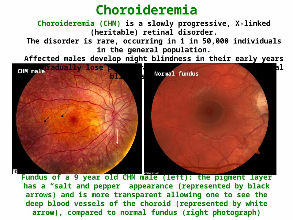
Fundus of a 9 year old CHM male (left): the pigment layer has a “salt and pepper” appearance (represented by black arrows) and is more transparent allowing one to see the deep blood vessels of the choroid (represented by white arrow), compared
to normal fundus (right photograph)
Choroideremia (CHM) is a slowly progressive, X-linked (heritable) retinal disorder.The disorder is rare, occurring in 1 in 50,000 individuals in the general population.
Affected males develop night blindness in their early years and gradually lose peripheral vision which results in legal blindness by age 40.
Choroideremia
CHM maleLeft eyeRight eye
Normal male
Normal maleNormal fundus
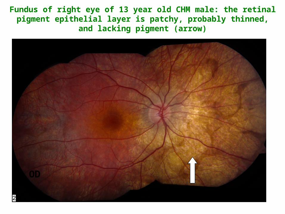
Fundus of right eye of 13 year old CHM male: the retinal pigment epithelial layer is patchy, probably thinned, and lacking pigment (arrow)
OD

Fundus of 18 year old CHM male: The pigment layer is now very thin and possibly absent (black arrow) except in the central area called the macula (white arrow), which remains healthy. The overlying retina remains intact,
but is transparent.
OD OS
Right eye Left eye
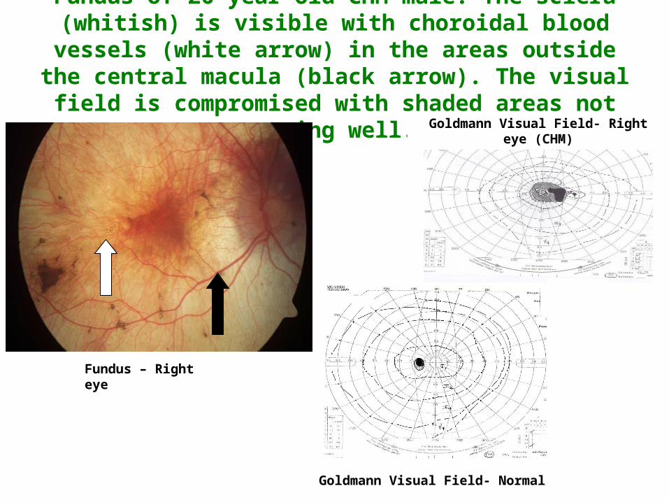
Fundus of 26 year old CHM male: The sclera (whitish) is visible with choroidal blood vessels (white arrow) in the areas outside
the central macula (black arrow). The visual field is compromised with shaded areas not seeing well.
Fundus – Right eye
Goldmann Visual Field- Normal
Goldmann Visual Field- Right eye (CHM)
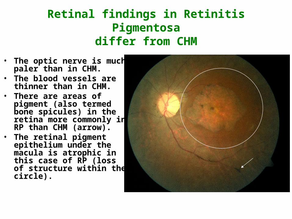
Retinal findings in Retinitis Pigmentosadiffer from CHM
• The optic nerve is much paler than in CHM.
• The blood vessels are thinner than in CHM.
• There are areas of pigment (also termed bone spicules) in the retina more commonly in RP than CHM (arrow).
• The retinal pigment epithelium under the macula is atrophic in this case of RP (loss of structure within the circle).

Female carriers of choroideremia
• In general female carriers experience a milder form of choroideremia.
• Some carriers experience a decline in visual function with difficulty seeing at night after age 50.
• In rare cases, female carriers may exhibit severe symptoms similar to those of affected males.
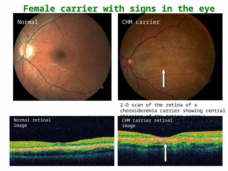
Female carrier with signs in the eye
2-D scan of the retina of a choroideremia carrier showing central thinning of the retina (arrow)
Right eye
CHM carrier
Normal retinal image CHM carrier retinal image
NormalCHM carrierNormal

Fundus of female carrier (left) and autofluorescent image of the same eye (right).
Note areas of speckling of the pigment layer representing its deterioration (arrow)

Additional Resources
• Choroideremia Research Foundation:USA: www.choroideremia.orgCanada: www.choroideremia.ca
• Foundation Fighting Blindness:USA: www.blindness.org
Canada: www.ffb.ca • Choroideremia patient survey:
English: http://choroideremiasurvey.questionpro.com/French: http://choroideremieenquete.questionpro.com/

Contact Us
• For questions or comments about this presentation please contact us at: [email protected]
• For information regarding care options appropriate for you and your family, please contact your ophthalmologist.