Embed Size (px)
Citation preview
2014; doi: 10.1101/cshperspect.a019729Cold Spring Harb Perspect Med Jigar V. Desai, Aaron P. Mitchell and David R. Andes Fungal Biofilms, Drug Resistance, and Recurrent Infection
Subject Collection Human Fungal Pathogens
Loss of HeterozygosityDiversity: Whole Ploidy Shifts, Aneuploidy, and Rapid Mechanisms for Generating Genome
Richard J. Bennett, Anja Forche and Judith Berman
InfectionFungal Biofilms, Drug Resistance, and Recurrent
AndesJigar V. Desai, Aaron P. Mitchell and David R.
Pathogenic Species ComplexCandidaThe Siobhán A. Turner and Geraldine Butler
Metabolism in Fungal Pathogenesis
et al.Iuliana V. Ene, Sascha Brunke, Alistair J.P. Brown,
MalasseziaFungi on the Skin: Dermatophytes and
Dawson, Jr., et al.Theodore C. White, Keisha Findley, Thomas L.
Sexual Reproduction of Human Fungal PathogensJoseph Heitman, Dee A. Carter, Paul S. Dyer, et al.
, the Etiologic Agents of CryptococcosisgattiiCryptococcus and Cryptococcus neoformans
Doering, et al.Kyung J. Kwon-Chung, James A. Fraser, Tamara L.
Fungal InfectionMendelian Genetics of Human Susceptibility to
HollandMichail S. Lionakis, Mihai G. Netea and Steven M.
Clinical Needs, and New ApproachesAntifungal Drug Development: Challenges, Unmet
Terry Roemer and Damian J. Krysan
Fungal DiagnosticsThomas R. Kozel and Brian Wickes
http://perspectivesinmedicine.cshlp.org/cgi/collection/ For additional articles in this collection, see
Copyright © 2014 Cold Spring Harbor Laboratory Press; all rights reserved
on October 9, 2014 - Published by Cold Spring Harbor Laboratory Press http://perspectivesinmedicine.cshlp.org/Downloaded from on October 9, 2014 - Published by Cold Spring Harbor Laboratory Press http://perspectivesinmedicine.cshlp.org/Downloaded from
Fungal Biofilms, Drug Resistance,and Recurrent Infection
Jigar V. Desai1, Aaron P. Mitchell1, and David R. Andes2
1Department of Biological Sciences, Carnegie Mellon University, Pittsburgh, Pennsylvania 152132Department of Medicine, University of Wisconsin, Madison, Wisconsin 53705
Correspondence: [email protected]
A biofilm is a surface-associated microbial community. Diverse fungi are capable of biofilmgrowth. The significance of this growth form for infection biology is that biofilm formation onimplanted devices is a major cause of recurrent infection. Biofilms also have limited drugsusceptibility, making device-associated infection extremely difficult to treat. Biofilm-likegrowth can occur during many kinds of infection, even when an implanted device is notpresent. Here we summarize the current understanding of fungal biofilm formation, itsgenetic control, and the basis for biofilm drug resistance.
Much of the global microbial communitygrows on surfaces, including rock, sedi-
ment, bark, skin, and mucosal tissues (Kolterand Greenberg 2006). The growth form of mi-croorganisms that is associated with a surface iscalled a biofilm. That definition is generally re-fined to include the specification that a truebiofilm is one in which the organisms grow asa community, rather than separate surface-ad-herent cells. In addition, the cells of a true bio-film produce their own extracellular matrix ma-terial and manifest phenotypes that are distinctfrom the phenotypes of cells growing in suspen-sion (called planktonic cells). It has long beenrealized that biofilm formation ability is wide-spread among bacteria, and although there arespecies-specific features of biofilm growth, thereare also common themes that have emerged. Inthis review, we highlight the fact that biofilmformation ability is widespread among fungi
as well, and discuss fungal biofilm structureand regulation.
The first question about fungal biofilmformation is “Why study it”? There are manyanswers of course, but the central significanceof biofilms in the context of infection biologycomes from the relationship of biofilms todevice-associated infection. Implanted devicessuch as central venous catheters, indwellingbladdercatheters, joint prostheses, and mechan-ical heart valves are in common use for the man-agement of diverse medical conditions (Raad1998; Darouiche 2001). The presence of suchdevices is a major risk factor for infection(Raad 1998; Costerton et al. 1999; Donlan2002). These infections arise from microbial at-tachment to device surfaces and their growthinto biofilms (Costerton et al. 1999; Hall-Stood-ley et al. 2004; Kuhn and Ghannoum 2004).These biofilms are invariably recalcitrant to
Editors: Arturo Casadevall, Aaron P. Mitchell, Judith Berman, Kyung J. Kwon-Chung, John R. Perfect, and Joseph Heitman
Additional Perspectives on Human Fungal Pathogens available at www.perspectivesinmedicine.org
Copyright # 2014 Cold Spring Harbor Laboratory Press; all rights reserved; doi: 10.1101/cshperspect.a019729
Cite this article as Cold Spring Harb Perspect Med 2014;4:a019729
1
ww
w.p
ersp
ecti
vesi
nm
edic
ine.
org
on October 9, 2014 - Published by Cold Spring Harbor Laboratory Press http://perspectivesinmedicine.cshlp.org/Downloaded from
drug treatment for a variety of reasons, and thuscan cause recurrent infections. Examination ofdevices isolated from infected patients has re-vealed that many bacterial and fungal speciesform biofilms that seed recurrent infections(Lynch and Robertson 2008). Among fungi, dif-ferent Candida species are the most commoncolonizers of implanted medical devices (Ku-mamoto and Vinces 2005; Ramage et al. 2005).Fusarium species and Malessezia pachydermatisalso form drug-resistant biofilms on implantedmedical devices (Martinez and Fries 2010; Ram-age et al. 2011). Table 1 describes biofilm char-acteristics for several fungi commonly associat-ed with device-related biofilm infections.
There are also situations in which the fungalgrowth form associated with an infection hasbiofilm-like properties, although there may notbe a rigorous demonstration that the growthform has all of the defining features of a biofilm(Cushion et al. 2009; Mowat et al. 2009; Fanningand Mitchell 2012). An excellent illustrationcomes from the mold Aspergillus fumigatus, acommon cause of nosocomial infection in im-munocompromised individuals (Mowat et al.2009). A. fumigatus is occasionally found to col-onize implanted medical devices. However,most infections begin when inhaled conidia ger-minate in the upper airway and in lung tissue.This growth state may lack a clear substrate, butresembles a biofilm nonetheless because hyphaeare enmeshed in an extracellular matrix (Lous-sert et al. 2010). Therefore, biofilm models havebeen developed for A. fumigatus that mightmimic the growth state that occurs duringinfection (Loussert et al. 2010). Indeed, tran-scriptional profiling of this biofilm model foundconsiderable similarity to the profile of A. fumi-gatus germlings in a lung infection model (Mc-Donagh et al. 2008; Gibbons et al. 2012), thusvalidating the concept. Another interesting ex-ample comes from the studyof Pneumocystis spe-cies, which cause pneumonia in immunocom-promised patients. These species are challengingto study because they cannot be cultured outsideof the host. Cushion and colleagues postulatedthat attachment of the organisms to tissue mightgenerate a biofilm-like physiology that promotesgrowth and division (Cushion et al. 2009). In-
deed, their in vitro model for biofilm develop-ment by Pneumocystis carinii and Pneumocystismurina enabled prolonged growth of the organ-isms in vitro compared with planktonic cultureconditions (Cushion et al. 2009). Finally, muco-sal infections, such as oropharyngeal candidiasisor vaginitis, are caused by true biofilms in whichtissue, rather than a device, serves as the sub-strate for biofilm growth (Dongari-Bagtzoglou2008; Ganguly and Mitchell 2011). In these sit-uations, the biofilm-associated phenotype ofdrug resistance has not been shown. Nonethe-less, the concept that a mucosal infection is abiofilm has proven useful by prompting the ac-curate prediction that known regulators of Can-dida albicans biofilm formation would also berequired for mucosal infection (Ganguly andMitchell 2011). These examples illustrate theprinciple that biofilm growth can provide a use-ful model for infection even if a canonical abi-otic-surface biofilm is not clearly associated withinfection.
GENETIC CONTROL OF FUNGAL BIOFILMDEVELOPMENT
What are the genes that govern biofilm forma-tion? One can imagine a broad spectrum of genefunctions that may be involved, affecting suchbiological properties as adherence, morphogen-esis, quorum sensing, matrix production, cellwall biosynthesis, and metabolism. Diverse ra-tionales have been used to choose candidategenes that may be required for normal biofilmformation. For example, investigators have cho-sen to analyze potential biofilm defects in mu-tants of random genes, mutants of genes with aparticular class of product, known functions re-lated to hyphal morphogenesis or azole drugresistance, and those differentially regulatedduring biofilm formation (Garcia-Sanchez etal. 2004; Murillo et al. 2005; Nobile et al. 2006,2012; Bonhomme et al. 2011; Desai et al. 2013).These approaches have been most extensivelyapplied to C. albicans, the focus of the discus-sion below, and have been implemented morerecently with non-albicans Candida species asgenetic tools have become available (Ramageet al. 2005; Finkel and Mitchell 2011).
J.V. Desai et al.
2 Cite this article as Cold Spring Harb Perspect Med 2014;4:a019729
ww
w.p
ersp
ecti
vesi
nm
edic
ine.
org
on October 9, 2014 - Published by Cold Spring Harbor Laboratory Press http://perspectivesinmedicine.cshlp.org/Downloaded from
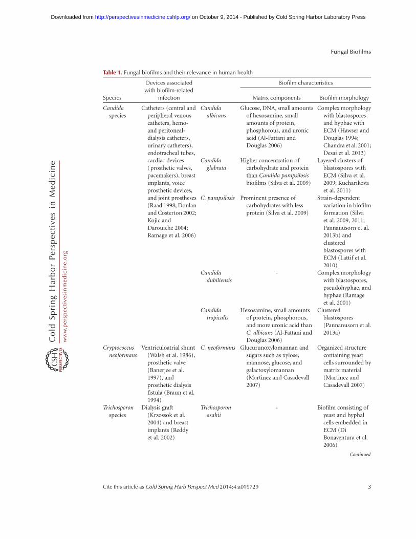
Table 1. Fungal biofilms and their relevance in human health
Species
Devices associated
with biofilm-related
infection
Biofilm characteristics
Matrix components Biofilm morphology
Candidaspecies
Catheters (central andperipheral venouscatheters, hemo-and peritoneal-dialysis catheters,urinary catheters),endotracheal tubes,cardiac devices(prosthetic valves,pacemakers), breastimplants, voiceprosthetic devices,and joint prostheses(Raad 1998; Donlanand Costerton 2002;Kojic andDarouiche 2004;Ramage et al. 2006)
Candidaalbicans
Glucose, DNA, small amountsof hexosamine, smallamounts of protein,phosphorous, and uronicacid (Al-Fattani andDouglas 2006)
Complex morphologywith blastosporesand hyphae withECM (Hawser andDouglas 1994;Chandra et al. 2001;Desai et al. 2013)
Candidaglabrata
Higher concentration ofcarbohydrate and proteinthan Candida parapsilosisbiofilms (Silva et al. 2009)
Layered clusters ofblastospores withECM (Silva et al.2009; Kucharikovaet al. 2011)
C. parapsilosis Prominent presence ofcarbohydrates with lessprotein (Silva et al. 2009)
Strain-dependentvariation in biofilmformation (Silvaet al. 2009, 2011;Pannanusorn et al.2013b) andclusteredblastospores withECM (Lattif et al.2010)
Candidadubiliensis
- Complex morphologywith blastospores,pseudohyphae, andhyphae (Ramageet al. 2001)
Candidatropicalis
Hexosamine, small amountsof protein, phosphorous,and more uronic acid thanC. albicans (Al-Fattani andDouglas 2006)
Clusteredblastospores(Pannanusorn et al.2013a)
Cryptococcusneoformans
Ventriculoatrial shunt(Walsh et al. 1986),prosthetic valve(Banerjee et al.1997), andprosthetic dialysisfistula (Braun et al.1994)
C. neoformans Glucurunoxylomannan andsugars such as xylose,mannose, glucose, andgalactoxylomannan(Martinez and Casadevall2007)
Organized structurecontaining yeastcells surrounded bymatrix material(Martinez andCasadevall 2007)
Trichosporonspecies
Dialysis graft(Krzossok et al.2004) and breastimplants (Reddyet al. 2002)
Trichosporonasahii
- Biofilm consisting ofyeast and hyphalcells embedded inECM (DiBonaventura et al.2006)
Continued
Fungal Biofilms
Cite this article as Cold Spring Harb Perspect Med 2014;4:a019729 3
ww
w.p
ersp
ecti
vesi
nm
edic
ine.
org
on October 9, 2014 - Published by Cold Spring Harbor Laboratory Press http://perspectivesinmedicine.cshlp.org/Downloaded from
One useful approach to define biofilm genefunctions has been to identify transcription fac-tors that are required for biofilm formation. Therationale behind the approach came from thesuspicion that many gene products—such asthe large families of ALS (agglutinin-like se-quence) proteins, CFEM (common in fungalextracellular membranes) proteins, and proteinsresembling Hyr1—might have compensatory oroverlapping functions in biofilm formation(Nobile and Mitchell 2005). The idea was thata single transcription factor is often required forexpression of many functionally related genes,and thus a defect in a transcription factor mu-tant may have a more pronounced phenotypicresult than a defect in a single target gene. (Theapproach was based on the analysis of meiot-ic gene regulation in Saccharomyces cerevisiae[Mitchell 1994], in which many meiotic reg-ulatory mutations had more dramatic pheno-types than target gene mutants.) Moreover, therelevant target genes could be identified throughexpression profiling (Nobile and Mitchell 2005)or, in subsequent studies, chromatin-immuno-precipitation analysis (Nobile et al. 2012). Theapproach of screening a panel of transcriptionfactor mutants for biofilm defects was produc-tive in yielding the first biofilm regulator, Bcr1(Nobile and Mitchell 2005), which was not re-quired for hyphal morphogenesis in the biofilm(see below). A more recent implementation ofthis approach has led to the finding that six keybiofilm regulators all govern each other’s expres-sion (Nobile et al. 2012), and a large spectrum ofregulators of adherence converge on the regula-
tion of a limited number of cell surface proteins(Finkel et al. 2012). We think that this approachremains an excellent strategy for dissection ofbiological processes.
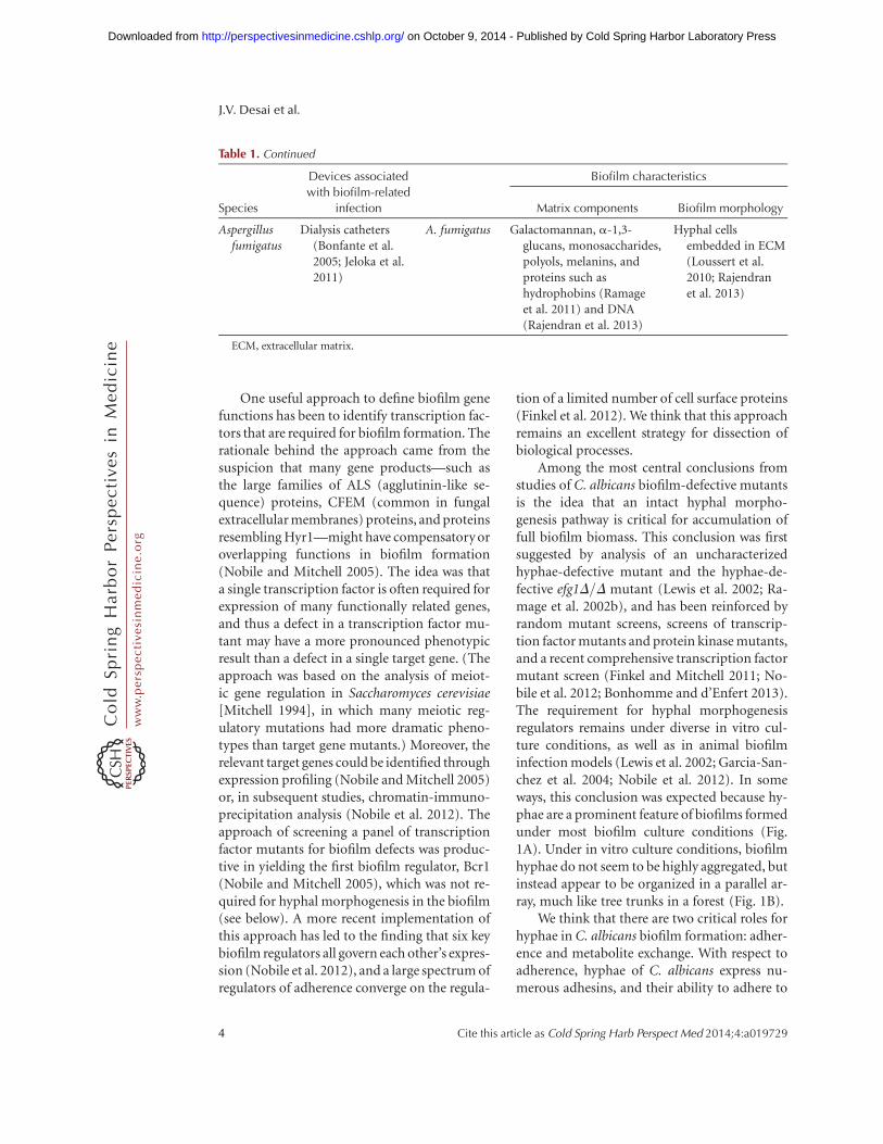
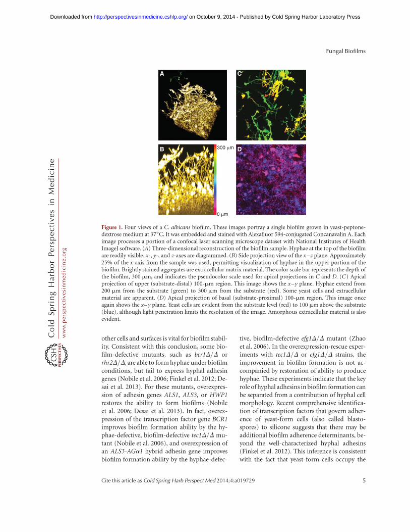
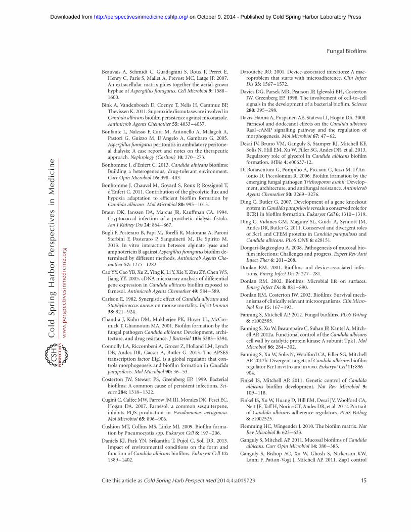
Among the most central conclusions fromstudies of C. albicans biofilm-defective mutantsis the idea that an intact hyphal morpho-genesis pathway is critical for accumulation offull biofilm biomass. This conclusion was firstsuggested by analysis of an uncharacterizedhyphae-defective mutant and the hyphae-de-fective efg1D/D mutant (Lewis et al. 2002; Ra-mage et al. 2002b), and has been reinforced byrandom mutant screens, screens of transcrip-tion factor mutants and protein kinase mutants,and a recent comprehensive transcription factormutant screen (Finkel and Mitchell 2011; No-bile et al. 2012; Bonhomme and d’Enfert 2013).The requirement for hyphal morphogenesisregulators remains under diverse in vitro cul-ture conditions, as well as in animal biofilminfection models (Lewis et al. 2002; Garcia-San-chez et al. 2004; Nobile et al. 2012). In someways, this conclusion was expected because hy-phae are a prominent feature of biofilms formedunder most biofilm culture conditions (Fig.1A). Under in vitro culture conditions, biofilmhyphae do not seem to be highly aggregated, butinstead appear to be organized in a parallel ar-ray, much like tree trunks in a forest (Fig. 1B).
We think that there are two critical roles forhyphae in C. albicans biofilm formation: adher-ence and metabolite exchange. With respect toadherence, hyphae of C. albicans express nu-merous adhesins, and their ability to adhere to
Table 1. Continued
Species
Devices associated
with biofilm-related
infection
Biofilm characteristics
Matrix components Biofilm morphology
Aspergillusfumigatus
Dialysis catheters(Bonfante et al.2005; Jeloka et al.2011)
A. fumigatus Galactomannan, a-1,3-glucans, monosaccharides,polyols, melanins, andproteins such ashydrophobins (Ramageet al. 2011) and DNA(Rajendran et al. 2013)
Hyphal cellsembedded in ECM(Loussert et al.2010; Rajendranet al. 2013)
ECM, extracellular matrix.
J.V. Desai et al.
4 Cite this article as Cold Spring Harb Perspect Med 2014;4:a019729
ww
w.p
ersp
ecti
vesi
nm
edic
ine.
org
on October 9, 2014 - Published by Cold Spring Harbor Laboratory Press http://perspectivesinmedicine.cshlp.org/Downloaded from
other cells and surfaces is vital for biofilm stabil-ity. Consistent with this conclusion, some bio-film-defective mutants, such as bcr1D/D orrhr2D/D, are able to form hyphae under biofilmconditions, but fail to express hyphal adhesingenes (Nobile et al. 2006; Finkel et al. 2012; De-sai et al. 2013). For these mutants, overexpres-sion of adhesin genes ALS1, ALS3, or HWP1restores the ability to form biofilms (Nobileet al. 2006; Desai et al. 2013). In fact, overex-pression of the transcription factor gene BCR1improves biofilm formation ability by the hy-phae-defective, biofilm-defective tec1D/D mu-tant (Nobile et al. 2006), and overexpression ofan ALS3-AGa1 hybrid adhesin gene improvesbiofilm formation ability by the hyphae-defec-
tive, biofilm-defective efg1D/D mutant (Zhaoet al. 2006). In the overexpression-rescue exper-iments with tec1D/D or efg1D/D strains, theimprovement in biofilm formation is not ac-companied by restoration of ability to producehyphae. These experiments indicate that the keyrole of hyphal adhesins in biofilm formation canbe separated from a contribution of hyphal cellmorphology. Recent comprehensive identifica-tion of transcription factors that govern adher-ence of yeast-form cells (also called blasto-spores) to silicone suggests that there may beadditional biofilm adherence determinants, be-yond the well-characterized hyphal adhesins(Finkel et al. 2012). This inference is consistentwith the fact that yeast-form cells occupy the
A
B
C
D300 µm
0 µm
Figure 1. Four views of a C. albicans biofilm. These images portray a single biofilm grown in yeast-peptone-dextrose medium at 37˚C. It was embedded and stained with Alexafluor 594-conjugated Concanavalin A. Eachimage processes a portion of a confocal laser scanning microscope dataset with National Institutes of HealthImageJ software. (A) Three-dimensional reconstruction of the biofilm sample. Hyphae at the top of the biofilmare readily visible. x-, y-, and z-axes are diagrammed. (B) Side projection view of the x–z plane. Approximately25% of the x-axis from the sample was used, permitting visualization of hyphae in the upper portion of thebiofilm. Brightly stained aggregates are extracellular matrix material. The color scale bar represents the depth ofthe biofilm, 300 mm, and indicates the pseudocolor scale used for apical projections in C and D. (C) Apicalprojection of upper (substrate-distal) 100-mm region. This image shows the x–y plane. Hyphae extend from200 mm from the substrate (green) to 300 mm from the substrate (red). Some yeast cells and extracellularmaterial are apparent. (D) Apical projection of basal (substrate-proximal) 100-mm region. This image onceagain shows the x–y plane. Yeast cells are evident from the substrate level (red) to 100 mm above the substrate(blue), although light penetration limits the resolution of the image. Amorphous extracellular material is alsoevident.
Fungal Biofilms
Cite this article as Cold Spring Harb Perspect Med 2014;4:a019729 5
ww
w.p
ersp
ecti
vesi
nm
edic
ine.
org
on October 9, 2014 - Published by Cold Spring Harbor Laboratory Press http://perspectivesinmedicine.cshlp.org/Downloaded from
basal biofilm layers in most in vitro–grown bio-films (Fig. 1D).
In addition, hyphal cells may have a func-tional role in biofilm formation that is indepen-dent of their adhesins. Specifically, the parallelhyphae of in vitro biofilms provide good solventaccess to the lower layer of the biofilm (Fig. 1B).Bacterial biofilm growth depends critically onsolvent channels that presumably facilitate re-moval of metabolic wastes and entry of nutri-ents (Davies et al. 1998; Wilking et al. 2013). Forexample, Pseudomonas aeruginosa biofilms area collection of mushroom-like microcolonieswhose spacing creates solvent channels (Davieset al. 1998). Perhaps filamentous hyphae andpseudohyphae in fungal biofilms function sim-ilarly to improve metabolite exchange amongbiofilm cells.
The possibility that there is a distinctivephysiology of biofilm cells was first indicatedby transcriptomic comparisons of biofilm andplanktonic cells. Among these studies we note,in particular, the foundational work of the d’En-fert laboratory, in which a core set of biofilm-induced genes was defined through multiplebiofilm-planktonic comparisons (Garcia-Sanchezet al. 2004). One large subset of these genes wasassociated with hyphal development, as mightbe expected from the discussion above. In ad-dition, a substantial proportion of these coreup-regulated genes were associated with physi-ological functions such as protein synthesis,amino acid synthesis, and glycolytic flux. Anal-ysis of suitable mutant strains (e.g., gcn4D/D,tye7D/D) has confirmed the functional require-ment for these physiological processes in opti-mal biofilm growth (Garcia-Sanchez et al. 2004;Bonhomme et al. 2011).
Revealing insight into biofilm cell physiolo-gy came in part from comparative transcrip-tomic studies (Rossignol et al. 2009). The Butlerand d’Enfert laboratories focused on Candidaparapsilosis, a highly significant cause of device-associated infection. C. parapsilosis, like C. albi-cans, is a member of the CTG clade, but it makesonly pseudohyphae, not true hyphae. The studyfound a highly significant correlation betweenthe gene-expression signature of biofilm cellsand the gene-expression response to hypoxia,
or low oxygen conditions. Functional analysisin C. albicans has extended this hypothesis withthe finding that adaptation to hypoxia is criticalfor biofilm integrity (Bonhomme et al. 2011).Thus, the hypoxia response is a major drivingforce behind the gene expression and physiolo-gy of biofilm cells.
Among the most perplexing observationsregarding C. albicans biofilm gene expressionis that biofilm cells up-regulate genes for sulfurassimilation and the biosynthesis of methionineand cysteine (Garcia-Sanchez et al. 2004; Mu-rillo et al. 2005). These genes are also up-regu-lated as part of the hypoxic response (Stichter-noth and Ernst 2009). It seems possible thatthese two sulfur amino acids may be overrepre-sented in biofilm translation products, or per-haps there are greater demands for the methyldonor S-adenosyl-methionine in biofilm cellsas compared with planktonic cells. It has alsobeen suggested that the reducing environmentcreated by cysteine may be necessary to detoxifyreactive oxygen species in biofilm cells (Stich-ternoth and Ernst 2009). In view of the key roleof hypoxic adaptation in biofilm formation, wepropose an additional metabolic explanationfor this regulatory response. C. albicans can-not ferment sugars, and thus it usually reducesoxygen through aerobic respiration to balancereducing equivalents from glycolysis. When ox-ygen is limiting, it may adapt by using otherreduction reactions to achieve this balance.The sulfur assimilation pathway includes mul-tiple reduction steps to convert sulfate to sul-fide. We suggest that C. albicans uses sulfate asan alternative electron acceptor under hypoxicconditions to balance reducing equivalents thatare generated from glycolysis.
Biofilms are not, simply, slightly hypoxicaggregates of yeast cells and hyphae; a biofilmcommunity has complex phenotypes. For ex-ample, biofilm cells produce an extracellularmatrix and are resistant to many antimicrobialagents, as discussed below. In addition, yeastcells are not restricted to the lower layers ofthe biofilm (Fig. 1C), and the yeast cells in theupper biofilm sites have distinct propertiesfrom planktonic yeast cells (see below) (Uppu-luri et al. 2010). Many other biofilm pheno-
J.V. Desai et al.
6 Cite this article as Cold Spring Harb Perspect Med 2014;4:a019729
ww
w.p
ersp
ecti
vesi
nm
edic
ine.
org
on October 9, 2014 - Published by Cold Spring Harbor Laboratory Press http://perspectivesinmedicine.cshlp.org/Downloaded from
types have been assayed in focused studies ormore broad-ranging screens. Among the mostremarkable findings in this context is the dis-covery that yeast-form cells released from C.albicans biofilms have properties that are dis-tinct from planktonic yeast-form cells (Uppu-luri et al. 2010). Most noteworthy is the fact thatreleased yeast-form cells are hypervirulent ina mouse-disseminated infection model com-pared with a planktonic inoculum (Uppuluriet al. 2010). Biofilm cell release is governed byseveral hyphal regulators, as well as HSP90(Robbins et al. 2011). This result has led to theproposal that HSP90 inhibitors could be used toreduce disseminated infection rates (Robbinset al. 2011). Therefore, it appears that C. albicansbiofilms promote infection not only by servingas a pathogen reservoir, but also through phys-iological changes that augment the virulence ofdispersed cells.
Gene-expression profiling of Candida bio-films has been useful not only for inferring keycellular processes, but also for defining specificgene functions that are required for normal bio-film formation (Bonhomme et al. 2011; Desaiet al. 2013). A recent comparison of A. fumiga-tus biofilm and planktonic transcriptomes hasthe potential to open the door to functionalanalysis in that organism (Gibbons et al. 2012;Muszkieta et al. 2013). Biofilm up-regulatedgenes were enriched for such functions as trans-lation, stress responses, transport, and cell wall/cell surface features. These are just the sorts ofgene functions that may reflect common themesin fungal biofilm formation. Interestingly, therewas also enrichment for secondary metabolismgenes, which may represent a unique feature ofA. fumigatus biofilm formation. Given the manydifferences between molds and yeasts, we antic-ipate that functional analysis of A. fumigatusbiofilm formation will reveal many distinctiveprocesses not evident in Candida biofilms.
The basidiomycete Cryptococcus neoformansis a distant relative of the ascomycetes such asCandida and Aspergillus, and several novel find-ings have emerged from the study of C. neofor-mans biofilms and cell–cell adherence. Cell–cell communication is critical for C. neoformansbiofilm formation and is coupled to adherence
in a novel way. Specifically, the surface/secretedprotein Cfl1 functions both as an adhesin and asignaling molecule that induces hyphal devel-opment (Wang et al. 2012, 2013). CFL1 is underthe control of the mating regulator Znf2 (Wanget al. 2012, 2013), and it has yet to be deter-mined whether it functions only in mating rath-er than in overall biofilm formation. Nonethe-less, a recent study revealed that conditionedmedium indeed stimulates C. neoformans bio-film formation (Albuquerque et al. 2013), inkeeping with the idea that secreted productspromote, rather than inhibit, biofilm forma-tion. In contrast, the most well-studied quo-rum-sensing molecule from C. albicans, farne-sol (Nickerson et al. 2006; Madhani 2011),inhibits biofilm formation when supplied exog-enously in pure form (Ramage et al. 2002a; Caoet al. 2005), although effects of farnesol fromendogenous biofilms may be more difficult topredict (Ganguly et al. 2011). Clearly, our un-derstanding of C. neoformans biofilm formationis in its infancy, and presents an opportunity forthe discovery of novel mechanisms that supportbiofilm formation.
Although we usually think of biofilm for-mation as a single process, it has become clearthat overall biofilm development depends great-ly on the environment. For example, Soll andcolleagues have shown that the structures ofbiofilms grown in different media have very dif-ferent structures (Daniels et al. 2013). Thus, it islikely that the relative prominence of many reg-ulatory relationships in biofilm formation maybe environmentally contingent. This conclu-sion was anticipated by the diverse biofilm tran-scriptomic datasets of the d’Enfert laboratory(Garcia-Sanchez et al. 2004), and the findingthat several mutant strains are biofilm defectiveunder some conditions, but not others (Richardet al. 2005; Nobile et al. 2006). In addition, bio-films of mating-competent C. albicans strains,which are homozygous at the MTL locus, createbiofilms with some genetic and environmentalrequirements that are distinct from those of themore frequently isolated MTLa/MTLa strains(Sahni et al. 2010; Park et al. 2013; Si et al. 2013).In fact, Fanning et al. (2012a) have shown thatthe biofilm regulator Bcr1 has distinct sets of
Fungal Biofilms
Cite this article as Cold Spring Harb Perspect Med 2014;4:a019729 7
ww
w.p
ersp
ecti
vesi
nm
edic
ine.
org
on October 9, 2014 - Published by Cold Spring Harbor Laboratory Press http://perspectivesinmedicine.cshlp.org/Downloaded from
responsive genes in the same strain during bio-film growth in vitro and on the oral mucosa invivo. Epistasis analysis of mutations in BCR1and the cAMP-dependent protein kinase geneTPK1 provide insight in this regard; loss ofTpk1 function permits biofilm formation by abcr1D/D strain. However, loss of Tpk1 functiondoes not restore expression of many majorBcr1-dependent genes, but instead activates al-ternative genes that can compensate for the de-fect in Bcr1 (Fanning et al. 2012a). Tpk1 andBcr1 thus govern alternative target gene sets,and either one can promote biofilm formation(Fanning et al. 2012a). The ability of fungi toform biofilms in diverse environments may re-flect reliance on a portfolio of gene functions,groups of which enable adaptation to a subset ofenvironmental conditions.
MULTISPECIES BIOFILMS
One of the most important environmental fac-tors that can modulate fungal biofilm formationand its consequences is the presence of bacteria.Clinical and experimental data underscore thesignificance of these interactions, as explainedin several excellent recent reviews (Morales andHogan 2010; Peleg et al. 2010, 2012a; Harriottand Noverr 2011). Some of the most dramaticresults come from studies with C. albicans andStaphylococcus aureus: coinfection causes muchgreater mortality than single-species infectionin a mouse model (Carlson 1982). In addition,C. albicans enables S. aureus biofilm formationin serum and decreases its susceptibility to van-comycin (Harriott and Noverr 2009). Thesesimple experiments underscore the need to un-derstand polymicrobial interactions to guide di-agnosis and therapy.
The distinct properties of mixed-speciesbiofilms arise from both direct cell–cell contactand the impact of secreted molecules. Amongthe best-characterized bacterial receptors onC. albicans is the surface adhesin Als3. Jenkin-son and colleagues used heterologous expres-sion to show that Als3 and Eap1 are sufficientto promote binding to the oral bacterium Strep-tococcus gordonii (Nobbs et al. 2010). Remark-ably, Als3 is also responsible for C. albicans
binding to S. aureus (Peters et al. 2012b). Giventhat ALS3 and EAP1 are both greatly inducedduring biofilm formation, a C. albicans biofilmwould provide an environment that couldmaintain these bacteria as coaggregates. Jenkin-son and colleagues underscore that S. gordonii isa pioneer colonizer of the oral cavity, and thusargue that coaggregate formation may increaselevels of C. albicans carriage (Nobbs et al. 2010).
Secreted small molecules, such as quorum-sensing molecules and metabolites, are also animportant factor in fungal–bacterial interac-tion. For example, the Hogan group has shownthat the C. albicans quorum-sensing molecule,farnesol, inhibits expression of P. aeruginosa vir-ulence factors by affecting its quinolone signalproduction machinery (Cugini et al. 2007). Ja-bra-Rizk et al. (2006) have shown that farnesolinhibits S. aureus biofilm formation, thus thisfungal quorum-sensing molecule impacts bothGram-negative and -positive bacteria. Bacteriaare all too happy to return the favor; the P.aeruginosa quorum-sensing molecule, homo-serinelactone, inhibits hyphal formation by C.albicans through inhibition of the Ras-adenyl-ate cyclase pathway (Davis-Hanna et al. 2008;Hall et al. 2011). Given that yeast-form cells aremost easily dispersed from C. albicans biofilms(Uppuluri et al. 2010), one would expect thepresence of P. aeruginosa to promote C. albicansbiofilm dispersal.
We have provided only a small sampling ofthe types of interactions that may confer uniqueproperties of polymicrobial biofilms. Clearly,there is much to learn, and clinical observationsserve as a useful guide to significant interactions(Morales and Hogan 2010; Peleg et al. 2010;Harriott and Noverr 2011; Peters et al. 2012a).It seems likely that mixed species interactionsare among the driving forces behind the evolu-tion of fungal biofilm gene functions.
BIOFILM COMPARATIVE GENOMICS
Can the detailed knowledge of C. albicans bio-film formation be applied to the biofilms ofother fungal pathogens? The Butler laboratoryhas pioneered the use of comparative genomicsto explore the functions of C. parapsilosis ortho-
J.V. Desai et al.
8 Cite this article as Cold Spring Harb Perspect Med 2014;4:a019729
ww
w.p
ersp
ecti
vesi
nm
edic
ine.
org
on October 9, 2014 - Published by Cold Spring Harbor Laboratory Press http://perspectivesinmedicine.cshlp.org/Downloaded from
logs of C. albicans biofilm regulators. They havefound that both Bcr1 and Efg1 are required forC. parapsilosis biofilm formation (Ding andButler 2007; Ding et al. 2011; Connolly et al.2013). Bcr1 has analogous functions, to someextent, in both species; the bcr1D/D mutantC. parapsilosis biofilms have reduced biomass,and there is no defect in morphogenesis of thefilamentous cells—pseudohyphae—that thisspecies produces (Ding and Butler 2007; Dinget al. 2011). However, there is limited overlapamong Bcr1 target genes in the two species. Theshared targets include cell surface CFEM pro-tein genes, an expanded gene family in C. para-psilosis that is involved in heme-iron acquisition(Ding and Butler 2007; Almeida et al. 2009).However, orthologs of the C. albicans biofilmadhesin genes ALS1, ALS3, and HWP1 are notdependent on Bcr1 for expression in C. para-psilosis (Ding and Butler 2007; Ding et al. 2011).In fact, the Bcr1-dependent genes that governC. parapsilosis biofilm formation are unknownat present; several CFEM protein mutants testeddo not affect biofilm biomass or structure (Dinget al. 2011). The Efg1 orthologs of C. parapsi-losis and C. albicans have greater overlap amongtheir target genes (Connolly et al. 2013), whichinclude several transcription factor genes thatare members of the C. albicans biofilm regula-tory network (Nobile et al. 2012). The emergingconclusion from these studies is that biofilmregulators are conserved, whereas their targetgenes are not. One caveat is that there may bemany examples of transcription factors whosetarget genes are contingent on the environment;as noted by the Butler group, the number ofBcr1- and Efg1-dependent genes detected indifferent studies of C. albicans varied greatly(Ding et al. 2011; Fanning et al. 2012b; Nobileet al. 2012; Connolly et al. 2013).
BIOFILM EXTRACELLULAR MATRIX
Production of an extracellular matrix is one ofthe distinguishing characteristics of both eu-karyotic and prokaryotic biofilms (Hawser etal. 1998; Donlan and Costerton 2002; O’Toole2003). In many organisms, this material is crit-ical for biofilm formation, providing the scaf-
fold for surface adhesion and cellular ag-gregation to maintain the biofilm architecture(Flemming and Wingender 2010). Extracellularmatrix has also been shown in some organismsto assist with retention of water and absorptionof nutrients. In addition, the material may bedegraded during nutrient-limiting conditionsby microbially produced enzymes to providea carbon or nitrogen source (Flemming andWingender 2010). One of the most medicallyimportant attributes of the extracellular matrixis its ability to provide protection from environ-mental insults, including host defenses andantimicrobial therapies (Costerton et al. 1999;Donlan 2001).
Hawser et al. (1998) first described the pres-ence of C. albicans biofilm matrix material andits variation with environmental conditions.The clinical relevance of this material is evidentby its presence on the catheters of patients withCandida biofilm (Paulitsch et al. 2009). Animalmodels of device-associated infections also re-veal abundant biofilm matrix material and pro-vide an opportunity to study matrix function(Andes et al. 2004; Nett et al. 2010b; Johnsonet al. 2012). In vivo, the matrix is postulatedto contain both the microbial-derived compo-nents identified in vitro, as well as host proteins,such as plasma and salivary proteins that adsorbto the surface of medical devices (Nett and An-des 2006).
The Douglas group has performed severalkey investigations to examine the content ofCandida biofilm matrix (Table 1) (Baillie andDouglas 2000). They then probed the functionsof matrix components in adherence throughenzymatic digestion and measurement of cellu-lar detachment (Al-Fattani and Douglas 2006).This study pointed to roles for matrix protein,chitin, extracellular DNA (eDNA), and b-1,3glucan in surface adhesion or cellular cohesion.
Matrix protein constituents have been iden-tified by Lopez-Ribot and colleagues through2D PAGE and mass spectrometry (Thomas etal. 2006). Surprisingly, the most abundant bio-film matrix proteins were very similar to thosein planktonic culture supernatants. Thus, ma-trix protein function during in vitro growth re-flects a change in the state of secreted proteins
Fungal Biofilms
Cite this article as Cold Spring Harb Perspect Med 2014;4:a019729 9
ww
w.p
ersp
ecti
vesi
nm
edic
ine.
org
on October 9, 2014 - Published by Cold Spring Harbor Laboratory Press http://perspectivesinmedicine.cshlp.org/Downloaded from
rather than a change in their identity. Matrixproteins included both predicted secretomecomponents, as well as many predicted cyto-plasmic proteins. It seems likely that cell lysiscontributes to the matrix through release ofproteins and DNA. Although matrix eDNAhas a role in biofilm structure and drug suscept-ibility (discussed below), the roles of the indi-vidual matrix proteins in biofilm properties isnot well understood.
Interestingly, the matrix content appears tovary significantly among Candida species (Al-Fattani and Douglas 2006; Silva et al. 2009). Forexample, compared with C. albicans, Candidatropicalis biofilms have higher hexosamine con-tent (27%) and are not disrupted by deoxyri-bonuclease (DNase) treatment (Al-Fattani andDouglas 2006). Also, matrix composition is de-pendent on environmental and media condi-tions. For example, C. albicans biofilms formedin RPMI (Roswell Park Memorial Institute) me-dium have an �1000�-fold higher concentra-tion of matrix eDNAwhen compared with yeastnitrogen base medium (Martins et al. 2010).
Like Candida, Aspergillus biofilms have beenlinked to infection of medical devices (Ramageet al. 2011). However, biofilm-like growth hasalso been associated with the most commonAspergillus infections, including invasive pul-monary aspergillosis and aspergilloma (Lous-sert et al. 2010). As a clinical niche, the lungvaries greatly from the environments for mostCandida biofilm infections. The ability of themost common Aspergillus species, A. fumigatus,to form a multicellular community with asurrounding extracellular matrix material hasbeen shown both in vitro and in vivo (Beauvaiset al. 2007; Loussert et al. 2010). Ultrastructureanalysis shows that these biofilms are composedof hyphae coalescing into a three-dimensionalstructure covered in dense extracellular materialwith embedded air channels (Beauvais et al.2007).
Beauvais et al. (2007) found that the extra-cellular matrix of in vitro A. fumigatus biofilmsis composed of galactomannan, a-1,3 glucan,monosaccharides, polyols, melanin, and pro-tein (Table 1). Immunolabeling showed thatgalactomannan was deposited throughout the
cell wall and matrix. In contrast, the a-1,3glucan label was the highest in the amorphousextracellular matrix material near the hyphalsurface. The protein fraction of matrix was small(2%) and included several major secreted anti-gens, as well as a group of hydrophobins. Thesehydrophobic surface-active proteins, common-ly associated with aerial growth, are presumedto play a role in cell–cell adhesion in the bio-film. Recent analysis also has shown that eDNAaccumulates in the matrix of A. fumigatus bio-films in vitro (Rajendran et al. 2013; Shopovaet al. 2013). This eDNA is identical to genomicDNA and is probably released through autolysis(Rajendran et al. 2013).
By studying the resected aspergillomas ofseveral patients, Loussert et al. (2010) shed lighton the in vivo composition of A. fumigatusbiofilms. Similar to in vitro biofilms, galacto-mannan was identified throughout the fungalcell wall and extracellular matrix of in vivo bio-films on immunolabeling. Also, a-1,3 glucanwas found in the matrix material located mostproximal to the cell wall surface. Compared within vitro biofilms, in vivo biofilms appeared tohave a higher concentration of the recently iden-tified cell wall polysaccharide galactosaminoga-lactan. Recent infection model studies indicatethat galactosaminogalactan mediates adher-ence, but also masks b-1,3 glucan to limit hostrecognition (Gravelat et al. 2013). The presenceof melanin in the biofilm matrix was confirmedin vivo, whereas antigenic proteins were not asapparent in vivo. When comparing the aspergil-lomas of patients with a murine model of inva-sive pulmonary aspergillosis, many similaritieswere noted, including the presence of both ma-trix galactomannan and galactosaminogalactan(Loussert et al. 2010). However, unlike in vitrobiofilms and the aspergilloma biofilm, the ex-tracellular matrix of the pulmonary aspergillosisbiofilms did not appear to have a high concen-tration of a-1,3 glucan.
Candida MATRIX AND BIOFILM DRUGRESISTANCE
Several investigations have examined the contri-bution of the biofilm extracellular matrix to the
J.V. Desai et al.
10 Cite this article as Cold Spring Harb Perspect Med 2014;4:a019729
ww
w.p
ersp
ecti
vesi
nm
edic
ine.
org
on October 9, 2014 - Published by Cold Spring Harbor Laboratory Press http://perspectivesinmedicine.cshlp.org/Downloaded from
drug resistance phenotype associated with theCandida biofilm lifestyle (Baillie and Douglas2000; Al-Fattani and Douglas 2004, 2006;Samaranayake et al. 2005). These studies haveused several experimental designs to test the hy-pothesis that matrix may prevent access of anti-fungals to the cells embedded in the biofilm,postulating that the material may either slowthe rate of drug transport or specifically bindantifungals extracellularly. Together, these in-vestigations found that an intact matrix is oneof the most influential factors promoting thedrug resistance of biofilms formed by differentCandida species.
Early studies from the Douglas group impli-cated the C. albicans biofilm matrix in drug re-sistance (Baillie and Douglas 2000; Al-Fattaniand Douglas 2004, 2006). To explore the possi-bility that matrix impedes drug access to biofilmcells, Samaranayake et al. (2005) measured drugpenetration using filter-grown biofilms. C. albi-cans biofilms inhibited the penetration of allantifungals tested, including fluconazole, am-photericin B, and flucytosine. C. parapsilosisand Candida krusei biofilms also impeded am-photericin B, but flucytosine and fluconazolepenetration was higher than for C. albicans bio-films. Hence, this investigation suggests that thematrix contributes biofilm antifungal resistanceby limiting drug diffusion, although it is unclearthat affecting the diffusion gradient would besufficient to abrogate drug sensitivity entirely.
Several key observations led to the discoveryof a drug sequestration activity for the Candidabiofilm matrix. The Andes group added isolatedCandida biofilm matrix to planktonic mini-mum growth inhibition concentration assaysand found that this material was able to providea degree of antifungal resistance to the nonbio-film cells (Nett et al. 2007). To test the hypoth-esis that the matrix interacts with antifungals,preventing drug penetration into the cells, ra-diolabeled fluconazole was tracked throughbiofilms. The vast majority of the drug wasfound in association with matrix, implicatingdrug sequestration. To explore the contributionof individual matrix components, the biofilmmatrix was exposed to enzymes targeting theconstituents. Degradation of matrix b-1,3 glu-
can impaired the biofilm-associated resistancemechanism, indicating a role for this matrixcomponent in protecting biofilms from antifun-gal drugs. The role for matrix b-1,3 glucan inbiofilm drug resistance during clinical infectionwas supported by the synergistic action of glu-canase and fluconazole on rat venous cathetersinfected with C. albicans (Nett et al. 2007).
Matrix b-1,3 glucan has been linked to re-sistance with other antifungals in distinct drugclasses. Vediyappan et al. (2010) have providedevidence that matrix b-1,3 glucan binds andsequesters amphotericin B. A similar resistancemechanism has also been shown for flucyto-sine (Nett et al. 2010a). Furthermore, biofilmsformed by other common Candida species, in-cluding Candida glabrata, C. parapsilosis, and C.tropicalis, also produce matrix b-1,3 glucan thatsequesters antifungal and enhances drug resis-tance (Mitchell et al. 2013). Therefore, drug se-questration by matrixb-1,3 glucan is a commonmechanism of biofilm-based resistance.
There are additional mechanisms that pro-mote C. albicans biofilm drug resistance. Forexample, several efflux pump genes are up-reg-ulated in biofilms (Mukherjee et al. 2003). Theefflux pumps have a detectable role in flucona-zole resistance in early biofilms, but their impactis more minor in mature biofilms with extensivematrix (Mukherjee et al. 2003). Finally, one ofthe most widespread biofilm resistance phe-nomena in bacteria, the presence of a small re-sistant subpopulation of “persister” cells (Lewis2010), occurs in C. albicans biofilms as well (La-Fleur et al. 2006). Interestingly, a study from theThevissen group showed that cell surface super-oxide dismutases are critical for the accumu-lation of persister cells (Bink et al. 2011). Thisstudy could be extended to other fungal biofilmsreadily through the use of superoxide dismutaseinhibitors (Bink et al. 2011). Thus, biofilm drugresistance is clearly multifactorial, a “perfectstorm” of matrix binding, efflux pump up-reg-ulation, and persister cell accumulation.
Candida MATRIX GLUCAN PRODUCTION
What are the genes that govern matrix b-1,3glucan production? In C. albicans, FKS1 en-
Fungal Biofilms
Cite this article as Cold Spring Harb Perspect Med 2014;4:a019729 11
ww
w.p
ersp
ecti
vesi
nm
edic
ine.
org
on October 9, 2014 - Published by Cold Spring Harbor Laboratory Press http://perspectivesinmedicine.cshlp.org/Downloaded from
codes a glucan synthase responsible for manu-facture of cell wall b-1,3 glucan during plank-tonic growth. Nett et al. (2010a,b) showedthe role of FKS1 in matrix-mediated drug se-questration with two key observations: reducedexpression of FKS1 expression causes reducedmatrix b-1,3 glucan and increased sensitivityof biofilm cells to fluconazole, and increasedexpression of FKS1 increased matrix b-1,3 glu-can and decreased sensitivity of biofilm cells tofluconazole. Fluconazole sensitivity was unaf-fected when the strains were grown under non-biofilm conditions (Nett et al. 2010a,b). Thephenotypes were shown both in vitro and in arat venous catheter model of biofilm infection.Thus, Fks1 is required to generate matrix b-1,3glucan and full levels of biofilm drug resistance.
To delineate the process by which glucan ismodified and delivered to the extracellular bio-film matrix, Taff et al. (2012) used a candidategene approach, examining a set of mutantsbased on transcriptional profiling of C. albicansin a rat venous catheter biofilm. They identifieda role for two predicted glucan transferases(Bgl2 and Phr1) and an exoglucanase (Xog1)in both the production of biofilm matrix anddevelopment of the resistance to antifungalsduring biofilm growth. Mutants with disrup-tion of one of these genes produced significantlyless matrixb-1,3 glucan and displayed increasedbiofilm susceptibility to azole drugs comparedwith the parent strain both in vitro and in a ratvenous catheter biofilm model. The isolatedmatrix of the mutant strains showed decreasedcapacity for antifungal sequestration. The geneproducts appear to have partially redundantfunctions, as the matrix sequestration defectwas more extreme in double mutants than insingle mutants. These gene products are pro-posed to modify matrix glucan generated byFks1 for development of functional matrix ca-pable of sequestering antifungal drugs and pro-moting biofilm antifungal drug resistance.
Several pathways regulate production ofCandida biofilm matrix and drug resistance.Nobile et al. (2009) discovered a critical negativeregulator of C. albicans biofilm matrix glucanproduction both in vitro and in vivo. In a screenof transcription factor mutants, the zap1 – / –
mutant was noted to have a glistening appear-ance that correlated with abundant matrixglucan. Through transcription profiling andchromatin immunoprecipitation, several groupsof target genes were identified. First, Zap1 wasfound to repress transcription of two glucoamy-lase genes (GCA1 and GCA2) that may have apositive role in matrix production, likely by hy-drolyzing insoluble carbohydrates. A secondgroup of Zap1 targets includes three alcohol de-hydrogenase genes (ADH5, CSH1, and LFD6)that may influence biofilm formation and ma-trix glucan production through quorum signalpathways. Interestingly, the expression of BGL2,PHR1, and XOG1 do not appear to be underZap1 control (Taff et al. 2012). Thus, Zap1may govern matrix production through mecha-nisms that feed into, or converge with, the FKS1pathway defined above.
The Cowen group discovered heat shockprotein, Hsp90, as novel regulator of C. albicansbiofilm matrix production and drug resistance(Robbins et al. 2011). Disruption of this path-way increased the effectiveness of triazole drugsboth in vitro and in a rat venous catheter model.Unlike planktonic conditions, this pathway wasnot modulated via calcineurin or Mkc1 path-ways. The finding that inhibition of Hsp90pactivity led to decreased b-1,3 glucan in thematrix of C. albicans biofilms suggests thatHsp90p contributes to the production of drugsequestering matrix. How Hsp90p specificallymodulates this activity is unknown, but it ishypothesized to stabilize one or more of theproteins involved in production and regulationof matrix production, such as Fks1 or Zap1.
A search for additional regulators of bio-film matrix-associated drug resistance identi-fied downstream components of the yeast pro-tein kinase C (PKC) pathway (Nett et al. 2011).C. albicans mutants with disruption of eitherSMI1 or RLM1 were deficient in both matrixglucan production and susceptibility to azoledrugs. These factors are thought to govern ma-trix-associated drug resistance through FKS1, asFKS1 overexpression restored the biofilm-asso-ciated drug-resistant phenotype. Surprisingly,upstream PKC pathway components do notappear to be involved in regulation of biofilm
J.V. Desai et al.
12 Cite this article as Cold Spring Harb Perspect Med 2014;4:a019729
ww
w.p
ersp
ecti
vesi
nm
edic
ine.
org
on October 9, 2014 - Published by Cold Spring Harbor Laboratory Press http://perspectivesinmedicine.cshlp.org/Downloaded from
matrix production and drug resistance. Thissuggests that matrix-associated drug resistanceand cell wall integrity are networked, but thebiofilm drug resistance is triggered through apathway distinct from the PKC pathway. Thenature of the crosstalk that engages PKC path-way downstream components independently ofPKC pathway upstream components is not un-derstood at this time.
The Soll laboratory has also explored thegenetic control of a panel of matrix-related phe-notypes. They found that a mating-competentMTLa/MTLa C. albicans biofilm differs from amating incompetent MTLa/MTLa biofilm inbeing more permeable to dyes and neutrophilsand more sensitive to fluconazole (Srikanthaet al. 2013). Their genetic studies indicatedthat in the MTLa/MTLa strain reduced activityof the Bcr1 transcription factor, a known posi-tive regulator of biofilm formation, was thecause of the three phenotypes of dye and neu-trophil permeability and fluconazole sensitivity(Yi et al. 2011). Accordingly, they developed anoverexpression approach to identify several Bcr1target genes that govern the three phenotypes(Srikantha et al. 2013). Notable among the geneproducts whose overexpression increases thelevel of dye and neutrophil permeability andfluconazole sensitivity were a set of cell surfaceproteins with the CFEM motif (Srikantha et al.2013), which has been shown to mediate ironacquisition. The three phenotypes are all prob-ably related to extracellular matrix accumula-tion or composition. Consistent with this idea,Noverr and colleagues have shown that Bcr1 isrequired for matrix accumulation in a vaginalcolonization model (Harriott et al. 2010). It willbe interesting to see whether the CFEM proteinshave a structural role in the extracellular matrix,or if their effects are more indirect.
MATRIX eDNA AND DRUG RESISTANCE
The presence of eDNA in the Candida bio-film matrix is critical for biofilm integrity andmaintenance (Martins et al. 2010). Degradationof matrix eDNA not only destroys biofilmarchitecture, but the addition of exogenousDNA promotes biofilm growth (Martins et al.
2010). Martins et al. (2012) explored the rela-tionship between eDNA and C. albicans biofilmdrug resistance through DNase treatment.Degradation of eDNA significantly improvedthe activity of amphotericin B against Candidabiofilms, based on either metabolic activitymeasurements or cell viability assays. However,biofilms treated with DNase remained resistantto both fluconazole and caspofungin. Thissuggests that there may be a specific eDNA–amphotericin B interaction. An alternative pos-sibility is that the matrix eDNA may interactwith a variety of antifungals, but other resistanceprocesses in play maintain biofilm resistance oneDNA degradation. How matrix eDNA contrib-utes to Candida biofilm resistance remains un-known.
Aspergillus BIOFILM MATRIXAND DRUG RESISTANCE
Considerably less is known about the process ofAspergillus biofilm matrix production and howit impacts drug susceptibility during biofilmgrowth. The matrix of Aspergillus biofilms isthought to contribute to antimicrobial resis-tance, through either slowing drug transit ordirect drug sequestration (Beauvais et al. 2007;Bugli et al. 2013; Rajendran et al. 2013). Extra-cellular matrix content increases as A. fumigatusbiofilms mature, so matrix is thought to pro-mote drug resistance during the later phases ofbiofilm growth (Beauvais et al. 2007).
One recently recognized matrix componentshown to impact drug resistance in A. fumigatisis eDNA. Rajendran et al. (2013) showed thatthe content of genomic DNA in the matrix in-creased as in vitro A. fumigatus biofilms ma-tured from 8–48 h. DNA release was linked tochitinase activity, suggesting that autolysis isthe source of eDNA. Degradation of eDNA byDNase decreases the biomass of the biofilm andrenders biofilms more susceptible both to am-photericin B and caspofungin, an echinocan-din. Interestingly, exogenous DNA also pro-motes structural integrity of the biofilm andincreases biofilm matrix carbohydrate produc-tion, suggesting a potential role for host DNAduring in vivo Aspergillus biofilm formation
Fungal Biofilms
Cite this article as Cold Spring Harb Perspect Med 2014;4:a019729 13
ww
w.p
ersp
ecti
vesi
nm
edic
ine.
org
on October 9, 2014 - Published by Cold Spring Harbor Laboratory Press http://perspectivesinmedicine.cshlp.org/Downloaded from
(Rajendran et al. 2013; Shopova et al. 2013).Therefore, matrix eDNA appears to be impor-tant for both biofilm structural integrity andresistance to antifungals in A. fumigatus.
Much remains unknown about Aspergillusbiofilm drug resistance and how the differentcomponents of the biofilm matrix influencethis phenotype. An intriguing study by Bugliet al. (2013) found that administration of algi-nate lyase increased in vitro A. fumigatus biofilmsusceptibility to amphotericin B. Alginate lyasedegrades uronic-acid-containing carbohydratesand is thought to act on the extracellular mat-rix carbohydrates of Aspergillus biofilms. Thisstudy suggests that a previously unidentifiedmatrix carbohydrate is contributing to antifun-gal drug resistance during biofilm growth. Fur-ther investigation is of interest to characterizethis material and its specific role in resistance toantifungal treatment.
CONCLUDING REMARKS
The ability to form biofilms is widespreadamong pathogenic fungi. It is relevant to infec-tion biology in the context of device-associatedinfection and as an in vivo growth form. Despitethe broad significance of fungal biofilms, ourunderstanding of the mechanisms that governtheir formation, physiology, and drug resistanceis limited. The genetic tractability of C. albicanshas driven biofilm studies in that organism, andwe now have candidate genes and processes thatmight be tested in other fungal pathogens.Molds and basidiomycetes may have distinctprocesses that mediate biofilm formation andour growing understanding of A. fumigatusand C. neoformans biofilms provides conceptsto fuel testable hypotheses with related fungi.
There are many major questions on the ho-rizon of fungal biofilm studies. For example, cellheterogeneity is an important attribute of allbiofilms but, apart from recognizing yeast andhyphae, we have little understanding of howextensive cell heterogeneity may be in biofilms,how biofilm growth generates heterogeneity,and how heterogeneity may contribute to bio-film persistence or functional adaptation. Ourunderstanding of mixed species interactions in
biofilms remains in its infancy, and we suspectthat computational analysis will be necessary todistill the bewildering spectrum of species andrelative abundances of different organisms intomeaningful scenarios for reductionist analysis.Relevant to both single-species and mixed-spe-cies biofilms, we have yet to define many of theextracellular small molecules that have a majorimpact on biofilm growth and integrity, yetsmall molecules are among the most promisingtargets for therapeutic intervention. Finally, theappreciation that biofilm growth is distinctfrom planktonic growth has driven new effortsto develop biofilm-based screens for antifungalactivities (see Srinivasan et al. 2013, for exam-ple). Such efforts will be critical as the useof implanted devices and immunosuppressivetherapies increases in the future.
ACKNOWLEDGMENTS
Biofilm research in our laboratories is support-ed by NIH Grants R01 AI073289 (D.R.A.), R01AI067703 (A.P.M), and a Stupakoff fellowship(J.V.D.).
REFERENCES
Albuquerque P, Nicola AM, Nieves E, Paes HC, WilliamsonPR, Silva-Pereira I, Casadevall A. 2013. Quorum sensing-mediated, cell density-dependent regulation of growthand virulence in Cryptococcus neoformans. MBio 5:e00986-13.
Al-Fattani MA, Douglas LJ. 2004. Penetration of Candidabiofilms by antifungal agents. Antimicrob Agents Chemo-ther 48: 3291–3297.
Al-Fattani MA, Douglas LJ. 2006. Biofilm matrix of Candidaalbicans and Candida tropicalis: Chemical compositionand role in drug resistance. J Med Microbiol 55: 999–1008.
Almeida RS, Wilson D, Hube B. 2009. Candida albicans ironacquisition within the host. FEMS Yeast Res 9: 1000–1012.
Andes D, Nett J, Oschel P, Albrecht R, Marchillo K, Pitula A.2004. Development and characterization of an in vivocentral venous catheter Candida albicans biofilm model.Infect Immun 72: 6023–6031.
Baillie GS, Douglas LJ. 2000. Matrix polymers of Candidabiofilms and their possible role in biofilm resistance toantifungal agents. J Antimicrob Chemother 46: 397–403.
Banerjee U, Gupta K, Venugopal P. 1997. A case of prostheticvalve endocarditis caused by Cryptococcus neoformansvar. neoformans. J Med Vet Mycol 35: 139–141.
J.V. Desai et al.
14 Cite this article as Cold Spring Harb Perspect Med 2014;4:a019729
ww
w.p
ersp
ecti
vesi
nm
edic
ine.
org
on October 9, 2014 - Published by Cold Spring Harbor Laboratory Press http://perspectivesinmedicine.cshlp.org/Downloaded from
Beauvais A, Schmidt C, Guadagnini S, Roux P, Perret E,Henry C, Paris S, Mallet A, Prevost MC, Latge JP. 2007.An extracellular matrix glues together the aerial-grownhyphae of Aspergillus fumigatus. Cell Microbiol 9: 1588–1600.
Bink A, Vandenbosch D, Coenye T, Nelis H, Cammue BP,Thevissen K. 2011. Superoxide dismutases are involved inCandida albicans biofilm persistence against miconazole.Antimicrob Agents Chemother 55: 4033–4037.
Bonfante L, Nalesso F, Cara M, Antonello A, Malagoli A,Pastori G, Guizzo M, D’Angelo A, Gambaro G. 2005.Aspergillus fumigatus peritonitis in ambulatory peritone-al dialysis: A case report and notes on the therapeuticapproach. Nephrology (Carlton) 10: 270–273.
Bonhomme J, d’Enfert C. 2013. Candida albicans biofilms:Building a heterogeneous, drug-tolerant environment.Curr Opin Microbiol 16: 398–403.
Bonhomme J, Chauvel M, Goyard S, Roux P, Rossignol T,d’Enfert C. 2011. Contribution of the glycolytic flux andhypoxia adaptation to efficient biofilm formation byCandida albicans. Mol Microbiol 80: 995–1013.
Braun DK, Janssen DA, Marcus JR, Kauffman CA. 1994.Cryptococcal infection of a prosthetic dialysis fistula.Am J Kidney Dis 24: 864–867.
Bugli F, Posteraro B, Papi M, Torelli R, Maiorana A, ParoniSterbini F, Posteraro P, Sanguinetti M, De Spirito M.2013. In vitro interaction between alginate lyase andamphotericin B against Aspergillus fumigatus biofilm de-termined by different methods. Antimicrob Agents Che-mother 57: 1275–1282.
Cao YY, Cao YB, Xu Z, Ying K, Li Y, Xie Y, Zhu ZY, Chen WS,Jiang YY. 2005. cDNA microarray analysis of differentialgene expression in Candida albicans biofilm exposed tofarnesol. Antimicrob Agents Chemother 49: 584–589.
Carlson E. 1982. Synergistic effect of Candida albicans andStaphylococcus aureus on mouse mortality. Infect Immun38: 921–924.
Chandra J, Kuhn DM, Mukherjee PK, Hoyer LL, McCor-mick T, Ghannoum MA. 2001. Biofilm formation by thefungal pathogen Candida albicans: Development, archi-tecture, and drug resistance. J Bacteriol 183: 5385–5394.
Connolly LA, Riccombeni A, Grozer Z, Holland LM, LynchDB, Andes DR, Gacser A, Butler G. 2013. The APSEStranscription factor Efg1 is a global regulator that con-trols morphogenesis and biofilm formation in Candidaparapsilosis. Mol Microbiol 90: 36–53.
Costerton JW, Stewart PS, Greenberg EP. 1999. Bacterialbiofilms: A common cause of persistent infections. Sci-ence 284: 1318–1322.
Cugini C, Calfee MW, Farrow JM III, Morales DK, Pesci EC,Hogan DA. 2007. Farnesol, a common sesquiterpene,inhibits PQS production in Pseudomonas aeruginosa.Mol Microbiol 65: 896–906.
Cushion MT, Collins MS, Linke MJ. 2009. Biofilm forma-tion by Pneumocystis spp. Eukaryot Cell 8: 197–206.
Daniels KJ, Park YN, Srikantha T, Pujol C, Soll DR. 2013.Impact of environmental conditions on the form andfunction of Candida albicans biofilms. Eukaryot Cell 12:1389–1402.
Darouiche RO. 2001. Device-associated infections: A mac-roproblem that starts with microadherence. Clin InfectDis 33: 1567–1572.
Davies DG, Parsek MR, Pearson JP, Iglewski BH, CostertonJW, Greenberg EP. 1998. The involvement of cell-to-cellsignals in the development of a bacterial biofilm. Science280: 295–298.
Davis-Hanna A, Piispanen AE, Stateva LI, Hogan DA. 2008.Farnesol and dodecanol effects on the Candida albicansRas1-cAMP signalling pathway and the regulation ofmorphogenesis. Mol Microbiol 67: 47–62.
Desai JV, Bruno VM, Ganguly S, Stamper RJ, Mitchell KF,Solis N, Hill EM, Xu W, Filler SG, Andes DR, et al. 2013.Regulatory role of glycerol in Candida albicans biofilmformation. MBio 4: e00637-12.
Di Bonaventura G, Pompilio A, Picciani C, Iezzi M, D’An-tonio D, Piccolomini R. 2006. Biofilm formation by theemerging fungal pathogen Trichosporon asahii: Develop-ment, architecture, and antifungal resistance. AntimicrobAgents Chemother 50: 3269–3276.
Ding C, Butler G. 2007. Development of a gene knockoutsystem in Candida parapsilosis reveals a conserved role forBCR1 in biofilm formation. Eukaryot Cell 6: 1310–1319.
Ding C, Vidanes GM, Maguire SL, Guida A, Synnott JM,Andes DR, Butler G. 2011. Conserved and divergent rolesof Bcr1 and CFEM proteins in Candida parapsilosis andCandida albicans. PLoS ONE 6: e28151.
Dongari-Bagtzoglou A. 2008. Pathogenesis of mucosal bio-film infections: Challenges and progress. Expert Rev AntiInfect Ther 6: 201–208.
Donlan RM. 2001. Biofilms and device-associated infec-tions. Emerg Infect Dis 7: 277–281.
Donlan RM. 2002. Biofilms: Microbial life on surfaces.Emerg Infect Dis 8: 881–890.
Donlan RM, Costerton JW. 2002. Biofilms: Survival mech-anisms of clinically relevant microorganisms. Clin Micro-biol Rev 15: 167–193.
Fanning S, Mitchell AP. 2012. Fungal biofilms. PLoS Pathog8: e1002585.
Fanning S, Xu W, Beaurepaire C, Suhan JP, Nantel A, Mitch-ell AP. 2012a. Functional control of the Candida albicanscell wall by catalytic protein kinase A subunit Tpk1. MolMicrobiol 86: 284–302.
Fanning S, Xu W, Solis N, Woolford CA, Filler SG, MitchellAP. 2012b. Divergent targets of Candida albicans biofilmregulator Bcr1 in vitro and in vivo. Eukaryot Cell 11: 896–904.
Finkel JS, Mitchell AP. 2011. Genetic control of Candidaalbicans biofilm development. Nat Rev Microbiol 9:109–118.
Finkel JS, Xu W, Huang D, Hill EM, Desai JV, Woolford CA,Nett JE, Taff H, Norice CT, Andes DR, et al. 2012. Portraitof Candida albicans adherence regulators. PLoS Pathog8: e1002525.
Flemming HC, Wingender J. 2010. The biofilm matrix. NatRev Microbiol 8: 623–633.
Ganguly S, Mitchell AP. 2011. Mucosal biofilms of Candidaalbicans. Curr Opin Microbiol 14: 380–385.
Ganguly S, Bishop AC, Xu W, Ghosh S, Nickerson KW,Lanni F, Patton-Vogt J, Mitchell AP. 2011. Zap1 control
Fungal Biofilms
Cite this article as Cold Spring Harb Perspect Med 2014;4:a019729 15
ww
w.p
ersp
ecti
vesi
nm
edic
ine.
org
on October 9, 2014 - Published by Cold Spring Harbor Laboratory Press http://perspectivesinmedicine.cshlp.org/Downloaded from
of cell-cell signaling in Candida albicans biofilms. Eukar-yot Cell 10: 1448–1454.
Garcia-Sanchez S, Aubert S, Iraqui I, Janbon G, Ghigo JM,d’Enfert C. 2004. Candida albicans biofilms: A develop-mental state associated with specific and stable gene ex-pression patterns. Eukaryot Cell 3: 536–545.
Gibbons JG, Beauvais A, Beau R, McGary KL, Latge JP,Rokas A. 2012. Global transcriptome changes underlyingcolony growth in the opportunistic human pathogenAspergillus fumigatus. Eukaryot Cell 11: 68–78.
Gravelat FN, Beauvais A, Liu H, Lee MJ, Snarr BD, Chen D,Xu W, Kravtsov I, Hoareau CM, Vanier G, et al. 2013.Aspergillus galactosaminogalactan mediates adherenceto host constituents and conceals hyphal b-glucan fromthe immune system. PLoS Pathog 9: e1003575.
Hall RA, Turner KJ, Chaloupka J, Cottier F, De Sordi L,Sanglard D, Levin LR, Buck J, Muhlschlegel FA. 2011.The quorum-sensing molecules farnesol/homoserinelactone and dodecanol operate via distinct modes of ac-tion in Candida albicans. Eukaryot Cell 10: 1034–1042.
Hall-Stoodley L, Costerton JW, Stoodley P. 2004. Bacterialbiofilms: From the natural environment to infectiousdiseases. Nat Rev Microbiol 2: 95–108.
Harriott MM, Noverr MC. 2009. Candida albicans andStaphylococcus aureus form polymicrobial biofilms: Ef-fects on antimicrobial resistance. Antimicrob AgentsChemother 53: 3914–3922.
Harriott MM, Noverr MC. 2011. Importance of Candida-bacterial polymicrobial biofilms in disease. Trends Micro-biol 19: 557–563.
Harriott MM, Lilly EA, Rodriguez TE, Fidel PL Jr, NoverrMC. 2010. Candida albicans forms biofilms on the vag-inal mucosa. Microbiology 156: 3635–3644.
Hawser SP, Douglas LJ. 1994. Biofilm formation by Candidaspecies on the surface of catheter materials in vitro. InfectImmun 62: 915–921.
Hawser SP, Baillie GS, Douglas LJ. 1998. Production of ex-tracellular matrix by Candida albicans biofilms. J MedMicrobiol 47: 253–256.
Jabra-Rizk MA, Meiller TF, James CE, Shirtliff ME. 2006.Effect of farnesol on Staphylococcus aureus biofilm for-mation and antimicrobial susceptibility. AntimicrobAgents Chemother 50: 1463–1469.
Jeloka TK, Shrividya S, Wagholikar G. 2011. Catheter out-flow obstruction due to an aspergilloma. Perit Dial Int31: 211–212.
Johnson CC, Yu A, Lee H, Fidel PL Jr, Noverr MC. 2012.Development of a contemporary animal model of Can-dida albicans-associated denture stomatitis using a novelintraoral denture system. Infect Immun 80: 1736–1743.
Kojic EM, Darouiche RO. 2004. Candida infections of med-ical devices. Clin Microbiol Rev 17: 255–267.
Kolter R, Greenberg EP. 2006. Microbial sciences: The super-ficial life of microbes. Nature 441: 300–302.
Krzossok S, Birck R, Henke S, Hof H, van der Woude FJ,Braun C. 2004. Trichosporon asahii infection of a dialysisPTFE arteriovenous graft. Clin Nephrol 62: 66–68.
Kucharikova S, Tournu H, Lagrou K, Van Dijck P, BujdakovaH. 2011. Detailed comparison of Candida albicans andCandida glabrata biofilms under different conditions and
their susceptibility to caspofungin and anidulafungin.J Med Microbiol 60: 1261–1269.
Kuhn DM, Ghannoum MA. 2004. Candida biofilms: Anti-fungal resistance and emerging therapeutic options. CurrOpin Investig Drugs 5: 186–197.
Kumamoto CA, Vinces MD. 2005. Alternative Candida al-bicans lifestyles: Growth on surfaces. Annu Rev Microbiol59: 113–133.
LaFleur MD, Kumamoto CA, Lewis K. 2006. Candida albi-cans biofilms produce antifungal-tolerant persister cells.Antimicrob Agents Chemother 50: 3839–3846.
Lattif AA, Mukherjee PK, Chandra J, Swindell K, LockhartSR, Diekema DJ, Pfaller MA, Ghannoum MA. 2010.Characterization of biofilms formed by Candida parapsi-losis, C. metapsilosis, and C. orthopsilosis. Int J Med Micro-biol 300: 265–270.
Lewis K. 2010. Persister cells. Annu Rev Microbiol 64: 357–372.
Lewis RE, Lo HJ, Raad II, Kontoyiannis DP. 2002. Lack ofcatheter infection by the efg1/efg1 cph1/cph1 double-null mutant, a Candida albicans strain that is defectivein filamentous growth. Antimicrob Agents Chemother46: 1153–1155.
Loussert C, Schmitt C, Prevost MC, Balloy V, Fadel E, Phi-lippe B, Kauffmann-Lacroix C, Latge JP, Beauvais A.2010. In vivo biofilm composition of Aspergillus fumiga-tus. Cell Microbiol 12: 405–410.
Lynch AS, Robertson GT. 2008. Bacterial and fungal biofilminfections. Annu Rev Med 59: 415–428.
Madhani HD. 2011. Quorum sensing in fungi: Q&A. PLoSPathog 7: e1002301.
Martinez LR, Casadevall A. 2007. Cryptococcus neoformansbiofilm formation depends on surface support and car-bon source and reduces fungal cell susceptibility to heat,cold, and UV light. Appl Environ Microbiol 73: 4592–4601.
Martinez LR, Fries BC. 2010. Fungal biofilms: Relevance inthe setting of human disease. Curr Fungal Infect Rep4: 266–275.
Martins M, Uppuluri P, Thomas DP, Cleary IA, HenriquesM, Lopez-Ribot JL, Oliveira R. 2010. Presence of extra-cellular DNA in the Candida albicans biofilm matrixand its contribution to biofilms. Mycopathologia 169:323–331.
Martins M, Henriques M, Lopez-Ribot JL, Oliveira R. 2012.Addition of DNase improves the in vitro activity of anti-fungal drugs against Candida albicans biofilms. Mycoses55: 80–85.
McDonagh A, Fedorova ND, Crabtree J, Yu Y, Kim S, ChenD, Loss O, Cairns T, Goldman G, Armstrong-James D, etal. 2008. Sub-telomere directed gene expression dur-ing initiation of invasive aspergillosis. PLoS Pathog 4:e1000154.
Mitchell AP. 1994. Control of meiotic gene expression inSaccharomyces cerevisiae. Microbiol Rev 58: 56–70.
Mitchell KF, Taff HT, Cuevas MA, Reinicke EL, Sanchez H,Andes DR. 2013. Role of matrix b-1,3 glucan in antifun-gal resistance of non-albicans Candida biofilms. Antimi-crob Agents Chemother 57: 1918–1920.
J.V. Desai et al.
16 Cite this article as Cold Spring Harb Perspect Med 2014;4:a019729
ww
w.p
ersp
ecti
vesi
nm
edic
ine.
org
on October 9, 2014 - Published by Cold Spring Harbor Laboratory Press http://perspectivesinmedicine.cshlp.org/Downloaded from
Morales DK, Hogan DA. 2010. Candida albicans interac-tions with bacteria in the context of human health anddisease. PLoS Pathog 6: e1000886.
Mowat E, Williams C, Jones B, McChlery S, Ramage G. 2009.The characteristics of Aspergillus fumigatus mycetomadevelopment: Is this a biofilm? Med Mycol 47 (Suppl1): S120–S126.
Mukherjee PK, Chandra J, Kuhn DM, Ghannoum MA.2003. Mechanism of fluconazole resistance in Candidaalbicans biofilms: Phase-specific role of efflux pumpsand membrane sterols. Infect Immun 71: 4333–4340.
Murillo LA, Newport G, Lan CY, Habelitz S, Dungan J,Agabian NM. 2005. Genome-wide transcription profil-ing of the early phase of biofilm formation by Candidaalbicans. Eukaryot Cell 4: 1562–1573.
Muszkieta L, Beauvais A, Pahtz V, Gibbons JG, AntonLeberre V, Beau R, Shibuya K, Rokas A, Francois JM,Kniemeyer O, et al. 2013. Investigation of Aspergillus fu-migatus biofilm formation by various “omics” approach-es. Front Microbiol 4: 13.
Nett J, Andes D. 2006. Candida albicans biofilm develop-ment, modeling a host-pathogen interaction. Curr OpinMicrobiol 9: 340–345.
Nett J, Lincoln L, Marchillo K, Massey R, Holoyda K, Hoff B,VanHandel M, Andes D. 2007. Putative role of b-1,3glucans in Candida albicans biofilm resistance. Antimi-crob Agents Chemother 51: 510–520.
Nett JE, Crawford K, Marchillo K, Andes DR. 2010a. Role ofFks1p and matrix glucan in Candida albicans biofilmresistance to an echinocandin, pyrimidine, and polyene.Antimicrob Agents Chemother 54: 3505–3508.
Nett JE, Sanchez H, Cain MT, Andes DR. 2010b. Geneticbasis of Candida biofilm resistance due to drug-seques-tering matrix glucan. J Infect Dis 202: 171–175.
Nett JE, Sanchez H, Cain MT, Ross KM, Andes DR. 2011.Interface of Candida albicans biofilm matrix-associateddrug resistance and cell wall integrity regulation. Eukar-yot Cell 10: 1660–1669.
Nickerson KW, Atkin AL, Hornby JM. 2006. Quorum sens-ing in dimorphic fungi: Farnesol and beyond. Appl En-viron Microbiol 72: 3805–3813.
Nobbs AH, Vickerman MM, Jenkinson HF. 2010. Heterol-ogous expression of Candida albicans cell wall-associatedadhesins in Saccharomyces cerevisiae reveals differentialspecificities in adherence and biofilm formation and inbinding oral Streptococcus gordonii. Eukaryot Cell 9:1622–1634.
Nobile CJ, Mitchell AP. 2005. Regulation of cell-surfacegenes and biofilm formation by the C. albicans transcrip-tion factor Bcr1p. Curr Biol 15: 1150–1155.
Nobile CJ, Andes DR, Nett JE, Smith FJ, Yue F, Phan QT,Edwards JE, Filler SG, Mitchell AP. 2006. Critical role ofBcr1-dependent adhesins in C. albicans biofilm forma-tion in vitro and in vivo. PLoS Pathog 2: e63.
Nobile CJ, Nett JE, Hernday AD, Homann OR, Deneault JS,Nantel A, Andes DR, Johnson AD, Mitchell AP. 2009.Biofilm matrix regulation by Candida albicans Zap1.PLoS Biol 7: e1000133.
Nobile CJ, Fox EP, Nett JE, Sorrells TR, Mitrovich QM,Hernday AD, Tuch BB, Andes DR, Johnson AD. 2012.A recently evolved transcriptional network controls bio-
film development in Candida albicans. Cell 148: 126–138.
O’Toole GA. 2003. To build a biofilm. J Bacteriol 185: 2687–2689.
Pannanusorn S, Fernandez V, Romling U. 2013a. Prevalenceof biofilm formation in clinical isolates of Candida spe-cies causing bloodstream infection. Mycoses 56: 264–272.
Pannanusorn S, Ramirez-Zavala B, Lunsdorf H, AgerberthB, Morschhauser J, Romling U. 2013b. Characterizationof biofilm formation and the role of BCR1 in clinicalisolates of Candida parapsilosis. Eukaryot Cell 13: 438–451.
Park YN, Daniels KJ, Pujol C, Srikantha T, Soll DR. 2013.Candida albicans forms a specialized “sexual” as well as“pathogenic” biofilm. Eukaryot Cell 12: 1120–1131.
Paulitsch AH, Willinger B, Zsalatz B, Stabentheiner E,Marth E, Buzina W. 2009. In vivo Candida biofilms inscanning electron microscopy. Med Mycol 47: 690–696.
Peleg AY, Hogan DA, Mylonakis E. 2010. Medically impor-tant bacterial–fungal interactions. Nat Rev Microbiol 8:340–349.
Peters BM, Jabra-Rizk MA, O’May GA, Costerton JW,Shirtliff ME. 2012a. Polymicrobial interactions: Impacton pathogenesis and human disease. Clin Microbiol Rev25: 193–213.
Peters BM, Ovchinnikova ES, Krom BP, Schlecht LM, ZhouH, Hoyer LL, Busscher HJ, van der Mei HC, Jabra-RizkMA, Shirtliff ME. 2012b. Staphylococcus aureus adher-ence to Candida albicans hyphae is mediated by the hy-phal adhesin Als3p. Microbiology 158: 2975–2986.
Raad I. 1998. Intravascular-catheter-related infections. Lan-cet 351: 893–898.
Rajendran R, Williams C, Lappin DF, Millington O, MartinsM, Ramage G. 2013. Extracellular DNA release acts as anantifungal resistance mechanism in mature Aspergillusfumigatus biofilms. Eukaryot Cell 12: 420–429.
Ramage G, Vande Walle K, Wickes BL, Lopez-Ribot JL. 2001.Biofilm formation by Candida dubliniensis. J Clin Micro-biol 39: 3234–3240.
Ramage G, Saville SP, Wickes BL, Lopez-Ribot JL. 2002a.Inhibition of Candida albicans biofilm formation by far-nesol, a quorum-sensing molecule. Appl Environ Micro-biol 68: 5459–5463.
Ramage G, VandeWalle K, Lopez-Ribot JL, Wickes BL.2002b. The filamentation pathway controlled by theEfg1 regulator protein is required for normal biofilmformation and development in Candida albicans. FEMSMicrobiol Lett 214: 95–100.
Ramage G, Saville SP, Thomas DP, Lopez-Ribot JL. 2005.Candida biofilms: An update. Eukaryot Cell 4: 633–638.
Ramage G, Martinez JP, Lopez-Ribot JL. 2006. Candida bio-films on implanted biomaterials: A clinically significantproblem. FEMS Yeast Res 6: 979–986.
Ramage G, Rajendran R, Gutierrez-Correa M, Jones B,Williams C. 2011. Aspergillus biofilms: Clinical and in-dustrial significance. FEMS Microbiol Lett 324: 89–97.
Reddy BT, Torres HA, Kontoyiannis DP. 2002. Breast im-plant infection caused by Trichosporon beigelii. ScandJ Infect Dis 34: 143–144.
Fungal Biofilms
Cite this article as Cold Spring Harb Perspect Med 2014;4:a019729 17
ww
w.p
ersp
ecti
vesi
nm
edic
ine.
org
on October 9, 2014 - Published by Cold Spring Harbor Laboratory Press http://perspectivesinmedicine.cshlp.org/Downloaded from
Richard ML, Nobile CJ, Bruno VM, Mitchell AP. 2005. Can-dida albicans biofilm-defective mutants. Eukaryot Cell 4:1493–1502.
Robbins N, Uppuluri P, Nett J, Rajendran R, Ramage G,Lopez-Ribot JL, Andes D, Cowen LE. 2011. Hsp90 gov-erns dispersion and drug resistance of fungal biofilms.PLoS Pathog 7: e1002257.
Rossignol T, Ding C, Guida A, d’Enfert C, Higgins DG,Butler G. 2009. Correlation between biofilm formationand the hypoxic response in Candida parapsilosis. Eukar-yot Cell 8: 550–559.
Sahni N, Yi S, Daniels KJ, Huang G, Srikantha T, Soll DR.2010. Tec1 mediates the pheromone response of the whitephenotype of Candida albicans: Insights into the evolu-tion of new signal transduction pathways. PLoS Biol 8:e1000363.
Samaranayake YH, Ye J, Yau JY, Cheung BP, SamaranayakeLP. 2005. In vitro method to study antifungal perfusion inCandida biofilms. J Clin Microbiol 43: 818–825.
Shopova I, Bruns S, Thywissen A, Kniemeyer O, BrakhageAA, Hillmann F. 2013. Extrinsic extracellular DNA leadsto biofilm formation and colocalizes with matrix poly-saccharides in the human pathogenic fungus Aspergillusfumigatus. Front Microbiol 4: 141.
Si H, Hernday AD, Hirakawa MP, Johnson AD, Bennett RJ.2013. Candida albicans white and opaque cells undergodistinct programs of filamentous growth. PLoS Pathog 9:e1003210.
Silva S, Henriques M, Martins A, Oliveira R, Williams D,Azeredo J. 2009. Biofilms of non-Candida albicans Can-dida species: Quantification, structure and matrix com-position. Med Mycol 47: 681–689.
Silva S, Negri M, Henriques M, Oliveira R, Williams DW,Azeredo J. 2011. Adherence and biofilm formation ofnon-Candida albicans Candida species. Trends Microbiol19: 241–247.
Srikantha T, Daniels KJ, Pujol C, Kim E, Soll DR. 2013.Identification of genes upregulated by the transcriptionfactor Bcr1 that are involved in impermeability, impene-trability, and drug resistance of Candida albicans a/abiofilms. Eukaryot Cell 12: 875–888.
Srinivasan A, Leung KP, Lopez-Ribot JL, RamasubramanianAK. 2013. High-throughput nano-biofilm microarray forantifungal drug discovery. MBio 4: pii: e00331-13.
Stichternoth C, Ernst JF. 2009. Hypoxic adaptation by Efg1regulates biofilm formation by Candida albicans. ApplEnviron Microbiol 75: 3663–3672.
Taff HT, Nett JE, Zarnowski R, Ross KM, Sanchez H, CainMT, Hamaker J, Mitchell AP, Andes DR. 2012. ACandidabiofilm-induced pathway for matrix glucan delivery: Im-plications for drug resistance. PLoS Pathog 8: e1002848.
Thomas DP, Bachmann SP, Lopez-Ribot JL. 2006. Proteo-mics for the analysis of the Candida albicans biofilmlifestyle. Proteomics 6: 5795–5804.
Uppuluri P, Chaturvedi AK, Srinivasan A, Banerjee M, Ram-asubramaniam AK, Kohler JR, Kadosh D, Lopez-RibotJL. 2010. Dispersion as an important step in the Candidaalbicans biofilm developmental cycle. PLoS Pathog 6:e1000828.
Vediyappan G, Rossignol T, d’Enfert C. 2010. Interaction ofCandida albicans biofilms with antifungals: Transcrip-tional response and binding of antifungals to b-glucans.Antimicrob Agents Chemother 54: 2096–2111.
Walsh TJ, Schlegel R, Moody MM, Costerton JW, SalcmanM. 1986. Ventriculoatrial shunt infection due to Crypto-coccus neoformans: An ultrastructural and quantitativemicrobiological study. Neurosurgery 18: 373–375.
Wang L, Zhai B, Lin X. 2012. The link between morphotypetransition and virulence in Cryptococcus neoformans.PLoS Pathog 8: e1002765.
Wang L, Tian X, Gyawali R, Lin X. 2013. Fungal adhesionprotein guides community behaviors and autoinductionin a paracrine manner. Proc Natl Acad Sci 110: 11571–11576.
Wilking JN, Zaburdaev V, De Volder M, Losick R, BrennerMP, Weitz DA. 2013. Liquid transport facilitated by chan-nels in Bacillus subtilis biofilms. Proc Natl Acad Sci 110:848–852.
Yi S, Sahni N, Daniels KJ, Lu KL, Srikantha T, Huang G,Garnaas AM, Soll DR. 2011. Alternative mating type con-figurations (a/a versus a/a or a/a) of Candida albicansresult in alternative biofilms regulated by different path-ways. PLoS Biol 9: e1001117.
Zhao X, Daniels KJ, Oh SH, Green CB, Yeater KM, Soll DR,Hoyer LL. 2006. Candida albicans Als3p is required forwild-type biofilm formation on silicone elastomer sur-faces. Microbiology 152: 2287–2299.
J.V. Desai et al.
18 Cite this article as Cold Spring Harb Perspect Med 2014;4:a019729
ww
w.p
ersp
ecti
vesi
nm
edic
ine.
org
on October 9, 2014 - Published by Cold Spring Harbor Laboratory Press http://perspectivesinmedicine.cshlp.org/Downloaded from