Embed Size (px)
Citation preview
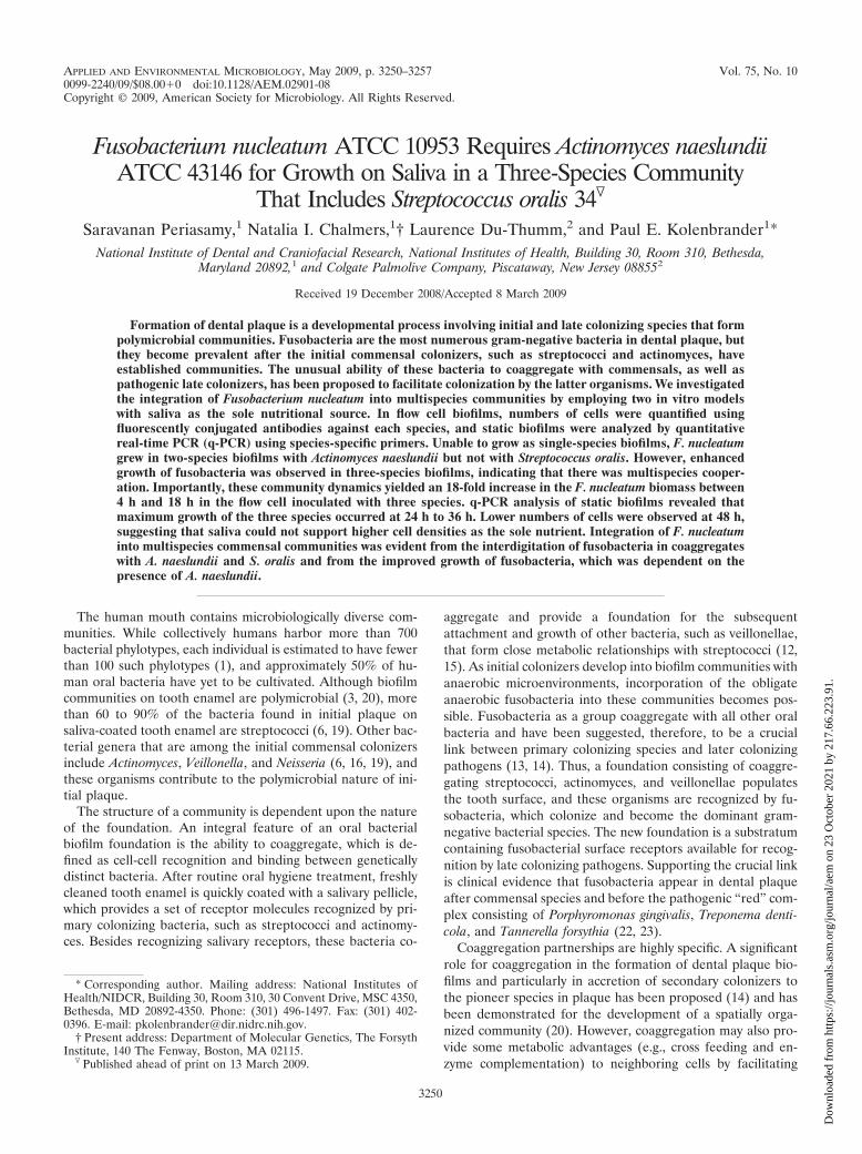
APPLIED AND ENVIRONMENTAL MICROBIOLOGY, May 2009, p. 3250–3257 Vol. 75, No. 100099-2240/09/$08.00�0 doi:10.1128/AEM.02901-08Copyright © 2009, American Society for Microbiology. All Rights Reserved.
Fusobacterium nucleatum ATCC 10953 Requires Actinomyces naeslundiiATCC 43146 for Growth on Saliva in a Three-Species Community
That Includes Streptococcus oralis 34�
Saravanan Periasamy,1 Natalia I. Chalmers,1† Laurence Du-Thumm,2 and Paul E. Kolenbrander1*National Institute of Dental and Craniofacial Research, National Institutes of Health, Building 30, Room 310, Bethesda,
Maryland 20892,1 and Colgate Palmolive Company, Piscataway, New Jersey 088552
Received 19 December 2008/Accepted 8 March 2009
Formation of dental plaque is a developmental process involving initial and late colonizing species that formpolymicrobial communities. Fusobacteria are the most numerous gram-negative bacteria in dental plaque, butthey become prevalent after the initial commensal colonizers, such as streptococci and actinomyces, haveestablished communities. The unusual ability of these bacteria to coaggregate with commensals, as well aspathogenic late colonizers, has been proposed to facilitate colonization by the latter organisms. We investigatedthe integration of Fusobacterium nucleatum into multispecies communities by employing two in vitro modelswith saliva as the sole nutritional source. In flow cell biofilms, numbers of cells were quantified usingfluorescently conjugated antibodies against each species, and static biofilms were analyzed by quantitativereal-time PCR (q-PCR) using species-specific primers. Unable to grow as single-species biofilms, F. nucleatumgrew in two-species biofilms with Actinomyces naeslundii but not with Streptococcus oralis. However, enhancedgrowth of fusobacteria was observed in three-species biofilms, indicating that there was multispecies cooper-ation. Importantly, these community dynamics yielded an 18-fold increase in the F. nucleatum biomass between4 h and 18 h in the flow cell inoculated with three species. q-PCR analysis of static biofilms revealed thatmaximum growth of the three species occurred at 24 h to 36 h. Lower numbers of cells were observed at 48 h,suggesting that saliva could not support higher cell densities as the sole nutrient. Integration of F. nucleatuminto multispecies commensal communities was evident from the interdigitation of fusobacteria in coaggregateswith A. naeslundii and S. oralis and from the improved growth of fusobacteria, which was dependent on thepresence of A. naeslundii.
The human mouth contains microbiologically diverse com-munities. While collectively humans harbor more than 700bacterial phylotypes, each individual is estimated to have fewerthan 100 such phylotypes (1), and approximately 50% of hu-man oral bacteria have yet to be cultivated. Although biofilmcommunities on tooth enamel are polymicrobial (3, 20), morethan 60 to 90% of the bacteria found in initial plaque onsaliva-coated tooth enamel are streptococci (6, 19). Other bac-terial genera that are among the initial commensal colonizersinclude Actinomyces, Veillonella, and Neisseria (6, 16, 19), andthese organisms contribute to the polymicrobial nature of ini-tial plaque.
The structure of a community is dependent upon the natureof the foundation. An integral feature of an oral bacterialbiofilm foundation is the ability to coaggregate, which is de-fined as cell-cell recognition and binding between geneticallydistinct bacteria. After routine oral hygiene treatment, freshlycleaned tooth enamel is quickly coated with a salivary pellicle,which provides a set of receptor molecules recognized by pri-mary colonizing bacteria, such as streptococci and actinomy-ces. Besides recognizing salivary receptors, these bacteria co-
aggregate and provide a foundation for the subsequentattachment and growth of other bacteria, such as veillonellae,that form close metabolic relationships with streptococci (12,15). As initial colonizers develop into biofilm communities withanaerobic microenvironments, incorporation of the obligateanaerobic fusobacteria into these communities becomes pos-sible. Fusobacteria as a group coaggregate with all other oralbacteria and have been suggested, therefore, to be a cruciallink between primary colonizing species and later colonizingpathogens (13, 14). Thus, a foundation consisting of coaggre-gating streptococci, actinomyces, and veillonellae populatesthe tooth surface, and these organisms are recognized by fu-sobacteria, which colonize and become the dominant gram-negative bacterial species. The new foundation is a substratumcontaining fusobacterial surface receptors available for recog-nition by late colonizing pathogens. Supporting the crucial linkis clinical evidence that fusobacteria appear in dental plaqueafter commensal species and before the pathogenic “red” com-plex consisting of Porphyromonas gingivalis, Treponema denti-cola, and Tannerella forsythia (22, 23).
Coaggregation partnerships are highly specific. A significantrole for coaggregation in the formation of dental plaque bio-films and particularly in accretion of secondary colonizers tothe pioneer species in plaque has been proposed (14) and hasbeen demonstrated for the development of a spatially orga-nized community (20). However, coaggregation may also pro-vide some metabolic advantages (e.g., cross feeding and en-zyme complementation) to neighboring cells by facilitating
* Corresponding author. Mailing address: National Institutes ofHealth/NIDCR, Building 30, Room 310, 30 Convent Drive, MSC 4350,Bethesda, MD 20892-4350. Phone: (301) 496-1497. Fax: (301) 402-0396. E-mail: [email protected].
† Present address: Department of Molecular Genetics, The ForsythInstitute, 140 The Fenway, Boston, MA 02115.
� Published ahead of print on 13 March 2009.
3250
Dow
nloa
ded
from
http
s://j
ourn
als.
asm
.org
/jour
nal/a
em o
n 23
Oct
ober
202
1 by
217
.66.
223.
91.
physical juxtaposition of partner cells, as has been shown forglucose metabolism of coaggregates of actinomyces and strep-tococci (7, 8). One aim of the present study was to examine thestructures of two- and three-species communities composed ofActinomyces naeslundii, Streptococcus oralis, and Fusobacte-rium nucleatum in model biofilm systems. The first two speciesare initial colonizers and are considered commensals, whereasfusobacteria are secondary colonizers and are postulated to bea coaggregation bridge between initial and late colonizers (14).Our second aim was to investigate the integration and growthof fusobacteria in polymicrobial communities.
A variety of experimental methods have been developed tostudy the formation of biofilms. Model systems often rely onthe flow of nutrients over a surface on which bacteria are ableto attach and grow. In the present study we used two distinct invitro models, a saliva-fed flow cell and a polystyrene peg im-mersed in static saliva. Biofilm communities form naturally andare undisturbed (3, 20, 21). The spatial organization of a mul-tispecies community resulting from colonization and growth ispreserved and can be examined noninvasively by confocal laserscanning microscopy (CLSM). In the static system, the amountof each species in multispecies biofilms formed on polystyrenepegs can be measured by real-time quantitative PCR (q-PCR).We show here with both models that fusobacteria are unable togrow as single species, but they integrate into commensalstreptococcus-actinomyces communities and grow. Integrationand growth are required for fusobacteria to become cruciallinks between commensal communities and later colonizingpathogenic communities. In the three-species community stud-ied here, A. naeslundii is required for F. nucleatum to integrateand grow.
MATERIALS AND METHODS
Bacterial strains and culture conditions. S. oralis 34 and A. naeslundii ATCC43146 were routinely cultured in Todd-Hewitt broth (Difco Laboratories, De-troit, MI) or on Todd-Hewitt agar. F. nucleatum ATCC 10953 was grown in brainheart infusion (Difco) broth supplemented with 0.25% L-glutamic acid. All spe-cies were grown in a Bactron anaerobic (N2-CO2-H2, 90:5:5) environmentalchamber (Sheldon Manufacturing Inc., Cornelius, OR) at 37°C.
Coaggregation assay. Coaggregation reactions were investigated by combiningF. nucleatum, S. oralis, and A. naeslundii pairwise and by mixing all three speciesin saliva. Equal volumes (0.1 ml) of microbial suspensions (about 1 � 109
cells/ml) were combined in glass tubes (12 by 75 mm) and mixed for 10 s with avortex mixer. After mixing, the suspensions were scored immediately for coag-gregation. The scoring system of Cisar et al. (4) was used to evaluate the degreeof coaggregation in the suspensions by viewing the tubes with the naked eye. Thescores ranged from 0 to �4, as follows: 0, no change in turbidity and no evidenceof coaggregates in the mixed suspensions; �1, turbid supernatant with finelydispersed coaggregates which did not precipitate immediately; �2, definite co-aggregates easily seen, but the suspension remained turbid and there was noimmediate settling of coaggregates; �3, slightly turbid supernatant and forma-tion of large precipitating coaggregates; and �4, clear supernatant and largecoaggregates that precipitated immediately. The coaggregating partners S. oralis34, A. naeslundii ATCC 43146, and F. nucleatum ATCC 10953 were used asinocula for formation of multispecies biofilms.
Saliva preparation. Saliva from at least six healthy individuals was collected onice, pooled on ice, and treated with 2.5 mM dithiothreitol for 10 min with stirringto reduce salivary protein aggregation. The saliva was then centrifuged andprocessed as previously described (5, 21). Briefly, the supernatant was dilutedwith distilled water to obtain 25% saliva and then was filtered through a 0.22-�m-pore-size SFCA low-protein-binding filter (Nalge Nunc International, Roch-ester, NY) and stored at �20°C. Prior to use, saliva was thawed and centrifugedto remove any precipitate that resulted from freezing and thawing.
Flow cell preparation. Two tracks (each track was 40 mm long, 3 mm wide, and2 mm deep) were milled into a high-density polyethylene block, resulting in two
chambers, each having a volume of 240 �l. A glass coverslip, which served as theattachment substratum for the growing biofilm, was secured to each of thereusable flow cells with a silicone adhesive. The flow cells were cleaned overnightwith 0.1 M HCl and rinsed with 5 ml of distilled water. To disinfect the flow cells,70% ethanol was injected into the flow cells and the flow cells were incubated for20 min. The flow cells were then treated with 25% sterile human saliva for 15 minat 37°C in an anaerobic chamber to condition the glass surface with salivarycomponents.
Biofilm growth conditions in flow cells. Overnight bacterial cultures wereharvested by centrifugation and washed twice with 25% sterile human saliva, andthe optical density at 600 nm was adjusted to 0.1. Flow cells were inoculated withone, two, or three species. Two-species and three-species inoculants were firstcoaggregated (by mixing 0.1-ml portions of appropriate combinations of S. oralis34, A. naeslundii ATCC 43146, and F. nucleatum ATCC 10953), which wasfollowed by incubation of the flow cell in the anaerobic chamber to provide anenvironment favorable for F. nucleatum, a strict anaerobe. Sterile 25% saliva wassupplied at a flow rate of 0.2 ml/min as the sole source of nutrients.
Biofilm staining. Cells were stained with BacLight Live/Dead (Invitrogen,Carlsbad, CA) when strains were grown as monocultures. When multiple specieswere inoculated, organisms were visualized by using primary immunofluores-cence with Alexa Fluor 633 (Invitrogen)-, Alexa Fluor 488 (Invitrogen)-, andAlexa Fluor 546 (Invitrogen)-conjugated immunoglobulin G of a polyclonalantiserum to S. oralis, A. naeslundii, and F. nucleatum, respectively. The immu-nofluorescence analysis was performed by injecting the antibody (5 �g/ml inphosphate-buffered saline) into the appropriate flow cell track and incubatingthe preparation for 20 min. A final wash with 1% bovine serum albumin inphosphate-buffered saline (filter sterilized with a 0.22-�m-pore-size SFCA low-protein-binding filter [Nalge Nunc International]) preceded CLSM. No cross-reactivity of staining by antibodies with nonhomologous strains was observed.
Image and statistical analysis. A TCS-SP2 confocal microscope (Leica, Exton,PA) with a 40�, 1.25 NA oil immersion lens was used to record confocal imagestacks in five random locations near the center of each flow cell, after whichbiofilm biovolumes were determined by volumetric analyses (IMARIS, version5.71; Bitplane AG, Zurich, Switzerland). Fluorescence intensity thresholds wereset manually for red, green, and blue pixels with cubic voxels used for thebiovolume determination. Five confocal data sets were analyzed for each timepoint, and the mean and standard deviation were calculated. A one-way analysisof variance at the 95% confidence level with a nonparametric Tukey’s pairwisecomparison test was used to determine if the means were statistically differentbetween the 4-h and 18-h time points for each species for each condition (one,two, or three species) in the flow cell studies and if the means were statisticallydifferent between the 12-h, 24-h, 36-h, and 48-h time points for each species foreach condition (one, two, or three species) in the peg studies with static saliva.All images presented below are maximum projections of the entire confocalimage stack produced by the Leica TCS software (Leica).
Biofilm growth conditions on polystyrene pegs. Static biofilms were grown in25% human saliva on polystyrene pegs (Nunc-Immuno TSP; Nunc catalog no.445497) (2, 17) mounted in U96 MicroWell plates (Nunc catalog no. 163320) (3,17). Microtiter plate wells were filled with 200 �l of 25% human saliva, and thepegs were then inserted for 30 min at room temperature to obtain a conditioningfilm. Overnight cultures (�20 �l) of S. oralis, A. naeslundii, and F. nucleatumwere added to the 200 �l of saliva in the wells to obtain an optical density at 600nm of 0.1, which was equivalent to about 1 � 107 to 3 � 107 cells of each speciesin the microtiter wells; the plates were placed in a humidity chamber andincubated anaerobically at 37°C for 48 h. After 12-h intervals, the pegs weretransferred to fresh, prereduced saliva in microtiter plate wells.
DNA extraction and quantification. DNA was extracted from biofilms by usinga modified alkaline lysis protocol (10). Biofilm-covered pegs were immersed in 40�l of sterile ultrapure water plus 160 �l of 0.05 M sodium hydroxide andincubated at 60°C for 45 to 60 min, after which 18.4 �l of 1 M Tris-HCl (pH 7.0)was added to neutralize the pH. The resulting extract was used as the templateDNA for the q-PCR analyses (3, 10). Bacterial genomic DNA for construction ofstandard curves was extracted from overnight cultures of S. oralis, A. naeslundii, andF. nucleatum with a DNA extraction kit (Qiagen, Valencia, CA) used according tothe manufacturer’s instructions. Genomic DNA was stored at �20°C.
Quantification of species in static biofilms by real-time q-PCR. Species-spe-cific primers were designed with AlleleID6 (PREMIER Biosoft International,Palo Alto, CA); no cross-reactivity of primers with nonhomologous species wasobserved. The primers specific for S. oralis were forward primer GATACATAGCCGACCTGAG and reverse primer TCCATTGCCGAAGATTCC, and theannealing temperature was 56°C. The primers specific for A. naeslundii wereforward primer GGCTGCGATACCGTGAGG and reverse primer TCTGCGATTACTAGCGACTCC, and the annealing temperature was 56°C. The primers
VOL. 75, 2009 GROWTH OF F. NUCLEATUM ON SALIVA 3251
Dow
nloa
ded
from
http
s://j
ourn
als.
asm
.org
/jour
nal/a
em o
n 23
Oct
ober
202
1 by
217
.66.
223.
91.
specific for F. nucleatum were forward primer CTTAGGAATGAGACAGAGATG and reverse primer TGATGGTAACATACGAAAGG, and the anneal-ing temperature was 56°C. Quantification of S. oralis, A. naeslundii, and F.nucleatum in the biofilms was performed by q-PCR using the SYBR green dye todetect the 16S rRNA gene amplicons. Each reaction mixture (final volume, 25�l) contained 3 �l of template, 3.5 �l of diethyl pyrocarbonate-treated ultrapurewater, 12.5 �l of Power SYBR green PCR master mixture (Applied Biosystems,Foster City, CA), and 3 �l each of the forward and reverse primers at a con-centration of 375 nM. q-PCR was performed with an MX3005P thermocycler(Stratagene, La Jolla, CA) using the thermocycle recommended for PowerSYBR green PCR master mixture (95°C for 10 min, followed by 40 cycles of 30 sat 95°C and 1 min at 56°C). Dissociation curves were generated by incubatingreaction products at 95°C for 1 min and at 56°C for 30 s and then incrementallyincreasing the temperature to 95°C. Fluorescence data were collected at the endof the 56°C primer annealing step for 40 amplification cycles and throughout thedissociation curve analysis. Analysis of the melting curves with each of the threeprimer sets revealed a single sharp peak. DNA concentrations (in ng/ml) werecalculated based on standard curves obtained by using 10-fold serial dilutions ofbacterial DNA isolated with a DNA extraction kit (Qiagen) and quantified withthe PicoGreen fluorescence assay (Invitrogen). To convert nanograms of DNAto numbers of cells, the following weights and genome sizes were used: 2.41fg/genome and 2.4 Mb for fusobacteria, 2.05 fg/genome and 2 Mb for strepto-cocci, and 3.08 fg/genome and 3.0 Mb for actinomyces (http://www.homd.org).The data presented below are data for three independent biofilms.
RESULTS
Coaggregation of three-species community members. Pair-wise coaggregation of F. nucleatum with A. naeslundii in salivayielded a coaggregation score of �2. A weak coaggregationscore, �1, was obtained for F. nucleatum and S. oralis, andexamination of the coaggregates by phase-contrast microscopyrevealed numerous corncob cellular arrangements consistingof long, slender fusobacterial cells surrounded by coccal strep-tococci. In three-species interactions F. nucleatum participatedin very strong coaggregations with a score of �4. Pairwisecoaggregations of S. oralis and A. naeslundii gave a score of �3.Thus, each species is a coaggregation partner of the other twospecies, and the ability of the species to form multispeciesbiofilm communities in two model systems was investigated.
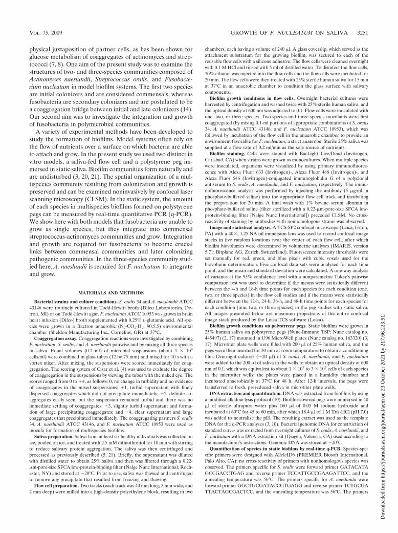
Flow cells inoculated with one species. S. oralis, A. naeslun-dii, and F. nucleatum monocultures were inoculated into flowcells, and images of the biofilms formed were obtained after4 h and 18 h of incubation (Fig. 1). The level of cell attachmentto the substratum was quantified by determining the biovolume(Fig. 2A). Each species attached to the saliva-conditioned sur-face within 4 h (Fig. 1, left panels). After 18 h of incubation,the number of F. nucleatum cells decreased, indicating thatthere was no growth (Fig. 1, top right panel). In most experi-ments in this anaerobic environment, S. oralis grew well (Fig. 1,middle right panel, and Fig. 2A). Occasionally, S. oralis grewpoorly, as we observed previously for aerobic environments(21). Only A. naeslundii grew consistently (Fig. 1, lower rightpanel, and Fig. 2A). To determine whether the cells in thebiofilms were viable after 4 h and 18 h, cells were stained withthe BacLight Live/Dead viability stain (Invitrogen), a fluores-cent marker of membrane integrity (Fig. 1). Most cells (90%)in biofilms containing S. oralis or A. naeslundii appeared tohave no membrane damage (green cells) at 4 h or 18 h. Also,at 4 h F. nucleatum appeared to have no membrane damage,which is consistent with the presence of a proper environmentfor the anaerobic physiology of this organism. However, after18 h in saliva, fewer F. nucleatum cells were present, and mostcells exhibited membrane damage (red cells), suggesting that
saliva did not provide nutrients for growth. Thus, while F.nucleatum was unable to grow by itself, the commensals strep-tococcus and actinomyces formed one-species biofilms.
Flow cells inoculated with two species. Saliva-conditionedflow cells were inoculated with pairwise combinations of coag-gregating species, and the species were distinguished by label-ing them with specific antibodies (Fig. 3). In the presence of A.naeslundii, F. nucleatum flourished (Fig. 3 and Fig. 2B). Al-though F. nucleatum attached to the substratum equally well inone-species biofilms (Fig. 1 and Fig. 2A) and in coaggregateswith A. naeslundii (Fig. 2B and Fig. 3), its biovolume increasedninefold when it was in coaggregates with A. naeslundii (Fig.2B), indicating that F. nucleatum required the actinomyces forcooperative growth. The A. naeslundii biovolume increasedfivefold (Fig. 2B). In the presence of S. oralis, F. nucleatum didnot grow, although the initial attachment to the substratum at4 h was excellent (Fig. 3), whereas the S. oralis biovolumeincreased ninefold (Fig. 3 and Fig. 2B). As determined byLive/Dead staining, fusobacteria were green at 4 h with either
FIG. 1. Confocal micrographs of biofilms formed in flow cells in-oculated with one species (F. nucleatum, S. oralis, or A. naeslundii)after 4 h and 18 h of growth on 25% saliva. BacLight Live/Dead stainwas used to assess the vitality of cells. Red cells have impaired mem-brane activity, whereas green cells have fully functional membranes.
3252 PERIASAMY ET AL. APPL. ENVIRON. MICROBIOL.
Dow
nloa
ded
from
http
s://j
ourn
als.
asm
.org
/jour
nal/a
em o
n 23
Oct
ober
202
1 by
217
.66.
223.
91.
coaggregation partner and at 18 h with A. naeslundii, but mostof the cells were red with S. oralis after 18 h (data not shown),suggesting that the membrane integrity of F. nucleatum cellswas compromised when they were paired with S. oralis. Thebiovolumes of S. oralis and A. naeslundii increased between 4 hand 18 h (Fig. 2B), and the increase in the A. naeslundii bio-
volume was statistically significant. Thus, the three speciesgrew in saliva in some but not all pairwise combinations, leav-ing open the possibility of unchanged pairwise relationships orunanticipated cooperative growth of all members in three-species communities.
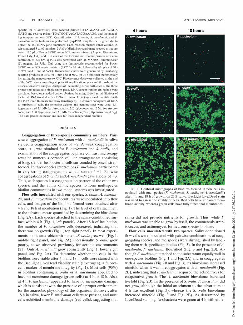
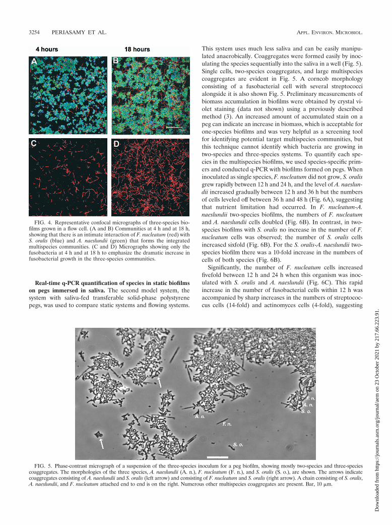
Flow cell inoculated with three species. Coaggregates of thethree species were inoculated into a flow cell, and all of thespecies attached to the saliva-conditioned substratum and grewbetween 4 h and 18 h during incubation (Fig. 4A and 4B). Adramatic increase in the biovolume of F. nucleatum was evi-dent (Fig. 4C and 4D). The biovolume of F. nucleatum in-creased 18-fold (Fig. 2C), indicating that the cooperation be-tween fusobacteria and actinomyces is stronger when the threespecies are present than when fusobacteria and actinomycesform two-species communities in the flowing saliva model sys-tem. Further, these data suggest that the outcome of interac-tions among three species cannot be predicted on the basis oftwo-species interactions.
FIG. 2. Time-resolved changes in the biovolume (�m3 per field ofview) of A. naeslundii (An), S. oralis (So), and F. nucleatum (Fn)following 4 h and 18 h of incubation in flow cells fed 25% saliva.(A) Biovolumes of single species (see Fig. 1). (B) Biovolumes of eachspecies in two-species biofilms (see Fig. 3). (C) Biovolumes of eachspecies in three-species biofilms (see Fig. 4). An asterisk indicatesstatistically significant growth.
FIG. 3. Representative confocal micrographs of 4-h and 18-h two-species biofilms grown in flow cells. The primary immunofluorescenceindicates S. oralis (blue), A. naeslundii (green), and F. nucleatum (red)and shows that there is intimate contact among species.
VOL. 75, 2009 GROWTH OF F. NUCLEATUM ON SALIVA 3253
Dow
nloa
ded
from
http
s://j
ourn
als.
asm
.org
/jour
nal/a
em o
n 23
Oct
ober
202
1 by
217
.66.
223.
91.
Real-time q-PCR quantification of species in static biofilmson pegs immersed in saliva. The second model system, thesystem with saliva-fed transferable solid-phase polystyrenepegs, was used to compare static systems and flowing systems.
This system uses much less saliva and can be easily manipu-lated anaerobically. Coaggregates were formed easily by inoc-ulating the species sequentially into the saliva in a well (Fig. 5).Single cells, two-species coaggregates, and large multispeciescoaggregates are evident in Fig. 5. A corncob morphologyconsisting of a fusobacterial cell with several streptococcialongside it is also shown Fig. 5. Preliminary measurements ofbiomass accumulation in biofilms were obtained by crystal vi-olet staining (data not shown) using a previously describedmethod (3). An increased amount of accumulated stain on apeg can indicate an increase in biomass, which is acceptable forone-species biofilms and was very helpful as a screening toolfor identifying potential target multispecies communities, butthis technique cannot identify which bacteria are growing intwo-species and three-species systems. To quantify each spe-cies in the multispecies biofilms, we used species-specific prim-ers and conducted q-PCR with biofilms formed on pegs. Wheninoculated as single species, F. nucleatum did not grow, S. oralisgrew rapidly between 12 h and 24 h, and the level of A. naeslun-dii increased gradually between 12 h and 36 h but the numbersof cells leveled off between 36 h and 48 h (Fig. 6A), suggestingthat nutrient limitation had occurred. In F. nucleatum-A.naeslundii two-species biofilms, the numbers of F. nucleatumand A. naeslundii cells doubled (Fig. 6B). In contrast, in two-species biofilms with S. oralis no increase in the number of F.nucleatum cells was observed; the number of S. oralis cellsincreased sixfold (Fig. 6B). For the S. oralis-A. naeslundii two-species biofilm there was a 10-fold increase in the numbers ofcells of both species (Fig. 6B).
Significantly, the number of F. nucleatum cells increasedfivefold between 12 h and 24 h when this organism was inoc-ulated with S. oralis and A. naeslundii (Fig. 6C). This rapidincrease in the number of fusobacterial cells within 12 h wasaccompanied by sharp increases in the numbers of streptococ-cus cells (14-fold) and actinomyces cells (4-fold), suggesting
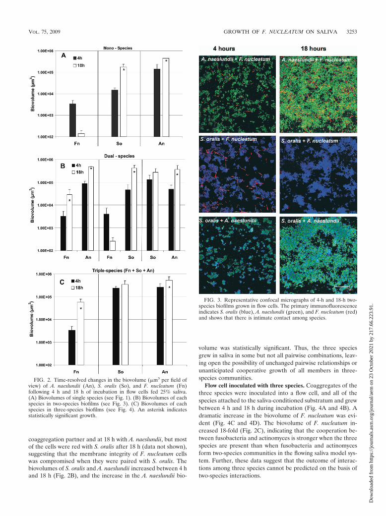
FIG. 4. Representative confocal micrographs of three-species bio-films grown in a flow cell. (A and B) Communities at 4 h and at 18 h,showing that there is an intimate interaction of F. nucleatum (red) withS. oralis (blue) and A. naeslundii (green) that forms the integratedmultispecies communities. (C and D) Micrographs showing only thefusobacteria at 4 h and at 18 h to emphasize the dramatic increase infusobacterial growth in the three-species communities.
FIG. 5. Phase-contrast micrograph of a suspension of the three-species inoculum for a peg biofilm, showing mostly two-species and three-speciescoaggregates. The morphologies of the three species, A. naeslundii (A. n.), F. nucleatum (F. n.), and S. oralis (S. o.), are shown. The arrows indicatecoaggregates consisting of A. naeslundii and S. oralis (left arrow) and consisting of F. nucleatum and S. oralis (right arrow). A chain consisting of S. oralis,A. naeslundii, and F. nucleatum attached end to end is on the right. Numerous other multispecies coaggregates are present. Bar, 10 �m.
3254 PERIASAMY ET AL. APPL. ENVIRON. MICROBIOL.
Dow
nloa
ded
from
http
s://j
ourn
als.
asm
.org
/jour
nal/a
em o
n 23
Oct
ober
202
1 by
217
.66.
223.
91.
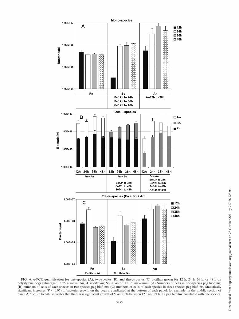
FIG. 6. q-PCR quantification for one-species (A), two-species (B), and three-species (C) biofilms grown for 12 h, 24 h, 36 h, or 48 h onpolystyrene pegs submerged in 25% saliva. An, A. naeslundii; So, S. oralis; Fn, F. nucleatum. (A) Numbers of cells in one-species peg biofilms;(B) numbers of cells of each species in two-species peg biofilms; (C) numbers of cells of each species in three-species peg biofilms. Statisticallysignificant increases (P � 0.05) in bacterial growth on the pegs are indicated at the bottom of each panel; for example, in the middle section ofpanel A, “So12h to 24h” indicates that there was significant growth of S. oralis 34 between 12 h and 24 h in a peg biofilm inoculated with one species.
3255
Dow
nloa
ded
from
http
s://j
ourn
als.
asm
.org
/jour
nal/a
em o
n 23
Oct
ober
202
1 by
217
.66.
223.
91.
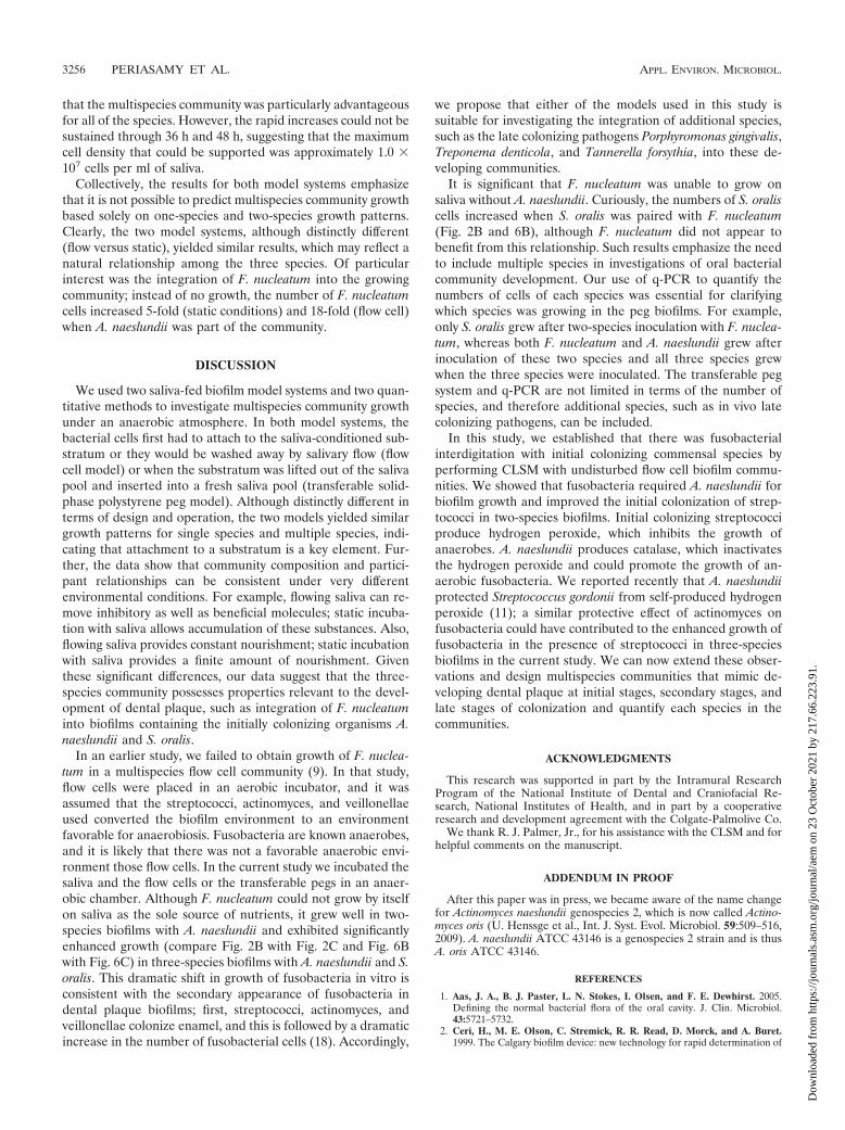
that the multispecies community was particularly advantageousfor all of the species. However, the rapid increases could not besustained through 36 h and 48 h, suggesting that the maximumcell density that could be supported was approximately 1.0 �107 cells per ml of saliva.
Collectively, the results for both model systems emphasizethat it is not possible to predict multispecies community growthbased solely on one-species and two-species growth patterns.Clearly, the two model systems, although distinctly different(flow versus static), yielded similar results, which may reflect anatural relationship among the three species. Of particularinterest was the integration of F. nucleatum into the growingcommunity; instead of no growth, the number of F. nucleatumcells increased 5-fold (static conditions) and 18-fold (flow cell)when A. naeslundii was part of the community.
DISCUSSION
We used two saliva-fed biofilm model systems and two quan-titative methods to investigate multispecies community growthunder an anaerobic atmosphere. In both model systems, thebacterial cells first had to attach to the saliva-conditioned sub-stratum or they would be washed away by salivary flow (flowcell model) or when the substratum was lifted out of the salivapool and inserted into a fresh saliva pool (transferable solid-phase polystyrene peg model). Although distinctly different interms of design and operation, the two models yielded similargrowth patterns for single species and multiple species, indi-cating that attachment to a substratum is a key element. Fur-ther, the data show that community composition and partici-pant relationships can be consistent under very differentenvironmental conditions. For example, flowing saliva can re-move inhibitory as well as beneficial molecules; static incuba-tion with saliva allows accumulation of these substances. Also,flowing saliva provides constant nourishment; static incubationwith saliva provides a finite amount of nourishment. Giventhese significant differences, our data suggest that the three-species community possesses properties relevant to the devel-opment of dental plaque, such as integration of F. nucleatuminto biofilms containing the initially colonizing organisms A.naeslundii and S. oralis.
In an earlier study, we failed to obtain growth of F. nuclea-tum in a multispecies flow cell community (9). In that study,flow cells were placed in an aerobic incubator, and it wasassumed that the streptococci, actinomyces, and veillonellaeused converted the biofilm environment to an environmentfavorable for anaerobiosis. Fusobacteria are known anaerobes,and it is likely that there was not a favorable anaerobic envi-ronment those flow cells. In the current study we incubated thesaliva and the flow cells or the transferable pegs in an anaer-obic chamber. Although F. nucleatum could not grow by itselfon saliva as the sole source of nutrients, it grew well in two-species biofilms with A. naeslundii and exhibited significantlyenhanced growth (compare Fig. 2B with Fig. 2C and Fig. 6Bwith Fig. 6C) in three-species biofilms with A. naeslundii and S.oralis. This dramatic shift in growth of fusobacteria in vitro isconsistent with the secondary appearance of fusobacteria indental plaque biofilms; first, streptococci, actinomyces, andveillonellae colonize enamel, and this is followed by a dramaticincrease in the number of fusobacterial cells (18). Accordingly,
we propose that either of the models used in this study issuitable for investigating the integration of additional species,such as the late colonizing pathogens Porphyromonas gingivalis,Treponema denticola, and Tannerella forsythia, into these de-veloping communities.
It is significant that F. nucleatum was unable to grow onsaliva without A. naeslundii. Curiously, the numbers of S. oraliscells increased when S. oralis was paired with F. nucleatum(Fig. 2B and 6B), although F. nucleatum did not appear tobenefit from this relationship. Such results emphasize the needto include multiple species in investigations of oral bacterialcommunity development. Our use of q-PCR to quantify thenumbers of cells of each species was essential for clarifyingwhich species was growing in the peg biofilms. For example,only S. oralis grew after two-species inoculation with F. nuclea-tum, whereas both F. nucleatum and A. naeslundii grew afterinoculation of these two species and all three species grewwhen the three species were inoculated. The transferable pegsystem and q-PCR are not limited in terms of the number ofspecies, and therefore additional species, such as in vivo latecolonizing pathogens, can be included.
In this study, we established that there was fusobacterialinterdigitation with initial colonizing commensal species byperforming CLSM with undisturbed flow cell biofilm commu-nities. We showed that fusobacteria required A. naeslundii forbiofilm growth and improved the initial colonization of strep-tococci in two-species biofilms. Initial colonizing streptococciproduce hydrogen peroxide, which inhibits the growth ofanaerobes. A. naeslundii produces catalase, which inactivatesthe hydrogen peroxide and could promote the growth of an-aerobic fusobacteria. We reported recently that A. naeslundiiprotected Streptococcus gordonii from self-produced hydrogenperoxide (11); a similar protective effect of actinomyces onfusobacteria could have contributed to the enhanced growth offusobacteria in the presence of streptococci in three-speciesbiofilms in the current study. We can now extend these obser-vations and design multispecies communities that mimic de-veloping dental plaque at initial stages, secondary stages, andlate stages of colonization and quantify each species in thecommunities.
ACKNOWLEDGMENTS
This research was supported in part by the Intramural ResearchProgram of the National Institute of Dental and Craniofacial Re-search, National Institutes of Health, and in part by a cooperativeresearch and development agreement with the Colgate-Palmolive Co.
We thank R. J. Palmer, Jr., for his assistance with the CLSM and forhelpful comments on the manuscript.
ADDENDUM IN PROOF
After this paper was in press, we became aware of the name changefor Actinomyces naeslundii genospecies 2, which is now called Actino-myces oris (U. Henssge et al., Int. J. Syst. Evol. Microbiol. 59:509–516,2009). A. naeslundii ATCC 43146 is a genospecies 2 strain and is thusA. oris ATCC 43146.
REFERENCES
1. Aas, J. A., B. J. Paster, L. N. Stokes, I. Olsen, and F. E. Dewhirst. 2005.Defining the normal bacterial flora of the oral cavity. J. Clin. Microbiol.43:5721–5732.
2. Ceri, H., M. E. Olson, C. Stremick, R. R. Read, D. Morck, and A. Buret.1999. The Calgary biofilm device: new technology for rapid determination of
3256 PERIASAMY ET AL. APPL. ENVIRON. MICROBIOL.
Dow
nloa
ded
from
http
s://j
ourn
als.
asm
.org
/jour
nal/a
em o
n 23
Oct
ober
202
1 by
217
.66.
223.
91.
antibiotic susceptibilities of bacterial biofilms. J. Clin. Microbiol. 37:1771–1776.
3. Chalmers, N. I., R. J. Palmer, Jr., J. O. Cisar, and P. E. Kolenbrander. 2008.Characterization of a Streptococcus sp.-Veillonella sp. community microma-nipulated from dental plaque. J. Bacteriol. 190:8145–8154.
4. Cisar, J. O., P. E. Kolenbrander, and F. C. McIntire. 1979. Specificity ofcoaggregation reactions between human oral streptococci and strains ofActinomyces viscosus or Actinomyces naeslundii. Infect. Immun. 24:742–752.
5. De Jong, M. H., and J. S. Van der Hoeven. 1987. The growth of oral bacteriaon saliva. J. Dent. Res. 66:498–505.
6. Diaz, P. I., N. I. Chalmers, A. H. Rickard, C. Kong, C. L. Milburn, R. J.Palmer, Jr., and P. E. Kolenbrander. 2006. Molecular characterization ofsubject-specific oral microflora during initial colonization of enamel. Appl.Environ. Microbiol. 72:2837–2848.
7. Distler, W., A. Kagermeier-Callaway, and A. Petschelt. 1992. Prevention ofcoaggregation by a dialysis membrane—influences on the glucose metabo-lism of mixtures of Actinomyces and streptococci. J. Dent. Res. 71:735.
8. Distler, W., A. Kagermeier, and A. Kroencke. 1991. Influence of coaggrega-tion on the glucose metabolism of mixtures of Actinomyces and streptococci.J. Dent. Res. 70:764.
9. Foster, J. S., and P. E. Kolenbrander. 2004. Development of a multispeciesoral bacterial community in a saliva-conditioned flow cell. Appl. Environ.Microbiol. 70:4340–4348.
10. Hoshino, T., T. Fujiwara, and M. Kilian. 2005. Use of phylogenetic andphenotypic analyses to identify nonhemolytic streptococci isolated from bac-teremic patients. J. Clin. Microbiol. 43:6073–6085.
11. Jakubovics, N. S., S. R. Gill, M. M. Vickerman, and P. E. Kolenbrander.2008. Role of hydrogen peroxide in competition and cooperation betweenStreptococcus gordonii and Actinomyces naeslundii. FEMS Microbiol. Ecol.66:637–644.
12. Jenkinson, H. F., and R. J. Lamont. 2005. Oral microbial communities insickness and in health. Trends Microbiol. 13:589–595.
13. Kolenbrander, P. E., R. N. Andersen, D. S. Blehert, P. G. Egland, J. S.Foster, and R. J. Palmer, Jr. 2002. Communication among oral bacteria.Microbiol. Mol. Biol. Rev. 66:486–505.
14. Kolenbrander, P. E., and J. London. 1993. Adhere today, here tomorrow:oral bacterial adherence. J. Bacteriol. 175:3247–3252.
15. Kolenbrander, P. E., R. J. Palmer, Jr., A. H. Rickard, N. S. Jakubovics, N. I.Chalmers, and P. I. Diaz. 2006. Bacterial interactions and successions duringplaque development. Periodontol. 2000 42:47–79.
16. Li, J., E. J. Helmerhorst, C. W. Leone, R. F. Troxler, T. Yaskell, A. D.Haffajee, S. S. Socransky, and F. G. Oppenheim. 2004. Identification of earlymicrobial colonizers in human dental biofilm. J. Appl. Microbiol. 97:1311–1318.
17. Mampel, J., T. Spirig, S. S. Weber, J. A. Haagensen, S. Molin, and H. Hilbi.2006. Planktonic replication is essential for biofilm formation by Legionellapneumophila in a complex medium under static and dynamic flow conditions.Appl. Environ. Microbiol. 72:2885–2895.
18. Moore, W. E., and L. V. Moore. 1994. The bacteria of periodontal diseases.Periodontol. 2000 5:66–77.
19. Nyvad, B., and M. Kilian. 1987. Microbiology of the early colonization ofhuman enamel and root surfaces in vivo. Scand. J. Dent. Res. 95:369–380.
20. Palmer, R. J., Jr., S. M. Gordon, J. O. Cisar, and P. E. Kolenbrander. 2003.Coaggregation-mediated interactions of streptococci and actinomyces de-tected in initial human dental plaque. J. Bacteriol. 185:3400–3409.
21. Palmer, R. J., Jr., K. Kazmerzak, M. C. Hansen, and P. E. Kolenbrander.2001. Mutualism versus independence: strategies of mixed-species oral bio-films in vitro using saliva as the sole nutrient source. Infect. Immun. 69:5794–5804.
22. Socransky, S. S., and A. D. Haffajee. 2005. Periodontal microbial ecology.Periodontol. 2000 38:135–187.
23. Socransky, S. S., A. D. Haffajee, M. A. Cugini, C. Smith, and R. L. Kent, Jr.1998. Microbial complexes in subgingival plaque. J. Clin. Periodontol. 25:134–144.
VOL. 75, 2009 GROWTH OF F. NUCLEATUM ON SALIVA 3257
Dow
nloa
ded
from
http
s://j
ourn
als.
asm
.org
/jour
nal/a
em o
n 23
Oct
ober
202
1 by
217
.66.
223.
91.