Embed Size (px)
Citation preview
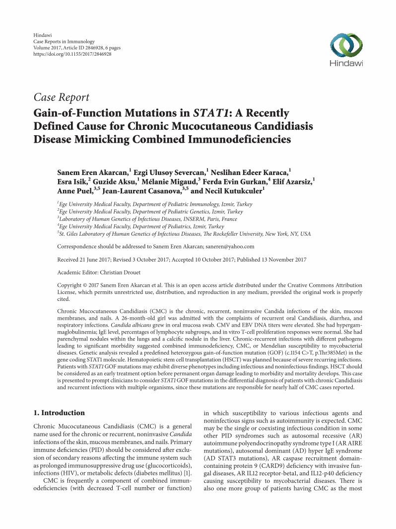
Case ReportGain-of-Function Mutations in STAT1: A RecentlyDefined Cause for Chronic Mucocutaneous CandidiasisDisease Mimicking Combined Immunodeficiencies
Sanem Eren Akarcan,1 Ezgi Ulusoy Severcan,1 Neslihan Edeer Karaca,1
Esra Isik,2 Guzide Aksu,1 Mélanie Migaud,3 Ferda Evin Gurkan,4 Elif Azarsiz,1
Anne Puel,3,5 Jean-Laurent Casanova,3,5 and Necil Kutukculer1
1Ege University Medical Faculty, Department of Pediatric Immunology, Izmir, Turkey2Ege University Medical Faculty, Department of Pediatric Genetics, Izmir, Turkey3Laboratory of Human Genetics of Infectious Diseases, INSERM, Paris, France4Ege University Medical Faculty, Department of Pediatrics, Izmir, Turkey5St. Giles Laboratory of Human Genetics of Infectious Diseases, The Rockefeller University, New York, NY, USA
Correspondence should be addressed to Sanem Eren Akarcan; [email protected]
Received 21 June 2017; Revised 3 October 2017; Accepted 10 October 2017; Published 13 November 2017
Academic Editor: Christian Drouet
Copyright © 2017 Sanem Eren Akarcan et al. This is an open access article distributed under the Creative Commons AttributionLicense, which permits unrestricted use, distribution, and reproduction in any medium, provided the original work is properlycited.
Chronic Mucocutaneous Candidiasis (CMC) is the chronic, recurrent, noninvasive Candida infections of the skin, mucousmembranes, and nails. A 26-month-old girl was admitted with the complaints of recurrent oral Candidiasis, diarrhea, andrespiratory infections. Candida albicans grew in oral mucosa swab. CMV and EBV DNA titers were elevated. She had hypergam-maglobulinemia; IgE level, percentages of lymphocyte subgroups, and in vitro T-cell proliferation responses were normal. She hadparenchymal nodules within the lungs and a calcific nodule in the liver. Chronic-recurrent infections with different pathogensleading to significant morbidity suggested combined immunodeficiency, CMC, or Mendelian susceptibility to mycobacterialdiseases. Genetic analysis revealed a predefined heterozygous gain-of-function mutation (GOF) (c.1154 C>T, p.Thr385Met) in thegene coding STAT1 molecule. Hematopoietic stem cell transplantation (HSCT) was planned because of severe recurring infections.Patients with STAT1GOFmutationsmay exhibit diverse phenotypes including infectious and noninfectious findings. HSCT shouldbe considered as an early treatment option before permanent organ damage leading to morbidity and mortality develops.This caseis presented to prompt clinicians to consider STAT1GOFmutations in the differential diagnosis of patients with chronic Candidiasisand recurrent infections with multiple organisms, since these mutations are responsible for nearly half of CMC cases reported.
1. Introduction
Chronic Mucocutaneous Candidiasis (CMC) is a generalname used for the chronic or recurrent, noninvasiveCandidainfections of the skin,mucousmembranes, andnails. Primaryimmune deficiencies (PID) should be considered after exclu-sion of secondary reasons affecting the immune system suchas prolonged immunosuppressive drug use (glucocorticoids),infections (HIV), or metabolic defects (diabetes mellitus) [1].
CMC is frequently a component of combined immun-odeficiencies (with decreased T-cell number or function)
in which susceptibility to various infectious agents andnoninfectious signs such as autoimmunity is expected. CMCmay be the single or coexisting infectious condition in someother PID syndromes such as autosomal recessive (AR)autoimmune polyendocrinopathy syndrome type I (ARAIREmutations), autosomal dominant (AD) hyper IgE syndrome(AD STAT3 mutations), AR caspase recruitment domain-containing protein 9 (CARD9) deficiency with invasive fun-gal diseases, AR IL12 receptor-beta1, and IL12-p40 deficiencycausing susceptibility to mycobacterial diseases. There isalso one more group of patients having CMC as the most
HindawiCase Reports in ImmunologyVolume 2017, Article ID 2846928, 6 pageshttps://doi.org/10.1155/2017/2846928
2 Case Reports in Immunology
(a) (b)
(c) (d)
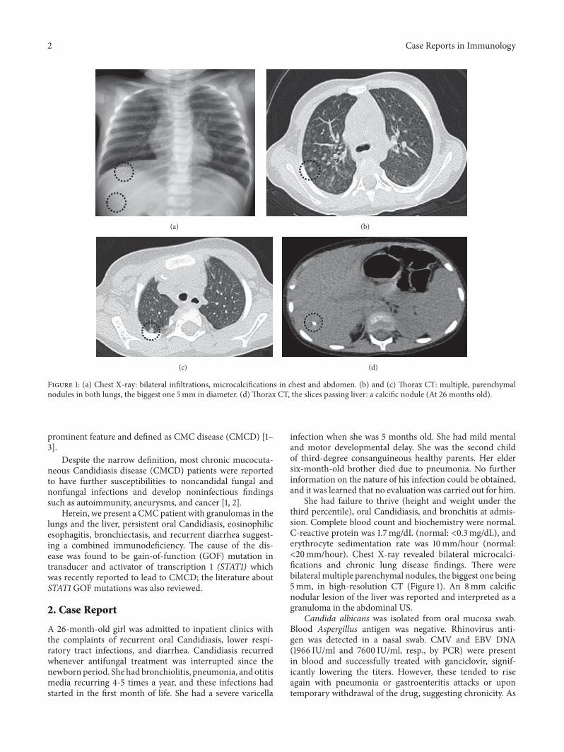
Figure 1: (a) Chest X-ray: bilateral infiltrations, microcalcifications in chest and abdomen. (b) and (c) Thorax CT: multiple, parenchymalnodules in both lungs, the biggest one 5mm in diameter. (d) Thorax CT, the slices passing liver: a calcific nodule (At 26 months old).
prominent feature and defined as CMC disease (CMCD) [1–3].
Despite the narrow definition, most chronic mucocuta-neous Candidiasis disease (CMCD) patients were reportedto have further susceptibilities to noncandidal fungal andnonfungal infections and develop noninfectious findingssuch as autoimmunity, aneurysms, and cancer [1, 2].
Herein, we present a CMCpatient with granulomas in thelungs and the liver, persistent oral Candidiasis, eosinophilicesophagitis, bronchiectasis, and recurrent diarrhea suggest-ing a combined immunodeficiency. The cause of the dis-ease was found to be gain-of-function (GOF) mutation intransducer and activator of transcription 1 (STAT1) whichwas recently reported to lead to CMCD; the literature aboutSTAT1 GOF mutations was also reviewed.
2. Case Report
A 26-month-old girl was admitted to inpatient clinics withthe complaints of recurrent oral Candidiasis, lower respi-ratory tract infections, and diarrhea. Candidiasis recurredwhenever antifungal treatment was interrupted since thenewborn period. She had bronchiolitis, pneumonia, and otitismedia recurring 4-5 times a year, and these infections hadstarted in the first month of life. She had a severe varicella
infection when she was 5 months old. She had mild mentaland motor developmental delay. She was the second childof third-degree consanguineous healthy parents. Her eldersix-month-old brother died due to pneumonia. No furtherinformation on the nature of his infection could be obtained,and it was learned that no evaluation was carried out for him.
She had failure to thrive (height and weight under thethird percentile), oral Candidiasis, and bronchitis at admis-sion. Complete blood count and biochemistry were normal.C-reactive protein was 1.7mg/dL (normal: <0.3mg/dL), anderythrocyte sedimentation rate was 10mm/hour (normal:<20mm/hour). Chest X-ray revealed bilateral microcalci-fications and chronic lung disease findings. There werebilateral multiple parenchymal nodules, the biggest one being5mm, in high-resolution CT (Figure 1). An 8mm calcificnodular lesion of the liver was reported and interpreted as agranuloma in the abdominal US.
Candida albicans was isolated from oral mucosa swab.Blood Aspergillus antigen was negative. Rhinovirus anti-gen was detected in a nasal swab. CMV and EBV DNA(1966 IU/ml and 7600 IU/ml, resp., by PCR) were presentin blood and successfully treated with ganciclovir, signif-icantly lowering the titers. However, these tended to riseagain with pneumonia or gastroenteritis attacks or upontemporary withdrawal of the drug, suggesting chronicity. As

Case Reports in Immunology 3
Table 1: Serum immunoglobulin, complement levels, lymphocyte subgroups as ratios, and absolute cell numbers with age-related referencevalues [4–6].
Patient Reference values(mean ± SD) Min–max
IgG (mg/dL) 1320 822.3 ± 208.4 430–1290IgA (mg/dL) 98.9 53.5 ± 26.8 23–130IgM (mg/dL) 149 92.5 ± 33.9 36–199IgE (IU/mL) 0.9 <100 2–199C3 (mg/dL) 171 120 ± 45 81–171C4 (mg/dL) 28.3 22 ± 13 9–36CD3+ T cells (%)(cells/mm3)
712620
70.0 ± 7.183220 ± 1180
48.2–81.4506–7267
CD19+ B cells (%)(cells/mm3)
23849
16.5 ± 5.70739 ± 329
6.7–30.4242–1459
CD3+CD4+Th cells (%)(cells/mm3)
401476
40.3 ± 7.271314 ± 542
23.2–59.5118–3245
CD3+CD8+ Tc cells (%)(cells/mm3)
281033
24.2 ± 5.48803 ± 417
15.2–39108–2367
CD3−CD1656+ NK cells (%)(cells/mm3)
5185
11.2 ± 4.85509 ± 295
3.4–26.4143–1599
CD3+HLA-DR+ active T cells (%)(cells/mm3)
18664
7.84 ± 3.7375 ± 235
2.1–16.222–954
oral fluconazole was not effective for Candidiasis, parenteralcaspofungin was initiated with good response. E. coli grewin urine culture which was responsive to proper antibacterialmedication. Tuberculin skin test was negative; interferon-gamma release assay (IGRA) was positive.
Mucopurulent secretions andmucous plaques on airwayswith normal anatomical structure were observed by bron-choscopy. Bronchoalveolar lavage fluid had benign cytology,and there was no evidence of bacteria, fungi, parasites, ormycobacteria. Repeated IGRA was found to be negative,and isoniazid (INH) prophylaxis was preferred instead ofmultidrug tuberculosis treatment. It was discontinued due toincreased liver enzymes after the second month.
Investigations for persistent diarrhea showed negativeresults for viruses, parasites, and bacteria. Recurrent vomitingwas also a problem in the patient. Upper gastrointestinalbarium scan and pH monitorization did not show gastroin-testinal reflux. The endoscopic evaluation revealed inflam-matory findings in esophagus and bulbus with a loose loweresophageal sphincter. There were no candidal plaques, prob-ably because she was under antifungal treatment at the timeof examination. Pathological studies showed eosinophilicesophagitis and fibrosis.
Vaccine responses to Hepatitis B, Rubella, and Mumpswere positive. Autoantibodies (antinuclear antibody,anti-gliadin antibodies, anti-thyroid peroxidase, and anti-thyroglobulin antibodies) and direct Coombs tests werenegative, and thyroid hormone levels were normal.
First line immunological workup revealed hypergamma-globulinemia with normal complement and IgE levels. Per-centages and numbers of lymphocyte subgroups were normalcompared to age-related healthy controls [4–6] (Table 1).The quantitative determination of oxidative burst, the foxp3expression on CD4+CD25+ T cells, and in vitro T-cellproliferation response to mitogens were normal.
She had chronic recurring infections with variouspathogens including fungi, viruses, and bacteria. She hadhypergammaglobulinemia, persistent oral Candidiasis, per-sistent granulomas, andnodules in the liver and the lungs. Shewas unresponsive to oral treatment and required parenteraltreatments with frequent, over one-week hospitalizationswarranting extensive workout for PID. Type and course of theinfections with substantial organ damage suggested the pres-ence of a severe combined immune deficiency, Mendeliansusceptibility to mycobacterial diseases (MSMD), or CMC aspreliminary diagnoses.
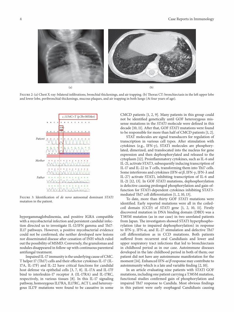
Regular intravenous immunoglobulin (IVIG) replace-ment was commenced when she was three years old, as asupportive measure to frequent hospitalizations for pneumo-nia and diarrhea attacks. Routine prophylactic antibacterialand antifungal therapies besides ganciclovir were continued.Despite these measures, she developed bronchiectasis at theage of four (Figure 2).
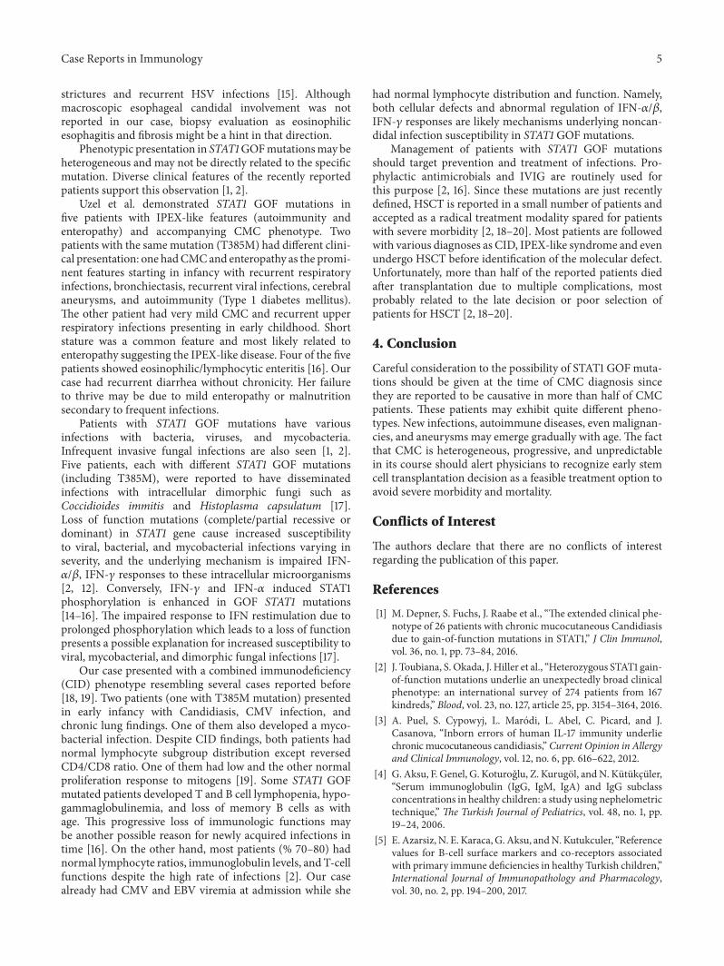
Molecular genetic analyses of IL12R/IFN gamma andTh17-IL17 pathways were planned and the former was foundto be negative. The investigation of the Th17/IL17 pathwayrevealed a heterozygous gain-of-function, de novo mutationin STAT1 gene, which was described recently (c.1154C>T,p.Thr385Met, T385M). Parents were wild-type for the gene(Figure 3). HSCT was planned because of ongoing infectionsdespite adequate supportivemeasures.There was nomatchedrelated donor. Thus, an unrelated donor search was initiated.
3. Discussion
Early-onset infections with diverse microorganisms, accom-panying CMC, warrant search for combined immunode-ficiency. Our case had normal lymphocyte subgroup dis-tribution and normal in vitro T-cell proliferation responseto mitogens. The granulomas in the lungs and the liver,
4 Case Reports in Immunology
(a) (b)
Figure 2: (a) Chest X-ray: bilateral infiltrations, bronchial thickenings, and air trapping. (b)Thorax CT: bronchiectasis in the left upper lobeand lower lobe, peribronchial thickenings, mucous plaques, and air trapping in both lungs (At four years of age).
Patient
Mother
Father
c.1154C>T (p.�r385Met)
Figure 3: Identification of de novo autosomal dominant STAT1mutation in the patient.
hypergammaglobulinemia, and positive IGRA compatiblewith a mycobacterial infection and persistent candidal infec-tion directed us to investigate IL-12/IFN gamma and Th17-IL17 pathways. However, a positive mycobacterial evidencecould not be confirmed; she neither developed new lesionsnor disseminated disease after cessation of INH which ruledout the possibility ofMSMD. Conversely, the granulomas andnodules disappeared in follow-up with continuous parenteralantifungal treatment.
Impaired IL-17 immunity is the underlying cause ofCMC.T helper 17 (Th17) cells and their effector cytokines IL-17 (IL-17A, IL-17F) and IL-22 have critical functions for candidalhost defense via epithelial cells [3, 7, 8]. IL-17A and IL-17Fbind to interleukin-17 receptor A (IL-17RA) and IL-17RC,respectively, in various tissues [8]. In this IL-17 signalingpathway, homozygous IL17RA, IL17RC, ACT 1, and heterozy-gous IL17F mutations were found to be causative in some
CMCD patients [1, 2, 9]. Many patients in this group couldnot be identified genetically until GOF heterozygous mis-sense mutations in the STAT1 molecule were defined in thisdecade [10, 11]. After that, GOF STAT1mutations were foundto be responsible for more than half of CMCD patients [1, 2].
STAT molecules are signal transducers for regulation oftranscription in various cell types. After stimulation withcytokines (e.g., IFN-𝛾), STAT1 molecules are phosphory-lated, dimerized, and translocated into the nucleus for geneexpression and then dephosphorylated and released to thecytoplasm [12]. Proinflammatory cytokines, such as IL-6 andIL-21, activate STAT3, subsequently inducing transcription ofIL-17 and IL-22 in T cells, transforming them intoTh17 cells.Some interferons and cytokines (IFN-𝛼/𝛽, IFN-𝛾, IFN-𝜆 andIL-27) activate STAT1, inhibiting transcription of IL-6 andIL-21 [12, 13]. In GOF STAT1 mutations, dephosphorylationis defective causing prolonged phosphorylation and gain-of-function for STAT1-dependent cytokines inhibiting STAT3-mediatedTh17 cell differentiation [1, 2, 10, 13].
To date, more than thirty GOF STAT1 mutations wereidentified. Early reported mutations were all in the coiled-coil domain (CCD) of STAT1 gene [1, 2, 10, 11]. Firstlydiscovered mutation in DNA binding domain (DBD) was aT385M mutation (as in our case) in two unrelated patientsfrom Japan. The investigators showed STAT1 hyperphospho-rylation (due to impaired dephosphorylation) in responseto IFN-𝛾, IFN-𝛼, and IL-27 stimulation and defective Th17cell differentiation as in CCD mutations. Both patientssuffered from recurrent oral Candidiasis and lower andupper respiratory tract infections that led to bronchiectasisin childhood period as in our case. Autoimmune diseasesdeveloped in the late childhood period in both of them; ourpatient did not have any autoimmune manifestation for themoment [14]. Enhanced IFN-𝛼/𝛽 response may contribute toautoimmunity which is a late and variable finding [2, 10].
In an article evaluating nine patients with STAT1 GOFmutations, including one patient carrying a T385Mmutation,functional studies confirmed gain of phosphorylation andimpaired Th17 response to Candida. Most obvious findingsin this patient were early esophageal Candidiasis causing
Case Reports in Immunology 5
strictures and recurrent HSV infections [15]. Althoughmacroscopic esophageal candidal involvement was notreported in our case, biopsy evaluation as eosinophilicesophagitis and fibrosis might be a hint in that direction.
Phenotypic presentation in STAT1GOFmutationsmay beheterogeneous and may not be directly related to the specificmutation. Diverse clinical features of the recently reportedpatients support this observation [1, 2].
Uzel et al. demonstrated STAT1 GOF mutations infive patients with IPEX-like features (autoimmunity andenteropathy) and accompanying CMC phenotype. Twopatients with the samemutation (T385M) had different clini-cal presentation: one hadCMCand enteropathy as the promi-nent features starting in infancy with recurrent respiratoryinfections, bronchiectasis, recurrent viral infections, cerebralaneurysms, and autoimmunity (Type 1 diabetes mellitus).The other patient had very mild CMC and recurrent upperrespiratory infections presenting in early childhood. Shortstature was a common feature and most likely related toenteropathy suggesting the IPEX-like disease. Four of the fivepatients showed eosinophilic/lymphocytic enteritis [16]. Ourcase had recurrent diarrhea without chronicity. Her failureto thrive may be due to mild enteropathy or malnutritionsecondary to frequent infections.
Patients with STAT1 GOF mutations have variousinfections with bacteria, viruses, and mycobacteria.Infrequent invasive fungal infections are also seen [1, 2].Five patients, each with different STAT1 GOF mutations(including T385M), were reported to have disseminatedinfections with intracellular dimorphic fungi such asCoccidioides immitis and Histoplasma capsulatum [17].Loss of function mutations (complete/partial recessive ordominant) in STAT1 gene cause increased susceptibilityto viral, bacterial, and mycobacterial infections varying inseverity, and the underlying mechanism is impaired IFN-𝛼/𝛽, IFN-𝛾 responses to these intracellular microorganisms[2, 12]. Conversely, IFN-𝛾 and IFN-𝛼 induced STAT1phosphorylation is enhanced in GOF STAT1 mutations[14–16]. The impaired response to IFN restimulation due toprolonged phosphorylation which leads to a loss of functionpresents a possible explanation for increased susceptibility toviral, mycobacterial, and dimorphic fungal infections [17].
Our case presented with a combined immunodeficiency(CID) phenotype resembling several cases reported before[18, 19]. Two patients (one with T385M mutation) presentedin early infancy with Candidiasis, CMV infection, andchronic lung findings. One of them also developed a myco-bacterial infection. Despite CID findings, both patients hadnormal lymphocyte subgroup distribution except reversedCD4/CD8 ratio. One of them had low and the other normalproliferation response to mitogens [19]. Some STAT1 GOFmutated patients developed T and B cell lymphopenia, hypo-gammaglobulinemia, and loss of memory B cells as withage. This progressive loss of immunologic functions maybe another possible reason for newly acquired infections intime [16]. On the other hand, most patients (% 70–80) hadnormal lymphocyte ratios, immunoglobulin levels, and T-cellfunctions despite the high rate of infections [2]. Our casealready had CMV and EBV viremia at admission while she
had normal lymphocyte distribution and function. Namely,both cellular defects and abnormal regulation of IFN-𝛼/𝛽,IFN-𝛾 responses are likely mechanisms underlying noncan-didal infection susceptibility in STAT1 GOF mutations.
Management of patients with STAT1 GOF mutationsshould target prevention and treatment of infections. Pro-phylactic antimicrobials and IVIG are routinely used forthis purpose [2, 16]. Since these mutations are just recentlydefined, HSCT is reported in a small number of patients andaccepted as a radical treatment modality spared for patientswith severe morbidity [2, 18–20]. Most patients are followedwith various diagnoses as CID, IPEX-like syndrome and evenundergo HSCT before identification of the molecular defect.Unfortunately, more than half of the reported patients diedafter transplantation due to multiple complications, mostprobably related to the late decision or poor selection ofpatients for HSCT [2, 18–20].
4. Conclusion
Careful consideration to the possibility of STAT1 GOF muta-tions should be given at the time of CMC diagnosis sincethey are reported to be causative in more than half of CMCpatients. These patients may exhibit quite different pheno-types. New infections, autoimmune diseases, even malignan-cies, and aneurysms may emerge gradually with age. The factthat CMC is heterogeneous, progressive, and unpredictablein its course should alert physicians to recognize early stemcell transplantation decision as a feasible treatment option toavoid severe morbidity and mortality.
Conflicts of Interest
The authors declare that there are no conflicts of interestregarding the publication of this paper.
References
[1] M. Depner, S. Fuchs, J. Raabe et al., “The extended clinical phe-notype of 26 patients with chronic mucocutaneous Candidiasisdue to gain-of-function mutations in STAT1,” J Clin Immunol,vol. 36, no. 1, pp. 73–84, 2016.
[2] J. Toubiana, S. Okada, J. Hiller et al., “Heterozygous STAT1 gain-of-function mutations underlie an unexpectedly broad clinicalphenotype: an international survey of 274 patients from 167kindreds,” Blood, vol. 23, no. 127, article 25, pp. 3154–3164, 2016.
[3] A. Puel, S. Cypowyj, L. Marodi, L. Abel, C. Picard, and J.Casanova, “Inborn errors of human IL-17 immunity underliechronic mucocutaneous candidiasis,” Current Opinion in Allergyand Clinical Immunology, vol. 12, no. 6, pp. 616–622, 2012.
[4] G. Aksu, F. Genel, G. Koturoglu, Z. Kurugol, andN. Kutukculer,“Serum immunoglobulin (IgG, IgM, IgA) and IgG subclassconcentrations in healthy children: a study using nephelometrictechnique,” The Turkish Journal of Pediatrics, vol. 48, no. 1, pp.19–24, 2006.
[5] E. Azarsiz, N. E. Karaca, G.Aksu, andN.Kutukculer, “Referencevalues for B-cell surface markers and co-receptors associatedwith primary immune deficiencies in healthy Turkish children,”International Journal of Immunopathology and Pharmacology,vol. 30, no. 2, pp. 194–200, 2017.
6 Case Reports in Immunology
[6] C. R. Jolliff, K. M. Cost, P. C. Stivrins et al., “Reference intervalsfor serum IgG, IgA, IgM, C3, and C4 as determined by ratenephelometry,” Clin Chem, vol. 28, no. 1, pp. 126–128, 1982.
[7] K. Eyerich, S. Eyerich, J. Hiller, H. Behrendt, and C. Traidl-Hoffmann, “Chronic mucocutaneous candidiasis, from benchto bedside,” European Journal of Dermatology, vol. 20, no. 3, pp.260–265, 2010.
[8] S. Eyerich, K. Eyerich, A. Cavani, and C. Schmidt-Weber, “IL-17and IL-22: siblings, not twins,” Trends in Immunology, vol. 31,no. 9, pp. 354–361, 2010.
[9] A. Puel, S. Cypowyj, J. Bustamante et al., “Chronic mucocuta-neous candidiasis in humans with inborn errors of interleukin-17 immunity,” Science, vol. 332, no. 6025, pp. 65–68, 2011.
[10] L. Liu, S. Okada, X. F. Kong et al., “Gain-of-function humanSTAT1 mutations impair IL-17 immunity and underlie chronicmucocutaneous Candidiasis,” J Exp Med, vol. 208, no. 8, pp.1635-1348, 2011.
[11] F. L. Van De Veerdonk, T. S. Plantinga, A. Hoischen et al.,“STAT1 mutations in autosomal dominant chronic mucocuta-neous candidiasis,” The New England Journal of Medicine, vol.365, no. 1, pp. 54–61, 2011.
[12] S. Boisson-Dupuis, X. F. Kong, S. Okada et al., “Inborn errorsof human STAT1: allelic heterogeneity governs the diversity ofimmunological and infectious phenotypes,” Current Opinion inImmunology, vol. 24, no. 4, pp. 364–378, 2012.
[13] D. C. Pichard, A. F. Freeman, and E. W. Cowen, “Primaryimmunodeficiency update: Part II. Syndromes associated withmucocutaneous candidiasis and noninfectious cutaneous man-ifestations,” Journal of the American Academy of Dermatology,vol. 73, no. 3, pp. 367–381, 2015.
[14] S. Takezaki, M. Yamada, M. Kato et al., “Chronic mucocuta-neous candidiasis caused by a gain-of-function mutation in theSTAT1 DNA-binding domain,”The Journal of Immunology, vol.189, no. 3, pp. 1521–1526, 2012.
[15] B. Soltesz, B. Toth, N. Shabashova et al., “New and recurrentgain-of-function STAT1 mutations in patients with chronicmucocutaneous candidiasis from Eastern and Central Europe,”Journal of Medical Genetics, vol. 50, no. 9, pp. 567–578, 2013.
[16] G. Uzel, E. P. Sampaio, M. G. Lawrence et al., “Dominant gain-of-function STAT1 mutations in FOXP3 wild-type immunedysregulation-polyendocrinopathy-enteropathy-X-linked-likesyndrome,” The Journal of Allergy and Clinical Immunology,vol. 131, no. 6, pp. 1611–1623, 2013.
[17] E. P. Sampaio, A. P. Hsu, J. Pechacek et al., “Signal transducerand activator of transcription 1 (STAT1) gain-of-functionmuta-tions and disseminated coccidioidomycosis and histoplasmo-sis,”The Journal of Allergy and Clinical Immunology, vol. 131, no.6, pp. 1624–1634, 2013.
[18] J. C. Aldave, E. Cachay, L. Nunez et al., “A 1-year-old girl witha gain-of-function STAT1 mutation treated with hematopoieticstem cell transplantation,” Journal of Clinical Immunology, vol.33, no. 8, pp. 1273–1275, 2013.
[19] S. Baris, F. Alroqi, A. Kiykim et al., “Severe Early-OnsetCombined Immunodeficiency due to Heterozygous Gain-of-Function Mutations in STAT1,” Journal of Clinical Immunology,vol. 36, no. 7, pp. 641–648, 2016.
[20] S. S. Kilic, A. Puel, and J.-L. Casanova, “Orf Infection in a Patientwith Stat1 Gain-of-Function,” Journal of Clinical Immunology,vol. 35, no. 1, pp. 80–83, 2015.

Submit your manuscripts athttps://www.hindawi.com
Stem CellsInternational
Hindawi Publishing Corporationhttp://www.hindawi.com Volume 2014
Hindawi Publishing Corporationhttp://www.hindawi.com Volume 2014
MEDIATORSINFLAMMATION
of
Hindawi Publishing Corporationhttp://www.hindawi.com Volume 2014
Behavioural Neurology
EndocrinologyInternational Journal of
Hindawi Publishing Corporationhttp://www.hindawi.com Volume 2014
Hindawi Publishing Corporationhttp://www.hindawi.com Volume 2014
Disease Markers
Hindawi Publishing Corporationhttp://www.hindawi.com Volume 2014
BioMed Research International
OncologyJournal of
Hindawi Publishing Corporationhttp://www.hindawi.com Volume 2014
Hindawi Publishing Corporationhttp://www.hindawi.com Volume 2014
Oxidative Medicine and Cellular Longevity
Hindawi Publishing Corporationhttp://www.hindawi.com Volume 2014
PPAR Research
The Scientific World JournalHindawi Publishing Corporation http://www.hindawi.com Volume 2014
Immunology ResearchHindawi Publishing Corporationhttp://www.hindawi.com Volume 2014
Journal of
ObesityJournal of
Hindawi Publishing Corporationhttp://www.hindawi.com Volume 2014
Hindawi Publishing Corporationhttp://www.hindawi.com Volume 2014
Computational and Mathematical Methods in Medicine
OphthalmologyJournal of
Hindawi Publishing Corporationhttp://www.hindawi.com Volume 2014
Diabetes ResearchJournal of
Hindawi Publishing Corporationhttp://www.hindawi.com Volume 2014
Hindawi Publishing Corporationhttp://www.hindawi.com Volume 2014
Research and TreatmentAIDS
Hindawi Publishing Corporationhttp://www.hindawi.com Volume 2014
Gastroenterology Research and Practice
Hindawi Publishing Corporationhttp://www.hindawi.com Volume 2014
Parkinson’s Disease
Evidence-Based Complementary and Alternative Medicine
Volume 2014Hindawi Publishing Corporationhttp://www.hindawi.com


![[Notes]STAT1 - Elementary Statistics](https://img.pdfslide.net/doc/110x75/577cdbee1a28ab9e78a976c0/notesstat1-elementary-statistics.jpg)