Embed Size (px)
Citation preview
Gastric Emptying Scintigraphy*
Mary Beth Farrell
Intersocietal Accreditation Commission
CE credit: For CE credit, you can access the test for this article, as well as additional JNMT CE tests, online at https://www.snmmilearningcenter.org.Complete the test online no later than June 2022. Your online test will be scored immediately. You may make 3 attempts to pass the test and mustanswer 80% of the questions correctly to receive 1.0 CEH (Continuing Education Hour) credit. SNMMI members will have their CEH credit added to theirVOICE transcript automatically; nonmembers will be able to print out a CE certificate upon successfully completing the test. The online test is free to SNMMImembers; nonmembers must pay $15.00 by credit card when logging onto the website to take the test.
In 1966, Griffith et al. used ‘‘a standard breakfast of or-dinary food’’ labeled with chromium-51 to report the first useof gastric emptying scintigraphy (GES). As the procedure wasrefined over the years, investigators realized the importance ofthe radiotracer binding to the meal to produce accurate results.To achieve the highest-efficiency labeling, early investiga-tors injected 99mTc–sulfur colloid (99mTc-SC) into a vein ona chicken’s wing, and it accumulated in the Kupffer cells in theliver. After 15 minutes, the investigators butchered the chickenand removed the liver. The livers were then cooked, mixed withstew—to make it more appetizing and increase the volume—and fed to the patient. Thankfully, better methods evolved.The literature shows the use of a variety of other foods for
GES, such as pancakes, cheese, milk, or oatmeal. A recentstudy by Farrell et al. found an assortment of unusual mealscurrently being used, including honey buns, corn flakes andmilk, peanut butter sandwiches, egg salad sandwiches, eggburritos, and McDonalds Egg McMuffins. In addition, a varietyof meal preparation methods was observed, such as addingthe tracer to eggs after cooking rather than before.Although GES using radiolabeled meals is considered the
gold standard for evaluating patients with gastrointestinalmotility disorders, gastroenterologists—who refer patientsfor the test and manage them based on the results—havequestioned the reliability of GES. The issue lies in inconsistentresults due to lack of standardized procedure for the type ofmeal used, patient positioning, image acquisition frequencyand duration, and quantitation method. Lack of standardi-zation affects reported normal values and, thus, comparisonof results between institutions. Discrepant test results com-plicate patient treatment decisions.To address the lack of consistency and standardization,
an expert panel of gastroenterologists from the American
Neurogastroenterology and Motility Society and nuclearmedicine physicians from the Society of Nuclear Medicineand Molecular Imaging (SNMMI) published a consensusguideline for solid meal GES in 2008. The guidelinesimplified the procedure and established standardized normalvalues. It also provided recommendations for precise patientpreparation, meal composition, acquisition method, and imageprocessing that should be adhered to by all laboratories.
RATIONALE/INDICATIONS/CONTRAINDICATIONS
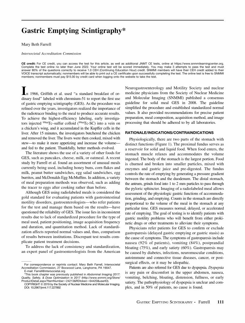
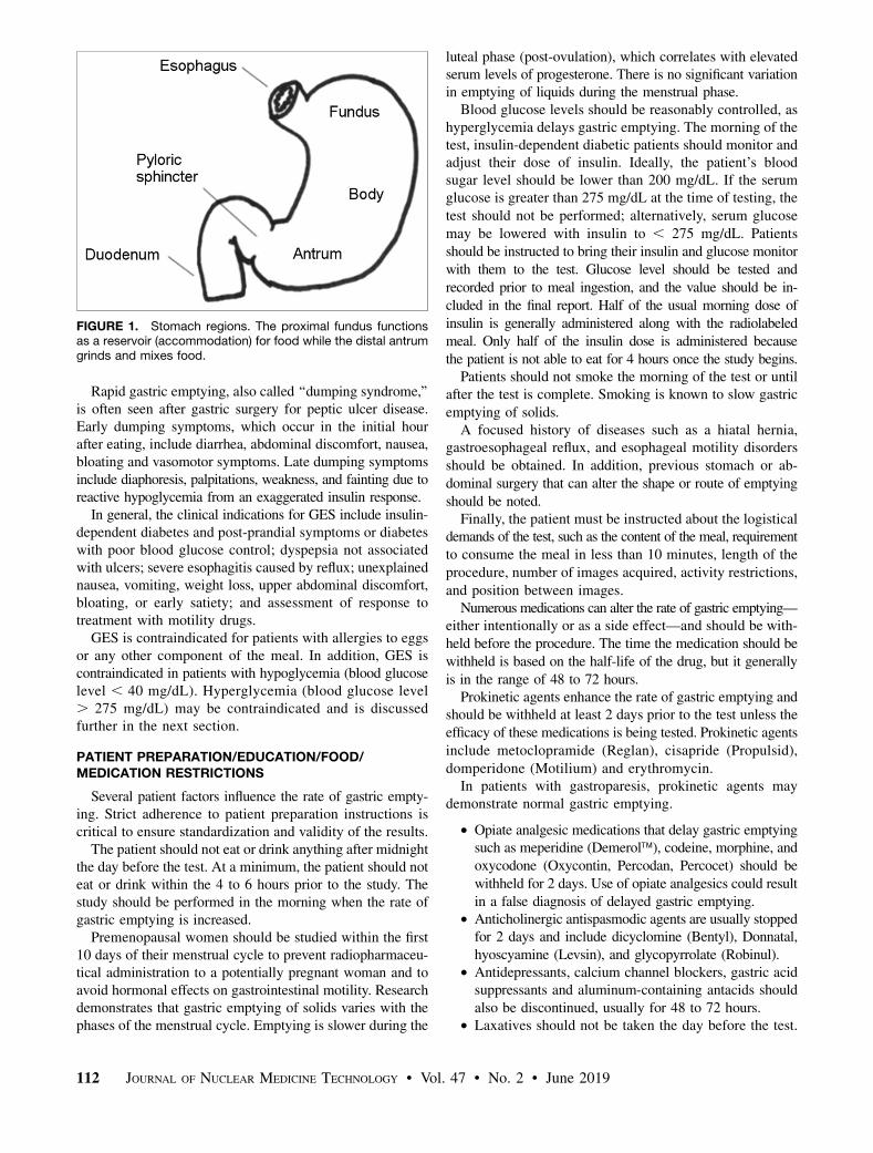
Physiologically, there are two parts of the stomach withdistinct functions (Figure 1). The proximal fundus serves asa reservoir for solid and liquid food. When food enters, thestomach muscle relaxes and accommodates the volumeingested. The body of the stomach is the largest portion. Foodis churned and broken into smaller particles, mixed withenzymes and gastric juice and pre-digested. The funduscontrols the rate of emptying by generating a pressure gradientbetween the stomach and the duodenum. The distal stomach,the antrum, grinds food into 1 to 2 mm particles to pass throughthe pyloric sphincter. Imaging of a radiolabeled meal allowsassessment of the physiologic gastric functions of accommoda-tion, grinding, and emptying. Counts in the stomach are directlyproportional to the volume of the meal in the stomach at anyparticular time. GES measures normal, delayed, or acceleratedrate of emptying. The goal of testing is to identify patients withgastric motility problems who will benefit from either proki-netic drugs or other treatments to alleviate their symptoms.
Physicians refer patients for GES to confirm or excludegastroparesis (delayed gastric emptying or gastric stasis) asthe cause of symptoms. The symptoms of gastroparesis includenausea (92% of patients), vomiting (84%), postprandialbloating (75%), and early satiety (60%). Gastroparesis maybe caused by diabetes, infections, neuromuscular conditions,autoimmune and connective tissue diseases, cancer, or post-surgical effects, or it may be idiopathic.
Patients are also referred for GES due to dyspepsia. Dyspepsiais any pain or discomfort in the upper abdomen, nausea,vomiting, belching, bloating, distension, fullness, or earlysatiety. The pathophysiology of dyspepsia is unclear and com-plex, and in 50% of patients, no cause is found.
For correspondence or reprints contact: Mary Beth Farrell, IntersocietalAccreditation Commission, 27 Boxwood Lane, Langhorne, PA 19047.E-mail: [email protected]*This book chapter was previously published in Abdominal Imaging 2017:
Quality, Safety, & Dose Optimization in 2017 (http://www.snmmi.org/Store/ProductDetail.aspx?ItemNumber524213&RDtoken564420&userID).COPYRIGHT© 2019 by the Society of Nuclear Medicine and Molecular Imaging.DOI: 10.2967/jnmt.117.227892
GASTRIC EMPTYING SCINTIGRAPHY • Farrell 111
Rapid gastric emptying, also called ‘‘dumping syndrome,’’is often seen after gastric surgery for peptic ulcer disease.Early dumping symptoms, which occur in the initial hourafter eating, include diarrhea, abdominal discomfort, nausea,bloating and vasomotor symptoms. Late dumping symptomsinclude diaphoresis, palpitations, weakness, and fainting due toreactive hypoglycemia from an exaggerated insulin response.In general, the clinical indications for GES include insulin-
dependent diabetes and post-prandial symptoms or diabeteswith poor blood glucose control; dyspepsia not associatedwith ulcers; severe esophagitis caused by reflux; unexplainednausea, vomiting, weight loss, upper abdominal discomfort,bloating, or early satiety; and assessment of response totreatment with motility drugs.GES is contraindicated for patients with allergies to eggs
or any other component of the meal. In addition, GES iscontraindicated in patients with hypoglycemia (blood glucoselevel , 40 mg/dL). Hyperglycemia (blood glucose level. 275 mg/dL) may be contraindicated and is discussedfurther in the next section.
PATIENT PREPARATION/EDUCATION/FOOD/MEDICATION RESTRICTIONS
Several patient factors influence the rate of gastric empty-ing. Strict adherence to patient preparation instructions iscritical to ensure standardization and validity of the results.The patient should not eat or drink anything after midnight
the day before the test. At a minimum, the patient should noteat or drink within the 4 to 6 hours prior to the study. Thestudy should be performed in the morning when the rate ofgastric emptying is increased.Premenopausal women should be studied within the first
10 days of their menstrual cycle to prevent radiopharmaceu-tical administration to a potentially pregnant woman and toavoid hormonal effects on gastrointestinal motility. Researchdemonstrates that gastric emptying of solids varies with thephases of the menstrual cycle. Emptying is slower during the
luteal phase (post-ovulation), which correlates with elevatedserum levels of progesterone. There is no significant variationin emptying of liquids during the menstrual phase.
Blood glucose levels should be reasonably controlled, ashyperglycemia delays gastric emptying. The morning of thetest, insulin-dependent diabetic patients should monitor andadjust their dose of insulin. Ideally, the patient’s bloodsugar level should be lower than 200 mg/dL. If the serumglucose is greater than 275 mg/dL at the time of testing, thetest should not be performed; alternatively, serum glucosemay be lowered with insulin to , 275 mg/dL. Patientsshould be instructed to bring their insulin and glucose monitorwith them to the test. Glucose level should be tested andrecorded prior to meal ingestion, and the value should be in-cluded in the final report. Half of the usual morning dose ofinsulin is generally administered along with the radiolabeledmeal. Only half of the insulin dose is administered becausethe patient is not able to eat for 4 hours once the study begins.
Patients should not smoke the morning of the test or untilafter the test is complete. Smoking is known to slow gastricemptying of solids.
A focused history of diseases such as a hiatal hernia,gastroesophageal reflux, and esophageal motility disordersshould be obtained. In addition, previous stomach or ab-dominal surgery that can alter the shape or route of emptyingshould be noted.
Finally, the patient must be instructed about the logisticaldemands of the test, such as the content of the meal, requirementto consume the meal in less than 10 minutes, length of theprocedure, number of images acquired, activity restrictions,and position between images.
Numerous medications can alter the rate of gastric emptying—either intentionally or as a side effect—and should be with-held before the procedure. The time the medication should bewithheld is based on the half-life of the drug, but it generallyis in the range of 48 to 72 hours.
Prokinetic agents enhance the rate of gastric emptying andshould be withheld at least 2 days prior to the test unless theefficacy of these medications is being tested. Prokinetic agentsinclude metoclopramide (Reglan), cisapride (Propulsid),domperidone (Motilium) and erythromycin.
In patients with gastroparesis, prokinetic agents maydemonstrate normal gastric emptying.
• Opiate analgesic medications that delay gastric emptyingsuch as meperidine (Demerol�), codeine, morphine, andoxycodone (Oxycontin, Percodan, Percocet) should bewithheld for 2 days. Use of opiate analgesics could resultin a false diagnosis of delayed gastric emptying.
• Anticholinergic antispasmodic agents are usually stoppedfor 2 days and include dicyclomine (Bentyl), Donnatal,hyoscyamine (Levsin), and glycopyrrolate (Robinul).
• Antidepressants, calcium channel blockers, gastric acidsuppressants and aluminum-containing antacids shouldalso be discontinued, usually for 48 to 72 hours.
• Laxatives should not be taken the day before the test.
FIGURE 1. Stomach regions. The proximal fundus functionsas a reservoir (accommodation) for food while the distal antrumgrinds and mixes food.
112 JOURNAL OF NUCLEAR MEDICINE TECHNOLOGY • Vol. 47 • No. 2 • June 2019
• Other medications that may affect gastric emptyinginclude atropine, nifedipine progesterone, octreotide,theophylline, benzodiazepine, and phenolamine.
The patient may take other medications with a smallamount of water prior to the test. If the patient has severe nauseaand vomiting at the time of the test, serotonin receptor (5-HT-3)antagonists such as ondansetron (Zofran) may be given.
IMAGING PROCEDURE
Gastric emptying is a complex physiologic process con-trolled by the physical and chemical composition of the GESmeal, sympathetic and parasympathetic innervation of thestomach, and circulating neuroendocrine transmitters. Thetype of food, volume, and caloric content significantly affectthe rate of gastric emptying. In order to have any value, theGES meal and protocol must be closely followed. Table 1details the factors that affect the gastric emptying rate. Nor-mal rates of gastric emptying have been established and val-idated for the recommended meal based on these factors.
Solid Meal Study
The standardized meal consists of 120 grams (4 oz.) ofliquid egg whites such as Egg Beaters (ConAgra Foods),which is the equivalent of the whites of two large eggs (seeTable 2). The liquid egg white is mixed with 18.5 to 37 MBq(0.5–1.0 mCi) of 99mTc-SC and cooked in the microwave oron a nonstick griddle. The egg mixture is stirred once or twiceduring cooking and cooked until it has the texture of a firmomelet.For accurate results, the radiotracer must bind tightly to
the solid component of the meal and remain within thegastrointestinal tract. 99mTc-SC is the preferred radiophar-maceutical because it is not absorbed within the gastroin-testinal tract and binds to the albumin (egg white protein),denaturing as it cooks. Note, 99mTc-SC does not bind to egg
yolks. The goal of labeling eggs whites with 99mTc-SC is tokeep the meal from being absorbed or binding to the mucousmembranes in either the stomach or intestine.
Egg substitute is preferred over fresh whole eggs becauseit has a high binding percentage and is less likely todisintegrate in gastric fluid. If the tracer separates from theprotein, the test results will vary because the meal becomesa part-solid, part-liquid mixture. Approximately 80% of99mTc-SC remains bound to the egg substitute at 3 hours.Egg substitute also maintains firmer consistency than wholeeggs. The labeling efficiency is approximately 85%.
The egg mixture is also served with two slices of toastedwhite bread spread with 30 g of strawberry jam and 120 ml(4 oz.) of water. The meal is usually served as a sandwich todecrease the time required for ingestion, but it may be eatenseparately if the patient desires. The entire meal has acaloric content of 255 kcal composed of 72% carbohydrate,24% protein, 2% fat, and 2% fiber.
The patient must eat the entire meal as quickly as possible,ideally in less than 10 minutes. If the patient is unable toingest the entire meal, a minimum of 50% of the meal mustbe consumed. If less than 50% of the meal is ingested, theresults may overestimate the rate of gastric emptying, so thestudy cannot be considered diagnostic. The technologistshould document the time it takes the patient to ingest themeal and the percentage of the meal consumed.
Acquisition. A large field-of-view camera with a low-energy, all-purpose collimator is used to acquire images inword mode with a 128 · 128 matrix. The energy window ispeaked at 140 keV6 20%. Images are acquired in the uprightor standing position for 1 minute in both anterior and pos-terior projections with the distal esophagus, stomach, andproximal small intestine in the field of view. The imagesmay be acquired simultaneously using a dual-head cameraor sequentially using a single-head camera. There is no sig-nificant difference in the results between images acquiredsimultaneously versus images acquired sequentially.
If the patient is unable to stand, images may be acquiredsupine in the left anterior oblique (LAO) position, althoughthe rate of gastric emptying may be significantly decreasedin the supine position. If the patient is imaged in the supineposition, a dual-head camera is positioned above and belowthe patient. If a single-head camera is used, the images areusually acquired in the LAO view.
The standardized consensus protocol recommends ac-quisition of images immediately upon ingestion of the meal
TABLE 2Standardized Gastric Emptying Meal
120 g (4 oz.) of liquid egg whites (99% real eggs, cholesterol
free, fat-free and low calorie)
2 slices of white bread30 g strawberry jam120 ml (4 oz.) water18.5–37 MBq (0.5–1.0 mCi) 99mTc-SC
TABLE 1Factors that Affect the Rate of Gastric Emptying
Factors that Increase
the Rate of Emptying
Factors that Decrease
the Rate of Emptying
Liquids SolidsSmall particle size Large particle sizeLow fiber or low residue High fiberProteins and carbohydrates FatsLow calorie High calorieLarge volume Small volumeAlkaline AcidicHot food Cool foodEarly in the day Late in the dayActivity SedentaryUpright Lying downAbsence of pain PainLying on right side Lying on left sideMale FemaleProkinetic, erythromycin Narcotics, anticholinergicReserpine, anticholinesterases,
guanethidine,
cholinergic agents
(Atropine), tricyclic
antidepressants,
phenothiazines
GASTRIC EMPTYING SCINTIGRAPHY • Farrell 113
and then repeated at hourly intervals of 1, 2, and 4 hours. Thesame camera must be used for all images. A 57Co markerplaced on the xiphoid process is helpful for repositioning thepatient and drawing regions of interest during later process-ing. Imaging at 30 minutes may be helpful if rapid gastricemptying or impaired fundal accommodation is suspected.Imaging can be discontinued prior to 4 hours if less than10% of the original stomach contents remains in the stomach.Recent research suggests the importance of obtaining
images for up to 4 hours. Delayed gastric emptying is detectedwith higher sensitivity at 4 hours than at 2 hours. Images at
4 hours detect gastroparesis 30% more often. Imaging at 0, 1,2, and 4 hours allows for the identification of both rapid anddelayed gastric emptying, which is important because they aretreated differently, although the symptoms are similar.
Between images, the patient should rest in the sittingposition, minimizing walking and activity. Stair climbing,other diagnostic imaging tests, and other appointmentsshould be avoided during the 4-hour test.
Variations
Liquid Study. Gastric emptying of liquids is a simpleprocess because the meal does not need to be ground intosmall particles to pass through the pyloric sphincter. Asliquid enters the stomach, the fundus relaxes to accommodatethe volume. Smooth muscle in the fundus contracts, creatinga pressure gradient between the fundus and pylorus. Liquidsbegin to leave the stomach almost immediately after ingestion.Liquids usually empty from the stomach in about 30 minutes.
The volume of liquid is the main determinant of the rateof liquid gastric emptying. The larger the volume of liquid, themore rapid the rate of emptying. As volume decreases, emptyingslows. The caloric content of the liquid also affects the rateof emptying. The liquid used should equilibrate quickly andnot be absorbed from the gastrointestinal tract.
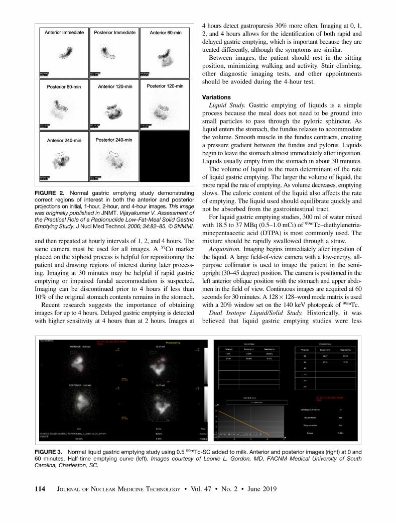
For liquid gastric emptying studies, 300 ml of water mixedwith 18.5 to 37 MBq (0.5–1.0 mCi) of 99mTc–diethylenetria-minepentaacetic acid (DTPA) is most commonly used. Themixture should be rapidly swallowed through a straw.
Acquisition. Imaging begins immediately after ingestion ofthe liquid. A large field-of-view camera with a low-energy, all-purpose collimator is used to image the patient in the semi-upright (30–45 degree) position. The camera is positioned in theleft anterior oblique position with the stomach and upper abdo-men in the field of view. Continuous images are acquired at 60seconds for 30 minutes. A 128 · 128–word mode matrix is usedwith a 20% window set on the 140 keV photopeak of 99mTc.
Dual Isotope Liquid/Solid Study. Historically, it wasbelieved that liquid gastric emptying studies were less
FIGURE 3. Normal liquid gastric emptying study using 0.5 99mTc-SC added to milk. Anterior and posterior images (right) at 0 and60 minutes. Half-time emptying curve (left). Images courtesy of Leonie L. Gordon, MD, FACNM Medical University of SouthCarolina, Charleston, SC.
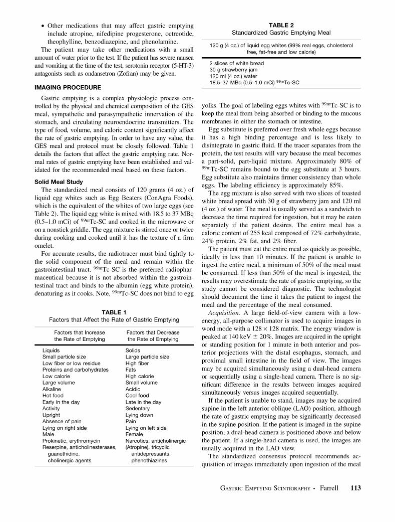
FIGURE 2. Normal gastric emptying study demonstratingcorrect regions of interest in both the anterior and posteriorprojections on initial, 1-hour, 2-hour, and 4-hour images. This imagewas originally published in JNMT. Vijayakumar V. Assessment ofthe Practical Role of a Radionuclide Low-Fat-Meal Solid GastricEmptying Study. J Nucl Med Technol. 2006; 34:82–85.© SNMMI.
114 JOURNAL OF NUCLEAR MEDICINE TECHNOLOGY • Vol. 47 • No. 2 • June 2019

sensitive for detecting gastroparesis than solid studies.However, recent studies comparing liquid-only studies withsolid-only studies found that liquid-only studies detectedgastroparesis more frequently. A patient may have normalsolid and liquid, abnormal liquid and normal solid, normalliquid and abnormal solid, or abnormal liquid and abnormalsolid results. Delayed liquid gastric emptying may be seenin 30% to 35% of patients with a normal solid gastricemptying study. Research has shown that postprandial full-ness and early satiety are associated with delayed gastricemptying of liquids; therefore, there is added diagnosticvalue in combining liquid and solid gastric emptyingstudies.When a liquid/solid study is performed, liquids empty
from the stomach more rapidly than the solids, but at aslower rate than if a liquid study were performed alone.When there is rapid passage of water from the stomach whilesolid materials are retained, this is known as ‘‘solid-liquiddiscrimination.’’Acquisition. Liquid and solid gastric emptying studies
can be performed sequentially on the same day. The liquidis performed first using 7.4 MBq (0.2 mCi) of 111In-DPTAin water followed by the solid meal using 99mTc-SC. Dualisotope liquid and solid gastric emptying studies may also beperformed simultaneously. Because 111In-DTPA is chemi-cally inert, it does not bind to the components of the solidmeal and does interfere with the results of the solid meal.A medium energy collimator is used with the camera, set
up for dual acquisition energy discrimination of the 99mTcphotopeak (20% window, 140 keV) and 111In (20% win-dow, 274 keV).Alternative Meals. For patients allergic to eggs or any of
the other meal components, gluten-sensitive patients, or patientswho will not eat the standardized meal, alternative mealsmay be used. Oatmeal or liquids such as milk or Ensure shakeshave been used; however, there is limited data on normalvalues available for these meals.
Radiation Exposure
The ICRP 106 model estimates that ingestion of 37 MBq(1.0 mCi) 99mTc-SC for a gastric emptying study wouldimpart an approximate effective dose of 0.8 mSv (0.08 rem)in an adult male. The critical organ for this study is the upperlarge intestine, which would receive 0.2 mGy (0.02 rad). In-gestion for an adult female of 37 MBq (1 mCi) 99mTc-SCwould impart an approximate effective dose of 1.0 mSv(0.10 rem). The critical organ for this study is the lungs, whichwould receive 0.3 mGy (0.03 rad). The effective dose for in-gestion of 37 MBq (1.0 mCi) 111In-DTPA is not available.
PROCESSING
Evaluation of the images alone is not useful for thedetermination of rapid or delayed gastric emptying. To quantifythe results, regions of interest (ROI) are drawn around thestomach on all anterior and posterior images. The ROI mustinclude the antrum and fundus regions of the stomach (Figure2). Care must be taken to ensure no activity from the adjacent
FIGURE 5. Normal gastric emptying curves. For solid meal(red), there is an initial 20-30 m lag period as the antrumreduces meal particle size and mixes with gastric acid. Afterthe lag period, the solid material empties from the stomach ina linear fashion. The liquid meal (purple) immediately begins toleave the stomach and empties in an exponential pattern.
FIGURE 4. Normal solid gastric emptying study. (Top) Anterior and posterior images at 0 and approximately 1, 2 and 4 hours.(Bottom) Region counts from the anterior and posterior images and geometric mean. The percent retention at 4 hours is 8.2%.Images courtesy of Leonie L. Gordon, MD, FACNM Medical University of South Carolina, Charleston, SC.
GASTRIC EMPTYING SCINTIGRAPHY • Farrell 115
small bowel is included within the region; however, if theimages demonstrate loops of small bowel on the initial image,then that area should be included in the region so that theentire ingested activity is used for comparison. Because thestudy is performed over 4 hours and 99mTc has a 6-hour half-life, radioactive decay correction must be performed.
The fundal portion of the stomach is relatively posteriorto the antral portion. Ingested material moves through thestomach superiorly to inferiorly, from left to right, andposterior to anterior. Therefore, the counts obtained fromthe regions of interest must be attenuation corrected. If theimages are not attenuation corrected, the rate of gastricemptying can be underestimated by 10% to 30%, mostcommonly in patients with large body habitus.
The most frequently used and easiest method of atten-uation correction is to use the geometric mean, whichresults in only a 3% to 4% error in counts. The geomet-ric mean is calculated by taking the square root of theanterior counts multiplied by the posterior counts for eachtime point.
Geometric mean 5ffiffiffiffiffiffiffiffiffiffiffiffiffiffiffiffiffiffiffiffiffiffiffiffiffiffiffiffiffiffiffiffiffiffiffiffiffiffiffiffiffiffiffiffiffiffiffiffiffiffiffiffiffiffiffiffiffiffiffiffiffiffiffi�Countsanterior x Countsposterior
�q
The results of the decay-corrected geometric means areused to determine the percent remaining in the stomach ateach time point (1, 2, and 4 hours) by dividing the totalcounts at the time point by the initial total counts. Thepercent remaining in the stomach is graphed over time.
If the images are acquired in the left anterior obliqueprojection, the geometric mean does not need to be calculatedbecause the movement of the stomach contents is roughlyparallel to the camera head and the attenuation effects are
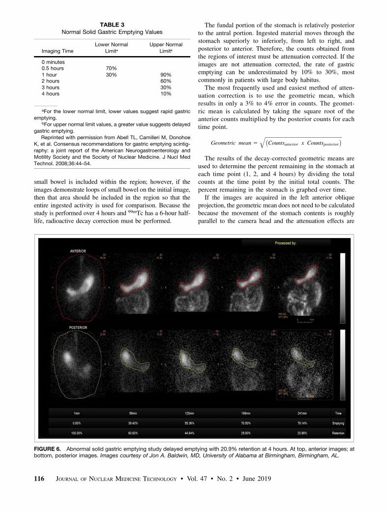
FIGURE 6. Abnormal solid gastric emptying study delayed emptying with 20.9% retention at 4 hours. At top, anterior images; atbottom, posterior images. Images courtesy of Jon A. Baldwin, MD, University of Alabama at Birmingham, Birmingham, AL.
TABLE 3Normal Solid Gastric Emptying Values
Imaging Time
Lower Normal
LimitaUpper Normal
Limitb
0 minutes0.5 hours 70%1 hour 30% 90%2 hours 60%3 hours 30%4 hours 10%
aFor the lower normal limit, lower values suggest rapid gastricemptying.
bFor upper normal limit values, a greater value suggests delayed
gastric emptying.
Reprinted with permission from Abell TL, Camilleri M, DonohoeK, et al. Consensus recommendations for gastric emptying scintig-
raphy: a joint report of the American Neurogastroenterology and
Motility Society and the Society of Nuclear Medicine. J Nucl MedTechnol. 2008;36:44–54.
116 JOURNAL OF NUCLEAR MEDICINE TECHNOLOGY • Vol. 47 • No. 2 • June 2019
minimized. No significant differences in emptying times havebeen demonstrated between images acquired in theanterior/posterior views versus the left anterior obliqueview; however, the geometric mean method is consideredthe most accurate.In the past, some facilities calculated the time for half of
the counts to leave the stomach (T½). This method is notrecommended in the consensus document, as T½ may bepotentially less accurate than percent retention, especially forpatients with delayed emptying, where extrapolation is needed tocalculate T½ if half of the meal does not leave the stomachduring the test.
Dual Isotope Liquid/Solid Study
When both solid and liquid gastric emptying are per-formed, scatter correction must be performed to correct fordown-scatter of the 111In photons into the 99mTc 140 keVwindow. However, scatter correction can be avoided if thedose ratio of 99mTc to 111In is at least 4:1.
Liquid Study
For liquid gastric emptying, a re-gion of interest is drawn around thestomach, and time-activity curves aregenerated. The emptying half time isdetermined as the time in minutes whencounts become half of the peak counts oran exponential mathematical fit of halftime can be calculated. Decay correctionand attenuation correction are not nec-essary. Normal values for liquid gastricemptying have not been well validated,and T½ of 23 minutes 6 3 standarddeviations is considered normal (Figure3).
IMAGE INTERPRETATION
Normal Results
Upon ingestion of the radiolabeledmeal, liquids rapidly diffuse throughoutthe stomach, while solids concentrateprimarily in the fundus (accommoda-tion) until they are moved down intothe antrum by fundal contractions. Theinitial localization of the solid material
in the fundus is apparent on the initial images. A transversephotopenic band between the fundus and antrum may beseen on later images (Figure 4). After the solids move intothe antrum, contractions of the antrum grind the solids into1 to 2 mm particles to pass through the pylorus. The time toaccomplish this is known as the lag period and normally lasts20 to 30 minutes, during which minimal gastric emptyingoccurs.
Once the small particles are mixed with gastric acid, theyempty from the stomach in a linear fashion at the same rate asliquids. This emptying results from the pressure gradient causedby the fundus (Figure 5). The results of a liquid gastric emptyingstudy demonstrate the liquid rapidly leaving the stomach in anexponential manner with no lag phase. As the volume of liquiddecreases, the rate of emptying slows.
Normal values for gastric emptying of solids for thestandardized protocol and meal were established by Tougaset al. using 123 normal subjects from 11 medical institu-tions in the United States, Canada, and Europe. The normal
values were set using the median and95th percentile (Table 3). A study isconsidered not delayed if the value at2 hours is less than 60% and at 4 hoursis less than 10%.
Abnormal Results
A study is considered to havedelayed gastric emptying if there ismore than 60% of the solid mealremaining at 2 hours or more than10% of the meal remaining at 4 hours.
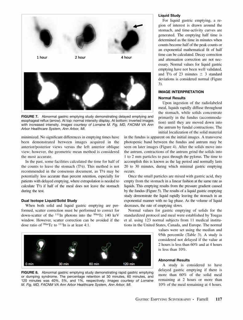
FIGURE 7. Abnormal gastric emptying study demonstrating delayed emptying andesophageal reflux (arrow). At top: normal intensity display. At bottom: inverted imageswith increased intensity. Images courtesy of Lorraine M. Fig, MD, FACNM VA AnnArbor Healthcare System, Ann Arbor, MI.
FIGURE 8. Abnormal gastric emptying study demonstrating rapid gastric emptyingor dumping syndrome. The percentage retention at 30 minutes, 60 minutes, and120 minutes was 40%, 5%, and 1%, respectively. Images courtesy of LorraineM. Fig, MD, FACNM VA Ann Arbor Healthcare System, Ann Arbor, MI.
GASTRIC EMPTYING SCINTIGRAPHY • Farrell 117
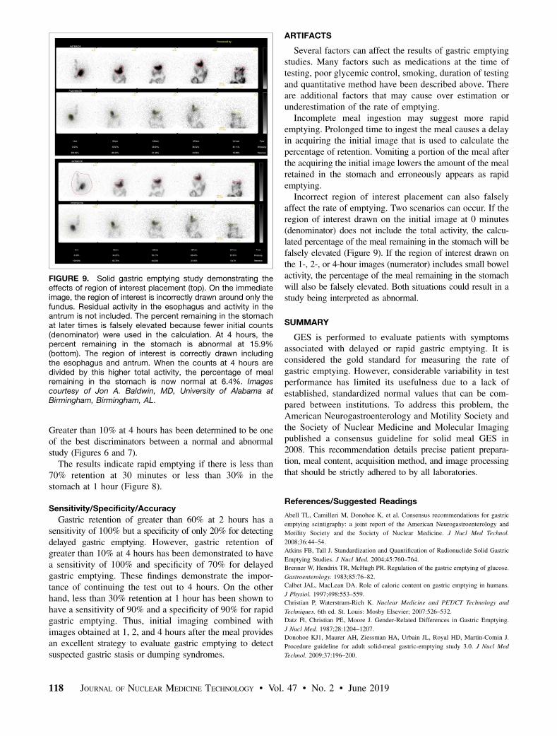
Greater than 10% at 4 hours has been determined to be oneof the best discriminators between a normal and abnormalstudy (Figures 6 and 7).The results indicate rapid emptying if there is less than
70% retention at 30 minutes or less than 30% in thestomach at 1 hour (Figure 8).
Sensitivity/Specificity/Accuracy
Gastric retention of greater than 60% at 2 hours has asensitivity of 100% but a specificity of only 20% for detectingdelayed gastric emptying. However, gastric retention ofgreater than 10% at 4 hours has been demonstrated to havea sensitivity of 100% and specificity of 70% for delayedgastric emptying. These findings demonstrate the impor-tance of continuing the test out to 4 hours. On the otherhand, less than 30% retention at 1 hour has been shown tohave a sensitivity of 90% and a specificity of 90% for rapidgastric emptying. Thus, initial imaging combined withimages obtained at 1, 2, and 4 hours after the meal providesan excellent strategy to evaluate gastric emptying to detectsuspected gastric stasis or dumping syndromes.
ARTIFACTS
Several factors can affect the results of gastric emptyingstudies. Many factors such as medications at the time oftesting, poor glycemic control, smoking, duration of testingand quantitative method have been described above. Thereare additional factors that may cause over estimation orunderestimation of the rate of emptying.
Incomplete meal ingestion may suggest more rapidemptying. Prolonged time to ingest the meal causes a delayin acquiring the initial image that is used to calculate thepercentage of retention. Vomiting a portion of the meal afterthe acquiring the initial image lowers the amount of the mealretained in the stomach and erroneously appears as rapidemptying.
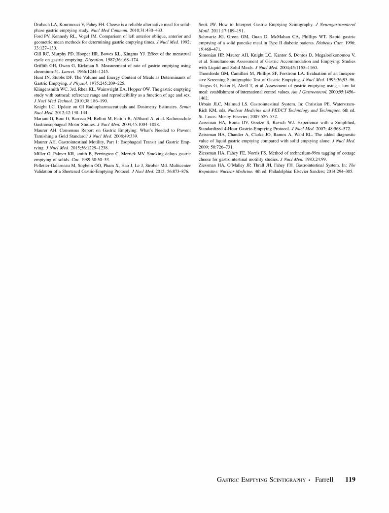
Incorrect region of interest placement can also falselyaffect the rate of emptying. Two scenarios can occur. If theregion of interest drawn on the initial image at 0 minutes(denominator) does not include the total activity, the calcu-lated percentage of the meal remaining in the stomach will befalsely elevated (Figure 9). If the region of interest drawn onthe 1-, 2-, or 4-hour images (numerator) includes small bowelactivity, the percentage of the meal remaining in the stomachwill also be falsely elevated. Both situations could result in astudy being interpreted as abnormal.
SUMMARY
GES is performed to evaluate patients with symptomsassociated with delayed or rapid gastric emptying. It isconsidered the gold standard for measuring the rate ofgastric emptying. However, considerable variability in testperformance has limited its usefulness due to a lack ofestablished, standardized normal values that can be com-pared between institutions. To address this problem, theAmerican Neurogastroenterology and Motility Society andthe Society of Nuclear Medicine and Molecular Imagingpublished a consensus guideline for solid meal GES in2008. This recommendation details precise patient prepara-tion, meal content, acquisition method, and image processingthat should be strictly adhered to by all laboratories.
References/Suggested Readings
Abell TL, Camilleri M, Donohoe K, et al. Consensus recommendations for gastric
emptying scintigraphy: a joint report of the American Neurogastroenterology and
Motility Society and the Society of Nuclear Medicine. J Nucl Med Technol.
2008;36:44–54.
Atkins FB, Tall J. Standardization and Quantification of Radionuclide Solid Gastric
Emptying Studies. J Nucl Med. 2004;45:760–764.
Brenner W, Hendrix TR, McHugh PR. Regulation of the gastric emptying of glucose.
Gastroenterology. 1983;85:76–82.
Calbet JAL, MacLean DA. Role of caloric content on gastric emptying in humans.
J Physiol. 1997;498:553–559.
Christian P, Waterstram-Rich K. Nuclear Medicine and PET/CT Technology and
Techniques. 6th ed. St. Louis: Mosby Elsevier; 2007:526–532.
Datz Fl, Christian PE, Moore J. Gender-Related Differences in Gastric Emptying.
J Nucl Med. 1987;28:1204–1207.
Donohoe KJ1, Maurer AH, Ziessman HA, Urbain JL, Royal HD, Martin-Comin J.
Procedure guideline for adult solid-meal gastric-emptying study 3.0. J Nucl Med
Technol. 2009;37:196–200.
FIGURE 9. Solid gastric emptying study demonstrating theeffects of region of interest placement (top). On the immediateimage, the region of interest is incorrectly drawn around only thefundus. Residual activity in the esophagus and activity in theantrum is not included. The percent remaining in the stomachat later times is falsely elevated because fewer initial counts(denominator) were used in the calculation. At 4 hours, thepercent remaining in the stomach is abnormal at 15.9%(bottom). The region of interest is correctly drawn includingthe esophagus and antrum. When the counts at 4 hours aredivided by this higher total activity, the percentage of mealremaining in the stomach is now normal at 6.4%. Imagescourtesy of Jon A. Baldwin, MD, University of Alabama atBirmingham, Birmingham, AL.
118 JOURNAL OF NUCLEAR MEDICINE TECHNOLOGY • Vol. 47 • No. 2 • June 2019
Drubach LA, Kourmouzi V, Fahey FH. Cheese is a reliable alternative meal for solid-
phase gastric emptying study. Nucl Med Commun. 2010;31:430–433.
Ford PV, Kennedy RL, Vogel JM. Comparison of left anterior oblique, anterior and
geometric mean methods for determining gastric emptying times. J Nucl Med. 1992;
33:127–130.
Gill RC, Murphy PD, Hooper HR, Bowes KL, Kingma YJ. Effect of the menstrual
cycle on gastric emptying. Digestion. 1987;36:168–174.
Griffith GH, Owen G, Kirkman S. Measurement of rate of gastric emptying using
chromium-51. Lancet. 1966:1244–1245.
Hunt JN, Stubbs DF. The Volume and Energy Content of Meals as Determinants of
Gastric Emptying. J Physiol. 1975;245:209–225.
Klingensmith WC, 3rd, Rhea KL, Wainwright EA, Hopper OW. The gastric emptying
study with oatmeal: reference range and reproducibility as a function of age and sex.
J Nucl Med Technol. 2010;38:186–190.
Knight LC. Update on GI Radiopharmaceuticals and Dosimetry Estimates. Semin
Nucl Med. 2012;42:138–144.
Mariani G, Boni G, Barreca M, Bellini M, Fattori B, AlSharif A, et al. Radionuclide
Gastroesophageal Motor Studies. J Nucl Med. 2004;45:1004–1028.
Maurer AH. Consensus Report on Gastric Emptying: What’s Needed to Prevent
Tarnishing a Gold Standard? J Nucl Med. 2008;49:339.
Maurer AH. Gastrointestinal Motility, Part 1: Esophageal Transit and Gastric Emp-
tying. J Nucl Med. 2015;56:1229–1238.
Miller G, Palmer KR, smith B, Ferrington C, Merrick MV. Smoking delays gastric
emptying of solids. Gut. 1989;30:50–53.
Pelletier-Galarneau M, Sogbein OO, Pham X, Hao J, Le J, Strober Md. Multicenter
Validation of a Shortened Gastric-Emptying Protocol. J Nucl Med. 2015; 56:873–876.
Seok JW. How to Interpret Gastric Emptying Scintigraphy. J Neurogastroenterol
Motil. 2011;17:189–191.
Schwartz JG, Green GM, Guan D, McMahan CA, Phillips WT. Rapid gastric
emptying of a solid pancake meal in Type II diabetic patients. Diabetes Care. 1996;
19:468–471.
Simonian HP, Maurer AH, Knight LC, Kantor S, Dontos D, Megalooikonomou V,
et al. Simultaneous Assessment of Gastric Accommodation and Emptying: Studies
with Liquid and Solid Meals. J Nucl Med. 2004;45:1155–1160.
Thomforde GM, Camilleri M, Phillips SF, Forstrom LA. Evaluation of an Inexpen-
sive Screening Scintigraphic Test of Gastric Emptying. J Nucl Med. 1995:36;93–96.
Tougas G, Eaker E, Abell T, et al Assessment of gastric emptying using a low-fat
meal: establishment of international control values. Am J Gastroenterol. 2000;95:1456–
1462.
Urbain JLC, Malmud LS. Gastrointestinal System. In: Christian PE, Waterstram-
Rich KM, eds. Nuclear Medicine and PET/CT Technology and Techniques. 6th ed.
St. Louis: Mosby Elsevier; 2007:526–532.
Zeissman HA, Bonta DV, Goetze S, Ravich WJ. Experience with a Simplified,
Standardized 4-Hour Gastric-Emptying Protocol. J Nucl Med. 2007; 48:568–572.
Zeissman HA, Chander A, Clarke JO, Ramos A, Wahl RL. The added diagnostic
value of liquid gastric emptying compared with solid emptying alone. J Nucl Med.
2009; 50:726–731.
Ziessman HA, Fahey FE, Norris FS. Method of technetium-99m tagging of cottage
cheese for gastrointestinal motility studies. J Nucl Med. 1983;24:99.
Ziessman HA, O’Malley JP, Thrall JH, Fahey FH. Gastrointestinal System. In: The
Requisites: Nuclear Medicine. 4th ed. Philadelphia: Elsevier Sanders; 2014:294–305.
GASTRIC EMPTYING SCINTIGRAPHY • Farrell 119