Embed Size (px)
Citation preview

Gating mechanisms of a naturalanion channelrhodopsinOleg A. Sineshchekov, Elena G. Govorunova, Hai Li, and John L. Spudich1
Center for Membrane Biology, Department of Biochemistry and Molecular Biology, The University of Texas Health Science Center at Houston, MedicalSchool, Houston, TX 77030
Edited by Peter H. Quail, University of California, Berkeley, Albany, CA, and approved September 28, 2015 (received for review July 10, 2015)
Anion channelrhodopsins (ACRs) are a class of light-gated channelsrecently identified in cryptophyte algae that provide unprecedentedfast and powerful hyperpolarizing tools for optogenetics. Analysis ofphotocurrents generated by Guillardia theta ACR 1 (GtACR1) andits mutants in response to laser flashes showed that GtACR1 gat-ing comprises two separate mechanisms with opposite dependen-cies on the membrane voltage and pH and involving differentamino acid residues. The first mechanism, characterized by slowopening and fast closing of the channel, is regulated by Glu-68.Neutralization of this residue (the E68Q mutation) specifically sup-pressed this first mechanism, but did not eliminate it completelyat high pH. Our data indicate the involvement of another, yet-unidentified pH-sensitive group X. Introducing a positive charge atthe Glu-68 site (the E68R mutation) inverted the channel gating sothat it was open in the dark and closed in the light, without alteringits ion selectivity. The second mechanism, characterized by fast open-ing and slow closing of the channel, was not substantially affected bythe E68Q mutation, but was controlled by Cys-102. The C102A muta-tion reduced the rate of channel closing by the second mechanism by∼100-fold, whereas it had only a twofold effect on the rate of thefirst. The results show that anion conductance by ACRs has a funda-mentally different structural basis than the relatively well studiedconductance by cation channelrhodopsins (CCRs), not attributableto simply a modification of the CCR selectivity filter.
ion transport | channel gating | channelrhodopsins | optogenetics
Recently we reported a class of rhodopsins from the crypto-phyte alga Guillardia theta that act as anion-conducting channels
when expressed in cultured animal cells and therefore can beused to suppress neuronal firing by light (1). These proteins—named anion channelrhodopsins (ACRs)—show distant sequencehomology to cation channelrhodopsins (CCRs) from chlorophyte(green) algae (2), but completely lack permeability for protons andmetal cations. This property, as well as large current amplitudes andfast kinetics, makes ACRs superior hyperpolarizing optogenetictools, compared with proton and chloride pumps (3, 4), or engi-neered Cl−-conducting CCR variants (5, 6). Two G. theta (Gt)ACRs differ in their spectral sensitivity and channel kinetics (1).GtACR1, with its absorption peak at 515 nm (Fig. S1), has anadvantage over a more blue-shifted GtACR2 with maximal ef-ficiency at 470 nm, because it allows using light of longerwavelengths that is less scattered by biological tissue.Because a CCR could be altered to exhibit Cl− channel activity
by mutation of a single amino acid residue (5), a fundamentalquestion about ACRs is whether they differ from CCRs only bytheir selectivity filter. In our search for an answer, we analyzedphotocurrents generated by GtACR1 in HEK293 cells undersingle-turnover conditions in which secondary photochemistrydoes not complicate the photocycle. We found that GtACR1conductance comprises two distinct mechanisms with oppositedependencies on the membrane voltage and bath pH, but thesame ionic selectivity. The first mechanism, observed by a fastcomponent of the photocurrent decay, depends on Glu-68, theposition occupied by a positively charged residue in the engineeredCl−-conducting CCR mutant. Substitution of Glu-68 with Arg in
GtACR1 did not change anion selectivity of the channel, but,rather, inverted its gating, rendering it constitutively open in thedark and closed in the light. The second mechanism, reflected by aslow component of the current decay, involves Cys-102. Our resultsdemonstrate that the structural basis for anion conductance inACRs is fundamentally different from that of Cl−-conductingCCR mutants.
ResultsThe rise and decay of photocurrents generated by GtACR1 re-sponse to a 6-ns laser flash under our standard ionic conditions(150 mM NaCl in the bath and 126 mM KCl in the pipette, pH7.4; for other components, see SI Materials and Methods) wereboth at least biphasic (Fig. 1A). Exponential functions were fit tothe data to derive amplitudes and time constants (τ) of eachcomponent. At the holding voltage (Eh) −60 mV, τ of the majorrise component (∼95%) was 86 ± 15 μs, accompanied by a minorcomponent with τ 1.8 ± 0.3 ms (mean ± SEM; n = 5). In somecases, this minor rise component could be further deconvolutedin two, but this additional complexity was not addressed in thepresent study. The two decay components had comparable am-plitudes and τ values of 48 ± 2 ms and 245 ± 20 ms (mean ± SEM;n = 5). A third, very slow decay component could also be resolved,but its contribution was ≤0.5% and was not considered here.Previously we reported that the voltage dependence of the
peak GtACR1 current recorded in response to a pulse of continu-ous light is linear (1), in contrast to inward rectification typical ofthe current–voltage relationships of CCRs (7). Deconvolution oflaser-flash–evoked currents revealed that the voltage dependenceof the amplitude of the fast decay showed outward rectification,
Significance
The recently discovered natural anion channelrhodopsins(ACRs) have exceptional properties for use as membrane-hyperpolarizing tools for neural silencing, but our knowledgeabout them is only rudimentary. We have gained, to our knowl-edge, first insights into the molecular mechanisms of their uniquelight-gated anion conductance by photocurrent measurements andmutant analysis. We identified a mutant constitutively open in thedark—to our knowledge, the first such mutant of any light-gatedchannel, invaluable for probing the structure of the channel openstate—and a “step function” ACR mutant useful in someoptogenetics protocols. Our results will help decipher themechanism of channelrhodopsin ion conductance and facilitaterational design of inhibitory tools tailored to the needs ofoptogenetic studies and potential clinical applications.
Author contributions: O.A.S., E.G.G., and J.L.S. designed research; O.A.S., E.G.G., and H.L.performed research; O.A.S., E.G.G., and J.L.S. analyzed data; and O.A.S., E.G.G., and J.L.S.wrote the paper.
The authors declare no conflict of interest.
This article is a PNAS Direct Submission.1To whom correspondence should be addressed. Email: [email protected].
This article contains supporting information online at www.pnas.org/lookup/suppl/doi:10.1073/pnas.1513602112/-/DCSupplemental.
14236–14241 | PNAS | November 17, 2015 | vol. 112 | no. 46 www.pnas.org/cgi/doi/10.1073/pnas.1513602112
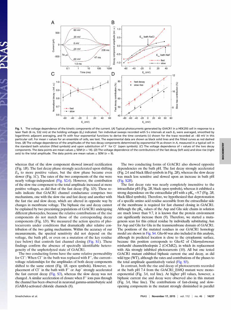
whereas that of the slow component showed inward rectification(Fig. 1B). The fast decay phase strongly accelerated upon shiftingEh to more positive values, but the slow phase became evenslower (Fig. 1C). The rates of the two components of the rise werenearly voltage-independent (Fig. S2A). However, the contributionof the slow rise component to the total amplitude increased at morepositive voltages, as did that of the fast decay (Fig. 1D). These re-sults indicate that GtACR1 channel conductance comprises twomechanisms, one with the slow rise and fast decay and another withthe fast rise and slow decay, which are altered in opposite way bychanges in membrane voltage. The biphasic rise and decay cannotbe explained by two preexisting populations ofGtACR1 undergoingdifferent photocycles, because the relative contributions of the risecomponents do not match those of the corresponding decaycomponents (Fig. 1D). We measured the action spectra of pho-tocurrents under conditions that dramatically alter relative con-tribution of the two gating mechanisms. Within the accuracy of ourmeasurements, the spectral sensitivity did not depend on thevoltage, the bath pH, or even on a mutation of the key residue(see below) that controls fast channel closing (Fig. S1). Thesefindings confirm the absence of spectrally identifiable hetero-geneity of the unphotolyzed state of GtACR1.The two conducting forms have the same relative permeability
for Cl−: When Cl− in the bath was replaced with F−, the current–voltage relationships for the amplitudes of both decay componentsshifted to the same extent (Fig. 1B, open symbols). However, re-placement of Cl− in the bath with F− or Asp− strongly acceleratedthe fast current decay (Fig. S3), whereas the slow decay was notchanged. A similar acceleration of closure when F− ions pass throughthe channel has been observed in neuronal gamma-aminobutyric acid(GABA)-activated chloride channels (8).
The two conducting forms of GtACR1 also showed oppositedependencies on the bath pH. The fast decay strongly accelerated(Fig. 2A and black filled symbols in Fig. 2B), whereas the slow decaywas much less sensitive and slowed upon an increase in bath pH(Fig. S2B).The fast decay rate was nearly completely insensitive to the
intracellular pH (Fig. 2B, black open symbols), whereas it exhibited astrong dependence on the extracellular pH with a pKa ∼9.7 (Fig. 2B,black filled symbols). Therefore, we hypothesized that deprotonationof a specific amino acid residue accessible from the extracellular sideof the membrane is required for fast channel closing in GtACR1.Although the pKa values of the Asp and Glu side chains in solutionare much lower than 9.7, it is known that the protein environmentcan significantly increase them (9). Therefore, we started a muta-genesis scan for this critical residue by individually substituting Asnfor Asp and Gln for Glu in the transmembrane domain of GtACR1.The positions of the mutated residues in our GtACR1 homologymodel are shown in Fig. S4. Glu-60 was also included in this analysis,although its predicted location is close to the cytoplasmic surface,because this position corresponds to Glu-82 of Chlamydomonasreinhardtii channelrhodopsin 2 (CrChR2), in which its replacementwith Ala strongly inhibited photocurrents (10). All but one testedGtACR1 mutant exhibited biphasic current rise and decay, as didwild type (WT), although the rates and contributions of the phases tothe total amplitude quantitatively varied (Fig. S5).In contrast, both the rise and decay of photocurrents recorded
at the bath pH 7.4 from the GtACR1_E68Q mutant were mono-exponential (Fig. 3A, red line). At higher pH values, however, abiphasic current rise and decay were observed also in this mutant(Fig. 3A, blue line). The contributions of fast-closing and slow-opening components in the mutant strongly diminished in parallel
Fig. 1. The voltage dependence of the kinetic components of the current. (A) Typical photocurrents generated by GtACR1 in a HEK293 cell in response to alaser flash (6 ns, 532 nm) at the holding voltages (Eh) indicated. Ten individual sweeps recorded with 5-s intervals at each Eh were averaged, smoothed bylogarithmic adjacent averaging, and fit with four exponential functions to derive the time constants (τ) shown for the trace recorded at −60 mV in thisparticular cell. For mean τ values for an ensemble of cells, see text. The experimental data are shown as black solid lines and the fitted curves as red dashedlines. (B) The voltage dependence of the amplitudes of the two decay components determined by exponential fit as shown in A, measured in a typical cell inthe standard bath solution (filled symbols) and upon substitution of F− for Cl− (open symbols). (C) The voltage dependence of τ values of the two decaycomponents. The data points are mean values ± SEM (n = 14). (D) The voltage dependence of the contributions of the fast decay (left axis) and slow rise (rightaxis) to the total amplitude. The data points are mean values ± SEM (n = 9).
Sineshchekov et al. PNAS | November 17, 2015 | vol. 112 | no. 46 | 14237
BIOCH
EMISTR
Y
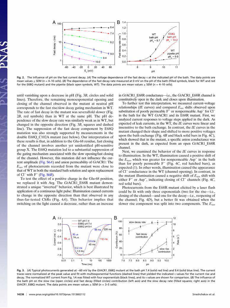
until vanishing upon a decrease in pH (Fig. 3B, circles and solidlines). Therefore, the remaining monoexponential opening andclosing of the channel observed in the mutant at neutral pHcorresponds to the fast rise/slow decay gating mechanism in WT.The rate of fast decay in the mutant was severalfold slower (Fig.2B, red symbols) than in WT at the same pH. The pH de-pendence of the slow decay rate was similarly weak as in WT, butchanged in the opposite direction (Fig. 3B, squares and dashedline). The suppression of the fast decay component by E68Qmutation was also strongly supported by measurements in thedouble E68Q_C102A mutant (see below). Our interpretation ofthese results is that, in addition to the Glu-68 residue, fast closingof the channel involves another yet unidentified pH-sensitivegroup X. The E68Q mutation led to a substantial suppression ofthe gating mechanism associated with the slow opening/fast closingof the channel. However, this mutation did not influence the cur-rent amplitude (Fig. S6A) and anion permeability of GtACR1: TheErev of photocurrents recorded from the mutant were close tothat of WT in both the standard bath solution and upon replacementof Cl− with F− (Fig. S6B).To test the effect of a positive charge in the Glu-68 position,
we replaced it with Arg. The GtACR1_E68R mutant demon-strated a unique “inverted” behavior, which is best illustrated byapplication of a continuous light pulse: Illumination caused currentsto change in the opposite direction than that observed in anythus-far-tested ChRs (Fig. 4A). This behavior implies thatswitching on the light caused a decrease, rather than an increase
in GtACR1_E68R conductance—i.e., theGtACR1_E68R channel isconstitutively open in the dark and closes upon illumination.To further test this interpretation, we measured current–voltage
relationships (IE curves) and compared Erev shifts observed uponsubstitution of poorly permeable F− or nonpermeable Asp− for Cl−
in the bath for the WT GtACR1 and its E68R mutant. First, weanalyzed current responses to voltage steps applied in the dark. Asexpected of leak currents, in the WT, the IE curves were linear andinsensitive to the bath exchange. In contrast, the IE curves in themutant changed their shape and shifted to more positive voltagesupon the bath exchange (Fig. 4B and black solid bars in Fig. 4C),which showed that in the mutant, a specific anion conductance waspresent in the dark, as expected from an open GtACR1_E68Rchannel.Next, we examined the behavior of the IE curves in response
to illumination. In the WT, illumination caused a positive shift ofthe Erev, which was greater for nonpermeable Asp− in the baththan for poorly permeable F− (Fig. 4C, red hatched bars), asexpected (1). In other words, illumination caused the appearanceof Cl− conductance in the WT (channel opening). In contrast, inthe mutant illumination caused a negative shift of Erev shift witheither F− or Asp−, indicating closing of Cl− channels (Fig. 4C,red solid bars).Photocurrents from the E68R mutant elicited by a laser flash
could be fit with only three exponentials (two for the rise—i.e.,closing of the channel—and one for the decay—i.e., reopening ofthe channel; Fig. 4D), but a better fit was obtained when theslower rise component was split into two components. The Erev
Fig. 2. The influence of pH on the fast current decay. (A) The voltage dependence of the fast decay τ at the indicated pH of the bath. The data points aremean values ± SEM (n = 4–10 cells). (B) The dependence of the fast decay rate measured at 0 mV on the pH of the bath (filled symbols, black for WT and redfor the E68Q mutant) and the pipette (black open symbols, WT). The data points are mean values ± SEM (n = 4–10 cells).
Fig. 3. (A) Typical photocurrents generated at −60 mV by the GtACR1_E68Q mutant at the bath pH 7.4 (solid red line) and 9.4 (solid blue line). The currenttraces were normalized at the peak value and fit with multiexponential functions (dashed lines) that yielded the indicated τ values for the current rise anddecay. The normalized WT current from Fig. 1A was fitted with four exponentials (black lines), and its τ values are shown for comparison. (B) The influence ofthe bath pH on the slow rise (open circles) and fast decay (filled circles) contribution (left axis) and the slow decay rate (filled squares; right axis) in theGtACR1_E68Q mutant. The data points are mean values ± SEM (n = 3–5 cells).
14238 | www.pnas.org/cgi/doi/10.1073/pnas.1513602112 Sineshchekov et al.
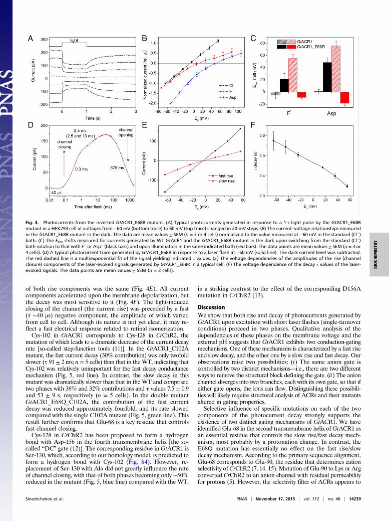
of both rise components was the same (Fig. 4E). All currentcomponents accelerated upon the membrane depolarization, butthe decay was most sensitive to it (Fig. 4F). The light-inducedclosing of the channel (the current rise) was preceded by a fast(τ ∼40 μs) negative component, the amplitude of which variedfrom cell to cell. Although its nature is not yet clear, it may re-flect a fast electrical response related to retinal isomerization.Cys-102 in GtACR1 corresponds to Cys-128 in CrChR2, the
mutation of which leads to a dramatic decrease of the current decayrate [so-called step-function tools (11)]. In the GtACR1_C102Amutant, the fast current decay (30% contribution) was only twofoldslower (τ 91 ± 2 ms; n = 5 cells) than that in the WT, indicating thatCys-102 was relatively unimportant for the fast decay conductancemechanism (Fig. 5, red line). In contrast, the slow decay in thismutant was dramatically slower than that in the WT and comprisedtwo phases with 38% and 32% contributions and τ values 7.5 ± 0.9and 53 ± 9 s, respectively (n = 5 cells). In the double mutantGtACR1_E68Q_C102A, the contribution of the fast currentdecay was reduced approximately fourfold, and its rate slowedcompared with the single C102A mutant (Fig. 5, green line). Thisresult further confirms that Glu-68 is a key residue that controlsfast channel closing.Cys-128 in CrChR2 has been proposed to form a hydrogen
bond with Asp-156 in the fourth transmembrane helix [the so-called “DC” gate (12)]. The corresponding residue in GtACR1 isSer-130, which, according to our homology model, is predicted toform a hydrogen bond with Cys-102 (Fig. S4). However, re-placement of Ser-130 with Ala did not greatly influence the rateof channel closing, with that of both phases becoming only ∼50%reduced in the mutant (Fig. 5, blue line) compared with the WT,
in a striking contrast to the effect of the corresponding D156Amutation in CrChR2 (13).
DiscussionWe show that both rise and decay of photocurrents generated byGtACR1 upon excitation with short laser flashes (single-turnoverconditions) proceed in two phases. Qualitative analysis of thedependencies of these phases on the membrane voltage and theexternal pH suggests that GtACR1 exhibits two conduction-gatingmechanisms. One of these mechanisms is characterized by a fast riseand slow decay, and the other one by a slow rise and fast decay. Ourobservations raise two possibilities: (i) The same anion gate iscontrolled by two distinct mechanisms—i.e., there are two differentways to remove the structural block defining the gate. (ii) The anionchannel diverges into two branches, each with its own gate, so that ifeither gate opens, the ions can flow. Distinguishing these possibili-ties will likely require structural analysis of ACRs and their mutantsaltered in gating properties.Selective influence of specific mutations on each of the two
components of the photocurrent decay strongly supports theexistence of two distinct gating mechanisms of GtACR1. We haveidentified Glu-68 in the second transmembrane helix ofGtACR1 asan essential residue that controls the slow rise/fast decay mech-anism, most probably by a protonation change. In contrast, theE68Q mutation has essentially no effect on the fast rise/slowdecay mechanism. According to the primary sequence alignment,Glu-68 corresponds to Glu-90, the residue that determines cationselectivity of CrChR2 (7, 14, 15). Mutation of Glu-90 to Lys or Argconverted CrChR2 to an anion channel with residual permeabilityfor protons (5). However, the selectivity filter of ACRs appears to
Fig. 4. Photocurrents from the inverted GtACR1_E68R mutant. (A) Typical photocurrents generated in response to a 1-s light pulse by the GtACR1_E68Rmutant in a HEK293 cell at voltages from −60 mV (bottom trace) to 60 mV (top trace) changed in 20-mV steps. (B) The current–voltage relationships measuredin the GtACR1_E68R mutant in the dark. The data are mean values ± SEM (n = 3 or 4 cells) normalized to the value measured at −60 mV in the standard (Cl−)bath. (C) The Erev shifts measured for currents generated by WT GtACR1 and the GtACR1_E68R mutant in the dark upon switching from the standard (Cl−)bath solution to that with F− or Asp− (black bars) and upon illumination in the same indicated bath (red bars). The data points are mean values ± SEM (n = 3 or4 cells). (D) A typical photocurrent trace generated by GtACR1_E68R in response to a laser flash at −60 mV (solid line). The dark current level was subtracted.The red dashed line is a multiexponential fit of the signal yielding indicated τ values. (E) The voltage dependencies of the amplitudes of the rise (channelclosure) components of the laser-evoked signals generated by GtACR1_E68R in a typical cell. (F) The voltage dependence of the decay τ values of the laser-evoked signals. The data points are mean values ± SEM (n = 3 cells).
Sineshchekov et al. PNAS | November 17, 2015 | vol. 112 | no. 46 | 14239
BIOCH
EMISTR
Y

be different from that of theCrChR2_E90K/Rmutants, because thepresence of the Glu-90 homolog in WT GtACRs is obviously not abarrier to anion permeation. Furthermore, replacement of Glu-68with Gln or Arg in GtACR1 did not change anion permeability ofthe channel, which confirmed that this residue does not determinethe selectivity filter in ACRs.Although the E68R mutation did not change the ionic selec-
tivity of the channel, it inverted its gating. The channel becameconstitutively open in the dark, whereas illumination caused itto close. A similar inversion of protein function was previouslyobserved in haloarchaeal sensory rhodopsin I (SRI), in which asingle point mutation either of the photoreceptor itself or of itscognate transducer converted SRI from an attractant to a re-pellent photoreceptor (16, 17). The SRI inversion is attributableto a switch in its conformation from the C (retinylidene Schiffbase accessible from the cytoplasm) to E (Schiff base accessiblefrom the extracellular space) conformer (18–20). The stable openchannel of GtACR1_E68R is likely to be particularly valuablefor structural comparisons with the WT by enabling identificationof residue determinants and structural changes distinguishing theclosed and open conformations of the channel by structural meth-ods such as molecular spectroscopy and crystallography.Our mutant analysis showed that Cys-102 controls only the
slow phase of the photocurrent decay, corresponding to the fastopening/slow closing gate, whereas its fast decay phase waspractically unaffected by the GtACR1_C102A mutation. Thisresidue (Cys-128 in CrChR2) is conserved in all thus-far-knownACRs and CCRs, but is substituted with Val in the third ho-mologous sequence from G. theta that generated no photocur-rents, despite good expression in HEK293 cells (1). The effectsof C128X mutations on channel activity have been best studiedin CrChR2 (11, 13), but a similar dramatic decrease in the cur-rent decay rate was also observed in the corresponding mutantsof other CCRs, such as Mesostigma viride channelrhodopsin 1(MvChR1) (21). This result has been attributed to a disruption of
the hydrogen bond (“DC gate”) that Cys-128 forms with Asp-156in CrChR2 (12). Indeed, mutation of Asp-156 yielded compa-rable or even greater extension of the channel open time, as didthat of Cys-128 (13, 22). In GtACR1, the position of Asp-156 isoccupied by Ser-130, which, according to our homology model,forms a hydrogen bond with Cys-102. However, in the S130Amutant, the slow decay phase was practically unchanged, whichsuggests that the effect of the C102A mutation inGtACR1 was notcaused by a disruption of the putative hydrogen bond.The two gating mechanisms of GtACR1 identified by kinetic
analysis of laser-evoked signals control conduction with the sameanion selectivity. None of the tested mutations that selectivelyinfluenced the kinetics of each form changed anion selectivity ofGtACR1. These observations show that inGtACR1 channel gatingis structurally independent of the selectivity filter. In contrast, inCrChR2 two conducting states have been recognized by theirdifferent relative permeability to H+ and Na+ (23, 24). A model oftwo interconnected photocycles, each of which contains a closed(nonconducting) state and an open (conducting) state, has beendeveloped for CCRs (25–27). However, single-turnover photo-currents generated by CrChR2 upon laser excitation decaymonoexponentially (28, 29), which means that the second con-ducting state accumulates only under continuous light, indicatingthat the second state requires secondary photochemistry. Two expo-nential phases have been resolved in the decay of laser-evoked pho-tocurrents from the Volvox carteri channelrhodopsin 1/Volvox carterichannelrhodopsin 2 (VcChR1/VcChR2) hybrid (30), but this phe-nomenon has not yet been incorporated into the four-state model.The importance of the Cys-128 homolog is not the only sim-
ilarity between the slow phase of the decay of GtACR1 photo-current and the monoexponential decay of the current generatedby CrChR2. The latter also became slower with membrane de-polarization (29, 31). However, in CrChR2, this voltage de-pendence was eliminated by the E123T mutation (29), whereasin WT GtACR1, the voltage dependence persisted in the pres-ence of a noncarboxylic residue (Ser-97) in the position of Glu-123 (corresponding to Asp-85 in bacteriorhodopsin).The many differences between ACR and CCR conductance
mechanisms evident in our results clearly indicate that ACRsdiffer from CCRs, not simply by their selectivity filter, but theyrepresent a distinct class of light-gated channels that evolved toconduct anions.
Materials and MethodsA DNA polynucleotide encoding the seven-transmembrane domain (residues1–295) of GtACR1 optimized for human codon use was synthesized andmutated as described in SI Materials and Methods. Photocurrents wereevoked by a Nd:YAG laser (532 nm, pulsewidth 6 ns) and recorded by whole-cell patch clamp from HEK293 cells transiently transfected with WT GtACR1or its mutants fused to enhanced YFP. A laser artifact measured with ablocked optical path was digitally subtracted from the recorded traces. Forfurther analysis, the signals were logarithmically averaged with a custom-created computer algorithm. For multiexponential curve fitting, Origin 7software was used. Monochromatic (10 nm half-band) light from a Poly-chrome IV light source was used in experiments with continuous light pulses.All current–voltage dependencies were corrected for liquid junction potentials(LJPs) calculated by using the ClampEx built-in LJP calculator. Action spectrawere constructed by correction of measured currents for quantum density ofthe stimulation light. SI Materials and Methods provides detailed descriptionof experimental procedures.
ACKNOWLEDGMENTS. This work was supported by NIH Grant R01GM027750,the Hermann Eye Fund, and Endowed Chair AU-0009 from the Robert A.Welch Foundation.
1. Govorunova EG, Sineshchekov OA, Janz R, Liu X, Spudich JL (2015) Natural light-gated
anion channels: A family of microbial rhodopsins for advanced optogenetics. Science
349(6248):647–650.2. Nagel G, et al. (2005) Channelrhodopsins: Directly light-gated cation channels. Biochem
Soc Trans 33(Pt 4):863–866.
3. Gradinaru V, Thompson KR, Deisseroth K (2008) eNpHR: A Natronomonas
halorhodopsin enhanced for optogenetic applications. Brain Cell Biol 36(1-4):
129–139.4. Chow BY, et al. (2010) High-performance genetically targetable optical neural si-
lencing by light-driven proton pumps. Nature 463(7277):98–102.
Fig. 5. The decay of typical photocurrents generated by GtACR1_C102A(solid red line), GtACR1_E68Q_C102A (solid green line), and GtACR1_S130A(solid blue line) at −60 mV in the standard bath solution. The traces werenormalized at the peak amplitude. The corresponding dashed lines showtwo- or three-exponential fits used to determine the decay time constantsfor the mutants, as shown on the graph.
14240 | www.pnas.org/cgi/doi/10.1073/pnas.1513602112 Sineshchekov et al.
5. Wietek J, et al. (2014) Conversion of channelrhodopsin into a light-gated chloridechannel. Science 344(6182):409–412.
6. Berndt A, Lee SY, Ramakrishnan C, Deisseroth K (2014) Structure-guided transformationof channelrhodopsin into a light-activated chloride channel. Science 344(6182):420–424.
7. Gradmann D, Berndt A, Schneider F, Hegemann P (2011) Rectification of the chan-nelrhodopsin early conductance. Biophys J 101(5):1057–1068.
8. Robertson B (1989) Characteristics of GABA-activated chloride channels in mammaliandorsal root ganglion neurones. J Physiol 411:285–300.
9. Li H, Govorunova EG, Sineshchekov OA, Spudich JL (2014) Role of a helix B lysineresidue in the photoactive site in channelrhodopsins. Biophys J 106(8):1607–1617.
10. Sugiyama Y, et al. (2009) Photocurrent attenuation by a single polar-to-nonpolarpoint mutation of channelrhodopsin-2. Photochem Photobiol Sci 8(3):328–336.
11. Berndt A, Yizhar O, Gunaydin LA, Hegemann P, Deisseroth K (2009) Bi-stable neuralstate switches. Nat Neurosci 12(2):229–234.
12. Nack M, et al. (2010) The DC gate in Channelrhodopsin-2: Crucial hydrogen bondinginteraction between C128 and D156. Photochem Photobiol Sci 9(2):194–198.
13. Bamann C, Gueta R, Kleinlogel S, Nagel G, Bamberg E (2010) Structural guidance ofthe photocycle of channelrhodopsin-2 by an interhelical hydrogen bond. Biochemistry49(2):267–278.
14. Ruffert K, et al. (2011) Glutamate residue 90 in the predicted transmembrane domain2 is crucial for cation flux through channelrhodopsin 2. Biochem Biophys Res Commun410(4):737–743.
15. Eisenhauer K, et al. (2012) In channelrhodopsin-2 Glu-90 is crucial for ion selectivityand is deprotonated during the photocycle. J Biol Chem 287(9):6904–6911.
16. Olson KD, Zhang XN, Spudich JL (1995) Residue replacements of buried aspartyl andrelated residues in sensory rhodopsin I: D201N produces inverted phototaxis signals.Proc Natl Acad Sci USA 92(8):3185–3189.
17. Jung KH, Spudich JL (1998) Suppressor mutation analysis of the sensory rhodopsinI-transducer complex: Insights into the color-sensing mechanism. J Bacteriol 180(8):2033–2042.
18. Sineshchekov OA, Sasaki J, Phillips BJ, Spudich JL (2008) A Schiff base connectivityswitch in sensory rhodopsin signaling. Proc Natl Acad Sci USA 105(42):16159–16164.
19. Sineshchekov OA, Sasaki J, Wang J, Spudich JL (2010) Attractant and repellent sig-naling conformers of sensory rhodopsin-transducer complexes. Biochemistry 49(31):6696–6704.
20. Sasaki J, Takahashi H, Furutani Y, Kandori H, Spudich JL (2011) Sensory rhodopsin-Ias a bidirectional switch: Opposite conformational changes from the same photo-isomerization. Biophys J 100(9):2178–2183.
21. Govorunova EG, Spudich EN, Lane CE, Sineshchekov OA, Spudich JL (2011) Newchannelrhodopsin with a red-shifted spectrum and rapid kinetics from Mesostigmaviride. MBio 2(3):e00115-11.
22. Dawydow A, et al. (2014) Channelrhodopsin-2-XXL, a powerful optogenetic tool forlow-light applications. Proc Natl Acad Sci USA 111(38):13972–13977.
23. Berndt A, Prigge M, Gradmann D, Hegemann P (2010) Two open states with pro-gressive proton selectivities in the branched channelrhodopsin-2 photocycle. BiophysJ 98(5):753–761.
24. Schneider F, Gradmann D, Hegemann P (2013) Ion selectivity and competition inchannelrhodopsins. Biophys J 105(1):91–100.
25. Hegemann P, Ehlenbeck S, Gradmann D (2005) Multiple photocycles of channelrho-dopsin. Biophys J 89(6):3911–3918.
26. Nikolic K, et al. (2009) Photocycles of channelrhodopsin-2. Photochem Photobiol 85(1):400–411.
27. Stehfest K, Hegemann P (2010) Evolution of the channelrhodopsin photocycle model.ChemPhysChem 11(6):1120–1126.
28. Tsunoda SP, Hegemann P (2009) Glu 87 of channelrhodopsin-1 causes pH-dependentcolor tuning and fast photocurrent inactivation. Photochem Photobiol 85(2):564–569.
29. Berndt A, et al. (2011) High-efficiency channelrhodopsins for fast neuronal stimula-tion at low light levels. Proc Natl Acad Sci USA 108(18):7595–7600.
30. Ernst OP, et al. (2008) Photoactivation of channelrhodopsin. J Biol Chem 283(3):1637–1643.
31. Bamann C, Kirsch T, Nagel G, Bamberg E (2008) Spectral characteristics of the pho-tocycle of channelrhodopsin-2 and its implication for channel function. J Mol Biol375(3):686–694.
Sineshchekov et al. PNAS | November 17, 2015 | vol. 112 | no. 46 | 14241
BIOCH
EMISTR
Y