Embed Size (px)
Citation preview
Fortunato et al. EvoDevo 2012, 3:14http://www.evodevojournal.com/content/3/1/14
RESEARCH Open Access
Genome-wide analysis of the sox family in thecalcareous sponge Sycon ciliatum: multiple geneswith unique expression patternsSofia Fortunato1,2, Marcin Adamski1, Brith Bergum1, Corina Guder1, Signe Jordal1, Sven Leininger1,Christin Zwafink1, Hans Tore Rapp2 and Maja Adamska1*
Abstract
Background: Sox genes are HMG-domain containing transcription factors with important roles in developmentalprocesses in animals; many of them appear to have conserved functions among eumetazoans. Demosponges havefewer Sox genes than eumetazoans, but their roles remain unclear. The aim of this study is to gain insight into theearly evolutionary history of the Sox gene family by identification and expression analysis of Sox genes in thecalcareous sponge Sycon ciliatum.
Methods: Calcaronean Sox related sequences were retrieved by searching recently generated genomic andtranscriptome sequence resources and analyzed using variety of phylogenetic methods and identification ofconserved motifs. Expression was studied by whole mount in situ hybridization.
Results: We have identified seven Sox genes and four Sox-related genes in the complete genome of Syconciliatum. Phylogenetic and conserved motif analyses showed that five of Sycon Sox genes represent groups B, C, E,and F present in cnidarians and bilaterians. Two additional genes are classified as Sox genes but cannot beassigned to specific subfamilies, and four genes are more similar to Sox genes than to other HMG-containinggenes. Thus, the repertoire of Sox genes is larger in this representative of calcareous sponges than in thedemosponge Amphimedon queenslandica. It remains unclear whether this is due to the expansion of the genefamily in Sycon or a secondary reduction in the Amphimedon genome. In situ hybridization of Sycon Sox genesrevealed a variety of expression patterns during embryogenesis and in specific cell types of adult sponges.
Conclusions: In this study, we describe a large family of Sox genes in Sycon ciliatum with dynamic expressionpatterns, indicating that Sox genes are regulators in development and cell type determination in sponges, asobserved in higher animals. The revealed differences between demosponge and calcisponge Sox genes repertoirehighlight the need to utilize models representing different sponge lineages to describe sponge development, aprerequisite for deciphering evolution of metazoan developmental mechanisms.
BackgroundThe Sox genes (Sry related high mobility group, HMGbox) are a family of transcription factors with importantroles in regulating development and cell fate determin-ation throughout the animal kingdom [1,2]. The Sox pro-teins are characterized by the HMG DNA binding domainof 79 amino acids, resembling the mammalian testis deter-mination factor, Sry, which was the first Sox domain
* Correspondence: [email protected] International Centre for Marine Molecular Biology, Thormøhlensgt. 55,Bergen 5008, NorwayFull list of author information is available at the end of the article
© 2012 Fortunato et al.; licensee BioMed CentCommons Attribution License (http://creativecreproduction in any medium, provided the or
identified [3]. There are 20 Sox genes in mammals [4]which have been classified in five groups of Sox proteins(B, C, D, E, and F) [5]. However, additional groups havebeen created to accommodate divergent genes with lim-ited taxonomic distribution, for instance group J [5].Groups B, C, E, and F are found in all eumetazoanlineages, but group D is found only in the bilaterians [5].No Sox genes are present in the sequenced genomes of
the unicellular choanoflagellate, Monosiga brevicollis [6],or the amoeboid holozoan Capsaspora owczarzaki [7].Since they are present in basal metazoans like sponges(that is, the demosponge Amphimedon queenslandica)
ral Ltd. This is an Open Access article distributed under the terms of the Creativeommons.org/licenses/by/2.0), which permits unrestricted use, distribution, andiginal work is properly cited.
Fortunato et al. EvoDevo 2012, 3:14 Page 2 of 11http://www.evodevojournal.com/content/3/1/14
[8,9] and placozoans (Trichoplax adhaerens) [10], theyhave likely arisen in the last common ancestor to theMetazoa [8]. There is a larger repertoire of Sox genes incnidarians [11-13] and the ctenophore Pleurobrachia pi-leus [14] than in the demosponges [8,9,15] and the pla-cozoans [10]. Previous phylogenetic analysis of cnidarianSox genes including the species Hydra magnipapillata,Nematostella vectensis, and Clytia hemisphaerica placedsome of these sequences into the previously identifiedgroups of Sox genes; however some of these genes cannotbe classified into any specific group [11-13]. The expres-sion patterns of cnidarian Sox genes suggest that they haveroles in a wide variety of developmental functions, such asgerm layer formation, organ development, cell type speci-fication, and neural development [11-13].Previous studies on Sox genes in sponges include the
two demosponges, Amphimedon queenslandica [8,9]and Ephydatia muelleri, as well as the calcareous spongeSycon raphanus [15]. In Amphimedon, four Sox geneshave been found, including two members of group B(AmqSoxB1 and AmqSoxB2) and single members ofgroups C and F [9]. Sox genes from Ephydatia and Syconraphanus could not be clearly classified due to incom-plete domain sequences included in the phylogeneticanalyses [15]. As a consequence, the complement of Soxgenes in calcareous sponges is still unclear. In addition,apart from an RT-PCR study suggesting dynamic expres-sion of Sox genes during embryonic development inAmphimedon [9], no expression patterns on a cellularlevel are published for this or any other sponge. For thisreason, more studies in sponges are required to fullyunderstand the function of Sox genes in the phylumPorifera in comparison with the Eumetazoa. The aims ofthis study were to analyze the repertoire of Sox genes inthe calcareous sponge Sycon ciliatum and to trace theirexpression during development.Sycon ciliatum is an attractive model system for devel-
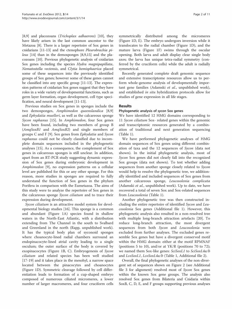
opmental biology studies [16]. This sponge is a commonand abundant (Figure 1A) species found in shallowwaters in the North-East Atlantic, with a distributionextending from The Channel in the south to Svalbardand Greenland in the north (Rapp, unpublished work).It has the typical body plan of syconoid spongeswhere choanocyte-lined radial chambers surround anendopinacocyte-lined atrial cavity leading to a singleosculum; the outer surface of the body is covered byexopinacocytes (Figure 1B, C). Embryogenesis of Syconciliatum and related species has been well studied[17-19] and it takes place in the mesohyl, a narrow spacelocated between the pinacocytes and choanocytes(Figure 1D). Symmetric cleavage followed by cell differ-entiation leads to formation of a cup-shaped embryocomposed of numerous ciliated micromeres, a lowernumber of larger macromeres, and four cruciform cells
symmetrically distributed among the micromeres(Figure 1D, E). The embryo undergoes inversion while ittranslocates to the radial chamber (Figure 1D), and themature larva (Figure 1F) swims through the oscularopening. Both larva and adult display clear single bodyaxes; the larva has unique tetra-radial symmetry (con-ferred by the cruciform cells) while the adult is radiallysymmetrical.Recently generated complete draft genomic sequence
and extensive transcriptome resources allow us to per-form whole-genome analysis of developmentally impor-tant gene families (Adamski et al., unpublished work),and established in situ hybridization protocols allow forstudies of gene expression in all life stages.
ResultsPhylogenetic analysis of sycon Sox genesWe have identified 12 HMG domains corresponding to11 Sycon ciliatum Sox- related genes within the genomicand transcriptomic resources generated by a combin-ation of traditional and next generation sequencing(Table 1).We have performed phylogenetic analyses of HMG
domain sequences of Sox genes using different combin-ation of taxa and the 12 sequences of Sycon (data notshown). In the initial phylogenetic analysis, most ofSycon Sox genes did not clearly fall into the recognizedSox groups (data not shown). To test whether addingsequences from another sponge closely related to Syconwould help to resolve the phylogenetic tree, we addition-ally identified and included sequences of Sox genes fromanother calcareous sponge, Leucosolenia complicata(Adamski et al., unpublished work). Up to date, we haverecovered a total of seven Sox and Sox-related sequencesfrom Leucosolenia (Table 1).Another phylogenetic tree was then constructed in-
cluding the entire repertoire of identified Sycon and Leu-cosolenia Sox genes (Additional file 1). However, thisphylogenetic analysis also resulted in a non-resolved treewith multiple long-branch attraction artefacts [20]. Toreduce long-branch attraction, the most divergentsequences from both Sycon and Leucosolenia wereexcluded from further analyses. The excluded genes re-semble Sox genes but have a divergent conserved motifwithin the HMG domain: either at the motif RPMNAF(positions 5 to 10), and/or at YK/R (positions 70 to 72);we named them Sox-like genes: SciSoxL1 to SciSoxL4a/band LcoSoxL1, LcoSoxL4a/b (Table 1, Additional file 2).Overall, the final phylogenetic analyses of the non-diver-
gent set of sequences shown on Figure 2 (see Additionalfile 3 for alignment) resolved most of Sycon Sox geneswithin the known Sox gene groups. The analysis alsoresolved Sox genes from Bilateria and Cnidaria withinSoxB, C, D, E, and F groups supporting previous analyses
Figure 1 Sycon ciliatum: morphology and embryonic development. (A) Environmental sample of multiple specimens of Sycon growing onstipe of the kelp Laminaria hyperborea. (B) Transverse section of Sycon ciliatum demonstrating radial symmetry. (C) Schematic representation ofSycon body plan. (D) Schematic representation of key stages in embryogenesis (after [17]): top; oocyte, early and late cleavage stage; bottom,pre-inversion stage, inversion and post-inversion. (E) Confocal image of an embryo during pre-inversion stage showing four cruciform cells (cc)among micromeres. Actin cytoskeleton is labeled green, DNA is blue. (F) Larvae. Cell types are abbreviated as follows: ac, accessory cells; cc,cruciform cells; ch, choanocytes; ma, macromeres; mi, micromeres; pin, pinacocytes.
Fortunato et al. EvoDevo 2012, 3:14 Page 3 of 11http://www.evodevojournal.com/content/3/1/14
[5,8,9,11-15]. However, SoxB group did not show a cleardivision into SoxB1 or SoxB2 clades. Five Sycon HMGdomains of Sox genes can be assigned to the known eume-tazoan Sox groups B, C, E, and F (Figure 2). Although thelist of Sox genes in Leucosolenia might still be incomplete,so far all of the identified sequences have clustered withSycon sequences. A SoxC gene in Leucosolenia has notbeen identified; this may be due to incomplete sequenceresources for this species or represent genuine gene loss inLeucosolenia. In addition, our analysis suggests that an ex-pansion of SoxF genes have occurred in the calcaroneansponges; we named these genes SoxF1 and SoxF2 (Table 1).Notably, our analysis did not reveal orthologous re-
lationships between Amphimedon and calcaronean se-quences even in cases where members of the samesubfamily are present in both sponges, such as SoxB or
SoxC. As reported by Larroux and colleagues [9] theAmphimedon SoxF gene did not cluster with other SoxFsequences in the maximum likelihood analysis. However,conserved motif analysis (see below) indicates that thisgene belongs to the SoxF subfamily.The remaining two Sycon Sox genes named SciSox6
and SciSox7 (Table 1) did not fall into any known Soxgroup, while clustering within the Sox family (Figure 2).One ortholog of SciSox6 was found in Leucosolenia, andit was named LcoSox6. In contrast, we have not found acounterpart of SciSox7 in Leucosolenia.
Motif conservation within sponge Sox genesWe compared full length Sox proteins from Sycon,Leucosolenia, and Amphimedon with their homologsfrom different taxa (Figure 2, Additional file 4) to find
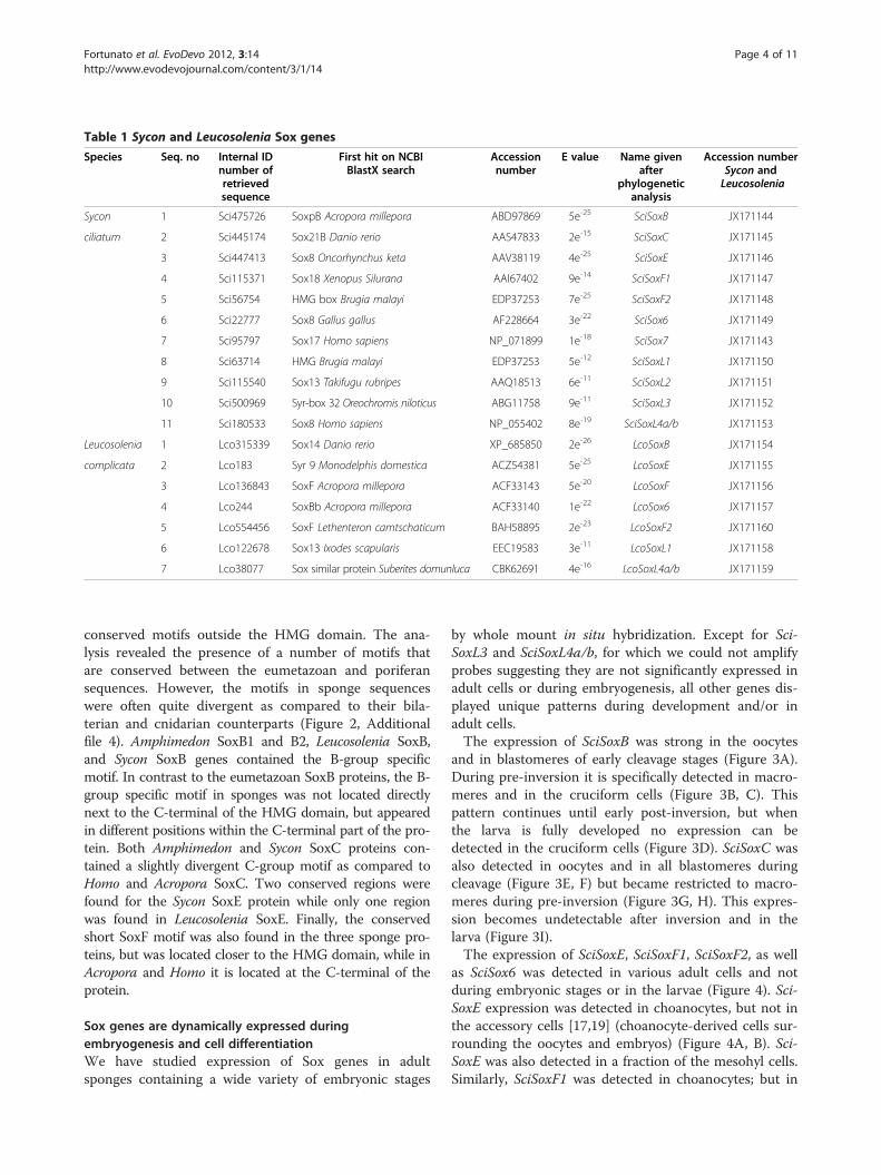
Table 1 Sycon and Leucosolenia Sox genes
Species Seq. no Internal IDnumber ofretrievedsequence
First hit on NCBIBlastX search
Accessionnumber
E value Name givenafter
phylogeneticanalysis
Accession numberSycon andLeucosolenia
Sycon 1 Sci475726 SoxpB Acropora millepora ABD97869 5e-25 SciSoxB JX171144
ciliatum 2 Sci445174 Sox21B Danio rerio AAS47833 2e-15 SciSoxC JX171145
3 Sci447413 Sox8 Oncorhynchus keta AAV38119 4e-25 SciSoxE JX171146
4 Sci115371 Sox18 Xenopus Silurana AAI67402 9e-14 SciSoxF1 JX171147
5 Sci56754 HMG box Brugia malayi EDP37253 7e-25 SciSoxF2 JX171148
6 Sci22777 Sox8 Gallus gallus AF228664 3e-22 SciSox6 JX171149
7 Sci95797 Sox17 Homo sapiens NP_071899 1e-18 SciSox7 JX171143
8 Sci63714 HMG Brugia malayi EDP37253 5e-12 SciSoxL1 JX171150
9 Sci115540 Sox13 Takifugu rubripes AAQ18513 6e-11 SciSoxL2 JX171151
10 Sci500969 Syr-box 32 Oreochromis niloticus ABG11758 9e-11 SciSoxL3 JX171152
11 Sci180533 Sox8 Homo sapiens NP_055402 8e-19 SciSoxL4a/b JX171153
Leucosolenia 1 Lco315339 Sox14 Danio rerio XP_685850 2e-26 LcoSoxB JX171154
complicata 2 Lco183 Syr 9 Monodelphis domestica ACZ54381 5e-25 LcoSoxE JX171155
3 Lco136843 SoxF Acropora millepora ACF33143 5e-20 LcoSoxF JX171156
4 Lco244 SoxBb Acropora millepora ACF33140 1e-22 LcoSox6 JX171157
5 Lco554456 SoxF Lethenteron camtschaticum BAH58895 2e-23 LcoSoxF2 JX171160
6 Lco122678 Sox13 Ixodes scapularis EEC19583 3e-11 LcoSoxL1 JX171158
7 Lco38077 Sox similar protein Suberites domunluca CBK62691 4e-16 LcoSoxL4a/b JX171159
Fortunato et al. EvoDevo 2012, 3:14 Page 4 of 11http://www.evodevojournal.com/content/3/1/14
conserved motifs outside the HMG domain. The ana-lysis revealed the presence of a number of motifs thatare conserved between the eumetazoan and poriferansequences. However, the motifs in sponge sequenceswere often quite divergent as compared to their bila-terian and cnidarian counterparts (Figure 2, Additionalfile 4). Amphimedon SoxB1 and B2, Leucosolenia SoxB,and Sycon SoxB genes contained the B-group specificmotif. In contrast to the eumetazoan SoxB proteins, the B-group specific motif in sponges was not located directlynext to the C-terminal of the HMG domain, but appearedin different positions within the C-terminal part of the pro-tein. Both Amphimedon and Sycon SoxC proteins con-tained a slightly divergent C-group motif as compared toHomo and Acropora SoxC. Two conserved regions werefound for the Sycon SoxE protein while only one regionwas found in Leucosolenia SoxE. Finally, the conservedshort SoxF motif was also found in the three sponge pro-teins, but was located closer to the HMG domain, while inAcropora and Homo it is located at the C-terminal of theprotein.
Sox genes are dynamically expressed duringembryogenesis and cell differentiationWe have studied expression of Sox genes in adultsponges containing a wide variety of embryonic stages
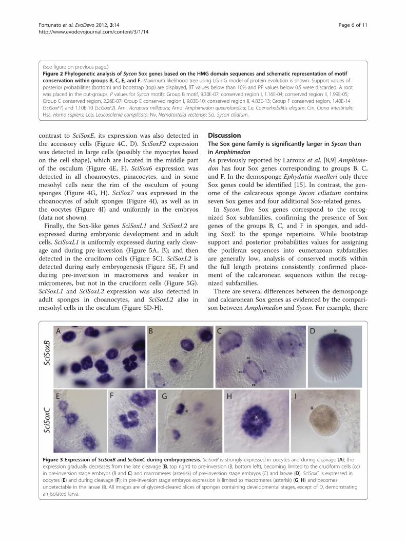
by whole mount in situ hybridization. Except for Sci-SoxL3 and SciSoxL4a/b, for which we could not amplifyprobes suggesting they are not significantly expressed inadult cells or during embryogenesis, all other genes dis-played unique patterns during development and/or inadult cells.The expression of SciSoxB was strong in the oocytes
and in blastomeres of early cleavage stages (Figure 3A).During pre-inversion it is specifically detected in macro-meres and in the cruciform cells (Figure 3B, C). Thispattern continues until early post-inversion, but whenthe larva is fully developed no expression can bedetected in the cruciform cells (Figure 3D). SciSoxC wasalso detected in oocytes and in all blastomeres duringcleavage (Figure 3E, F) but became restricted to macro-meres during pre-inversion (Figure 3G, H). This expres-sion becomes undetectable after inversion and in thelarva (Figure 3I).The expression of SciSoxE, SciSoxF1, SciSoxF2, as well
as SciSox6 was detected in various adult cells and notduring embryonic stages or in the larvae (Figure 4). Sci-SoxE expression was detected in choanocytes, but not inthe accessory cells [17,19] (choanocyte-derived cells sur-rounding the oocytes and embryos) (Figure 4A, B). Sci-SoxE was also detected in a fraction of the mesohyl cells.Similarly, SciSoxF1 was detected in choanocytes; but in
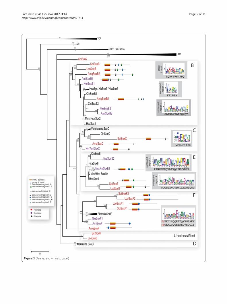
Figure 2 (See legend on next page.)
Fortunato et al. EvoDevo 2012, 3:14 Page 5 of 11http://www.evodevojournal.com/content/3/1/14
(See figure on previous page.)Figure 2 Phylogenetic analysis of Sycon Sox genes based on the HMG domain sequences and schematic representation of motifconservation within groups B, C, E, and F. Maximum likelihood tree using LG+G model of protein evolution is shown. Support values ofposterior probabilities (bottom) and bootstrap (top) are displayed, BT values below than 10% and PP values below 0.5 were discarded. A rootwas placed in the out-groups. P values for Sycon motifs: Group B motif, 9.30E-07; conserved region I, 1.16E-04; conserved region II, 1.99E-05;Group C conserved region, 2.26E-07; Group E conserved region I, 9.03E-10; conserved region II, 4.83E-13; Group F conserved region, 1.40E-14(SciSoxF1) and 1.10E-10 (SciSoxF2). Ami, Acropora millepora; Amq, Amphimedon queenslandica; Ce, Caenorhabditis elegans; Cin, Ciona intestinalis;Hsa, Homo sapiens; Lco, Leucosolenia complicata; Nv, Nematostella vectensis; Sci, Sycon ciliatum.
Fortunato et al. EvoDevo 2012, 3:14 Page 6 of 11http://www.evodevojournal.com/content/3/1/14
contrast to SciSoxE, its expression was also detected inthe accessory cells (Figure 4C, D). SciSoxF2 expressionwas detected in large cells (possibly the myocytes basedon the cell shape), which are located in the middle partof the osculum (Figure 4E, F). SciSox6 expression wasdetected in all choanocytes, pinacocytes, and in somemesohyl cells near the rim of the osculum of youngsponges (Figure 4G, H). SciSox7 was expressed in thechoanocytes of adult sponges (Figure 4I), as well as inthe oocytes (Figure 4J) and uniformly in the embryos(data not shown).Finally, the Sox-like genes SciSoxL1 and SciSoxL2 are
expressed during embryonic development and in adultcells. SciSoxL1 is uniformly expressed during early cleav-age and during pre-inversion (Figure 5A, B); and thendetected in the cruciform cells (Figure 5C). SciSoxL2 isdetected during early embryogenesis (Figure 5E, F) andduring pre-inversion in macromeres and weaker inmicromeres, but not in the cruciform cells (Figure 5G).SciSoxL1 and SciSoxL2 expression was also detected inadult sponges in choanocytes, and SciSoxL2 also inmesohyl cells in the osculum (Figure 5D-H).
Figure 3 Expression of SciSoxB and SciSoxC during embryogenesis. Scexpression gradually decreases from the late cleavage (B, top right) to pre-in pre-inversion stage embryos (B and C) and macromeres (asterisk) of pre-oocytes (E) and during cleavage (F); in pre-inversion stage embryos expresundetectable in the larvae (I). All images are of glycerol-cleared slices of span isolated larva.
DiscussionThe Sox gene family is significantly larger in Sycon thanin AmphimedonAs previously reported by Larroux et al. [8,9] Amphime-don has four Sox genes corresponding to groups B, C,and F. In the demosponge Ephydatia muelleri only threeSox genes could be identified [15]. In contrast, the gen-ome of the calcareous sponge Sycon ciliatum containsseven Sox genes and four additional Sox-related genes.In Sycon, five Sox genes correspond to the recog-
nized Sox subfamilies, confirming the presence of Soxgenes of the groups B, C, and F in sponges, and add-ing SoxE to the sponge repertoire. While bootstrapsupport and posterior probabilities values for assigningthe poriferan sequences into eumetazoan subfamiliesare generally low, analysis of conserved motifs withinthe full length proteins consistently confirmed place-ment of the calcaronean sequences within the recog-nized subfamilies.There are several differences between the demosponge
and calcaronean Sox genes as evidenced by the compari-son between Amphimedon and Sycon. For example, there
iSoxB is strongly expressed in oocytes and during cleavage (A); theinversion (B, bottom left), becoming limited to the cruciform cells (cc)inversion stage embryos (C) and larvae (D). SciSoxC is expressed insion is limited to macromeres (asterisk) (G, H) and becomesonges containing developmental stages, except of D, demonstrating
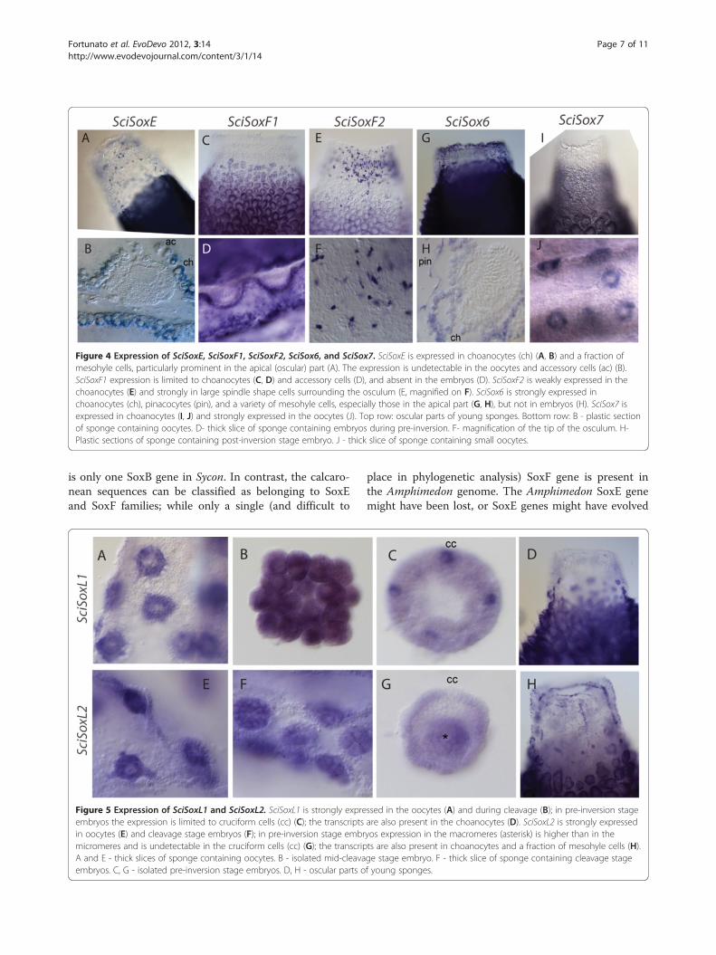
Figure 4 Expression of SciSoxE, SciSoxF1, SciSoxF2, SciSox6, and SciSox7. SciSoxE is expressed in choanocytes (ch) (A, B) and a fraction ofmesohyle cells, particularly prominent in the apical (oscular) part (A). The expression is undetectable in the oocytes and accessory cells (ac) (B).SciSoxF1 expression is limited to choanocytes (C, D) and accessory cells (D), and absent in the embryos (D). SciSoxF2 is weakly expressed in thechoanocytes (E) and strongly in large spindle shape cells surrounding the osculum (E, magnified on F). SciSox6 is strongly expressed inchoanocytes (ch), pinacocytes (pin), and a variety of mesohyle cells, especially those in the apical part (G, H), but not in embryos (H). SciSox7 isexpressed in choanocytes (I, J) and strongly expressed in the oocytes (J). Top row: oscular parts of young sponges. Bottom row: B - plastic sectionof sponge containing oocytes. D- thick slice of sponge containing embryos during pre-inversion. F- magnification of the tip of the osculum. H-Plastic sections of sponge containing post-inversion stage embryo. J - thick slice of sponge containing small oocytes.
Fortunato et al. EvoDevo 2012, 3:14 Page 7 of 11http://www.evodevojournal.com/content/3/1/14
is only one SoxB gene in Sycon. In contrast, the calcaro-nean sequences can be classified as belonging to SoxEand SoxF families; while only a single (and difficult to
Figure 5 Expression of SciSoxL1 and SciSoxL2. SciSoxL1 is strongly expreembryos the expression is limited to cruciform cells (cc) (C); the transcriptsin oocytes (E) and cleavage stage embryos (F); in pre-inversion stage embrmicromeres and is undetectable in the cruciform cells (cc) (G); the transcripA and E - thick slices of sponge containing oocytes. B - isolated mid-cleavaembryos. C, G - isolated pre-inversion stage embryos. D, H - oscular parts o
place in phylogenetic analysis) SoxF gene is present inthe Amphimedon genome. The Amphimedon SoxE genemight have been lost, or SoxE genes might have evolved
ssed in the oocytes (A) and during cleavage (B); in pre-inversion stageare also present in the choanocytes (D). SciSoxL2 is strongly expressedyos expression in the macromeres (asterisk) is higher than in thets are also present in choanocytes and a fraction of mesohyle cells (H).ge stage embryo. F - thick slice of sponge containing cleavage stagef young sponges.
Fortunato et al. EvoDevo 2012, 3:14 Page 8 of 11http://www.evodevojournal.com/content/3/1/14
after demosponges diverged. It is impossible to differen-tiate between these two scenarios until the issue ofsponge monophyly vs. paraphyly is resolved. On theother hand, our result indicates that SoxF genes in Syconand Leucosolenia are likely to be a result of lineage-specific duplication.Interestingly, the Amphimedon genome does not ap-
pear to contain the large number of Sox-related genesthat we have identified in the two calcaronean genomes.It remains unclear whether this is a result of significantgene loss in Amphimedon, or rather of expansion of theSox family in the Calcaronea. Only analysis of additionalporiferan genomes representing a range of clades (espe-cially homoscleromorphs, calcineans, and a range ofdemosponges) will help to shed light into this issue.
Dynamic expression of Sox genes in syconThe expression patterns of Sycon Sox genes fall into twocategories: embryonic (SciSoxB and SciSoxC) or predom-inantly in differentiated adult cells (SciSoxE, SciSoxF1,SciSoxF2, and SciSox6). Sox-like genes are expressedboth during development and in adult tissues (Summaryon Table 2).Until functional data are obtained in sponges, the spe-
cific roles of the identified genes will remain unclear.However, we can hypothesize on their putative functionin Sycon and on hypothetical ancestral roles in the meta-zoan ancestor, by comparing the expression patterns ofSycon and the eumetazoan Sox genes. This is particularlytempting for genes belonging to subfamilies that appearto have a conserved function throughout the Eumetazoa,such as the SoxB group. At least one Sox gene belongingto Group B is expressed in the embryonic ectoderm andthe neurogenic region of embryos in early developmentin most bilaterians (for a review see [21]), cnidarians[12,13], and in the ctenophore P. pileus [14].Sycon SoxB expression is restricted to two cell types of
the embryo, the macromeres and the cruciform cells.During settlement and metamorphosis, the macromeresbecome the outer cells of the post-larva and subse-quently differentiate into exopinacocytes, the outer epi-thelium of the sponge [22,23]. The SciSoxB expression inthe macromeres provides support for the notion that theexopinacoderm of the sponges might be homologous tothe ectoderm of higher metazoans.The cruciform cells are characteristic cells of the calcaro-
nean sponge larvae [19,24]. They form from four cyto-plasm regions segregated during cleavage and differentiateat the pre-inversion stage; they are present in the swim-ming larva, to later degenerate during settlement andmetamorphosis. Their role is not yet clear, but these fourcells are the only candidate cells suggested to play a role inlarval photoreception [24]. If the cruciform cells are indeedinvolved in photoreception, the SoxB expression during
their differentiation would indicate conservation of SoxBfunctions in broadly defined neurogenesis and sensoryorgan formation [25].The expression of Sycon SoxC is very prominent in
macromeres during pre-inversion, while expression was notdetected in larvae. In the cnidarians Acropora and Nema-tostella, SoxC is expressed during embryogenesis in celltypes that are suspected to be sensory neurons [11,12].However in Clytia, SoxC (ChSox15) is expressed in stemcells [13]. Therefore it appears that there is no clear conser-vation of expression pattern among these organisms.While there is no strong conservation of expression
for SoxE and SoxF genes, SoxE genes in bilaterian inver-tebrates tend to have a role in sex-specific aspects ofgonad development, and SoxF genes tend to be asso-ciated with endoderm formation [21,26]. In the cnidar-ians Nematostella and Acropora, SoxE and SoxF areexpressed in endodermal lineages; while in Clytia SoxEis expressed in germline cells, stem cells, and nemato-blasts [13], indicating once again no clear conservationamong cnidarians within this group. However, expres-sion in the endoderm (in Anthozoan cnidarians) andmesodermal derivatives (gonads) of bilaterians, togetherwith the observed expression of Sycon SoxE and SoxF inchoanocytes and some mesohyl cells, could be used tosupport a concept of homology of the choanoderm+mesohyl with endomesoderm. Otherwise, these twogenes might play roles in cell differentiation in Sycon, asevidenced by the fact that expression of SoxE disappearsin choanocytes that transdifferentiate into accessorycells, while expression of SoxF1 becomes stronger inthese cells during the process.
ConclusionsSponges are relatively simple organisms with few celltypes, thus the limited number of transcription fac-tors representing conserved metazoan families in thedemosponge Amphimedon quenslandica fits neatlywith the concept of a simple developmental tool kitpatterning a simple body. This study demonstratesthat Sycon ciliatum has multiple Sox genes which aredynamically expressed during development and inpatterns consistent with governing adult cell differen-tiation. This indicates that Sox genes were involvedin development and cell differentiation from the be-ginning of multicellular animal evolution. Furtheranalyses of this and other developmental gene fam-ilies in the Calcarea and in other sponge group arenecessary to test whether the identified differencesbetween Sycon and Amphimedon are indicative ofglobal differences in the developmental toolkits. Suchstudies, now underway in our laboratory and in othergroups, will provide insight into the evolutionary his-tory of the animal developmental toolkit.
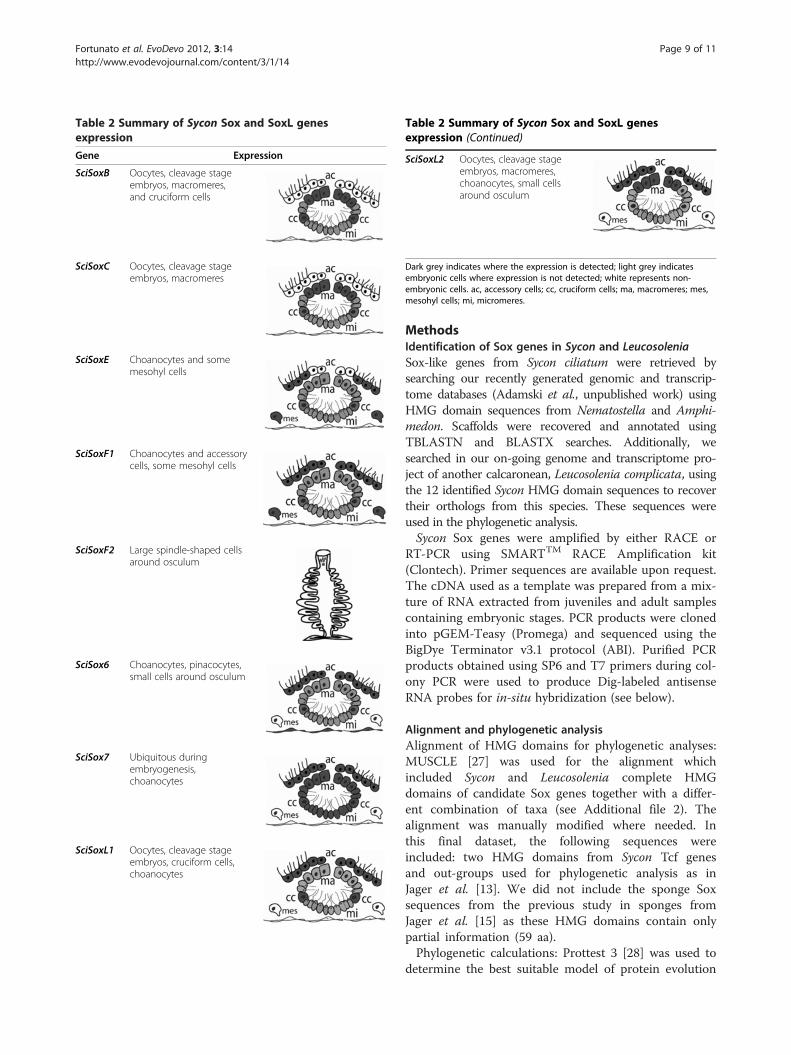
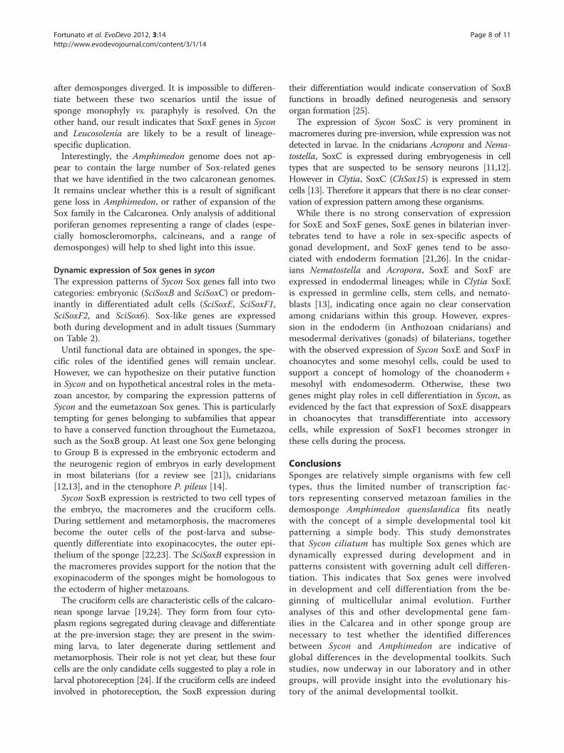
Table 2 Summary of Sycon Sox and SoxL genesexpression
Gene Expression
SciSoxB Oocytes, cleavage stageembryos, macromeres,and cruciform cells
SciSoxC Oocytes, cleavage stageembryos, macromeres
SciSoxE Choanocytes and somemesohyl cells
SciSoxF1 Choanocytes and accessorycells, some mesohyl cells
SciSoxF2 Large spindle-shaped cellsaround osculum
SciSox6 Choanocytes, pinacocytes,small cells around osculum
SciSox7 Ubiquitous duringembryogenesis,choanocytes
SciSoxL1 Oocytes, cleavage stageembryos, cruciform cells,choanocytes
Table 2 Summary of Sycon Sox and SoxL genesexpression (Continued)
SciSoxL2 Oocytes, cleavage stageembryos, macromeres,choanocytes, small cellsaround osculum
Dark grey indicates where the expression is detected; light grey indicatesembryonic cells where expression is not detected; white represents non-embryonic cells. ac, accessory cells; cc, cruciform cells; ma, macromeres; mes,mesohyl cells; mi, micromeres.
Fortunato et al. EvoDevo 2012, 3:14 Page 9 of 11http://www.evodevojournal.com/content/3/1/14
MethodsIdentification of Sox genes in Sycon and LeucosoleniaSox-like genes from Sycon ciliatum were retrieved bysearching our recently generated genomic and transcrip-tome databases (Adamski et al., unpublished work) usingHMG domain sequences from Nematostella and Amphi-medon. Scaffolds were recovered and annotated usingTBLASTN and BLASTX searches. Additionally, wesearched in our on-going genome and transcriptome pro-ject of another calcaronean, Leucosolenia complicata, usingthe 12 identified Sycon HMG domain sequences to recovertheir orthologs from this species. These sequences wereused in the phylogenetic analysis.Sycon Sox genes were amplified by either RACE or
RT-PCR using SMARTTM RACE Amplification kit(Clontech). Primer sequences are available upon request.The cDNA used as a template was prepared from a mix-ture of RNA extracted from juveniles and adult samplescontaining embryonic stages. PCR products were clonedinto pGEM-Teasy (Promega) and sequenced using theBigDye Terminator v3.1 protocol (ABI). Purified PCRproducts obtained using SP6 and T7 primers during col-ony PCR were used to produce Dig-labeled antisenseRNA probes for in-situ hybridization (see below).
Alignment and phylogenetic analysisAlignment of HMG domains for phylogenetic analyses:MUSCLE [27] was used for the alignment whichincluded Sycon and Leucosolenia complete HMGdomains of candidate Sox genes together with a differ-ent combination of taxa (see Additional file 2). Thealignment was manually modified where needed. Inthis final dataset, the following sequences wereincluded: two HMG domains from Sycon Tcf genesand out-groups used for phylogenetic analysis as inJager et al. [13]. We did not include the sponge Soxsequences from the previous study in sponges fromJager et al. [15] as these HMG domains contain onlypartial information (59 aa).Phylogenetic calculations: Prottest 3 [28] was used to
determine the best suitable model of protein evolution
Fortunato et al. EvoDevo 2012, 3:14 Page 10 of 11http://www.evodevojournal.com/content/3/1/14
for our alignment. We used two phylogenetic analyses ofHMG domains:
(1) Two independent runs of PhyML [29] wereperformed. Each run searched for five randomstarting trees using SPR moves. The tree with thebest log likelihood value was selected (Loglikelihood =−5686.2). From this tree a bootstrapanalysis using 100 replicates was performed.
(2) Bayesian analysis [30] under LG model, with5,000,000 generations sampled every 500generations using four chains. Convergence wasreached before 5,000,000 generations. A majorityrule of consensus tree of 12,500 trees wasgenerated and posterior probabilities values werecalculated from this tree.
Finding conserved motifs within sponge Sox sequencesMEME 3.5.7 [31] was used to find conserved motifs out-side the HMG domain within Sycon and LeucosoleniaSox proteins and their closest homologues from Acro-pora, Homo, Nematostella, and Amphimedon. The fol-lowing parameters were used for searching possibleconserved motifs: minimum motif width, six; maximumwidth, 100; maximum motifs to find, six. Completesequences were aligned and their motif locations werecompared with previous studies [4,12]. ‘My domainimage creator’ tool included in Prosite [32] was used tovisualize the locations of motifs in Sox proteins.
Specimen collection and whole mount in-situhybridizationAdult Sycon specimens were collected from fjordslocated near Bergen, Norway (+60° 27' 33", +4° 56' 1")during the reproductive season from May to September(2008 to 2011). For in-situ hybridization, samples wereimmediately fixed in 100 mM MOPS, pH 7.5; 0.5 M so-dium chloride; 2 mM MgSO4; 4% paraformaldehyde;0.05% glutaraldehyde over night at 4°C, stepped into andextensively washed in 70% EtOH and stored at −20°Cuntil processing. Macro sections of sponges in 24 wellplates (Nunc) were rehydrated and washed in PBS/0.1%Tween (PTw). Samples were pretreated with 7.5 μg/mLproteinase K for 10 minutes at 37°C, followed byquenching with glycine (2 mg/mL PTw). Acetylation wasperformed by serial treatment with 0.1 M triethanola-mine containing 0, 1.5, and 3 μl/mL acetic anhydride.Re-fixation was done in 4% paraformaldehyde/0.05% glu-taraldehyde in PBS for 1 h at room temperature, fol-lowed by extensive washing in PTw. Tissue wasprehybridized as previously described [33] in 2 mL-tubesfor 90 to 180 min at 51°C. Probe hybridization was donewith denatured RNA probe (0.1-0.3 ng/μL, approxi-mately 1 kb) for 12 to 18 h at 51°C. Stringent washes
were carried out at 55°C as following: 1 × 10 min inhybridization buffer; 2 × 10 min 50% formamide/4 × SSC/0.1%; 2 × 10 min 50% formamide/2 × SSC/0.1% Tween;2 × 10 min 25% formamide/2 × SSC/0.1% Tween, fol-lowed by 3 × 15 min 2 × SSC/0.1% Tween at roomtemperature. Samples were transferred to maleic acidbuffer and incubated in 2% (w/v) Blocking Reagent(Roche) for 60 min at room temperature. After overnightincubation with AP-coupled anti-Digoxigenin-Fab frag-ments (Sigma, 1:5,000) at 4°C, samples were washed inmaleic acid buffer at least 6 × 30 min. Probe wasdetected using NBT/BCIP as substrate (Roche) with tis-sue equilibrated in alkaline phosphatase buffer (100 mMsodium chloride, 50 mM MgCl2, 100 mM Tris pH 9.5,0.1% Tween, 1 mM Levamisole). The staining reaction(0.5 to 3 days) was stopped with PBS/0.5% Tween, sam-ples were transferred to 100% glycerol for microscopy orethanol-dehydrated and embedded in epoxy resin(Sigma) for sectioning. Pictures of whole mount samplesand sections were taken using a Nikon DS-U3 micro-scope and processed in Photoshop.
Additional files
Additional file 1: Maximum likelihood phylogenetic tree of HMGsequences found in Sycon ciliatum and Leucosolenia complicata. Aphylogenetic analysis which includes the entire repertoire of HMGdomains sequences found in Sycon (twelve sequences) and Leucosolenia(seven sequences). PhyMl tree using LG +G model of protein evolution isshown. Bootstrap support values are displayed. Taxa names: Ami,Acropora millepora; Amq, Amphimedon queenslandica; Ce, Caenorhabditiselegans; Ci, Ciona intestinalis; Gdo, Gallus domesticus; Hsa, Homo sapiens;Lco, Leucosolenia complicata; Mm, Mus musculus; Ncr, Neutrospora crassa;Omy, Oncorhynchus mykis; Sci, Sycon ciliatum; Xle, Xenopus laevis.
Additional file 2: HMG domains recovered from Sycon andLeucosolenia. Alignment of Sycon and Leucosolenia HMG domains of thecomplete repertoire of sox and sox-like genes recovered for this study.Sequences were compared with: Acropora millepora (Ami); andAmphimedon queenslandica (Amq).
Additional file 3: Alignment of HMG domains used for thephylogenetic analysis. Includes the HMG domain sequence alignmentused for the phylogenetic analysis in Figure 2.
Additional file 4: Calculation of conserved motifs. This file includesall taxa used for finding conserved motifs within sponge sequences.P values are shown and conserved regions are highlighted in red.
Competing interestsThe authors declare that they have no competing interests.
Authors’ contributionsConceived and designed the study: MajA and SF. Suggested the modelsystem and provided knowledge about its biology: H-TR. Assembledgenomes and transcriptomes and created sequence databases: MarA. Carriedout sampling and experiments: SF, BB, MarA, CZ, SL, CG, SJ, and MajA.Analyzed data: SF and MajA. Drafted the manuscript: SF. Edited themanuscript: MajA and SF with input from co-authors. All authors read andapproved the final manuscript.
AcknowledgementsThis study was funded by the core budget of the Sars International Centrefor Marine Molecular Biology. Sequencing has been performed at TheNorwegian High-Throughput Sequencing Centre funded by the Research
Fortunato et al. EvoDevo 2012, 3:14 Page 11 of 11http://www.evodevojournal.com/content/3/1/14
Council of Norway. We thank Lucas Leclère for helpful comments on themanuscript.
Author details1Sars International Centre for Marine Molecular Biology, Thormøhlensgt. 55,Bergen 5008, Norway. 2Department of Biology and Centre for Geobiology,University of Bergen, Thormøhlensgt. 55, Bergen 5008, Norway.
Received: 29 March 2012 Accepted: 22 June 2012Published: 23 July 2012
References1. Guth SI, Wegner M: Having it both ways: sox protein function between
conservation and innovation. Cell Mol Life Sci 2008, 65:3000–3018.2. Lefebvre V, Dumitriu B, Penzo-Méndez A, Han Y, Pallavi B: Control of cell
fate and differentitation by Sry-related high-mobility-group box (Sox)transcription factors. Int J Biochem Cell B 2007, 39:2195–2214.
3. Gubbay J, Collignon J, Koopman P, Capel B, Economou A, Munsterberg A,Vivian N, Goodfellow P, Lovell-Badge R: A gene mapping to the sex-determining region of the mouse Y chromosome is a member of a novelfamily of embryonically expressed genes. Nature 1990, 346:245–250.
4. Schepers GE, Teasdale RD, Koopman P: Twenty pairs of sox: extent,homology, and nomenclature of the mouse and human soxtranscription factor gene families. Dev Cell 2002, 3:167–170.
5. Bowles J, Schepers G, Koopman P: Phylogeny of the SOX family ofdevelopmental transcription factors based on sequence and structuralindicators. Dev Biol 2000, 227:239–255.
6. King N, Westbrook MJ, Young S, Kuo A, Abedin M, Chapman J, Fairclough S,Hellsten U, Isogai Y, Letunic I, Marr M, Pincus D, Putnam N, Rokas A, WrightKJ, Zuzow R, Dirks W, Good M, Goodstein D, Lemons D, Li W, Lyons JB,Morris A, Nichols S, Richter DJ, Salamov A, Sequencing JG, Bork P, Lim WA,Manning G, et al: The genome of the choanoflagellate Monosigabrevicollis and the origin of metazoans. Nature 2008, 451:783–788.
7. Sebé-Pedrós A, de Mendoza A, Lang BF, Degnan BM, Ruiz-Trillo I: Unexpectedrepertoire of metazoan transcription factors in the unicellular holozoancapsaspora owczarzaki. Mol Biol Evol 2011, 28:1241–1254.
8. Larroux C, Fahey B, Liubicich D, Hinman VF, Gauthier M, Gongora M, GreenK, Wörheide G, Leys SP, Degnan BM: Developmental expression oftranscription factor genes in a demosponge: insights into the origin ofmetazoan multicellularity. Evol Dev 2006, 8:150–173.
9. Larroux C, Luke GN, Koopman P, Rokhsar DS, Shimeld SM, Degnan BM:Genesis and expansion of metazoan transcription factor gene classes.Mol Biol Evol 2008, 25:980–996.
10. Srivastava M, Begovic E, Chapman J, Putnam NH, Hellsten U, Kawashima T,Kuo A, Mitros T, Salamov A, Carpenter ML, Signorovitch AY, Moreno MA,Kamm K, Grimwood J, Schmutz J, Shapiro H, Grigoriev IV, Buss LW,Schierwater B, Dellaporta SL, Rokhsar DS: The Trichoplax genome and thenature of placozoans. Nature 2008, 454:955–960.
11. Magie CR, Pang K, Martindale MQ: Genomic inventory and expression ofSox and Fox genes in the cnidarian Nematostella vectensis. Dev Genes Evol2005, 215:618–630.
12. Shinzato C, Iguchi A, Hayward DC, Technau U, Ball EE, Miller DJ: Sox genesin the coral Acropora millepora: divergent expression patterns reflectdifferences in developmental mechanisms within the Anthozoa. BMCEvol Biol 2008, 8:311.
13. Jager M, Queinnec E, Le Guyarde H, Manuel M: Multiple Sox genes areexpressed in stem cells or in differentiating neuro-sensory cells in thehydrozoan Clytia hemisphaerica. EvoDevo 2011, 2:12.
14. Jager M, Queinnec E, Chiori R, Le Guyader H, Manuel M: Insights into theearly evolution of SOX genes from expression analyses in a ctenophore.J Exp Zool B Mol Dev Evol 2008, 310:650–667.
15. Jager M, Queinnec E, Houliston E, Manuel M: Expansion of the SOX genefamily predated the emergence of the Bilateria. Mol Phylogenet Evol 2006,39:468–477.
16. Adamska M, Degnan B, Green K, Zwafink C: What sponges can tell usabout the evolution of developmental processes. Zoology 2011, 114:1–10.
17. Franzen W: Oogenesis and larval development of Scypha ciliata (Porifera,Calcarea). Zoomorphology 1988, 107:349–357.
18. Leys SP, Eerkes-Medrano D: Gastrulation in Calcareous Sponges: in Searchof Haeckel’s Gastraea. Integr Comp Biol 2005, 45:342–351.
19. Ereskovsky AV: The Comparative Embryology of Sponges. Netherlands:Springer; 2010.
20. Bergsten J: A review of long-branch attraction. Cladistics 2005, 21:163–193.21. Phochanukul N, Russell S: No backbone but lots of SOX: the invertebrate
SOX family. Int J Biochem Cell Biol 2009, 42:453–464.22. Amano S, Hori I: Metamorphosis of calcareous sponges I. Ultrastructure
of free-swimming larvae. Invertebr Reprod Dev 1992, 21:81–90.23. Gallissian MF, Vacelet J: Ultrastructure of the oocyte and embryo of the
calcified sponge (Petrobiona massiliana Porifera, Calcarea).Zoomorphology 1992, 112:133–141.
24. Tuzet O: Éponges calcaires. In Traité de Zoologie. Anatomie, Systématique,Biologie. Spongiaires. Edited by Grassé P-P. Paris: Masson et Cie; 1973:27–132.
25. Meulemans D, Bronner-Fraser M: The amphioxus soxB family: implicationsfor the evolution of vertebrate placodes. Int J Biol Sci 2007, 3:356–364.
26. Nanda T, DeFalco SHY, Phochanukul LN, Camara N, VanDoren M, Russell S:Sox100B, a drosophila group e sox-domain gene, is required for somatictestis differentiation. Sexual Development 2009, 3:26–37.
27. Edgar RC: MUSCLE: a multiple sequence alignment method with reducedtime and space complexity. BMC Bioinform 2004, 5:113.
28. Abascal F, Zardoya R, Posada D: ProtTest: Selection of best-fit models ofprotein evolution. Bioinformatics 2005, 21:2104–2105.
29. Guindon S, Dufayard JF, Lefort V, Anisimova M, Hordijk W, Gascuel O: Newalgorithms and methods to estimate maximum-likelihood phylogenies:assessing the performance of phyML 3.0. Syst Biol 2010, 59:307–321.
30. Ronquist F, Huelsenbeck JP, van der Mark P: MrBayes 3.1. 2005,[http://mrbayes.csit.fsu.edu/index.php].
31. Bailey TL, Boden M, Buske FA, Frith M, Grant CE, Clementi L, Ren J, Li WW,Noble WS: MEME Suite: tools for motif discovery and searching. NucleicAcids Research 2010, suppl 2:W202–W208.
32. Sigrist CJA, Cerutti L, de Castro E, Langendijk-Genevaux PS, Bulliard V,Bairoch A, Hulo N: PROSITE, a protein domain database for functionalcharacterization and annotation. Nucleic Acids Research 2010, 38:161–166.
33. Larroux C, Fahey B, Adamska M, Richards GS, Gauthier M, Green K, Lovas E,Degnanet BM: Whole-Mount In Situ Hybridization in Amphimedon. ColdSpring Harbor Protocols 2008, doi:10.1101/pdb.prot5096.
doi:10.1186/2041-9139-3-14Cite this article as: Fortunato et al.: Genome-wide analysis of the soxfamily in the calcareous sponge Sycon ciliatum: multiple genes withunique expression patterns. EvoDevo 2012 3:14.
Submit your next manuscript to BioMed Centraland take full advantage of:
• Convenient online submission
• Thorough peer review
• No space constraints or color figure charges
• Immediate publication on acceptance
• Inclusion in PubMed, CAS, Scopus and Google Scholar
• Research which is freely available for redistribution
Submit your manuscript at www.biomedcentral.com/submit


![Occurrence of Fibonacci numbers in development and structure … · 2013. 12. 24. · Calcareous sponge spicules (note (b) and (f)). After BEPS [20]. Hydra, a single sessile tube-like](https://img.pdfslide.net/doc/110x75/60f69ba3ac8d7423511eaecc/occurrence-of-fibonacci-numbers-in-development-and-structure-2013-12-24-calcareous.jpg)


![Calcareous sponge genomes reveal complex -carbonic … · 2017. 8. 29. · or characterize CA-proteins from the calcareous sponge S. ciliatum have not been successful [22]. Only recently,](https://img.pdfslide.net/doc/110x75/60d35117c3bc180d086fdbcc/calcareous-sponge-genomes-reveal-complex-carbonic-2017-8-29-or-characterize.jpg)