Embed Size (px)
Citation preview
Malaysian Orthopaedic Journal 2020 Vol 14 No 3 Singaravadivelu V, et al
42
ABSTRACTIntroduction: Giant cell tumour (GCT) of the bone is abenign tumour with a high tendency to recur after surgery.This study aimed to analyse prospectively the rate of localrecurrence following management of giant cell tumours bycurettage, using intravenous zoledronic acid as an adjuvant,and fibular struts to support the empty cavity after curettage. Materials and Methods: This study was carried out in tencases of biopsy-proven GCTs: five males and five females, inthe age group between 18 and 39 years. All patients weregiven three doses of zoledronic acid, one pre-operative andtwo post-operative. Extended curettage was done threeweeks after the pre-operative dose of zoledronate. The cavitywas left empty in all the cases. Fibular struts were used tosupport the cavity from collapse. Patients were followed-upfor post-operative local recurrence. The functional status ofthe patients was assessed during each visit using theMusculoskeletal Tumour Society (MSTS) score.Results: There were no recurrences at a follow-up of twoyears. All patients had a stable knee and were able to bearweight fully. The average knee flexion was 75º. The averageMSTS score of the study was 92%.Conclusion: Extended curettage using hydrogen peroxide,systemic zoledronic acid adjuvant and leaving the cavityempty without using cancellous bone graft did not lead to arecurrence of GCT. Non-vascularised fibular strut providedadequate support while the cavity left empty after curettagedid not collapse and there was good knee function.
Keywords:GCT, zoledronic acid, fibula strut, recurrence
INTRODUCTIONGiant cell tumour is a common bone tumour accounting for5% of all primary bone tumours. Although the mortality rate
associated with the disease is low, the tumour is locallyaggressive and has a high tendency to recur1. With theadvancement in treatment options, the recurrence rateassociated with GCT has fallen from an excess of 40% to lessthan 20% with extended curettage and use of adjuvants2-4.
The cavity left behind following the curettage is commonlyfilled with a bone graft or bone cement. Studies in literature5,6had reported higher recurrence rates when iliac crest bonegraft was used to fill the cavity. Bone cement is an inertmaterial and does not get incorporated or remodelled alongthe lines of stress.
In this study, we postulated that leaving the cavity emptyfollowing curettage reduced the recurrence rates while thecavity did not collapse and the cortical struts used to supportthe empty cavity were incorporated with the host cortex.
MATERIALS AND METHODSTen consecutive cases of biopsy-proven GCT admitted in ourunit between 2015 and 2017 were enrolled in the study.Primary GCT, recurrent GCT and GCT with pathologicalfractures were all included. Informed written consent wasobtained from all the patients. MRI was done to confirm theintramedullary extent of the tumour and possible soft tissueextension. CT chest was done to rule out pulmonarymetastasis. All the patients were available for a final follow-up. Ethical clearance was obtained from the InstitutionalEthical Committee.
All the cases in our study were treated in the same manner.A creatinine clearance of 60ml per minute was taken as theminimum value for the administration of 4mg of zoledronicacid as per FDA standards. Zoledronic acid was administeredin 100ml normal saline over 15 minutes after adequate fluidpreloading.
Giant Cell Tumour Around Knee Managed by Curettageand Zoledronic Acid with Structural Support by Fibula
Cortical Struts
Singaravadivelu V, PhD, Kavinkumar V, MS
Institute of Orthopaedics and Traumatology, Rajiv Gandhi Government General Hospital, Chennai, India
This is an open-access article distributed under the terms of the Creative Commons Attribution License, which permits unrestricted use, distribution, and reproduction in any medium, provided the original work is properly cited
Date of submission: 31st March 2020Date of acceptance: 09th July 2020
Corresponding Author: Kavinkumar Vembanan, c/o Prof V Singaravadivelu, Institute of Orthopaedics and Traumatology, Rajiv GandhiGovernment General Hospital, GH Post Office, 3, Poonamallee High Road, Near Chennai Central, near Park Town, Chennai, Tamil Nadu600003, India Email: [email protected]
doi: https://doi.org/10.5704/MOJ.2011.008
7-OR3-180_OA1 11/26/20 1:56 PM Page 42

Management of GCT Around Knee
43
Extended curettage was done three weeks after the firstdose of zoledronic acid. The surgery was performed undertourniquet control. The cavity was well visualised, and acurettage of the lesion was done. Power burrs were used toenhance surgical clearance. The cavity was thoroughlyirrigated at the end of the procedure to wash away the tumourcells. The cavity was then treated with 3% hydrogenperoxide for three minutes. A total of three hydrogenperoxide washes were given.
The dimensions of the cavity were measured to calculate thelength of fibula needed to be resected. Proximal tibia cavitieswere generally supported by two struts, one mediolateral andone superoinferior strut. Distal femur cavities were given anadditional anteroposterior strut when the tumour involved alarge portion of the posterior femoral condyle. Themeasured length of the fibula was resected using aposterolateral approach. The ipsilateral fibula was harvestedin lesions involving the distal femur while the contralateralfibula was used in proximal tibia lesions. This protocol wasfollowed to prevent further compromise in the stability of theleg when a fibular defect was made on the same side as thetibial cavity. The fibula was harvested sparing the proximaland distal 8cm.
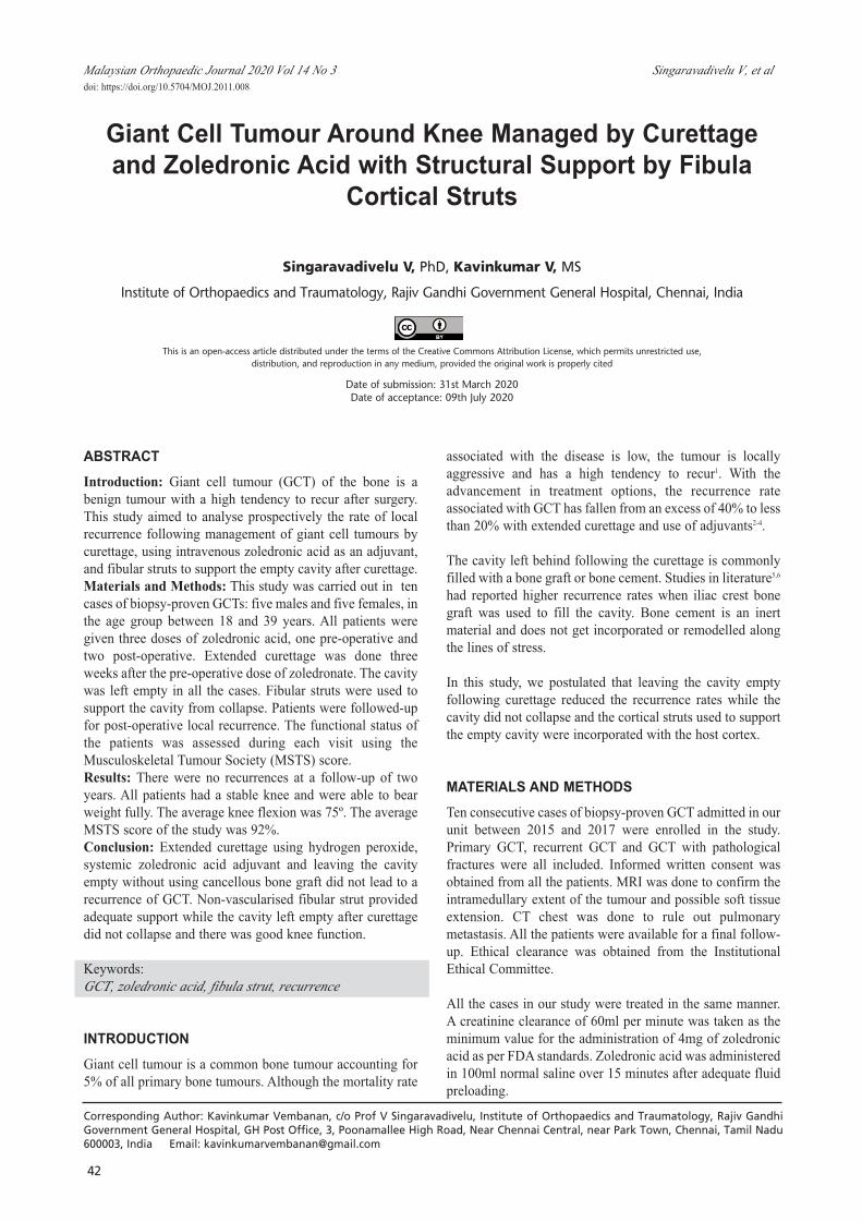
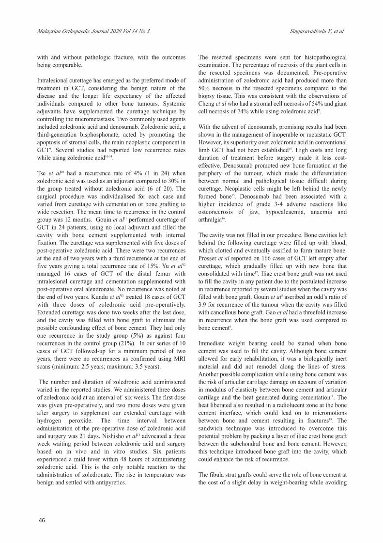
The resected piece of fibula was split into multiple struts.The mediolateral strut was placed first. The superoinferiorstrut was positioned over the above strut and hitched againstthe cortex proximally or distally. This construct providedenough support to the cavity (Fig. 1). In one case, the fibulargraft was secured to the host cortex using a 3.5mm corticalscrew (Fig. 2). The breadth of the graft was more than halfthe width of the tibial plateau to prevent point loading of thegraft.
Patients were given above-knee casts in the immediate post-operative period. Three doses of antibiotics were given.Suture removal was done on the 12th post-operative day. Thepatients were kept non-weight bearing with above-kneecasts. The first follow-up visit was at three weeks post-surgery, during which the second dose of zoledronic acid wasgiven. The final dose of zoledronic acid was given afteranother six weeks.
Routine radiographs were taken at six weeks, twelve weeks,three months, six months, one year; and at six-month-intervals thereafter. MRI was taken two years post-operativeto detect recurrences. The decision to discontinue plasterimmobilisation and commence knee mobilisation andweight-bearing was individualised for every patientdepending on the consolidation of the graft. The patientswere evaluated at six-month intervals using theMusculoskeletal Tumour Society (MSTS) score.
RESULTSThe average age of the patient in the study was 30 years(range 18 - 39 years). There were five men and five women(sex ratio - one: one). The distal femur (five) and proximaltibia (five) were equally involved in our study. Eight patientshad a primary giant cell tumour while two patients had arecurrent giant cell tumour. The primary tumours werestaged radiologically using the Campanacci grading. Therewere five cases of Grade II tumour and three cases of GradeIII tumour. All patients were available for the final follow-up. The longest follow-up was 3.5 years, while the shortestfollow-up was for 2.5 years. No recurrence was seen in anyof the patients as confirmed with MRI scans two years afterthe surgery. Radiographs revealed good consolidation of thefibular struts and gradual filling up of the cavity. The jointspace was maintained in all cases.
Patients were immobilised post-operatively in above-kneecasts. The average period of immobilisation was 10 weeks (4- 16 weeks). The patients were started on toe-touch weight-bearing at the time of plaster removal. The average timetaken to start full weight-bearing was 18 weeks (12 - 22weeks). Four patients regained a knee flexion of more than90° while six patients had a knee flexion between 60º and90º. The mean MSTS score of our study was 92%. Twopatients had extensor hallucis longus (EHL) weakness on theside of fibular resection which eventually improved.
DISCUSSIONTen cases were included in our study, as outlined in Table I.We had eight cases of the primary tumour and two cases ofrecurrent tumour. Of the two recurrent cases, the first casehad a recurrence one year after curettage and cementation.The second case had a recurrence ten years after the firstsurgery, which was managed with curettage and cementationfollowed by a second recurrence two years later. Therecurrent tumours were all operated primarily at othercentres. The use of adjuvant therapy was not documented inboth cases. A radical surgery like wide resection offered nosignificant additional disease control over extendedcurettage in the management of recurrent GCT. Steyern et al7did not find any significant difference in recurrence ratewhile managing primary and recurrent tumours withextended curettage.
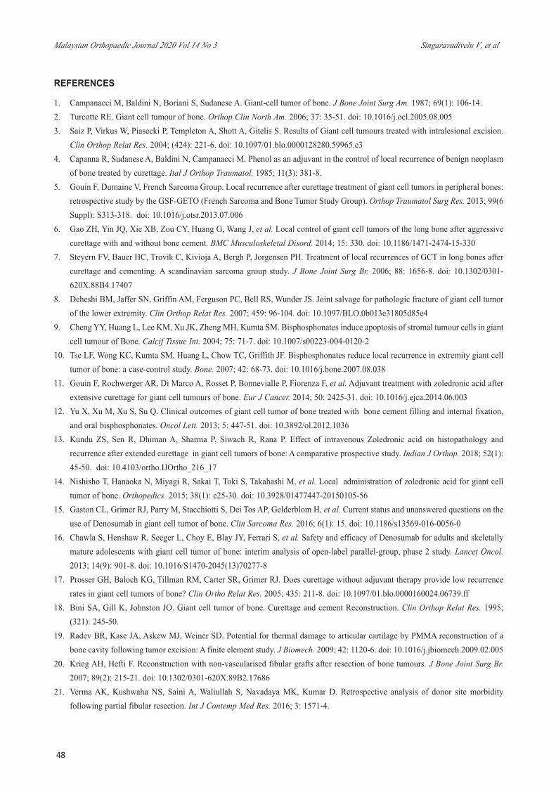
The presence of a pathological fracture was not acontraindication for inclusion in our study. Two patients inour series presented with a pathological fracture; oneinvolving the distal femur (Fig. 3), and the other involvingthe proximal tibia. Intra-operatively, the tumour was found tobe well contained within a pseudocapsule, and anintralesional curettage was done with resection of thepseudocapsule. Deheshi et al8 compared recurrence-freesurvival and functional outcome after curettage in patients
7-OR3-180_OA1 11/26/20 1:56 PM Page 43

Malaysian Orthopaedic Journal 2020 Vol 14 No 3 Singaravadivelu V, et al
44
Table I: Master chart summarising the findings of the study.
S. Age/ Diagnosis Campanacci Period of Time to Duration Knee MSTS ComplicationsNo Sex Grading immobilisation full weight of flexion Score at
bearing follow-up final follow-up
1. 35 M Primary GCT III 16 weeks 22 weeks 3 years 120° 29 Nilproximal tibia – 6 months
left side2. 34 M Primary GCT II 16 weeks 24 weeks 3 years 6° 26 Nil
distal femur – 6 monthsleft side
3. 28 M Primary GCT III 10 weeks 16 weeks 3 years 75° 28 Nildistal femur – 3 monthsRight side
4. 34 F Primary GCT III 14 weeks 20 weeks 3 years 60º 27 Nildistal femur with pathological fracture- Right side
5. 28 F Primary GCT II 10 weeks 18 weeks 3 years 60º 26 Nilproximal tibia
with pathological fracture – Right side
6. 18 F Primary GCT II 10 weeks 16 weeks 3 years 120º 29 Nilproximal tibia –
Right side7. 38 M Recurrent GCT - 4 weeks 12 weeks 2 years 75º 27 Nil
proximal tibia – 10 monthsRight side
8. 23 F Recurrent GCT - 6 weeks 18 weeks 2 years 110º 29 EHL Weaknessproximal tibia – 9 months
Right side9. 39 M Primary GCT II 8 weeks 14 weeks 2 years 75º 27 EHL Weakness
distal femur – 6 monthsRight side
10. 23 F Primary GCT II 8 weeks 18 weeks 2 years 100º 28 Nildistal femur – 6 months
Left side
Fig. 1: (a) Cavity after curettage (b) Cavity supported with fibula cortical struts.
(a) (b)
7-OR3-180_OA1 11/26/20 1:56 PM Page 44

Management of GCT Around Knee
45
Fig. 2: (a) Giant cell tumour involving the lateral femoral condyle in 24-year-old. (b) Cavity was supported with three struts aftercurettage. Arrow indicates a 3.5mm cortical screw used to secure the superoinferior strut to the lateral femoral cortex. (c,d) Goodconsolidation of the graft at two years post-operation.
(a) (b) (c) (d)
Fig. 4: (a) Giant cell tumour of the medial femoral condyle in a 38-year-old. (b) Immediate post-operation radiograph shows cavitysupported with two struts. (c,d) Good hypertrophy and incorporation of the graft at two years post-operation.
(a) (b) (c) (d)
Fig. 3: (a,b) Giant cell tumour of distal femur presenting with a pathological fracture. (c) Immediate post-operative radiograph. (d) Twoyears post-operative radiograph.
(a) (b) (c) (d)
7-OR3-180_OA1 11/26/20 1:56 PM Page 45
Malaysian Orthopaedic Journal 2020 Vol 14 No 3 Singaravadivelu V, et al
46
with and without pathologic fracture, with the outcomesbeing comparable.
Intralesional curettage has emerged as the preferred mode oftreatment in GCT, considering the benign nature of thedisease and the longer life expectancy of the affectedindividuals compared to other bone tumours. Systemicadjuvants have supplemented the curettage technique bycontrolling the micrometastasis. Two commonly used agentsincluded zoledronic acid and denosumab. Zoledronic acid, athird-generation bisphosphonate, acted by promoting theapoptosis of stromal cells, the main neoplastic component inGCT9. Several studies had reported low recurrence rateswhile using zoledronic acid10-14.
Tse et al10 had a recurrence rate of 4% (1 in 24) whenzoledronic acid was used as an adjuvant compared to 30% inthe group treated without zoledronic acid (6 of 20). Thesurgical procedure was individualised for each case andvaried from curettage with cementation or bone grafting towide resection. The mean time to recurrence in the controlgroup was 12 months. Gouin et al11 performed curettage ofGCT in 24 patients, using no local adjuvant and filled thecavity with bone cement supplemented with internalfixation. The curettage was supplemented with five doses ofpost-operative zoledronic acid. There were two recurrencesat the end of two years with a third recurrence at the end offive years giving a total recurrence rate of 15%. Yu et al12managed 16 cases of GCT of the distal femur withintralesional curettage and cementation supplemented withpost-operative oral alendronate. No recurrence was noted atthe end of two years. Kundu et al13 treated 18 cases of GCTwith three doses of zoledronic acid pre-operatively.Extended curettage was done two weeks after the last dose,and the cavity was filled with bone graft to eliminate thepossible confounding effect of bone cement. They had onlyone recurrence in the study group (5%) as against fourrecurrences in the control group (21%). In our series of 10cases of GCT followed-up for a minimum period of twoyears, there were no recurrences as confirmed using MRIscans (minimum: 2.5 years; maximum: 3.5 years).
The number and duration of zoledronic acid administeredvaried in the reported studies. We administered three dosesof zoledronic acid at an interval of six weeks. The first dosewas given pre-operatively, and two more doses were givenafter surgery to supplement our extended curettage withhydrogen peroxide. The time interval betweenadministration of the pre-operative dose of zoledronic acidand surgery was 21 days. Nishisho et al14 advocated a threeweek waiting period between zoledronic acid and surgerybased on in vivo and in vitro studies. Six patientsexperienced a mild fever within 48 hours of administeringzoledronic acid. This is the only notable reaction to theadministration of zoledronate. The rise in temperature wasbenign and settled with antipyretics.
The resected specimens were sent for histopathologicalexamination. The percentage of necrosis of the giant cells inthe resected specimens was documented. Pre-operativeadministration of zoledronic acid had produced more than50% necrosis in the resected specimens compared to thebiopsy tissue. This was consistent with the observations ofCheng et al who had a stromal cell necrosis of 54% and giantcell necrosis of 74% while using zoledronic acid9.
With the advent of denosumab, promising results had beenshown in the management of inoperable or metastatic GCT.However, its superiority over zoledronic acid in conventionallimb GCT had not been established15. High costs and longduration of treatment before surgery made it less cost-effective. Denosumab promoted new bone formation at theperiphery of the tumour, which made the differentiationbetween normal and pathological tissue difficult duringcurettage. Neoplastic cells might be left behind the newlyformed bone15. Denosumab had been associated with ahigher incidence of grade 3-4 adverse reactions likeosteonecrosis of jaw, hypocalcaemia, anaemia andarthralgia16.
The cavity was not filled in our procedure. Bone cavities leftbehind the following curettage were filled up with blood,which clotted and eventually ossified to form mature bone.Prosser et al reported on 166 cases of GCT left empty aftercurettage, which gradually filled up with new bone thatconsolidated with time17. Iliac crest bone graft was not usedto fill the cavity in any patient due to the postulated increasein recurrence reported by several studies when the cavity wasfilled with bone graft. Gouin et al5 ascribed an odd’s ratio of3.9 for recurrence of the tumour when the cavity was filledwith cancellous bone graft. Gao et al had a threefold increasein recurrence when the bone graft was used compared tobone cement6.
Immediate weight bearing could be started when bonecement was used to fill the cavity. Although bone cementallowed for early rehabilitation, it was a biologically inertmaterial and did not remodel along the lines of stress.Another possible complication while using bone cement wasthe risk of articular cartilage damage on account of variationin modulus of elasticity between bone cement and articularcartilage and the heat generated during cementation18. Theheat liberated also resulted in a radiolucent zone at the bonecement interface, which could lead on to micromotionsbetween bone and cement resulting in fractures19. Thesandwich technique was introduced to overcome thispotential problem by packing a layer of iliac crest bone graftbetween the subchondral bone and bone cement. However,this technique introduced bone graft into the cavity, whichcould enhance the risk of recurrence.
The fibula strut grafts could serve the role of bone cement atthe cost of a slight delay in weight-bearing while avoiding
7-OR3-180_OA1 11/26/20 1:56 PM Page 46

Management of GCT Around Knee
47
the possible joint destruction. These struts had the addedadvantage of incorporating with the host cortex andremodelling along the lines of stress. The anteroposterior andmediolateral struts were strategically placed over thecondyles to ensure coverage over at least two-thirds of thesubchondral surface area. Intra-operative varus-valgus stresstesting was done to ensure no movement of the graft andcollapse of the cavity. Post-operatively patients wereimmobilised with above-knee slabs till there was adequatenew bone formation. By three months, all the cavitiesshowed good consolidation of the graft with adequatesupport to the subchondral bone. Hypertrophy andincorporation of the fibular graft were noted in all the cases.Krieg et al used non-vascularised fibular grafts to bridge 30cases of tumour cavity post-resection. They concluded thatnon-vascularised fibular grafts also showed biologicalactivity, evident by their hypertrophy and fracture healingpotential through the formation of callus20.
Two patients had post-operative extensor hallucis longusweakness on the side of fibular resection. EHL weakness hadcommonly been reported in literature following resection oflong segments of the fibula. Verma et al21 reported EHLweakness in 43 out of 85 cases of fibular resection (50%).Singhade et al22 had 10 cases (38%) of EHL weaknessfollowing fibular resection. Consistent with the observationsof other authors, the weakness was partial and completelyrecovered within six months in both cases. The distal eightcm of the fibula was preserved to ensure ankle stability.Similarly, the proximal third of the fibula was left intact toprotect the common peroneal nerve and knee stability. Noneof our patients had any functional limitations due to painover donor site or sensory disturbance.
The average time taken to commence knee mobilisation was10.2 weeks. The patients were advised to do isometricquadriceps strengthening exercises during this period. Thepatients were then started on physiotherapy, emphasising onquadriceps strengthening and knee flexion. The prolongedperiod of knee immobilisation did not affect the post-operative knee range of movement significantly to deternormal activities. Four patients were able to achieve a kneeflexion beyond 90º while the other six patients achievedbetween 60º and 90º of knee flexion.
The average time taken for the patients to fully weight bearwithout support was 18 weeks (12 - 24 weeks). All thepatients were able to resume their pre-surgery work function.Consolidation of the graft was achieved in all the cases. (Fig.4) The knee was stable, and the alignment achieved intra-operatively was maintained until the final follow-up. Thewafer-thin subchondral bone supported only with fibulastruts did not collapse, and there was no radiographicevidence of arthritis at the final follow-up.
The mean MSTS score of our series was 92% which iscomparable to the results obtained by other surgeons usingother modes of treatment. Saibaba et al23 had an MSTS scoreof 92% in their series of 36 patients managed with curettageand reconstruction using the sandwich technique. Gao et al6had a mean MSTS score of 94.7% in 31 patients managedwith curettage and cementation. The major limitations of thestudy included a short follow-up period, a small sample sizeand lack of a control group.
CONCLUSIONSeveral methods have been tried by surgeons to decrease therecurrence of GCT. We believed that by not addingcancellous bone graft to the cavity after curettage, with localadjuvant hydrogen peroxide and systemic zoledronic acid tosupplement the curettage with power burrs, would decreasethe recurrence rates in GCT. Fibula struts provided adequatemechanical stability to the empty cavity. We had norecurrence in any of the cases over a follow-up period ofmore than two years.
CONFLICT OF INTERESTThe authors declare no potential conflicts of interest.
7-OR3-180_OA1 11/26/20 1:56 PM Page 47
Malaysian Orthopaedic Journal 2020 Vol 14 No 3 Singaravadivelu V, et al
48
REFERENCES
1. Campanacci M, Baldini N, Boriani S, Sudanese A. Giant-cell tumor of bone. J Bone Joint Surg Am. 1987; 69(1): 106-14.2. Turcotte RE. Giant cell tumour of bone. Orthop Clin North Am. 2006; 37: 35-51. doi: 10.1016/j.ocl.2005.08.0053. Saiz P, Virkus W, Piasecki P, Templeton A, Shott A, Gitelis S. Results of Giant cell tumours treated with intralesional excision.
Clin Orthop Relat Res. 2004; (424): 221-6. doi: 10.1097/01.blo.0000128280.59965.e34. Capanna R, Sudanese A, Baldini N, Campanacci M. Phenol as an adjuvant in the control of local recurrence of benign neoplasm
of bone treated by curettage. Ital J Orthop Traumatol. 1985; 11(3): 381-8.5. Gouin F, Dumaine V, French Sarcoma Group. Local recurrence after curettage treatment of giant cell tumors in peripheral bones:
retrospective study by the GSF-GETO (French Sarcoma and Bone Tumor Study Group). Orthop Traumatol Surg Res. 2013; 99(6Suppl): S313-318. doi: 10.1016/j.otsr.2013.07.006
6. Gao ZH, Yin JQ, Xie XB, Zou CY, Huang G, Wang J, et al. Local control of giant cell tumors of the long bone after aggressivecurettage with and without bone cement. BMC Musculoskeletal Disord. 2014; 15: 330. doi: 10.1186/1471-2474-15-330
7. Steyern FV, Bauer HC, Trovik C, Kivioja A, Bergh P, Jorgensen PH. Treatment of local recurrences of GCT in long bones aftercurettage and cementing. A scandinavian sarcoma group study. J Bone Joint Surg Br. 2006; 88: 1656-8. doi: 10.1302/0301-620X.88B4.17407
8. Deheshi BM, Jaffer SN, Griffin AM, Ferguson PC, Bell RS, Wunder JS. Joint salvage for pathologic fracture of giant cell tumorof the lower extremity. Clin Orthop Relat Res. 2007; 459: 96-104. doi: 10.1097/BLO.0b013e31805d85e4
9. Cheng YY, Huang L, Lee KM, Xu JK, Zheng MH, Kumta SM. Bisphosphonates induce apoptosis of stromal tumour cells in giantcell tumour of Bone. Calcif Tissue Int. 2004; 75: 71-7. doi: 10.1007/s00223-004-0120-2
10. Tse LF, Wong KC, Kumta SM, Huang L, Chow TC, Griffith JF. Bisphosphonates reduce local recurrence in extremity giant celltumor of bone: a case-control study. Bone. 2007; 42: 68-73. doi: 10.1016/j.bone.2007.08.038
11. Gouin F, Rochwerger AR, Di Marco A, Rosset P, Bonnevialle P, Fiorenza F, et al.Adjuvant treatment with zoledronic acid afterextensive curettage for giant cell tumours of bone. Eur J Cancer. 2014; 50: 2425-31. doi: 10.1016/j.ejca.2014.06.003
12. Yu X, Xu M, Xu S, Su Q. Clinical outcomes of giant cell tumor of bone treated with bone cement filling and internal fixation,and oral bisphosphonates. Oncol Lett. 2013; 5: 447-51. doi: 10.3892/ol.2012.1036
13. Kundu ZS, Sen R, Dhiman A, Sharma P, Siwach R, Rana P. Effect of intravenous Zoledronic acid on histopathology andrecurrence after extended curettage in giant cell tumors of bone: A comparative prospective study. Indian J Orthop. 2018; 52(1):45-50. doi: 10.4103/ortho.IJOrtho_216_17
14. Nishisho T, Hanaoka N, Miyagi R, Sakai T, Toki S, Takahashi M, et al. Local administration of zoledronic acid for giant celltumor of bone. Orthopedics. 2015; 38(1): e25-30. doi: 10.3928/01477447-20150105-56
15. Gaston CL, Grimer RJ, Parry M, Stacchiotti S, Dei Tos AP, Gelderblom H, et al. Current status and unanswered questions on theuse of Denosumab in giant cell tumor of bone. Clin Sarcoma Res. 2016; 6(1): 15. doi: 10.1186/s13569-016-0056-0
16. Chawla S, Henshaw R, Seeger L, Choy E, Blay JY, Ferrari S, et al. Safety and efficacy of Denosumab for adults and skeletallymature adolescents with giant cell tumor of bone: interim analysis of open-label parallel-group, phase 2 study. Lancet Oncol.2013; 14(9): 901-8. doi: 10.1016/S1470-2045(13)70277-8
17. Prosser GH, Baloch KG, Tillman RM, Carter SR, Grimer RJ. Does curettage without adjuvant therapy provide low recurrencerates in giant cell tumors of bone? Clin Ortho Relat Res. 2005; 435: 211-8. doi: 10.1097/01.blo.0000160024.06739.ff
18. Bini SA, Gill K, Johnston JO. Giant cell tumor of bone. Curettage and cement Reconstruction. Clin Orthop Relat Res. 1995;(321): 245-50.
19. Radev BR, Kase JA, Askew MJ, Weiner SD. Potential for thermal damage to articular cartilage by PMMA reconstruction of abone cavity following tumor excision: A finite element study. J Biomech. 2009; 42: 1120-6. doi: 10.1016/j.jbiomech.2009.02.005
20. Krieg AH, Hefti F. Reconstruction with non-vascularised fibular grafts after resection of bone tumours. J Bone Joint Surg Br.2007; 89(2): 215-21. doi: 10.1302/0301-620X.89B2.17686
21. Verma AK, Kushwaha NS, Saini A, Waliullah S, Navadaya MK, Kumar D. Retrospective analysis of donor site morbidityfollowing partial fibular resection. Int J Contemp Med Res. 2016; 3: 1571-4.
7-OR3-180_OA1 11/26/20 1:56 PM Page 48

Management of GCT Around Knee
49
22. Shingade VU, Jagtap SM, Ranade AB. Weakness of extensor hallucis longus after removal of non-vascularised fibula as anautograft. J Bone Joint Surg Br. 2004; 86(3): 384-7. doi: 10.1302/0301-620x.86b3.14748
23. Saibaba B, Chouhan DK, Kumar V, Dhillon MS, Rajoli SR. Curettage and reconstruction by the sandwich technique for giantcell tumours around the knee. J Orthop Surg. 2014; 22: 351-5. doi: 10.1177/230949901402200317
7-OR3-180_OA1 11/26/20 1:56 PM Page 49