Embed Size (px)
Citation preview
Ann. rheum. Dis. (1974), 33, 333
Giant cell tumour associated withrheumatoid arthritis
ANTONIO REGINATO,*t VIRGINIA MARTINEZ,* H. RALPH SCHUMACHER,tAND JOSE TORRES*From the Arthritis Section, Departments of Medicine, Pathology, and Surgery, San Juan de Dios Hospitaland University of Chile, Santiago, Chile,* the Arthritis Section, Department of Medicine, University ofPennsylvania School of Medicine, and the Veterans Administration Hospital, Philadelphia, Pennsylvaniat
Pigmented villonodular synovitis is commonly con-sidered in the differential diagnosis of chronic mono-articular joint disease (Rodnan, 1966). Some authorshave considered it a neoplasm (Bennett, 1950;Wright, 1951), while others believe it is a chronicinflammatory process of unknown aetiology (Jaffe,Lichtenstein, and Sutro, 1941). A histologicallyidentical process localized to a single nodular tumour-like mass in one portion of a joint or tendon sheathhas variously been termed giant cell tumour of thetendon sheaths, fibrous xanthoma of synovium, ornodular pigmented villonodular synovitis (Byers,Cotton, Deacon, Lowy, Newman, Sissons, andThompson, 1968; Galloway, Broders, and Ghormley,1940; Jones, Soule, and Coventry, 1969; Larmon,1965). The purpose of this report is to document 5instances of the association of rheumatoid arthritisand giant cell tumour of the tendon sheaths or joint.
Materials and methods
Four patients with classical rheumatoid arthritis (RA) andone patient with definite RA by the A.R.A. criteria (Ropes,Bennett, Cobb, Jacox, and Jessar, 1959) with giant celltumours of the tendon sheaths of the hand have beenstudied in the Arthritis Clinic of the San Juan de DiosHospital, Santiago, Chile, from April 1969 to December1972. All tumours fulfilled the criteria for giant cell tumouror nodular villonodular synovitis of the tendon sheathsas described by Jaffe and others (1941).
Samples of all tumours were fixed in buffered formalinand sections were examined by light microscopy afterhaematoxylin and eosin, van Gieson, Sudan III, and Perls's(iron) stains. For electron microscopy the tumour speci-men was placed immediately in formaldehyde-glutaralde-hyde fixative (Karnovsky, 1965) diluted 1:1 with 0 1mol/l. cacodylate buffer at pH 7-4, and processed with atechnique previously reported (Schumacher, 1968).
Cases 1 to 3 had Parker-Pearson needle biopsies of theknee synovium (Schumacher and Kulka, 1972).Accepted for publication November 15, 1973Reprint requests to Dr. A. J. Reginato, Vitacura 641 1, Santiago, Chile.
Case reportsCASE 1A 65-year-old white woman one year before admis-sion noted progressive symmetrical polyarthritis withinvolvement of distal and proximal interphalangeal (DIP,PIP)joints, metacarpophalangeal (MCP)joints, shoulders,knees, and ankles. She was treated at another hospital withprednisone 20 mg daily, with some slight improvement ofher joint inflammation; but 6 months before admissionshe noticed prominent swelling extending beyond the jointspace superimposed on the previously inflamed right thirdPIP joint. This was not associated with more severe inflam-mation in other joints.
Three months before her admission, coincidental with asevere flare-up of her polyarthritis, she developed multiplesubcutaneous nodules and painful redness of both eyeswith progressive loss of vision.
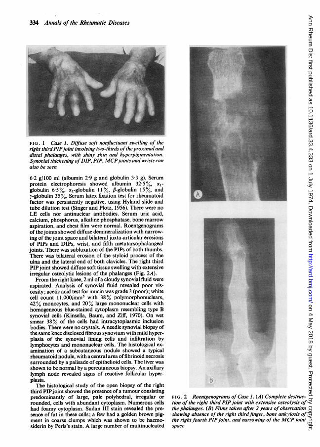
Physical examination revealed a chronically ill patientwho was unable to walk due to severe active polyarthritis.Subcutaneous nodules were palpated on the occipitalarea, helix of the right ear, dorsal aspect of both secondMCP joints, knees, and lateral malleoli. Painless lymphnodes about 1 cm in diameter were palpable in bothaxillae. There was almost complete loss of vision, thinningof sclerae, episcleral nodules, and signs of anterior uveitis.There was mild hepatomegaly and splenomegaly. Jointexamination revealed tender thickening of the synoviumwith limitation ofrange ofmotion of wrists and PIPjoints.There was subluxation of the PIPs of both thumbs. Therewas a diffuse, soft, symmetrical, well-demarcated, verylarge, nonfluctuant swelling of the right third PIP jointextending beyond the joint to the proximal and distalphalanges. There was shiny skin, increase in local tem-perature, and hyperpigmentation (Fig. 1). The right kneewas swollen with a small effusion and painful limitation offull flexion. The motion of shoulders, hips, and cervicalspine was painful and restricted.
Laboratory studies included a haematocrit of 31%,white cell count of 11,700/mm3 with basophils 2%,eosinophils 12%, neutrophils 71%, lymphocytes 13%,and monocytes 2%. The Westergren erythrocyte sedi-mentation rate was 65 mm/hr and the total protein was
on 4 May 2018 by guest. P
rotected by copyright.http://ard.bm
j.com/
Ann R
heum D
is: first published as 10.1136/ard.33.4.333 on 1 July 1974. Dow
nloaded from
334 Annals of the Rheumatic Diseases
FIG. 1 Case 1. Diffuse soft nonfluctuant swelling of theright thirdPIPjoint involving two-thirds oftheproximalanddistal phalanges, with shiny skin and hyperpigmentation.Synovial thickening ofDIP, PIP, MCPjoints and wrists canalso be seen
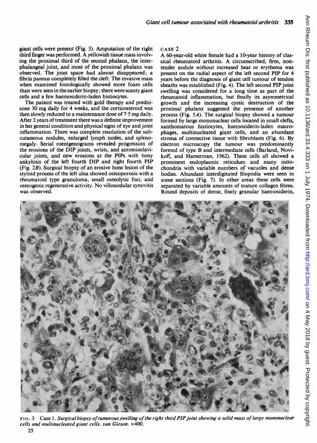
6-2 g/100 ml (albumin 2-9 g and globulin 3-3 g). Serumprotein electrophoresis showed albumin 32 5%, ax-globulin 65%, a2-globulin 11 %, fl-globulin 15%, andy-globulin 35 %. Serum latex fixation test for rheumatoidfactor was persistently negative, using Hyland slide andtube dilution test (Singer and Plotz, 1956). There were noLE cells nor antinuclear antibodies. Serum uric acid,calcium, phosphorus, alkaline phosphatase, bone marrowaspiration, and chest film were normal. Roentgenogramsof the joints showed diffuse demineralization with narrow-ing of the joint space and bilateral juxta-articular erosionsof PIPs and DIPs, wrist, and fifth metatarsophalangealjoints. There was subluxation of the PIPs of both thumbs.There was bilateral erosion of the styloid process of theulna and the lateral end of both clavicles. The right thirdPIP joint showed diffuse soft tissue swelling with extensiveirregular osteolytic lesions of the phalanges (Fig. 2A).From the right knee, 2 ml ofa cloudy synovial fluid were
aspirated. Analysis of synovial fluid revealed poor vis-cosity; acetic acid test for mucin was grade 3 (poor); whitecell count 11,000/mm3 with 38% polymorphonuclears,42% monocytes, and 20% large mononuclear cells withhomogeneous blue-stained cytoplasm resembling type Bsynovial cells (Kinsella, Baum, and Ziff, 1970). On wetsmear 38% of the cells had intracytoplasmic inclusionbodies. There were no crystals. A needle synovial biopsy ofthe same knee disclosed fibrous synovium with mild hyper-plasia of the synovial lining cells and infiltration bylymphocytes and mononuclear cells. The histological ex-amination of a subcutaneous nodule showed a typicalrheumatoid nodule, with a central area offibrinoid necrosissurrounded by a palisade of epithelioid cells. The liver wasshown to be normal by a percutaneous biopsy. An axillarylymph node revealed signs of reactive follicular hyper-plasia.The histological study of the open biopsy of the right
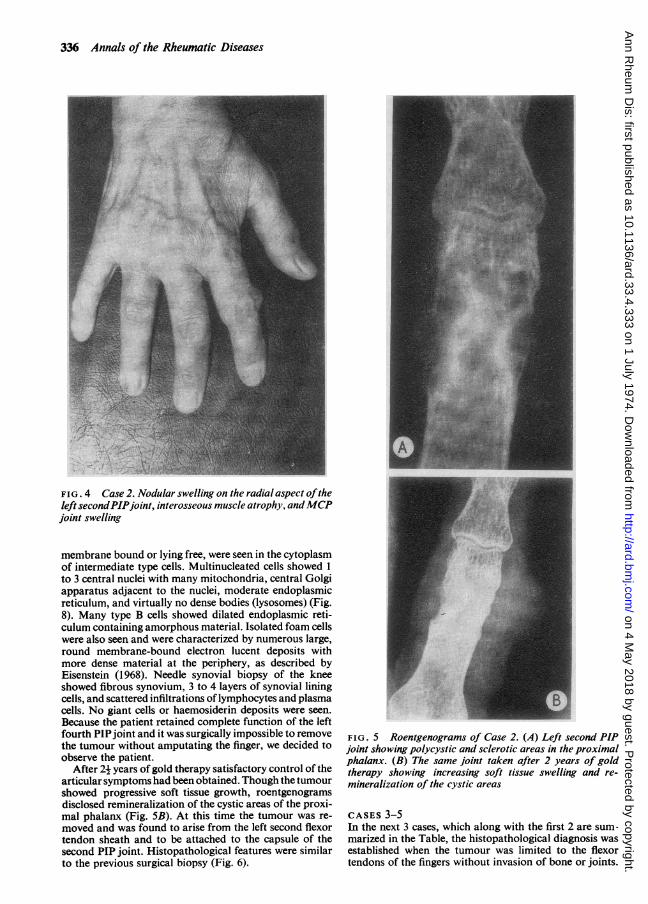
third PIPjoint showed the presence ofa tumour consistingpredominantly of large, pale polyhedral, irregular orrounded, cells with abundant cytoplasm. Numerous cellshad foamy cytoplasm. Sudan III stain revealed the pre-sence of fat in these cells; a few had a golden brown pig-ment in coarse clumps which was shown to be haemo-siderin by Perls's stain. A large number of multinucleated
r
FIG. 2 Roentgenograms of Case 1. (A) Complete destruc-tion of the right thirdPIP joint with extensive osteolysis ofthe phalanges. (B) Films taken after 2 years of observationshowing absence of the right thirdfinger, bone ankylosis ofthe right fourth PIP joint, and narrowing of the MCPjointspace
on 4 May 2018 by guest. P
rotected by copyright.http://ard.bm
j.com/
Ann R
heum D
is: first published as 10.1136/ard.33.4.333 on 1 July 1974. Dow
nloaded from
Giant cell tumour associated with rheumatoid arthritis 335
giant cells were present (Fig. 3). Amputation of the rightthird finger was performed. A yellowish tissue mass involv-ing the proximal third of the second phalanx, the inter-phalangeal joint, and most of the proximal phalanx wasobserved. The joint space had almost disappeared; afibrin pannus completely filled the cleft. The invasive masswhen examined histologically showed more foam cellsthan were seen in the earlier biopsy; there were scanty giantcells and a few haemosiderin-laden histiocytes.The patient was treated with gold therapy and predni-
sone 30 mg daily for 4 weeks, and the corticosteroid wasthen slowly reduced to a maintenance dose of 7-5 mg daily.After 2 years oftreatment there was a definite improvementin her general condition and physical signs of eye and jointinflammation. There was complete resolution of the sub-cutaneous nodules, enlarged lymph nodes, and spleno-megaly. Serial roentgenograms revealed progression ofthe erosions of the DIP joints, wrists, and acromioclavi-cular joints, and new erosions at the PIPs with bonyankylosis of the left fourth DIP and right fourth PIP(Fig. 2B). Surgical biopsy of an erosive bone lesion of thestyloid process of the left ulna showed osteoporosis with arheumatoid type granuloma, small osteolytic foci, andosteogenic regenerative activity. No villonodular synovitiswas observed.
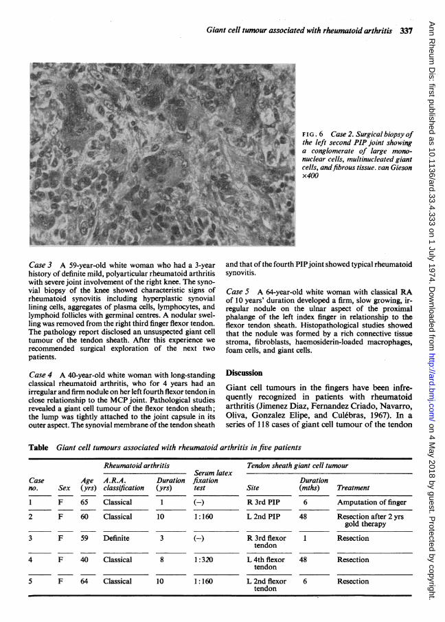
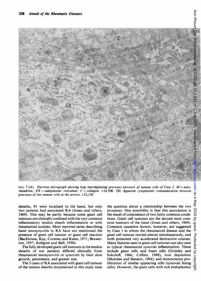
CASE 2A 60-year-old white female had a 10-year history of clas-sical rheumatoid arthritis. A circumscribed, firm, non-tender nodule without increased heat or erythema waspresent on the radial aspect of the left second PIP for 4years before the diagnosis of giant cell tumour of tendonsheaths was established (Fig. 4). The left second PIP jointswelling was considered for a long time as part of therheumatoid inflammation, but finally its asymmetricalgrowth and the increasing cystic destruction of theproximal phalanx suggested the presence of anotherprocess (Fig. 5A). The surgical biopsy showed a tumourformed by large mononuclear cells located in small clefts,xanthomatous histiocytes, haemosiderin-laden macro-phages, multinucleated giant cells, and an abundantstroma of connective tissue with fibroblasts (Fig. 6). Byelectron microscopy the tumour was predominantlyformed of type B and intermediate cells (Barland, Novi-koff, and Hamerman, 1962). These cells all showed aprominent endoplasmic reticulum and many mito-chondria with variable numbers of vacuoles and densebodies. Abundant interdigitated filopodia were seen insome sections (Fig. 7). In other areas these cells wereseparated by variable amounts of mature collagen fibres.Round deposits of dense, finely granular haemosiderin,_E~~~ Af* ss
w
FIG. 3 Case I. Surgical biopsy oftumorous swelling ofthe right thirdPIPjoint showing a solid mass oflarge mononuclearcells and multinucleated giant cells. van Gieson. x400.
25
on 4 May 2018 by guest. P
rotected by copyright.http://ard.bm
j.com/
Ann R
heum D
is: first published as 10.1136/ard.33.4.333 on 1 July 1974. Dow
nloaded from
336 Annals of the Rheumatic Diseases
FIG. 4 Case 2. Nodular swelling on the radial aspect oftheleft secondPIPjoint, interosseous muscle atrophy, andMCPjoint swelling
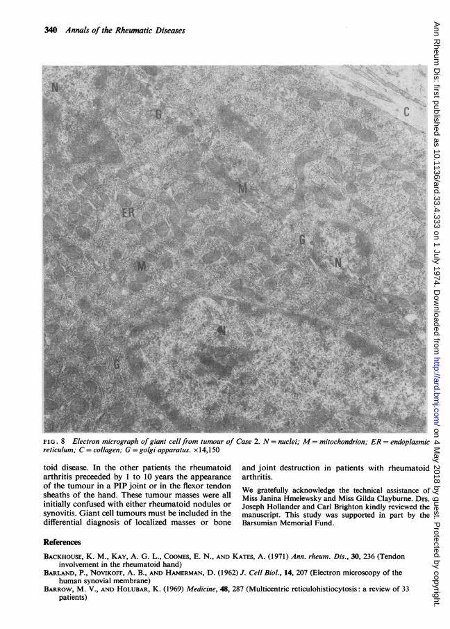
membrane bound or lying free, were seen in the cytoplasmof intermediate type cells. Multinucleated cells showed 1to 3 central nuclei with many mitochondria, central Golgiapparatus adjacent to the nuclei, moderate endoplasmicreticulum, and virtually no dense bodies (lysosomes) (Fig.8). Many type B cells showed dilated endoplasmic reti-culum containing amorphous material. Isolated foam cellswere also seen and were characterized by numerous large,round membrane-bound electron lucent deposits withmore dense material at the periphery, as described byEisenstein (1968). Needle synovial biopsy of the kneeshowed fibrous synovium, 3 to 4 layers of synovial liningcells, and scattered infiltrations of lymphocytes and plasmacells. No giant cells or haemosiderin deposits were seen.Because the patient retained complete function of the leftfourth PIPjoint and it was surgically impossible to removethe tumour without amputating the finger, we decided toobserve the patient.
After 2j years of gold therapy satisfactory control of thearticularsymptoms had been obtained. Though the tumourshowed progressive soft tissue growth, roentgenogramsdisclosed remineralization of the cystic areas of the proxi-mal phalanx (Fig. 5B). At this time the tumour was re-moved and was found to arise from the left second flexortendon sheath and to be attached to the capsule of thesecond PIP joint. Histopathological features were similarto the previous surgical biopsy (Fig. 6).
FIG. 5 Roentgenograms of Case 2. (A) Left second PIPjoint showing polycystic and sclerotic areas in the proximalphalanx. (B) The same joint taken after 2 years of goldtherapy showing increasing soft tissue swelling and re-mineralization of the cystic areas
CASES 3-5In the next 3 cases, which along with the first 2 are sum-marized in the Table, the histopathological diagnosis wasestablished when the tumour was limited to the flexortendons of the fingers without invasion of bone or joints.
on 4 May 2018 by guest. P
rotected by copyright.http://ard.bm
j.com/
Ann R
heum D
is: first published as 10.1136/ard.33.4.333 on 1 July 1974. Dow
nloaded from
Giant cell tumour associated with rheumatoid arthritis 337
FIG. 6 Case 2. Surgical biopsy ofthe left second PIP joint showinga conglomerate of large mono-nuclear cells, multinucleated giant
j cells, andfibrous tissue. van Gieson. G ~~~~X40
Case 3 A 59-year-old white woman who had a 3-yearhistory of definite mild, polyarticular rheumatoid arthritiswith severe joint involvement of the right knee. The syno-vial biopsy of the knee showed characteristic signs ofrheumatoid synovitis including hyperplastic synoviallining cells, aggregates of plasma cells, lymphocytes, andlymphoid follicles with germinal centres. A nodular swel-ling was removed from the right third finger flexor tendon.The pathology report disclosed an unsuspected giant celltumour of the tendon sheath. After this experience werecommended surgical exploration of the next twopatients.
Case 4 A 40-year-old white woman with long-standingclassical rheumatoid arthritis, who for 4 years had anirregular and firm nodule on her left fourth flexor tendon inclose relationship to the MCP joint. Pathological studiesrevealed a giant cell tumour of the flexor tendon sheath;the lump was tightly attached to the joint capsule in itsouter aspect. The synovial membrane of the tendon sheath
and that of the fourth PIPjoint showed typical rheumatoidsynovitis.
Case 5 A 64-year-old white woman with classical RAof 10 years' duration developed a firm, slow growing, ir-regular nodule on the ulnar aspect of the proximalphalange of the left index finger in relationship to theflexor tendon sheath. Histopathological studies showedthat the nodule was formed by a rich connective tissuestroma, fibroblasts, haemosiderin-loaded macrophages,foam cells, and giant cells.
Discussion
Giant cell tumours in the fingers have been infre-quently recognized in patients with rheumatoidarthritis (Jimenez Diaz, Fernandez Criado, Navarro,Oliva, Gonzalez Elipe, and Culebras, 1967). In aseries of 1 18 cases of giant cell tumour of the tendon
Table Giant cell tumours associated with rheumatoid arthritis in five patients
Rheumatoid arthritis
A.R.A.classification
Classical
Classical
Serum latexDuration fixation(yrs) test
1 (-)
10 1:160
Tendon sheath giant cell tumour
Site
R 3rd PIP
L 2nd PIP
Duration(mths)
6
48
Treatment
Amputation of finger
Resection after 2 yrsgold therapy
3 F 59 Definite 3 (-)
4 F 40 Classical 8 1:320
R 3rd flexor 1tendon
L 4th flexor 48tendon
L 2nd flexor 6tendon
5 F 64 Classical
Caseno.
1
2
Age(yrs)
65
60
Sex
F
F
Resection
Resection
ff ? :Sf
10 1:160 Resection
on 4 May 2018 by guest. P
rotected by copyright.http://ard.bm
j.com/
Ann R
heum D
is: first published as 10.1136/ard.33.4.333 on 1 July 1974. Dow
nloaded from
338 Annals of the Rheumatic Diseases
...........-....
W% piv
Wj.~~~~~~~f -,
~~~~ p~~~~Ai
Q,
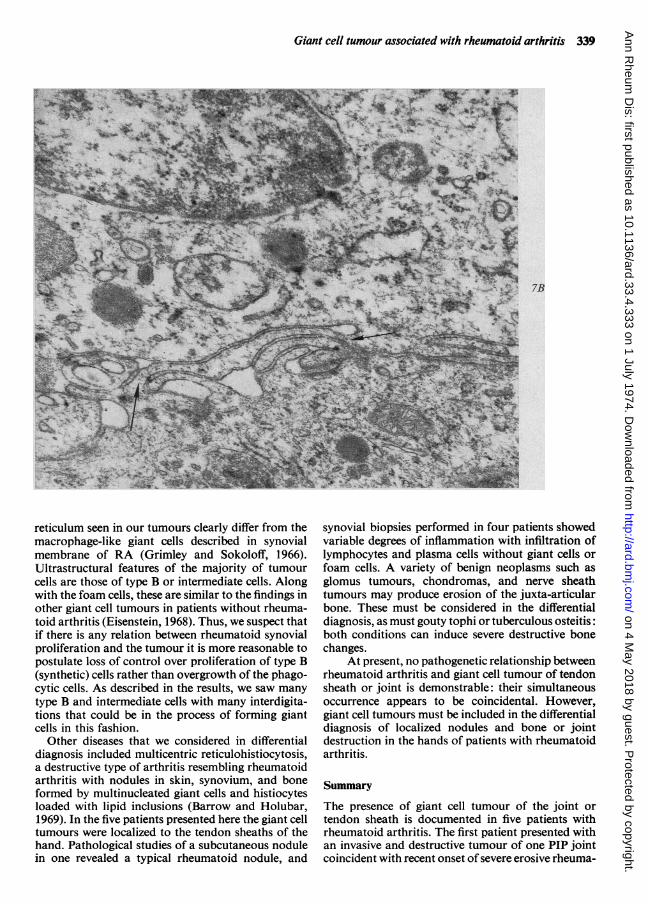
FIG7(A)Elctronmicroraph showin long interigitating prcesses (arrws) of tumor cells of ase 2 M mitchondrionER endoplasmic reticulum C~~~~~~~~~~~~1 .f.colae x24 500- (B) App~2arncyoasicm uiatnbew nprocessesoftwotumourcellsat the arrows x32 150~~ ~~~ ~~~ ~~~ ~~~~~~~~~~~~~~~~~~~~~~~~~~~~~~~~~~~~~~~~~~~~~~~~~~~~~~~~~~~~~~~~~~~~~~~~~~~~~~~~~~~~~~~~~~~~~~~~~~~~~~~~~~~~~~~~~~~~~~~~~~~~~~~~~~~~~~~~~~~~~~~~~~~~~~~~~~~~~~~~~~~~~~~~~~~~~~~~~~~~~~~~~~~~~~~~~~~~~~~~~~~~~~~~~~~~~
sheaths, 91 were localized to the hand, but onlytwo patients had associated RA (Jones and others,1969). This may be partly because some giant celltumours are clinically confused with the very commoninflammatory tendon sheath inflammation or withrheumatoid nodules. Most reported series describinghand tenosynovitis in RA have not mentioned thepresence of giant cell tumour or giant cell reaction(Backhouse, Kay, Coomes, and Kates, 1971 ; Brewer-ton, 1957; Kellgren and Ball, 1950).The fully developed giant cell tumours in the tendon
sheaths of our patients differed* clinically fromrheumatoid tenosynovitis or synovitis by their slowgrowth, persistence, and greater size.The 5 cases ofRA associated with giant cell tumour
of the tendon sheaths documented in this study raise
the question about a relationship between the twoprocesses. One possibility is that this association isthe result of coincidence of two fairly common condi-tions. Giant cell tumours are the second most com-mon tumours of the hand (Jones and others, 1969).Common causative factors, however, are suggestedby Case 1 in whom the rheumatoid disease and thegiant cell tumour started almost simultaneously, andboth presented very accelerated destructive courses.Many features seen in giant cell tumours are also seenin typical rheumatoid synovial inflammation. Theseinclude giant cells and foam cells (Grimley andSokoloff, 1966; Collins, 1949), iron deposition(Muirden and Senator, 1968), and monotonous pro-liferation of similar appearing cells (synovial liningcells). However, the giant cells with rich endoplasmic
on 4 May 2018 by guest. P
rotected by copyright.http://ard.bm
j.com/
Ann R
heum D
is: first published as 10.1136/ard.33.4.333 on 1 July 1974. Dow
nloaded from
Giant cell tumour associated with rheumatoid arthritis 339
A~~~~~~~~~1
reticulum seen in our tumours clearly differ from themacrophage-like giant cells described in synovialmembrane of RA (Grimley and Sokoloff, 1966).Ultrastructural features of the majority of tumourcells are those of type B or intermediate cells. Alongwith the foam cells, these are similar to the findings inother giant cell tumours in patients without rheuma-toid arthritis (Eisenstein, 1968). Thus, we suspect thatif there is any relation between rheumatoid synovialproliferation and the tumour it is more reasonable topostulate loss of control over proliferation of type B(synthetic) cells rather than overgrowth of the phago-cytic cells. As described in the results, we saw manytype B and intermediate cells with many interdigita-tions that could be in the process of forming giantcells in this fashion.
Other diseases that we considered in differentialdiagnosis included multicentric reticulohistiocytosis,a destructive type of arthritis resembling rheumatoidarthritis with nodules in skin, synovium, and boneformed by multinucleated giant cells and histiocytesloaded with lipid inclusions (Barrow and Holubar,1969). In the five patients presented here the giant celltumours were localized to the tendon sheaths of thehand. Pathological studies of a subcutaneous nodulein one revealed a typical rheumatoid nodule, and
7B
VA
lymphocytes and plasma cells without giant cells orfoam cells. A variety of benign neoplasms such asglomus tumours, chondromas, and nerve sheathtumours may produce erosion of the juxta-articularbone. These must be considered in the differentialdiagnosis, as must gouty tophi or tuberculous osteitis:both conditions can induce severe destructive bonechanges.
At present, no pathogenetic relationship betweenrheumatoid arthritis and giant cell tumour of tendonsheath or joint is demonstrable: their simultaneousoccurrence appears to be coincidental. However,giant cell tumours must be included in the differentialdiagnosis of localized nodules and bone or jointdestruction in the hands of patients with rheumatoidarthritis.
SummaryThe presence of giant cell tumour of the joint ortendon sheath is documented in five patients withrheumatoid arthritis. The first patient presented withan invasive and destructive tumour of one PIP jointcoincident with recent onset of severe erosive rheuma-
on 4 May 2018 by guest. P
rotected by copyright.http://ard.bm
j.com/
Ann R
heum D
is: first published as 10.1136/ard.33.4.333 on 1 July 1974. Dow
nloaded from
340 Annals of the Rheumatic Diseases
kXvs......... '::4
Swi.:_.....
,£gkf.j,.,': ji .:,. :i.7.
X1 stz 2|- -; ; tZC*
4i;,;. 1S'r' 0'i',;; is>,. | ., :}! ......... i P; a | ........... *~4 2 .,...
....: t ............... .te ,' Z .0! ..
a°*|-rer :fl- ->ySwsC;Py=-_j e*;- ,:- - >4<<mffiffiw5e_-rb>:s<;ffi</:-/w-*v;e ; ~~~~~~w".,:~~ t I. . ^we e,
FIG. 8 Electron micrograph ofgiant cellfrom tumour of Case 2. N=nuclei; M=mitochondrion; ER=endoplasmicreticulum; C = collagen; G = golgi apparatus. x 14,150
toid disease. In the other patients the rheumatoidarthritis preceeded by 1 to 10 years the appearanceof the tumour in a PIP joint or in the flexor tendonsheaths of the hand. These tumour masses were allinitially confused with either rheumatoid nodules orsynovitis. Giant cell tumours must be included in thedifferential diagnosis of localized masses or bone
and joint destruction in patients with rheumatoidarthritis.We gratefully acknowledge the technical assistance ofMiss Janina Hmelewsky and Miss Gilda Clayburne. Drs.Joseph Hollander and Carl Brighton kindly reviewed themanuscript. This study was supported in part by theBarsumian Memorial Fund.
References
BACKHOUSE, K. M., KAY, A. G. L., CooMEs, E. N., AND KATES, A. (1971) Ann. rheum. Dis., 30, 236 (Tendoninvolvement in the rheumatoid hand)
BARLAND, P., NOVIKOFF, A. B., AND HAMERMAN, D. (1962) J. Cell Biol., 14, 207 (Electron microscopy of thehuman synovial membrane)
BARROW, M. V., AND HOLUBAR, K. (1969) Medicine, 48, 287 (Multicentric reticulohistiocytosis: a review of 33patients)
on 4 May 2018 by guest. P
rotected by copyright.http://ard.bm
j.com/
Ann R
heum D
is: first published as 10.1136/ard.33.4.333 on 1 July 1974. Dow
nloaded from
Giant cell tumour associated with rheumatoid arthritis 341
BENNETT, G. A. (1950) Proc. Inst. Med. Chicago, 18, 26 (Reactive and neoplastic changes in synovial tissues)BREWERTON, D. A. (1957) Ann. rheum. Dis., 16, 183 (Hand deformities in rheumatoid arthritis)BYERS, P. D., COTTON, R. E., DEACON, 0. W., Lowy, M., NEWMAN, P. H., SISSONS, H. A., AND THOMPSON, A. D.
(1968) J. Bone Jt Surg., 50-B, 290 (The diagnosis and treatment of pigmented villonodular synovitis)COLLINS, D. H. (1949) In 'The Pathology of Articular and Spinal Diseases', 179. Arnold, LondonEISENSTEIN, R. (1968) J. Bone Jt Surg., 50A, 476 (Giant cell tumor of tendon sheath. Its histogenesis as studied in
the electron microscope)GALLOWAY, J. D. B., BRODERS, A. C., AND GHORMLEY, R. (1940) Arch. Surg., 40, 485 (Xanthoma of tendon
sheaths and synovial membranes: a clinical and pathologic study)GRIMLEY, P. M., AND SOKOLOFF, L. (1966) Amer. J. Path., 49, 931 (Synovial giant cells in rheumatoid arthritis)JAFFE, H. L., LICHTENSTEIN, L., AND SUTRO, C. J. (1941) Arch. Path., 31, 731 (Pigmented villonodular synovitis,
bursitis and tenosynovitis)JIMENEZ-DIAZ, C., FERNANDEZ-CRIADO, M., NAVARRO, V., OLIVA, H., GONZALEZ ELIPE, J., AND CULEBRAS, A.
(1967) Rev. Rhum., 34, 11 (Sur une forme polyarticulaire de synovite villonodulaire pigmentaire. Polyarticularform of pigmented villonodular synovitis)
JONES, F. E., SOULE, E. H., AND COVENTRY, M. B. (1969) J. Bone Jt. Surg., 51A, 76 (Fibrous xanthoma ofsynovium. Giant-cell tumor of tendon sheath, pigmented villonodular synovitis)
KARNOVSKY, M. J. (1965) J. Cell Biol., 27, 137A, Abstract 270 (A formaldehyde-glutaraldehyde fixative of highosmolality for use in electron microscopy)
KAY, A. G. L. (1971) Ann. rheum. Dis., 30, 98 (The natural history of synovial hypertrophy in the rheumatoid hand)KELLGREN, J. H., AND BALL, J. (1950) Ibid., 9, 48 (Tendon lesions in rheumatoid arthritis. A clinico-pathological
study)KINSELLA, T. D., BAUM, J., AND ZIFF, M. (1970) Arthr. and Rheum., 13, 734 (Studies of isolated synovial lining cells
of rheumatoid and nonrheumatoid synovial membranes)LARMON, W. A. (1965) Med. Clin. N. Amer., 49, 141 (Pigmented villonodular synovitis)MUIRDEN, K. D., AND SENATOR, G. B. (1968) Ann. rheum. Dis., 27, 38 (Iron in the synovial membrane in
rheumatoid arthritis and other joint disease)RODNAN, G. D. (1966) Tumors of synovial joints, bursae and tendon sheaths', in 'Arthritis and Allied Conditions'.
ed. J. L. Hollander, 7th ed., p. 1135. Lea and Febiger, PhiladelphiaROPES, M. W., BENNETT, G. A., COBB, S., JACOX, R., AND JESSAR, R. A. (1959) Ann. rheum. Dis., 18, 49
(Diagnostic criteria for rheumatoid arthritis; 1958 revision)SCHUMACHER, H. R. (1968) Arthr. and Rheum., 11, 426 (The synovitis of pseudogout: electron microscopic
observations), AND KULKA, J. P. (1972) New Engl. J. Med., 286, 416 (Needle biopsy of the synovial membrane. Experiencewith the Parker-Pearson technic)
SINGER, J. M., AND PLOTZ, C. M. (1956) Amer. J. Med., 21, 888 (The latex fixation test: application to theserologic diagnosis of rheumatoid arthritis)
WRIGHT, C. J. E. (1951) Brit. J. Surg., 38, 257 (Benign giant cell synovioma: an investigation of 85 cases)
on 4 May 2018 by guest. P
rotected by copyright.http://ard.bm
j.com/
Ann R
heum D
is: first published as 10.1136/ard.33.4.333 on 1 July 1974. Dow
nloaded from