Embed Size (px)
Citation preview
7/31/2019 Glomus Tumor of the Cheek, M 51
http://slidepdf.com/reader/full/glomus-tumor-of-the-cheek-m-51 1/5
Glomus tumor of the cheek, M 51
Deba P Sarma, MD, Omaha
M 51, asymptomatic red right lower cheek lesion, clinically “venous lake”.
Diagnosis:
Glomus tumor of the cheek
Comment:
Glomus tumor is a rare benign painful tumor commonly found in the corium and subcutaneous tissue, mostly in the subungual
region of the fingers.
It arises from the rests of glomus bodies, which are specialized structures at arteriovenous anastomosis functioning in thermal
regulation.
Extradigital glomus tumor is a rare entity, especially in the location, such as cheek. In a reported study of 56 extradigital glomus
tumors seen in Mayo Clinic over a period of twenty years (1985-2005), the authors found a single case occurring in the cheek 1.
Other reported sites for extradigital glomus tumors include face 2, colon 3, stomach, lung, bone, nervous system, and fallopian
tubes 1.
Extradigital glomus tumors can be a diagnostic challenge for the clinicians. The characteristic symptoms of digital glomus tumors,
e.g., pain, pinpoint tenderness with blunt palpation, and hypersensitivity to cold, may not be present.
Excisional biopsy is usually necessary for the diagnosis as well as the treatment
REF:
Wang B, Wang J, Shehan J, Sarma DP.(2008). Glomus tumor of the cheek. The Internet J of Dermatology 6 (2).
Indexed by Google Scholar
Glomus Tumor of the Cheek
Bo Wang M.D. Assistant Professor of Pathology, Creighton University Medical Center Omaha, Nebraska
USA
7/31/2019 Glomus Tumor of the Cheek, M 51
http://slidepdf.com/reader/full/glomus-tumor-of-the-cheek-m-51 2/5
Jeff Wang MD Resident in Pathology, Creighton University Medical Center Omaha, Nebraska USA
James Shehan MD Assistant Professor of Dermatology, Creighton University Medical Center Omaha,
Nebraska USA
Deba P. Sarma MD Professor of Pathology, Creighton University Medical Center Omaha, Nebraska USA
Citation: B. Wang, J. Wang, J. Shehan, D.P. Sarma: Glomus Tumor of the Cheek. The Internet Journal of
Dermatology . 2008 Volume 6 Number 2
Keywords: Glomus tumor, glomangioma, extradigital glomus tumor, glomus tumor of cheek
Abstract
We are reporting two cases of glomus tumor of the cheek that we had recently encounteredduring the last six months. English literature is briefly reviewed.
Case Reports
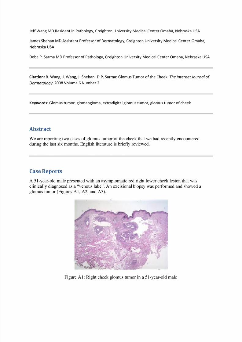
A 51-year-old male presented with an asymptomatic red right lower cheek lesion that wasclinically diagnosed as a “venous lake”. An excisional biopsy was performed and showed aglomus tumor (Figures A1, A2, and A3).
Figure A1: Right check glomus tumor in a 51-year-old male
7/31/2019 Glomus Tumor of the Cheek, M 51
http://slidepdf.com/reader/full/glomus-tumor-of-the-cheek-m-51 3/5
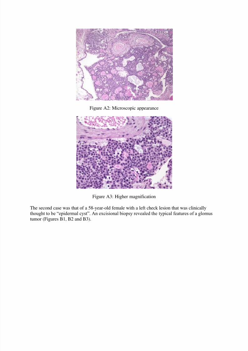
Figure A2: Microscopic appearance
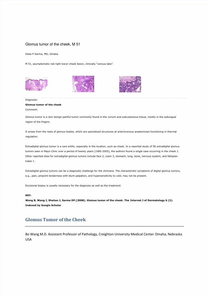
Figure A3: Higher magnification
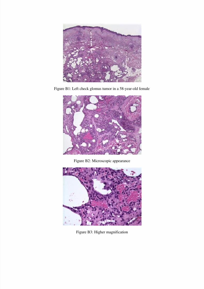
The second case was that of a 58-year-old female with a left check lesion that was clinically
thought to be “epidermal cyst”. An excisional biopsy revealed the typical features of a glomustumor (Figures B1, B2 and B3).
7/31/2019 Glomus Tumor of the Cheek, M 51
http://slidepdf.com/reader/full/glomus-tumor-of-the-cheek-m-51 4/5
Figure B1: Left check glomus tumor in a 58-year-old female
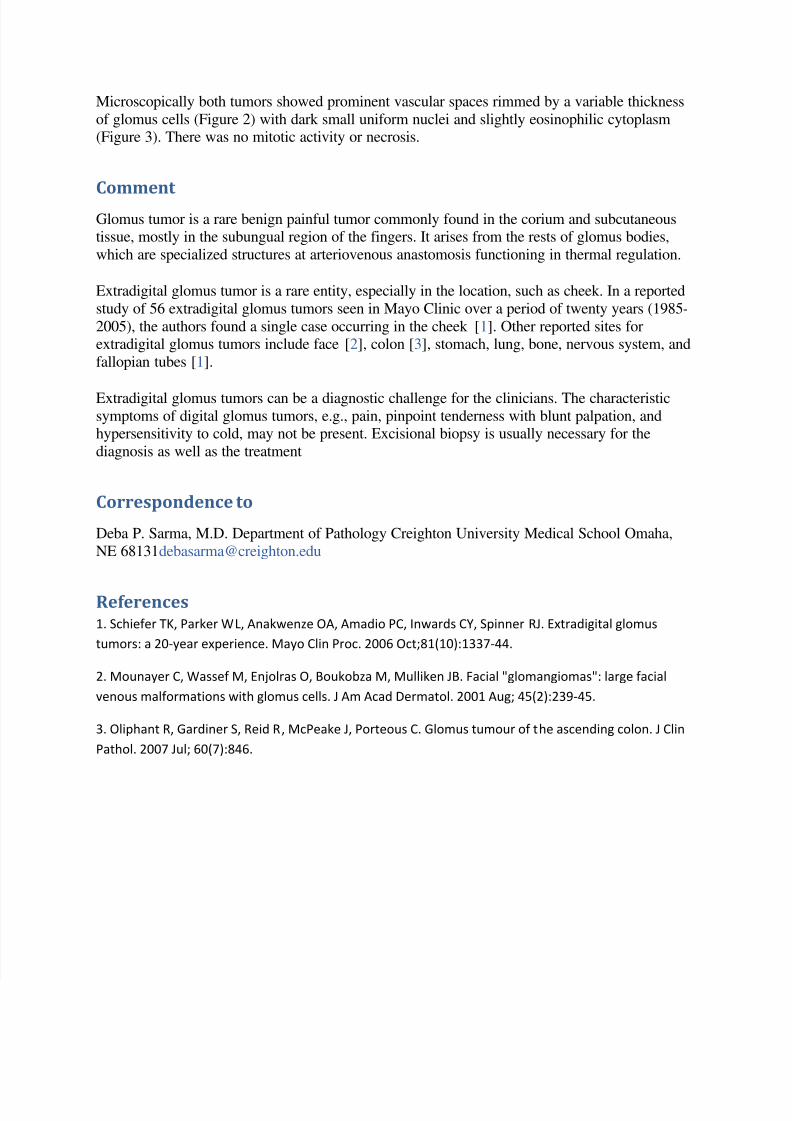
Figure B2: Microscopic appearance
Figure B3: Higher magnification
7/31/2019 Glomus Tumor of the Cheek, M 51
http://slidepdf.com/reader/full/glomus-tumor-of-the-cheek-m-51 5/5
Microscopically both tumors showed prominent vascular spaces rimmed by a variable thickness
of glomus cells (Figure 2) with dark small uniform nuclei and slightly eosinophilic cytoplasm(Figure 3). There was no mitotic activity or necrosis.
Comment Glomus tumor is a rare benign painful tumor commonly found in the corium and subcutaneoustissue, mostly in the subungual region of the fingers. It arises from the rests of glomus bodies,
which are specialized structures at arteriovenous anastomosis functioning in thermal regulation.
Extradigital glomus tumor is a rare entity, especially in the location, such as cheek. In a reported
study of 56 extradigital glomus tumors seen in Mayo Clinic over a period of twenty years (1985-
2005), the authors found a single case occurring in the cheek [1]. Other reported sites forextradigital glomus tumors include face [2], colon [3], stomach, lung, bone, nervous system, and
fallopian tubes [1].
Extradigital glomus tumors can be a diagnostic challenge for the clinicians. The characteristicsymptoms of digital glomus tumors, e.g., pain, pinpoint tenderness with blunt palpation, andhypersensitivity to cold, may not be present. Excisional biopsy is usually necessary for the
diagnosis as well as the treatment
Correspondence to
Deba P. Sarma, M.D. Department of Pathology Creighton University Medical School Omaha,
References1. Schiefer TK, Parker WL, Anakwenze OA, Amadio PC, Inwards CY, Spinner RJ. Extradigital glomus
tumors: a 20-year experience. Mayo Clin Proc. 2006 Oct;81(10):1337-44.
2. Mounayer C, Wassef M, Enjolras O, Boukobza M, Mulliken JB. Facial "glomangiomas": large facial
venous malformations with glomus cells. J Am Acad Dermatol. 2001 Aug; 45(2):239-45.
3. Oliphant R, Gardiner S, Reid R, McPeake J, Porteous C. Glomus tumour of the ascending colon. J Clin
Pathol. 2007 Jul; 60(7):846.