Embed Size (px)
Citation preview

Open Journal of Urology, 2016, 6, 80-85 Published Online May 2016 in SciRes. http://www.scirp.org/journal/oju http://dx.doi.org/10.4236/oju.2016.65015
How to cite this paper: Lazor, J.W., Guzzo, T.J., Bing, Z.Y., Lal, P., Ramchandani, P. and Torigian, D.A. (2016) Glomus Tumor of the Kidney: A Case Report with CT, MRI, and Histopathological Findings. Open Journal of Urology, 6, 80-85. http://dx.doi.org/10.4236/oju.2016.65015
Glomus Tumor of the Kidney: A Case Report with CT, MRI, and Histopathological Findings Jillian W. Lazor1, Thomas J. Guzzo2, Zhanyong Bing3, Priti Lal3, Parvati Ramchandani1, Drew A. Torigian1* 1Department of Radiology, Hospital of the University of Pennsylvania, Philadelphia, PA, USA 2Department of Surgery, Hospital of the University of Pennsylvania, Philadelphia, PA, USA 3Department of Pathology and Laboratory Medicine, Hospital of the University of Pennsylvania, Philadelphia, PA, USA
Received 1 April 2016; accepted 15 May 2016; published 18 May 2016
Copyright © 2016 by authors and Scientific Research Publishing Inc. This work is licensed under the Creative Commons Attribution International License (CC BY). http://creativecommons.org/licenses/by/4.0/
Abstract We describe the computed tomographic (CT) and magnetic resonance imaging (MRI) features of a very rare renal neoplasm, a glomus tumor. Our patient was a 68-year-old woman with a history of high grade T1 stage bladder cancer, status post intravesical Bacillus Calmette-Guérin (BCG) ther-apy and left ureteral stent placement, who presented for routine follow-up imaging evaluation of the urothelial tract. Computed tomographic urography (CTU) incidentally demonstrated a 1.7 cm well-circumscribed, non-calcified, non-fat containing lesion in the left renal cortex with arterial phase continuous peripheral rim enhancement and central hypoattenuation relative to enhanced renal parenchyma. Subsequent MRI showed the lesion to be isointense in signal intensity relative to the renal parenchyma on T1-weighted imaging and hyperintense on T2-weighted imaging. No macroscopic fat or microscopic lipid was seen within the lesion, and there were no foci of suscep-tibility artifact on T1-weighted images. Diffusion-weighted and apparent diffusion coefficient im-ages demonstrated no restricted diffusion. Contrast-enhanced images demonstrated continuous peripheral rim enhancement in the arterial phase and persistent rim enhancement with partial centripetal fill in of enhancement on venous phase images, similar to the pattern seen on CT. Par-tial left nephrectomy was performed for the suspected solid renal neoplasm. Histopathological assessment was diagnostic of a renal glomus tumor.
Keywords Glomus Tumor, Kidney, Renal, Computed Tomography (CT), Magnetic Resonance Imaging (MRI)
*Corresponding author.
J. W. Lazor et al.
81
1. Introduction Glomus tumors are perivascular myoid neoplasms that originate from the glomus body, a specialized dermal ar-teriovenous structure with a rich nerve supply that is involved in thermoregulation. These tumors are typically found in the subungual region, although they can rarely be found in the visceral organs. Herein, we describe a case of a renal glomus tumor and report the computed tomography (CT), magnetic resonance imaging (MRI), and histopathological characteristics of this rare neoplasm.
2. Case Report A 68-year-old woman with a history of high grade T1 stage bladder cancer complicated by obstruction of the distal left ureter presented for routine follow-up CT urography (CTU). She had previously completed 6 cycles of intravesical Bacillus Calmette-Guérin (BCG) therapy and placement of a left-sided ureteral stent.
CTU incidentally revealed a 1.7 cm lesion in the lateral interpolar left renal cortex. Non-contrast CT images demonstrated that the lesion had slight hypoattenuation (19 HU) relative to the surrounding renal parenchyma (mean attenuation 29 HU) (Figure 1(a)). No foci of macroscopic fat or calcification were seen in the lesion. Ar-terial phase contrast-enhanced CT images revealed continuous peripheral rim enhancement (110 HU) and cen-tral hypoattenuation (37 HU) of the well-circumscribed lesion (Figure 1(b)). Excretory phase contrast-enhanced CT images acquired 10 minutes after intravenous contrast administration showed homogeneous hypoattenuation (62 HU) of the lesion relative to the surrounding renal parenchyma (Figure 1(c)). No prior cross-sectional im-aging was available for comparison.
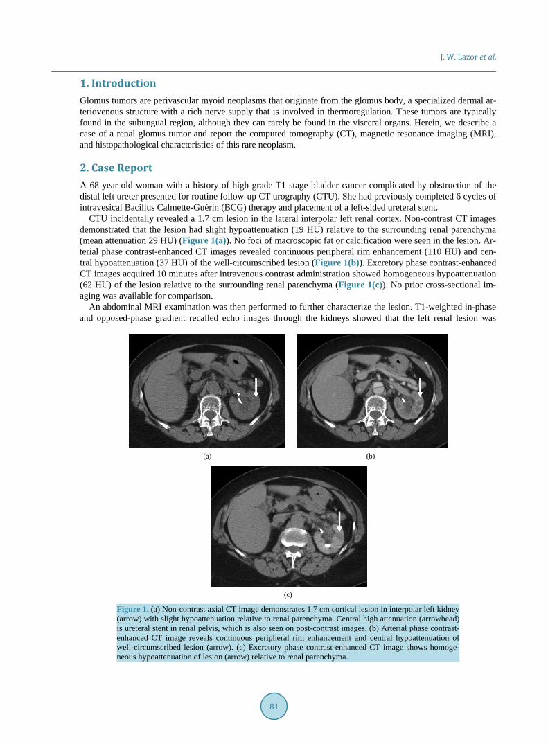
An abdominal MRI examination was then performed to further characterize the lesion. T1-weighted in-phase and opposed-phase gradient recalled echo images through the kidneys showed that the left renal lesion was
(a) (b)
(c)
Figure 1. (a) Non-contrast axial CT image demonstrates 1.7 cm cortical lesion in interpolar left kidney (arrow) with slight hypoattenuation relative to renal parenchyma. Central high attenuation (arrowhead) is ureteral stent in renal pelvis, which is also seen on post-contrast images. (b) Arterial phase contrast- enhanced CT image reveals continuous peripheral rim enhancement and central hypoattenuation of well-circumscribed lesion (arrow). (c) Excretory phase contrast-enhanced CT image shows homoge-neous hypoattenuation of lesion (arrow) relative to renal parenchyma.
J. W. Lazor et al.
82
isointense to the renal parenchyma and did not have macroscopic or microscopic lipid or foci of susceptibility artifact (Figure 2(a) and Figure 2(b)). Fat-suppressed T2-weighted fast spin echo images demonstrated homo-genous high signal intensity of the lesion relative to renal parenchyma, with a thin peripheral low signal intensi-ty rim (Figure 2(c)). Low b-value and high b-value diffusion-weighted gradient recalled echo images revealed loss of high signal intensity of the lesion with increasing b-value (Figure 2(d) and Figure 2(e)), and the corres-ponding apparent diffusion coefficient parametric map images demonstrated high signal intensity of the lesion, indicating a lack of restriction of water molecule diffusion (Figure 2(f)). Fat-suppressed T1-weighted gradient recalled echo images obtained after intravenous administration of gadolinium-based contrast material revealed arterial phase continuous peripheral rim enhancement of the lesion, followed by venous phase persistent rim en-hancement and partial centripetal fill in of enhancement of the lesion (Figure 2(g) and Figure 2(h)). No renal sinus involvement, left renal vein thrombosis, or lymphadenopathy was seen in the abdomen. Based on the CT and MRI findings, the major differential diagnostic considerations for this lesion included renal cell carcinoma, oncocytoma, and mesenchymal lesions such as juxtaglomerular cell tumor and hemangioma.
Subsequently, the patient underwent an uneventful open partial left nephrectomy. On gross pathological
(a) (b) (c)
(d) (e) (f)
(g) (h)
Figure 2. (a) and (b) Axial T1-weighted in-phase and opposed-phase gradient recalled echo images show that left renal lesion (arrows) is isointense to renal parenchyma and does not contain microscopic lipid or foci of susceptibility arti-fact; (c) Fat-suppressed T2-weighted fast spin echo image demonstrates homogeneous high signal intensity of lesion (arrow) relative to renal parenchyma along with thin peripheral low signal intensity rim; (d) and (e) Low b-value and high b-value diffusion-weighted gradient recalled echo images reveal loss of high signal intensity of lesion (arrows) with increasing b-value; (f) Corresponding apparent diffusion coefficient parametric map image demonstrates high signal intensity of lesion (arrow), indicating lack of restriction of water molecule diffusion; (g) and (h) Fat-suppressed T1-weighted gradient recalled echo images obtained after intravenous administration of gadolinium-based contrast ma-terial reveal arterial phase continuous peripheral rim enhancement of lesion (arrow), followed by venous phase persis-tent rim enhancement of lesion (arrow) with partial centripetal fill in.

J. W. Lazor et al.
83
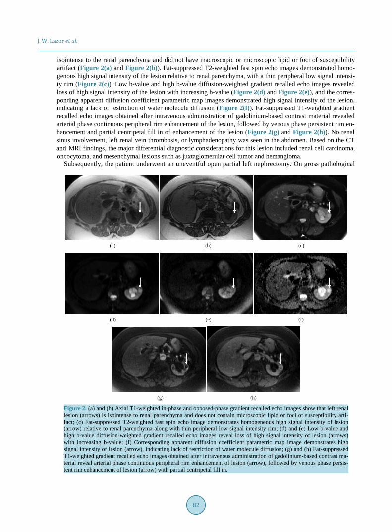
examination, a 1.0 cm tan-white well-circumscribed soft tissue mass was identified in the left kidney. Histopa-thologically, the lesion was composed of sheets of small round to oval cells with moderate amount of eosino-philic to amphophilic cytoplasm. The cells were fairly uniform with no evidence of pleomorphism or increased mitotic activity. The nuclei revealed a finely speckled chromatin and smooth nuclear contours. The background stroma was composed of delicate fibrovascular stroma (Figure 3(a) and Figure 3(b)). A differential diagnosis of myoid predominant angiomyolipoma, leiomyoma, and the remote possibility of gastrointestinal stromal tumor, lymphangioma, or hemangioma was considered. Accordingly, a panel of immunohistochemical staining was performed. The tumor cells were strongly and diffusely positive for smooth muscle actin, and CD31 highlighted the capillary network. The tumor was negative for all other markers including HMB-45, CD10, RCC, EMA, CD117, synaptophysin, chromogranin, and D2-40. All together, these findings were in keeping with a diagnosis of a renal glomus tumor.
At follow-up clinical evaluation 7 months later, the patient was noted to be doing well. Follow-up cross- sectional imaging did not demonstrate the presence of tumor recurrence or of a new mass in either kidney.
3. Discussion The glomus tumor, first described in 1924 by Masson [1], is a perivascular myoid neoplasm that originates from the modified smooth muscle cells found in the walls of the glomus body, a specialized arteriovenous structure with a rich nerve supply that is involved in thermoregulation [2]. Glomus bodies are found within the stratum reticularis of the dermis, predominantly in the subungual region, the lateral aspects of the digits, and the palm. Glomus bodies are also found in the precoccygeal soft tissue [3].
Glomus tumors account for less than 2% of soft tissue tumors. The tumors occur in both sexes with equal frequency, primarily within the age range of 20 to 40 years. Ninety percent occur as solitary neoplasms, and fa-milial cases have been described [4]. Most glomus tumors present as paroxysmally painful, subcutaneous blue-red nodules of the distal extremities, usually in the subungual region [5] [6]. About one quarter of glomus tumors are found in visceral organs that typically do not express glomus bodies [4] and have been reported to occur in the gastrointestinal tract [7]-[9], bone [10], lung [11], liver [12], pancreas [13], oral cavity [14], and si-nonasal region [15]. Glomus tumors in the genitourinary tract are uncommon and primarily involve the clitoris, vagina, cervix, and periurethral soft tissue [16]-[18]. To date, there have been 11 previously reported cases of renal glomus tumors; our case is the 12th such case in the literature [2] [3] [5] [19]-[24].
Histopathologically, glomus tumors are well circumscribed and composed of varying proportions of glomus cells, blood vessels, and smooth muscle. Based on the relative proportions of these cell types, glomus tumors are divided into three subgroups: glomus tumor proper, glomangioma, and glomangiomyoma [2].
Glomus tumors are generally considered to be benign and are treated by local excision [6]. However, rare ma-lignant glomus tumors have been reported [6] [25] [26]. These more aggressive tumors arise both de novo and
(a) (b)
Figure 3. (a) H&E stain of surgically resected renal specimen at 50× magnification demonstrates central hypocellular (*) and peripheral cellular (**) portions of lesion (**) surrounding capillary- sized vessels, along with surrounding normal renal parenchyma (K); (b) H&E stain of peripheral aspect of lesion at 400× shows extensive capillary-sized vessels (V) surrounded by uniform small glomus cells (arrows) with eosinophilic cytoplasm and rounded nuclei, without findings of nuc-lear atypia or increased mitoses.
J. W. Lazor et al.
84
from preexisting benign glomus tumors [25]. Folpe et al. retrospectively analyzed 52 cases of atypical glomus tumors to establish histological criteria for malignancy. The authors proposed that deep location, size greater than 2 cm, presence of moderate to high grade nuclear atypia, or presence of 5 or more mitoses per 50 high- powered fields should be considered as criteria for malignancy [26]. However, each of the neoplasms reviewed arose in the extremity [26]; the applicability of the criteria to glomus neoplasms of the visceral organs is there-fore uncertain. Given the rarity of visceral glomus tumors and limited follow-up, the biological behavior of glomus tumors of the internal organs has not been fully characterized. Nine of the 11 previously described renal glomus tumors were considered to be benign, without recurrent or metastatic disease seen after short-term fol-low-up [2] [19]-[24]. One of the 11 cases was considered to be an infiltrative tumor of uncertain malignant po-tential [3]. Another one of the 11 was classified as malignant with metastatic disease identified at time of pres-entation [5].
To our knowledge, the present case report is the first in the literature to describe the CT and MR imaging characteristics of a renal glomus tumor. Although the reported imaging findings are likely insufficient for the purpose of definitive prospective diagnosis (given their overlap with the features of other renal neoplasms), an interesting and potentially distinctive imaging feature of this lesion was the presence of centripetal fill in of en-hancement from arterial phase to venous phase contrast-enhanced images. Although this pattern of enhancement is characteristic of hepatic and splenic hemangiomas [27] [28], it has not been previously described in renal he-mangiomas [29]. Renal mixed epithelial and stromal tumor (MEST) and renal leiomyosarcoma may occasional-ly contain areas of delayed enhancement on CT imaging; however, these tumors enhance heterogeneously and do not exhibit centripetal fill-in of enhancement from arterial phase to venous phase contrast-enhanced images [29]-[31].
4. Conclusion In conclusion, renal glomus tumor is a rare perivascular myoid neoplasm that may mimic other renal neoplasms. We describe a case of renal glomus tumor and report the CT, MRI, and histopathological characteristics of this rare neoplasm.
References [1] Masson, P. (1924) Le glomus neuromyoarterial des regions tactiles et ses tumeurs. Lyon Chirurgical, 21, 257-280. [2] Herawi, M., Parwani, A.V., Edlow, D., Smolev, J.K. and Epstein, J.I. (2005) Glomus Tumor of Renal Pelvis: A Case
Report and Review of the Literature. Human Pathology, 36, 299-302. http://dx.doi.org/10.1016/j.humpath.2004.10.005 [3] Gill, J. and Van Vliet, C. (2010) Infiltrating Glomus Tumor of Uncertain Malignant Potential Arising in the Kidney.
Human Pathology, 41, 145-149. http://dx.doi.org/10.1016/j.humpath.2009.08.003 [4] Enzinger, F.M. and Weiss, S.W. (2001) Perivascular Tumors. In: Enzinger, F.M. and Weiss, S.W., Eds., Enzinger and
Weiss’ Soft Tissue Tumors, 4th Edition, St Louis, MO, Mosby, 985-1035. [5] Lamba, G., Rafiyath, S.M., Kaur, H., Khan, S., Singh, P., Hamilton, A.M. and Ang, D.C. (2011) Malignant Glomus
Tumor of Kidney: The First Reported Case and Review of Literature. Human Pathology, 42, 1200-1203. http://dx.doi.org/10.1016/j.humpath.2010.11.009
[6] Brathwaite, C.D. and Poppiti Jr., R.J. (1996) Malignant Glomus Tumor. A Case of Widespread Metastasis in a Patient with Multiple Glomus Body Hamartomas. American Journal of Surgical Pathology, 20, 233-238. http://dx.doi.org/10.1097/00000478-199602000-00012
[7] Haque, S., Modlin, I.M. and West, A.B. (1992) Multiple Glomus Tumors of the Stomach with Intravascular Spread. American Journal of Surgical Pathology, 16, 291-299. http://dx.doi.org/10.1097/00000478-199203000-00010
[8] Almagro, U.A., Schulte, W.J., Norback, D.H. and Turcotte, J.K. (1981) Glomus Tumor of the Stomach: Histologic and Ultrastructural Features. American Journal of Clinical Pathology, 75, 415-419. http://dx.doi.org/10.1093/ajcp/75.3.415
[9] Miettinen, M., Paal, E., Lasota, J. and Sobin, L.H. (2002) Gastrointestinal Glomus Tumors: A Clinicopathologic, Im-munohistochemical, and Molecular Genetic Study of 32 Cases. American Journal of Surgical Pathology, 26, 301-311. http://dx.doi.org/10.1097/00000478-200203000-00003
[10] Rozmaryn, L.M., Sadler, A.H. and Dorfman, H.D. (1987) Intraosseous Glomus Tumor in the Ulna. A Case Report. Clinical Orthopedics and Related Research, 220, 126-129.
[11] Koss, M.N., Hochholzer, L. and Moran, C.A. (1998) Primary Pulmonary Glomus Tumor: A Clinicopathologic and Immunohistochemical Study of Two Cases. Modern Pathology, 11, 253-258.
J. W. Lazor et al.
85
[12] Gassel, H.J., Klein, I., Timmermann, W., Kenn, W., Gassel, A.M. and Thiede, A. (2002) Presentation of an Unusual Benign Liver Tumor: Primary Hepatic Glomangioma. Scandinavian Journal of Gastroenterology, 37, 1237-40. http://dx.doi.org/10.1080/003655202760373489
[13] Miliauskas, J.R., Worthley, C. and Allen, P.W. (2002) Glomangiomyoma (Glomus Tumour) of the Pancreas: A Case Report. Pathology, 34, 193-195. http://dx.doi.org/10.1080/003130201201118034
[14] Tajima, Y., Weather, D.R., Neville, B.W., Benoit, P.W. and Pedley, D.M. (1981) Glomus Tumor (Golomangioma) of the Tongue. A Light and Electron Microscopic Study. Oral Surgery, Oral Medicine, and Oral Pathology, 52, 288-293. http://dx.doi.org/10.1016/0030-4220(81)90268-1
[15] Chu, P.G., Chang, K.L., Wu, A.Y. and Weiss, L.M. (1999) Nasal Glomus Tumors: Report of Two Cases with Empha-sis on Immunohistochemical Features and Differential Diagnosis. Human Pathology, 30, 1259-1261. http://dx.doi.org/10.1016/S0046-8177(99)90047-3
[16] Sonobe, H., Ro, J.Y., Ramos, M., Diaz, I., Mackay, B., Ordonez, N.G. and Ayala, A.G. (1994) Glomus Tumor of the Female External Genitalia: A Report of Two Cases. International Journal of Gynecological Pathology, 13, 359-364. http://dx.doi.org/10.1097/00004347-199410000-00010
[17] Albores-Saavedra, J. and Gilcrease, M. (1999) Glomus Tumor of the Uterine Cervix. International Journal of Gyneco-logical Pathology, 18, 69-72. http://dx.doi.org/10.1097/00004347-199901000-00010
[18] Blandamura, S., Florea, G., Brotto, M., Salmaso, R. and Castellan, L. (2000) Periurethral Glomangiomyoma in Women: Case Report and Review of the Literature. Histopathology, 36, 571-572. http://dx.doi.org/10.1046/j.1365-2559.2000.00918-5.x
[19] Schwartz, R. (1957) A Case of Renal Glomus Tumor. Zentralblatt fur Chirurgie, 82, 1516-1520. [20] Billard, F., Dumollard, J.M., Cucherousset, J., Boucheron, S. and Baril, A. (1991) Two Benign Vascular Tumors of the
Kidney Capsule. Annales de Pathologie, 11, 266-270. [21] Siddiqui, N., Rogalska, A. and Basil, I.S. (2005) Glomangiomyoma (Glomus tumor) of the Kidney. Archives of Pa-
thology and Laboratory Medicine, 129, 1172-1174. [22] Al-Ahmadie, H.A., Yilmaz, A., Olgac, S. and Reuter, V.E. (2007) Glomus Tumor of the Kidney: A Report of 3 Cases
Involving Renal Parenchyma and Review of the Literature. The American Journal of Surgical Pathology, 31, 585-591. http://dx.doi.org/10.1097/01.pas.0000213373.64053.41
[23] Nuwayhid, Z., Rodriguez, M.M., Prescott, A., Ciancio, G., Rojas, C.P., Casillas, J. and Sola, J.E. (2010) Renal Glomus Tumor in an Adolescent: A Conservative Approach. Journal of Pediatric Surgery, 45, e23-e26. http://dx.doi.org/10.1016/j.jpedsurg.2010.03.029
[24] Sasaki, K., Bastacky, S.I., Hrebinko, R.L., Parwani, A.V. and Zynger, D.L. (2011) Glomus Tumor of the Kidney: Case Report and Literature Review. International Journal of Surgical Pathology, 19, 393-397. http://dx.doi.org/10.1177/1066896908331233
[25] Gould, E.W., Manivel, J.C., Albores-Saavedra, J. and Monforte, H. (1990) Locally Infiltrative Glomus Tumors and Glomangiosarcomas. A Clinical, Ultrastructural, and Immunohistochemical Study. Cancer, 65, 310-318. http://dx.doi.org/10.1002/1097-0142(19900115)65:2<310::AID-CNCR2820650221>3.0.CO;2-Q
[26] Folpe, A.L., Fanburg-Smith, J.C., Miettinen, M. and Weiss, S.W. (2001) Atypical and Malignant Glomus Tumors. Analysis of 52 Cases, with a Proposal for the Reclassification of Glomus Tumors. The American Journal of Surgical Pathology, 25, 1-12. http://dx.doi.org/10.1097/00000478-200101000-00001
[27] Mitchell, D.G., Saini, S., Weinreb, J., De Lange, E.E., Runge, V.M., Kuhlman, J.E., Parisky, Y., Johnson, C.D., Brown, J.J., Schnall, M., Herfkens, R.J., Davis, P.L., Gorczyca, D., Sica, G., Foster, G.S. and Bernardino, M.E. (1994) Hepatic Metastases and Cavernous Hemangiomas: Distinction with Standard and Triple-Dose Gadoteridol-Enhanced MR Im-aging. Radiology, 193, 49-57. http://dx.doi.org/10.1148/radiology.193.1.8090921
[28] Ramani, M., Reinhold, C., Semelka, R.C., Siegelman, E.S., Liang, L., Ascher, S.M., Brown, J.J., Eisen, R.N. and Bret, P.M. (1997) Splenic Hemangiomas and Hamartomas: MR Imaging Characteristics of 28 Lesions. Radiology, 202, 166- 172. http://dx.doi.org/10.1148/radiology.202.1.8988207
[29] Lee, H.S., Koh, B.H., Kim, J.W., Kim, Y.S., Rhim, H.C., Cho, O.K., Hahm, C.K., Woo, Y.N. and Park, M.H. (2000) Radiologic Findings of Renal Hemangioma: Report of Three Cases. Korean Journal of Radiology, 1, 60-63. http://dx.doi.org/10.3348/kjr.2000.1.1.60
[30] Chu, L.C., Hruban, R.H., Horton, K.M., and Fishman, E.K. (2010) Mixed Epithelial and Stromal Tumor of the Kidney: Radiologic-Pathologic Correlation. Radiographics, 30, 1541-1551. http://dx.doi.org/10.1148/rg.306105503
[31] Ochiai, K., Onitsuka, H., Honda, H., Kawamoto, K., Uozui, J., Kumazawa, J. and Masuda, K. (1993) Leiomyosarcoma of the Kidney: CT and MR Appearance. Journal of Computer Assisted Tomography, 17, 656-658. http://dx.doi.org/10.1097/00004728-199307000-00027