Embed Size (px)
Citation preview
39
Graham-Little syndrome
Acta Dermatoven APA Vol 19, 2010, No 3
C a s e r e p o r t
Graham-Little syndromeB. Zegarska, D. Kallas, R. A. Schwartz, R. Czajkowski, G. Uchanska, and W. Placek
Graham-Little syndrome, also know as Graham-Little-Piccardi-Lassueur syndrome, is an unusual form of lichen planopilaris, characterized by the presence of cicatricial alopecia on the scalp, ke-ratosis pilaris of the trunk and extremities, and non-cicatricial hair loss of the pubis and axillae. We present the case of a 47-year-old woman whose condition was unusual in that there was a promi-nence of scalp findings. Her treatment included a topical steroid plus systemic prednisone begin-ning at 30 mg every morning, which rendered her skin smooth, but did not alter her scalp lopecia.
K E YW O R D S
lichen planus, lichen
planopilaris, alo-pecia,
keratosis pilaris, lichenoid
dermatosis, Graham-Little
syndrome, Graham-Little-
Piccardi-Lasseur syndrome
IntroductionGraham-Little syndrome (GLS) was described in
1914 by Piccardi (1) in a patient with progressive cica-tricial alopecia of the scalp, non-cicatricial alopecia of the axillae and groin, and follicular lichen planus (LP) on the trunk and extremities, to which he gave the name cheratosi spinulosa (keratotic spinulosa). In 1915, Ernst Graham-Little (2) (1867–1950) published a similar case study that had been referred by Las-seur of Lausanne. GLS is an unusual type of lichen planus called lichen planopilaris that affects the hair follicles (3–10). This rare lichenoid dermatosis is char-acterized by scarring alopecia, the loss of pubic and axillary hairs, and the progressive development of spinous or accuminate follicular papules on the trunk and extremities. GLS predominantly affects women, the duration of the illness varies from 6 months to
10 years. We present the case of a 47-year-old woman with GLS.
Case reportA 47-year-old woman was referred for evaluation of
scalp alopecia and a perifollicular eruption on her trunk and extremities. The illness had begun to present three years prior. Its onset was noted on the skin near the wrists. After about three weeks the papules had intensi-fied and spread to the trunk, including the lumbosacral area and mammary folds, as well as to the lower extrem-ities, especially the thighs. Two to three months after onset, the patient observed gradual hair loss, resulting later in the alopecia foci. Her skin changes were accom-panied by a marked and intensified cutaneous pruritus. Her past medical history was non-contributory. There was no family history of a similar eruption.
A B S T R A C T
40
Graham-Little syndrome
Acta Dermatoven APA Vol 19, 2010, No 3
C a s e r e p o r t
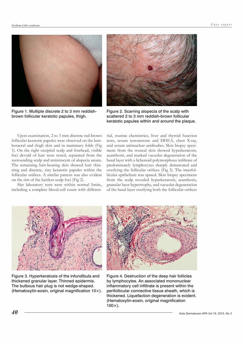
Upon examination, 2 to 3 mm discrete red-brown follicular keratotic papules were observed on the lum-bosacral and thigh skin and in mammary folds (Fig 1). On the right occipital scalp and forehead, visible foci devoid of hair were noted, separated from the surrounding scalp and reminiscent of alopecia areata. The remaining hair-bearing skin showed hair thin-ning and discrete, tiny keratotic papules within the follicular orifices. A similar pattern was also evident on the rim of the hairless scalp foci (Fig 2).
Her laboratory tests were within normal limits, including a complete blood-cell count with differen-
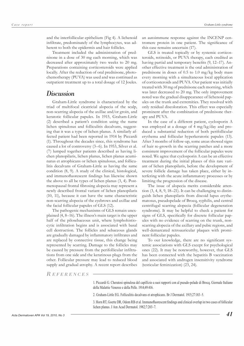
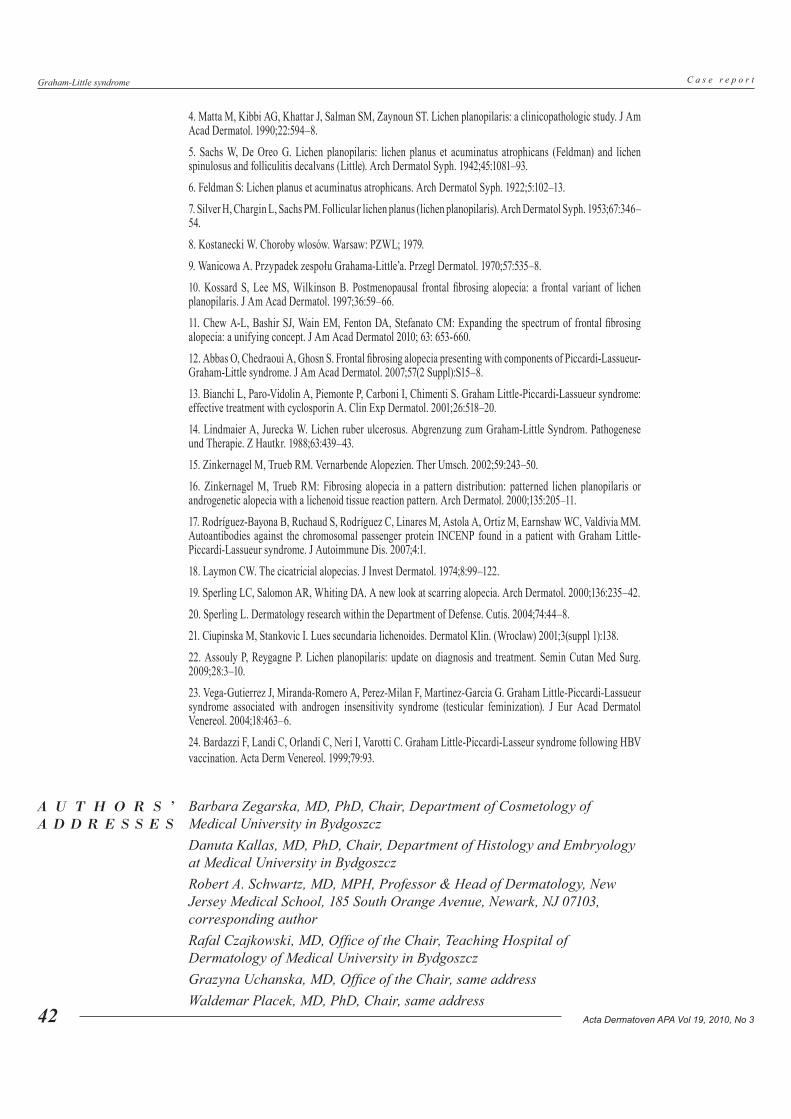
tial, routine chemistries, liver and thyroid function tests, serum testosterone and DHEA, chest X-ray, and serum antinuclear antibodies. Skin biopsy speci-mens from the truncal skin showed hyperkeratosis, acanthosis, and marked vacuolar degeneration of the basal layer with a lichenoid polymorphous infiltrate of predominately lymphocytes sharply demarcated and overlying the follicular orifices (Fig 3). The interfol-licular epithelium was spared. Skin biopsy specimens from the scalp revealed hyperkeratosis, acanthosis, granular layer hypertrophy, and vacuolar degeneration of the basal layer overlying both the follicular orifices
Figure 1. Multiple discrete 2 to 3 mm reddish-brown follicular keratotic papules, thigh.
Figure 2. Scarring alopecia of the scalp with scattered 2 to 3 mm reddish-brown follicular keratotic papules within and around the plaque.
Figure 3. Hyperkeratosis of the infundibula and thickened granular layer. Thinned epidermis. The bulbous hair plug is not wedge-shaped. (Hematoxylin-eosin, original magnification 10×).
Figure 4. Destruction of the deep hair follicles by lymphocytes. An associated mononuclear inflammatory cell infiltrate is present within the perifollicular connective tissue sheath, which is thickened. Liquefaction degeneration is evident. (Hematoxylin-eosin, original magnification 100×).
41
Graham-Little syndrome
Acta Dermatoven APA Vol 19, 2010, No 3
C a s e r e p o r t
and the interfollicular epithelium (Fig 4). A lichenoid infiltrate, predominately of the lymphocytes, was ad-herent to both the epidermis and hair follicles.
Treatment included the administration of pred-nisone in a dose of 30 mg each morning, which was decreased after approximately two weeks to 20 mg. Preparations containing corticosteroids were applied locally. After the reduction of oral prednisone, photo-chemotherapy (PUVA) was used and was continued as outpatient treatment up to a total dosage of 12 Joules.
DiscussionGraham-Little syndrome is characterized by the
triad of multifocal cicatricial alopecia of the scalp, non-scarring alopecia of the axillae and/or groin, and keratotic follicular papules. In 1915, Graham-Little (2) described a patient’s condition using the name lichen spinulosus and folliculitis decalvans, suggest-ing that it was a type of lichen planus. A similarly af-fected patient had been reported in 1914 by Piccardi (1). Throughout the decades since, this syndrome has caused a lot of controversy (3–6). In 1953, Silver et al. (7) lumped together patients described as having li-chen planopilaris, lichen planus, lichen planus acumi-natus et atrophicans or lichen spinulosus, and follicu-litis decalvans of Graham-Little as having the same condition (8, 9). A study of the clinical, histological, and immunofluorescent findings has likewise shown the above to all be types of lichen planus (3, 4). Post-menopausal frontal fibrosing alopecia may represent a newly described frontal variant of lichen planopilaris (10, 11), because it can have the same characteristic non-scarring alopecia of the eyebrows and axillae and the facial follicular papules of GLS (12).
The pathogenic mechanisms of GLS remain unex-plained (4, 8–16). The illness’s main target is the upper half of the pilosebaceous unit, where lymphohistio-cytic infiltration begins and is associated with basal cell destruction. The follicles and sebaceous glands are gradually damaged by inflammatory infiltrates and are replaced by connective tissue, this change being represented by scarring. Damage to the follicles may be caused by pressure from the perifollicular infiltra-tions from one side and the keratinous plugs from the other. Follicular pressure may lead to reduced blood supply and gradual atrophy. A recent report describes
an autoimmune response against the INCENP cen-tromere protein in one patient. The significance of this case remains uncertain (17).
GLS is treated topically or by systemic corticos-teroids, retinoids, or PUVA therapy, each credited as having partial and temporary benefits (9, 12–17). An-other effective treatment is the oral administration of prednisone in doses of 0.5 to 1.0 mg/kg body mass every morning with a simultaneous local application of corticosteroids and PUVA. Our patient was initially treated with 30 mg of prednisone each morning, which was later decreased to 20 mg. The only improvement noted was the gradual disappearance of lichenoid pap-ules on the trunk and extremities. They resolved with only residual discoloration. This effect was especially prominent after the combination of prednisone ther-apy and PUVA.
In the case of a different patient, cyclosporin A was employed at a dosage of 4 mg/kg/day and pro-duced a substantial reduction of both perifollicular erythema and follicular hyperkeratotic papules (13). After 3 months of follow-up, some areas showed signs of hair re-growth in the scarring patches and a more consistent improvement of the follicular papules were noted. We agree that cyclosporin A can be an effective treatment during the initial phases of this rare vari-ant of lichen planopilaris, before the development of severe follicle damage has taken place, either by in-terfering with the acute inflammatory processes or by limiting the progression of the disease.
The issue of alopecia merits considerable atten-tion (3, 4, 8, 9, 18–21). It can be challenging to distin-guish lichen planopilaris from discoid lupus erythe-matosus, pseudopelade of Brocq, syphilis, and central centrifugal scarring alopecia (follicular degeneration syndrome). It may be helpful to check a patient for signs of GLS, specifically for discrete follicular pap-ules with no evidence of scarring on the trunk, non-scarring alopecia of the axillary and pubic regions, and well-demarcated retroauricular plaques with promi-nent follicular papules.
To our knowledge, there are no significant sys-temic associations with GLS except for psychological ones (22). It may be noteworthy, however, that GLS has been connected with the hepatitis B vaccination and associated with androgen insensitivity syndrome (testicular feminization) (23, 24).
R E F E R E N C E S
1. Piccardi G. Cheratosi spinulosa del capillizio a suoi rapporti con al pseudo-pelade di Brocq. Giornale Italiano della Malattie Veneree e della Pelle. 1914;49:416.
2. Graham-Little EG. Folliculitis decalvans et atrophicans. Br J Dermatol. 1915;27:183–5.
3. Horn RT, Goette DR, Odom RB et al. Immunofluorescent findings and clinical overlap in two cases of follicular lichen planus. J Am Acad Dermatol. 1982;7:203–7.
42
Graham-Little syndrome
Acta Dermatoven APA Vol 19, 2010, No 3
C a s e r e p o r t
4. Matta M, Kibbi AG, Khattar J, Salman SM, Zaynoun ST. Lichen planopilaris: a clinicopathologic study. J Am Acad Dermatol. 1990;22:594–8.5. Sachs W, De Oreo G. Lichen planopilaris: lichen planus et acuminatus atrophicans (Feldman) and lichen spinulosus and folliculitis decalvans (Little). Arch Dermatol Syph. 1942;45:1081–93.6. Feldman S: Lichen planus et acuminatus atrophicans. Arch Dermatol Syph. 1922;5:102–13.7. Silver H, Chargin L, Sachs PM. Follicular lichen planus (lichen planopilaris). Arch Dermatol Syph. 1953;67:346–54.8. Kostanecki W. Choroby wlosów. Warsaw: PZWL; 1979.9. Wanicowa A. Przypadek zespołu Grahama-Little’a. Przegl Dermatol. 1970;57:535–8.10. Kossard S, Lee MS, Wilkinson B. Postmenopausal frontal fibrosing alopecia: a frontal variant of lichen planopilaris. J Am Acad Dermatol. 1997;36:59–66.11. Chew A-L, Bashir SJ, Wain EM, Fenton DA, Stefanato CM: Expanding the spectrum of frontal fibrosing alopecia: a unifying concept. J Am Acad Dermatol 2010; 63: 653-660.12. Abbas O, Chedraoui A, Ghosn S. Frontal fibrosing alopecia presenting with components of Piccardi-Lassueur-Graham-Little syndrome. J Am Acad Dermatol. 2007;57(2 Suppl):S15–8.13. Bianchi L, Paro-Vidolin A, Piemonte P, Carboni I, Chimenti S. Graham Little-Piccardi-Lassueur syndrome: effective treatment with cyclosporin A. Clin Exp Dermatol. 2001;26:518–20.14. Lindmaier A, Jurecka W. Lichen ruber ulcerosus. Abgrenzung zum Graham-Little Syndrom. Pathogenese und Therapie. Z Hautkr. 1988;63:439–43.15. Zinkernagel M, Trueb RM. Vernarbende Alopezien. Ther Umsch. 2002;59:243–50.16. Zinkernagel M, Trueb RM: Fibrosing alopecia in a pattern distribution: patterned lichen planopilaris or androgenetic alopecia with a lichenoid tissue reaction pattern. Arch Dermatol. 2000;135:205–11.17. Rodríguez-Bayona B, Ruchaud S, Rodríguez C, Linares M, Astola A, Ortiz M, Earnshaw WC, Valdivia MM. Autoantibodies against the chromosomal passenger protein INCENP found in a patient with Graham Little-Piccardi-Lassueur syndrome. J Autoimmune Dis. 2007;4:1.18. Laymon CW. The cicatricial alopecias. J Invest Dermatol. 1974;8:99–122.19. Sperling LC, Salomon AR, Whiting DA. A new look at scarring alopecia. Arch Dermatol. 2000;136:235–42.20. Sperling L. Dermatology research within the Department of Defense. Cutis. 2004;74:44–8.21. Ciupinska M, Stankovic I. Lues secundaria lichenoides. Dermatol Klin. (Wroclaw) 2001;3(suppl 1):138.22. Assouly P, Reygagne P. Lichen planopilaris: update on diagnosis and treatment. Semin Cutan Med Surg. 2009;28:3–10.23. Vega-Gutierrez J, Miranda-Romero A, Perez-Milan F, Martinez-Garcia G. Graham Little-Piccardi-Lassueur syndrome associated with androgen insensitivity syndrome (testicular feminization). J Eur Acad Dermatol Venereol. 2004;18:463–6.24. Bardazzi F, Landi C, Orlandi C, Neri I, Varotti C. Graham Little-Piccardi-Lasseur syndrome following HBV vaccination. Acta Derm Venereol. 1999;79:93.
Barbara Zegarska, MD, PhD, Chair, Department of Cosmetology of Medical University in BydgoszczDanuta Kallas, MD, PhD, Chair, Department of Histology and Embryology at Medical University in BydgoszczRobert A. Schwartz, MD, MPH, Professor & Head of Dermatology, New Jersey Medical School, 185 South Orange Avenue, Newark, NJ 07103, corresponding authorRafal Czajkowski, MD, Office of the Chair, Teaching Hospital of Dermatology of Medical University in BydgoszczGrazyna Uchanska, MD, Office of the Chair, same addressWaldemar Placek, MD, PhD, Chair, same address
A U T H O R S ’A D D R E S S E S