Embed Size (px)
DESCRIPTION
KGD
Citation preview
“PROBLEM 2B”Group 17
Thursday, Oct 10th,2013

Group 17 “Membership”TUTOR Dr. Haming
Silvia Isditya 405080088 – scriber
Ludolfus Bertolomeus Temu 405080175
Stephanie 405090231
Amran Cleo Petra Sinaga 405090267
Queencia Editha Morin 405090269
Hotris Anandita Vitalli 405100002 – secretary
Theresia Veronika 405100017
Yowendru 405100111
Karina Eda Clearesta 405100120 – leader
Vivi Anggelia 405100154
Theresia Jasmine 405100157
Billy Anthonio Khuana 405100279
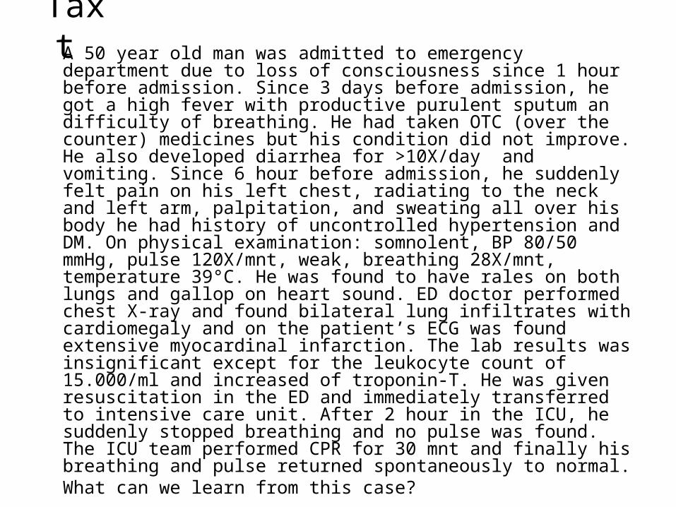
Taxt A 50 year old man was admitted to emergency department due to loss of consciousness since 1 hour before admission. Since 3 days before admission, he got a high fever with productive purulent sputum an difficulty of breathing. He had taken OTC (over the counter) medicines but his condition did not improve. He also developed diarrhea for >10X/day and vomiting. Since 6 hour before admission, he suddenly felt pain on his left chest, radiating to the neck and left arm, palpitation, and sweating all over his body he had history of uncontrolled hypertension and DM. On physical examination: somnolent, BP 80/50 mmHg, pulse 120X/mnt, weak, breathing 28X/mnt, temperature 39°C. He was found to have rales on both lungs and gallop on heart sound. ED doctor performed chest X-ray and found bilateral lung infiltrates with cardiomegaly and on the patient’s ECG was found extensive myocardinal infarction. The lab results was insignificant except for the leukocyte count of 15.000/ml and increased of troponin-T. He was given resuscitation in the ED and immediately transferred to intensive care unit. After 2 hour in the ICU, he suddenly stopped breathing and no pulse was found. The ICU team performed CPR for 30 mnt and finally his breathing and pulse returned spontaneously to normal.What can we learn from this case?
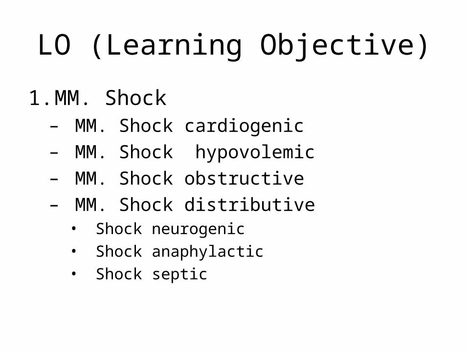
LO (Learning Objective)
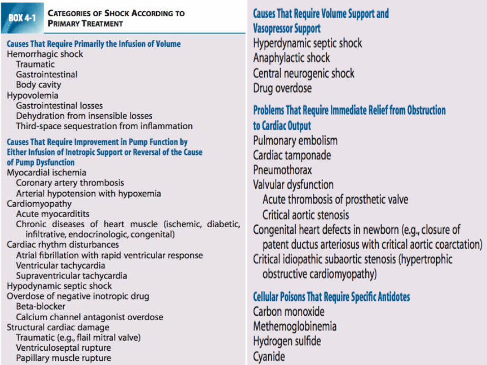
1. MM. Shock– MM. Shock cardiogenic– MM. Shock hypovolemic– MM. Shock obstructive– MM. Shock distributive
• Shock neurogenic• Shock anaphylactic• Shock septic
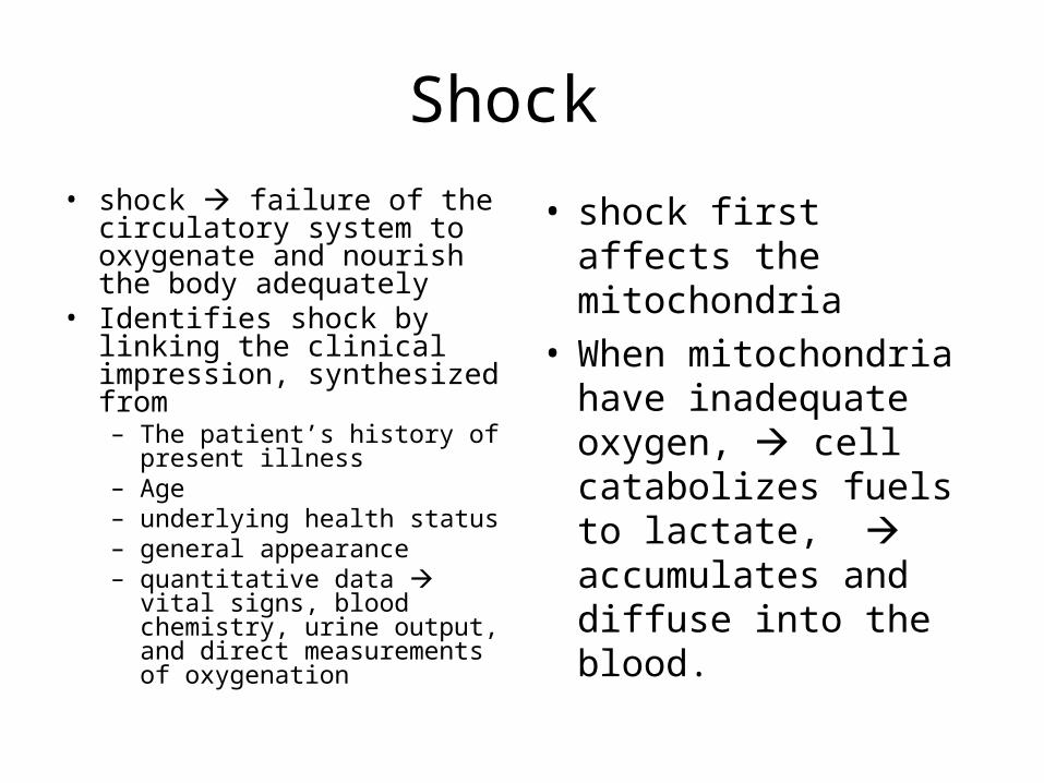
Shock • shock failure of the
circulatory system to oxygenate and nourish the body adequately
• Identifies shock by linking the clinical impression, synthesized from – The patient’s history of present
illness– Age– underlying health status– general appearance– quantitative data vital signs,
blood chemistry, urine output, and direct measurements of oxygenation
• shock first affects the mitochondria
• When mitochondria have inadequate oxygen, cell catabolizes fuels to lactate, accumulates and diffuse into the blood.
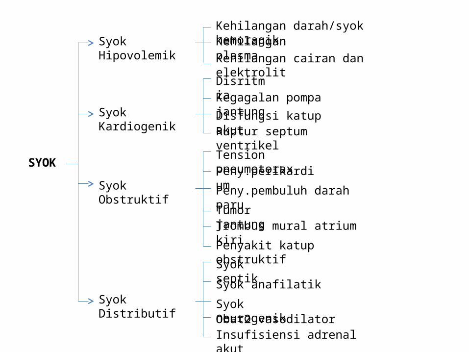
SYOK
Syok HipovolemikKehilangan darah/syok hemoragikKehilangan plasmaKehilangan cairan dan elektrolit
Syok Kardiogenik
Disritmia Kegagalan pompa jantung
Disfungsi katup akutRuptur septum ventrikel
Syok Obstruktif
Tension pneumotoraxPeny.perikardium
Peny.pembuluh darah paru
Tumor jantungTrombus mural atrium kiri
Penyakit katup obstruktif
Syok Distributif
Syok septik
Syok anafilatik
Syok neurogenikObat2 vasodilatorInsufisiensi adrenal akut
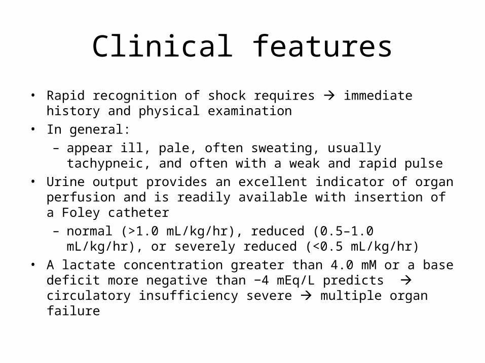
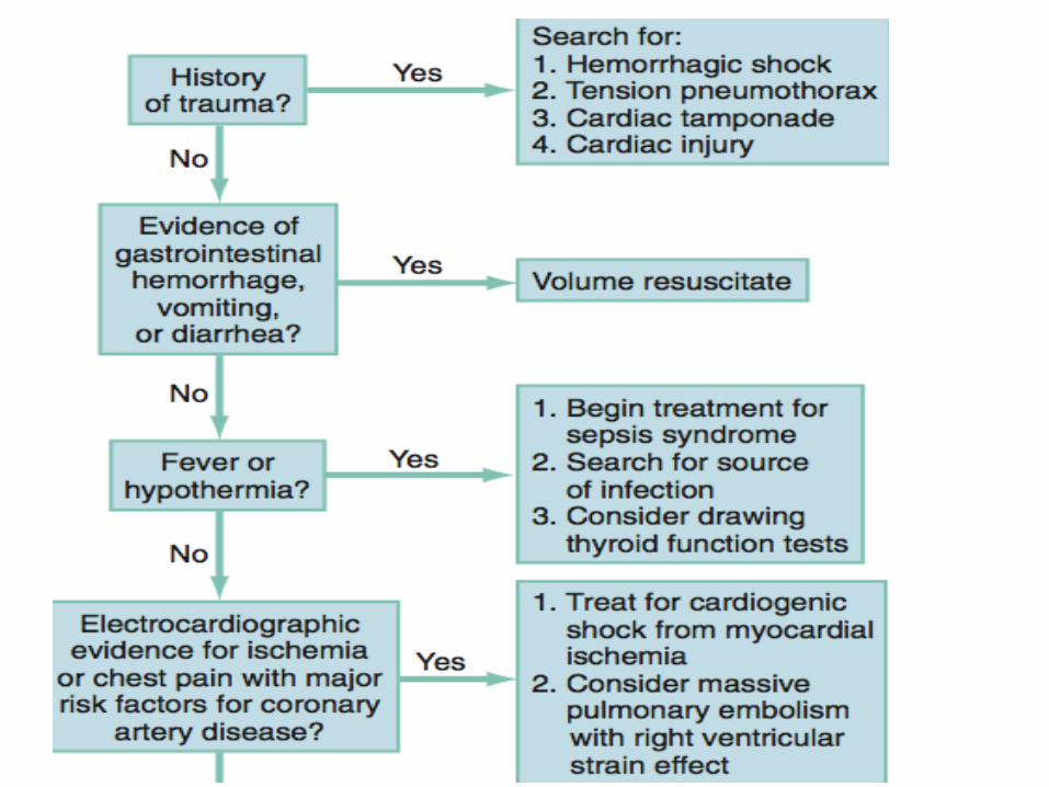
Clinical features• Rapid recognition of shock requires immediate history and physical
examination• In general:
– appear ill, pale, often sweating, usually tachypneic, and often with a weak and rapid pulse
• Urine output provides an excellent indicator of organ perfusion and is readily available with insertion of a Foley catheter– normal (>1.0 mL/kg/hr), reduced (0.5–1.0 mL/kg/hr), or severely
reduced (<0.5 mL/kg/hr)• A lactate concentration greater than 4.0 mM or a base deficit more
negative than −4 mEq/L predicts circulatory insufficiency severe multiple organ failure
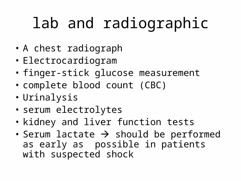
lab and radiographic
• A chest radiograph• Electrocardiogram• finger-stick glucose measurement• complete blood count (CBC)• Urinalysis• serum electrolytes• kidney and liver function tests • Serum lactate should be performed as early as
possible in patients with suspected shock
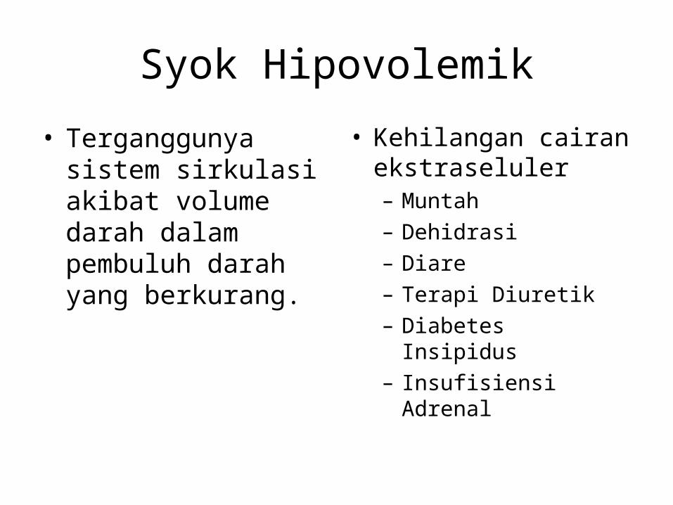
Syok Hipovolemik
• Terganggunya sistem sirkulasi akibat volume darah dalam pembuluh darah yang berkurang.
• Kehilangan cairan ekstraseluler– Muntah– Dehidrasi– Diare– Terapi Diuretik– Diabetes Insipidus– Insufisiensi Adrenal
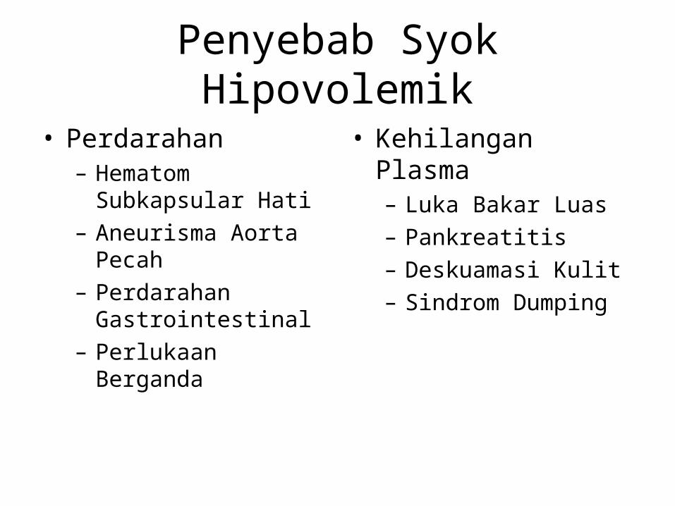
Penyebab Syok Hipovolemik
• Perdarahan– Hematom Subkapsular
Hati– Aneurisma Aorta Pecah– Perdarahan
Gastrointestinal– Perlukaan Berganda
• Kehilangan Plasma– Luka Bakar Luas– Pankreatitis– Deskuamasi Kulit– Sindrom Dumping
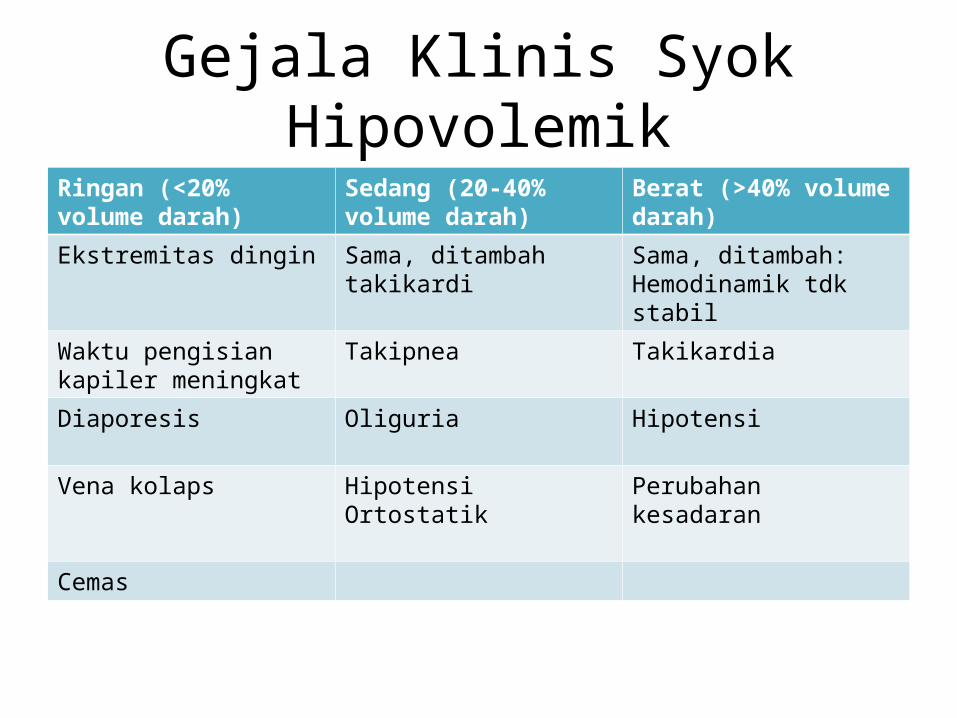
Gejala Klinis Syok HipovolemikRingan (<20% volume darah)
Sedang (20-40% volume darah)
Berat (>40% volume darah)
Ekstremitas dingin Sama, ditambah takikardi Sama, ditambah: Hemodinamik tdk stabil
Waktu pengisian kapiler meningkat
Takipnea Takikardia
Diaporesis Oliguria Hipotensi
Vena kolaps Hipotensi Ortostatik Perubahan kesadaran
Cemas
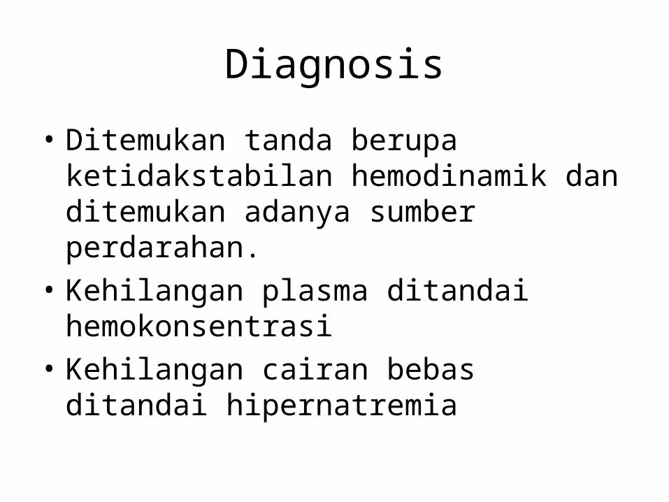
Diagnosis
• Ditemukan tanda berupa ketidakstabilan hemodinamik dan ditemukan adanya sumber perdarahan.
• Kehilangan plasma ditandai hemokonsentrasi• Kehilangan cairan bebas ditandai
hipernatremia
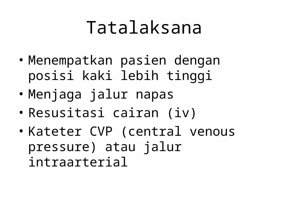
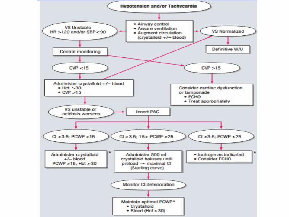
Tatalaksana
• Menempatkan pasien dengan posisi kaki lebih tinggi
• Menjaga jalur napas• Resusitasi cairan (iv)• Kateter CVP (central venous pressure) atau
jalur intraarterial

Syok Kardiogenik
• Definisi : Gangguan yang disebabkan oleh penurunan curah jantung sistemik pada keadaan volume intravaskular yang cukup, dan dapat mengakibatkan hipoksia jaringan.
• Penyebab terbanyak infark miokard akut (elevasi segmen ST)
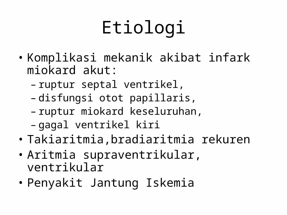
Etiologi
• Komplikasi mekanik akibat infark miokard akut: – ruptur septal ventrikel, – disfungsi otot papillaris, – ruptur miokard keseluruhan, – gagal ventrikel kiri
• Takiaritmia,bradiaritmia rekuren• Aritmia supraventrikular, ventrikular• Penyakit Jantung Iskemia
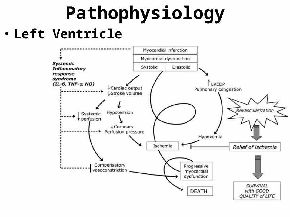
Pathophysiology• Left Ventricle
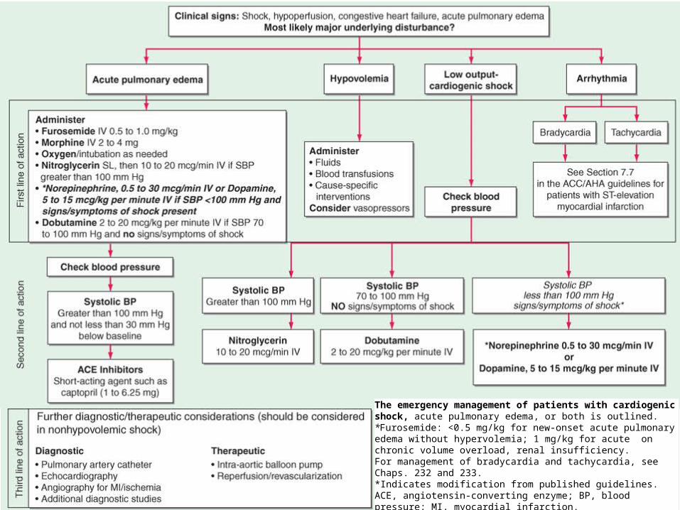
The emergency management of patients with cardiogenic shock, acute pulmonary edema, or both is outlined. *Furosemide: <0.5 mg/kg for new-onset acute pulmonary edema without hypervolemia; 1 mg/kg for acute on chronic volume overload, renal insufficiency.
For management of bradycardia and tachycardia, see Chaps. 232 and 233. *Indicates modification from published guidelines. ACE, angiotensin-converting enzyme; BP, blood pressure; MI, myocardial infarction.
Prediktor
• Klasifikasi Killip berdasarkan gambaran klinis :– Tanda gagal jantung kongestif– Suara s3 gallop, ronki– Edema paru– Syok kardiogenik
• Klasifikasi Forrester berdasarkan keadaan hemodinamik :– Angka PCWP (Pulmonary Capillary Wedge Pressure)
& CI (Cardiac Index)– Semakin tinggi nilain PCWP & semakin rendah CI
mortalitas meningkat

The emergency management of patients with cardiogenic shock, acute pulmonary edema, or both is outlined. *Furosemide: <0.5 mg/kg for new-onset acute pulmonary edema without hypervolemia; 1 mg/kg for acute on chronic volume overload, renal insufficiency.
For management of bradycardia and tachycardia, see Chaps. 232 and 233. *Indicates modification from published guidelines. ACE, angiotensin-converting enzyme; BP, blood pressure; MI, myocardial infarction.
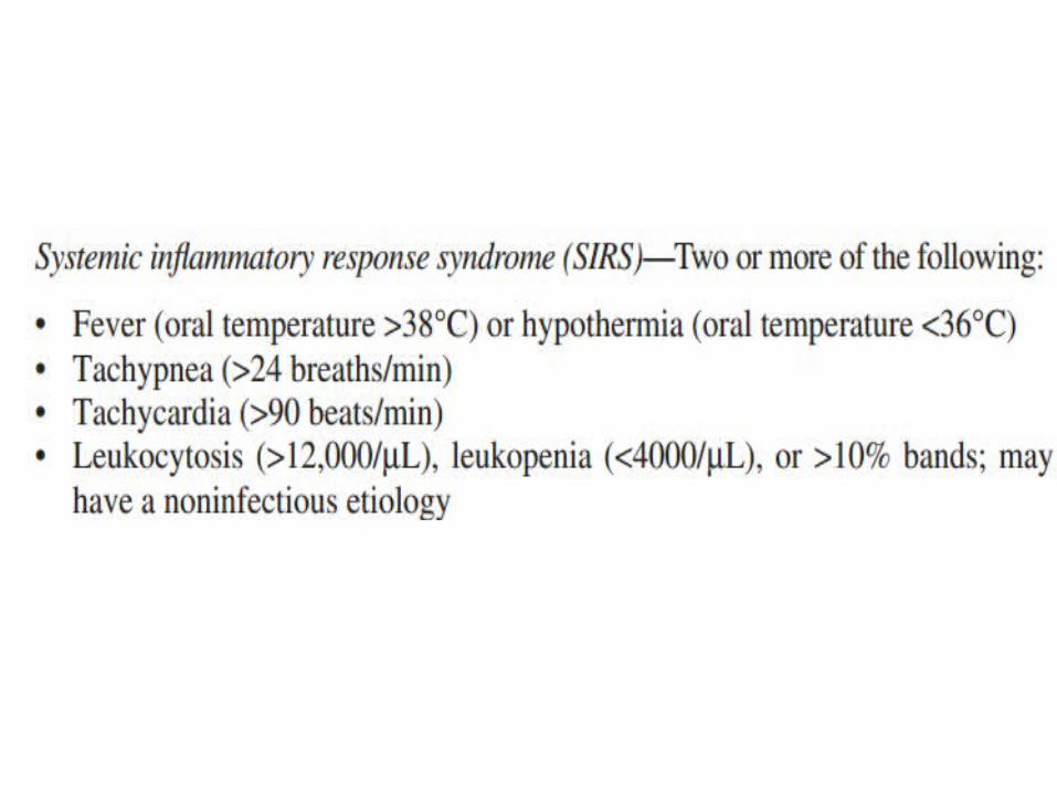
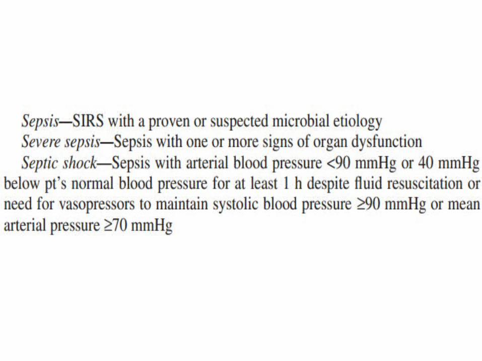
SShock septikhock septik
Definisi• Merupakan suatu kegagalan sirkulasi perifer dengan perfusi
jaringan yang tidak adekuat akibat septikemia (terdapatnya multiplikasi bakteri dalam darah).
• Syok septik mengenai :– Jantung– Sistem vaskular– Hampir semua organ
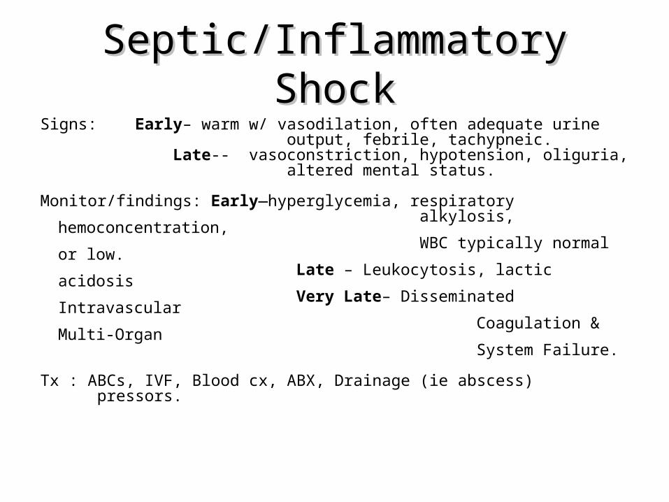
Septic/Inflammatory ShockSeptic/Inflammatory ShockSigns: Early– warm w/ vasodilation, often adequate urine output, febrile, tachypneic. Late-- vasoconstriction, hypotension, oliguria, altered mental status.
Monitor/findings: Early—hyperglycemia, respiratory alkylosis, hemoconcentration, WBC typically normal or low. Late – Leukocytosis, lactic acidosis Very Late– Disseminated Intravascular Coagulation & Multi-Organ System Failure.
Tx : ABCs, IVF, Blood cx, ABX, Drainage (ie abscess) pressors.
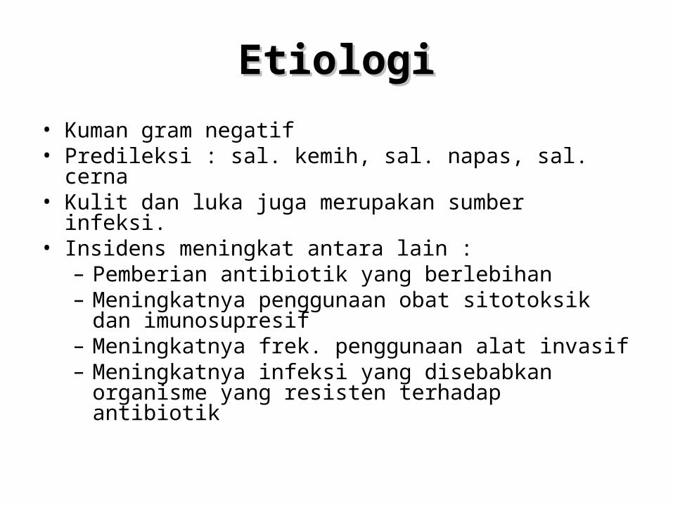
EtiologiEtiologi
• Kuman gram negatif• Predileksi : sal. kemih, sal. napas, sal. cerna• Kulit dan luka juga merupakan sumber infeksi.• Insidens meningkat antara lain :
– Pemberian antibiotik yang berlebihan– Meningkatnya penggunaan obat sitotoksik dan
imunosupresif– Meningkatnya frek. penggunaan alat invasif– Meningkatnya infeksi yang disebabkan organisme yang
resisten terhadap antibiotik
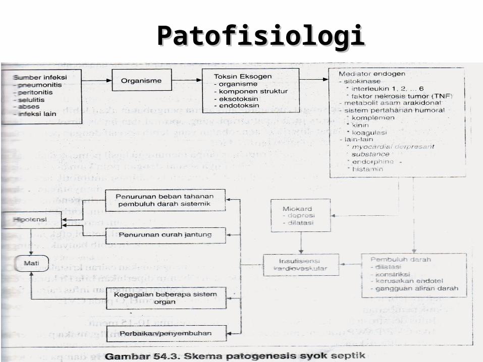
PatofisiologiPatofisiologi
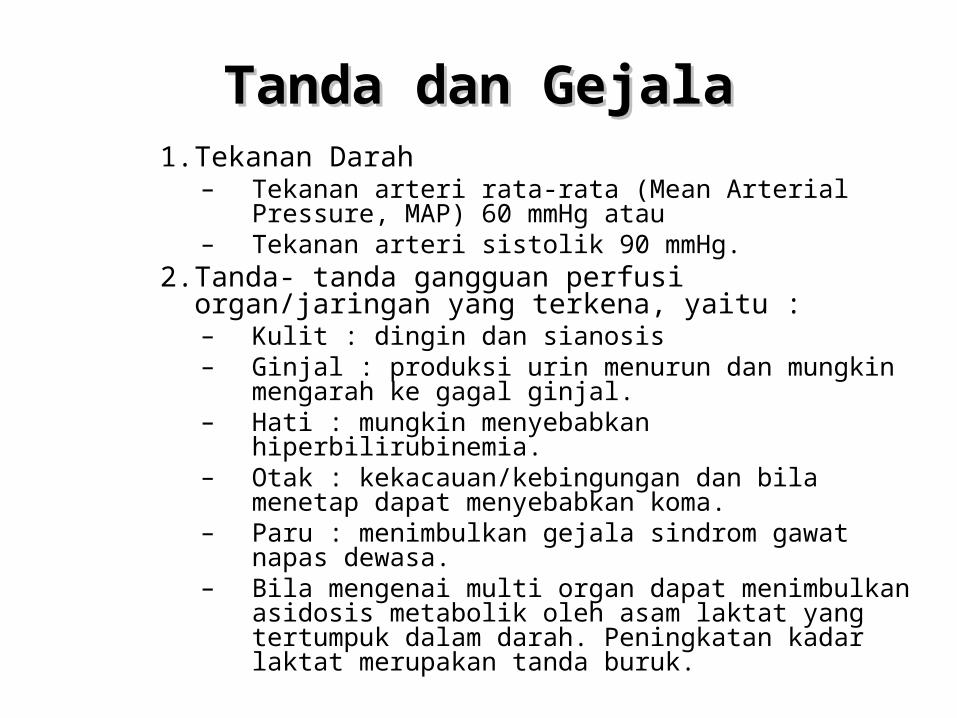
Tanda dan Tanda dan GejalaGejala1. Tekanan Darah
– Tekanan arteri rata-rata (Mean Arterial Pressure, MAP) 60 mmHg atau
– Tekanan arteri sistolik 90 mmHg.2. Tanda- tanda gangguan perfusi organ/jaringan yang terkena,
yaitu :– Kulit : dingin dan sianosis– Ginjal : produksi urin menurun dan mungkin mengarah ke
gagal ginjal.– Hati : mungkin menyebabkan hiperbilirubinemia.– Otak : kekacauan/kebingungan dan bila menetap dapat
menyebabkan koma.– Paru : menimbulkan gejala sindrom gawat napas dewasa.– Bila mengenai multi organ dapat menimbulkan asidosis
metabolik oleh asam laktat yang tertumpuk dalam darah. Peningkatan kadar laktat merupakan tanda buruk.
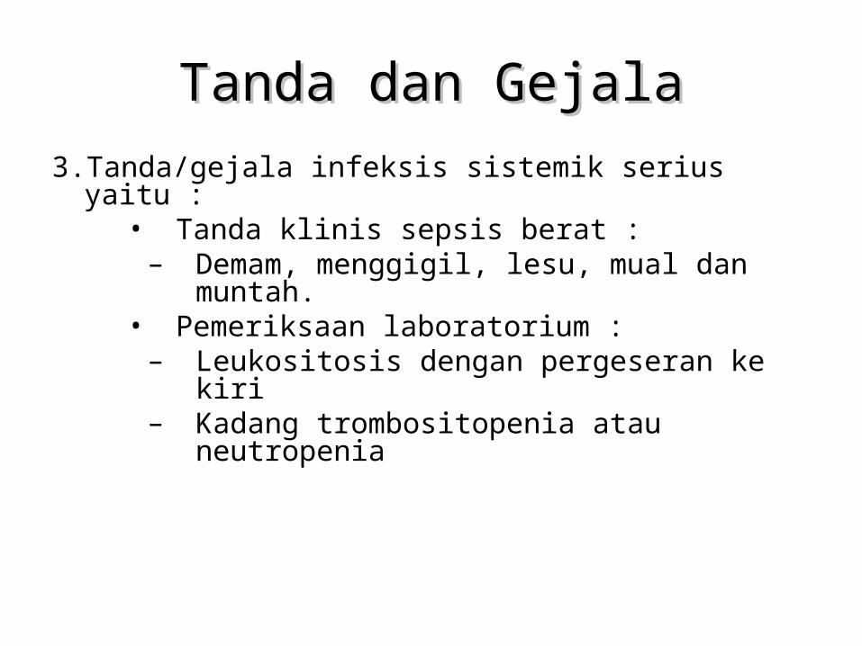
Tanda dan GejalaTanda dan Gejala3. Tanda/gejala infeksis sistemik serius yaitu :
• Tanda klinis sepsis berat :– Demam, menggigil, lesu, mual dan muntah.
• Pemeriksaan laboratorium :– Leukositosis dengan pergeseran ke kiri– Kadang trombositopenia atau neutropenia
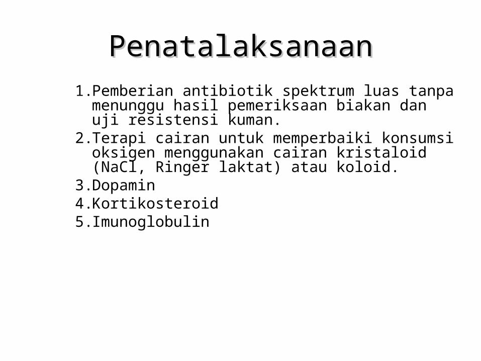
PenatalaksanaanPenatalaksanaan1. Pemberian antibiotik spektrum luas tanpa menunggu
hasil pemeriksaan biakan dan uji resistensi kuman.2. Terapi cairan untuk memperbaiki konsumsi oksigen
menggunakan cairan kristaloid (NaCl, Ringer laktat) atau koloid.
3. Dopamin4. Kortikosteroid5. Imunoglobulin
Penatalaksanaan Penatalaksanaan
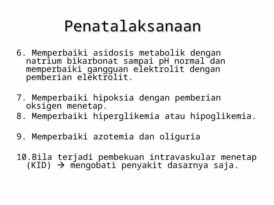
6. Memperbaiki asidosis metabolik dengan natrium bikarbonat sampai pH normal dan memperbaiki gangguan elektrolit dengan pemberian elektrolit.
7. Memperbaiki hipoksia dengan pemberian oksigen menetap.8. Memperbaiki hiperglikemia atau hipoglikemia.
9. Memperbaiki azotemia dan oliguria
10.Bila terjadi pembekuan intravaskular menetap (KID) mengobati penyakit dasarnya saja.
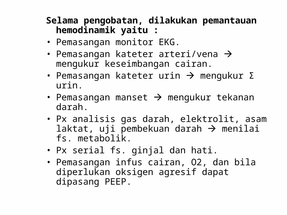
Selama pengobatan, dilakukan pemantauan hemodinamik yaitu :
• Pemasangan monitor EKG.• Pemasangan kateter arteri/vena mengukur
keseimbangan cairan.• Pemasangan kateter urin mengukur Σ urin.• Pemasangan manset mengukur tekanan darah.• Px analisis gas darah, elektrolit, asam laktat, uji
pembekuan darah menilai fs. metabolik.• Px serial fs. ginjal dan hati.• Pemasangan infus cairan, O2, dan bila diperlukan
oksigen agresif dapat dipasang PEEP.
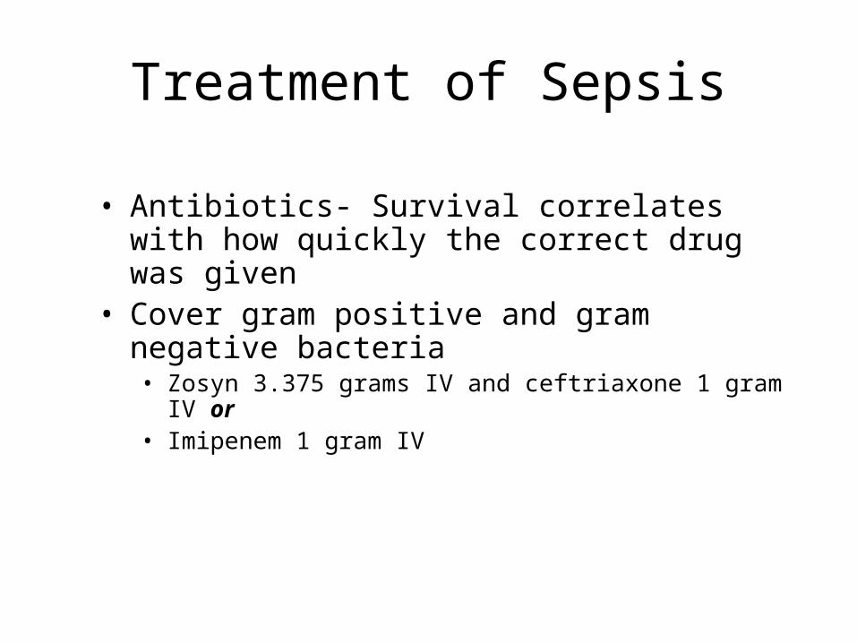
Treatment of Sepsis
• Antibiotics- Survival correlates with how quickly the correct drug was given
• Cover gram positive and gram negative bacteria• Zosyn 3.375 grams IV and ceftriaxone 1 gram IV or• Imipenem 1 gram IV
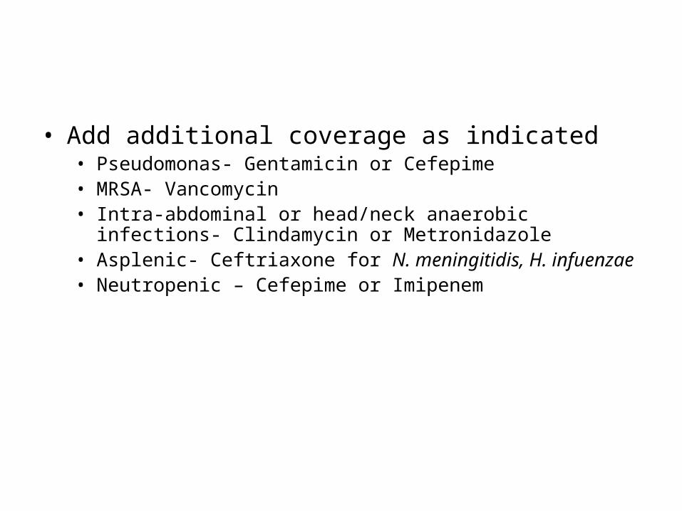
• Add additional coverage as indicated• Pseudomonas- Gentamicin or Cefepime• MRSA- Vancomycin • Intra-abdominal or head/neck anaerobic infections- Clindamycin or
Metronidazole • Asplenic- Ceftriaxone for N. meningitidis, H. infuenzae• Neutropenic – Cefepime or Imipenem
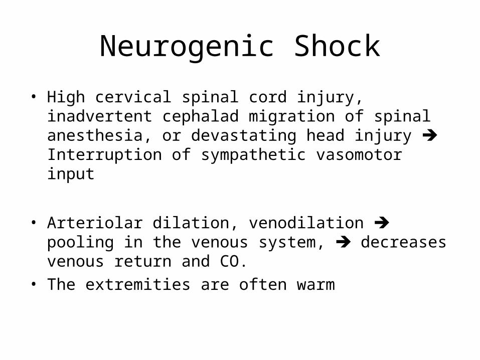
Neurogenic Shock
• High cervical spinal cord injury, inadvertent cephalad migration of spinal anesthesia, or devastating head injury Interruption of sympathetic vasomotor input
• Arteriolar dilation, venodilation pooling in the venous system, decreases venous return and CO.
• The extremities are often warm
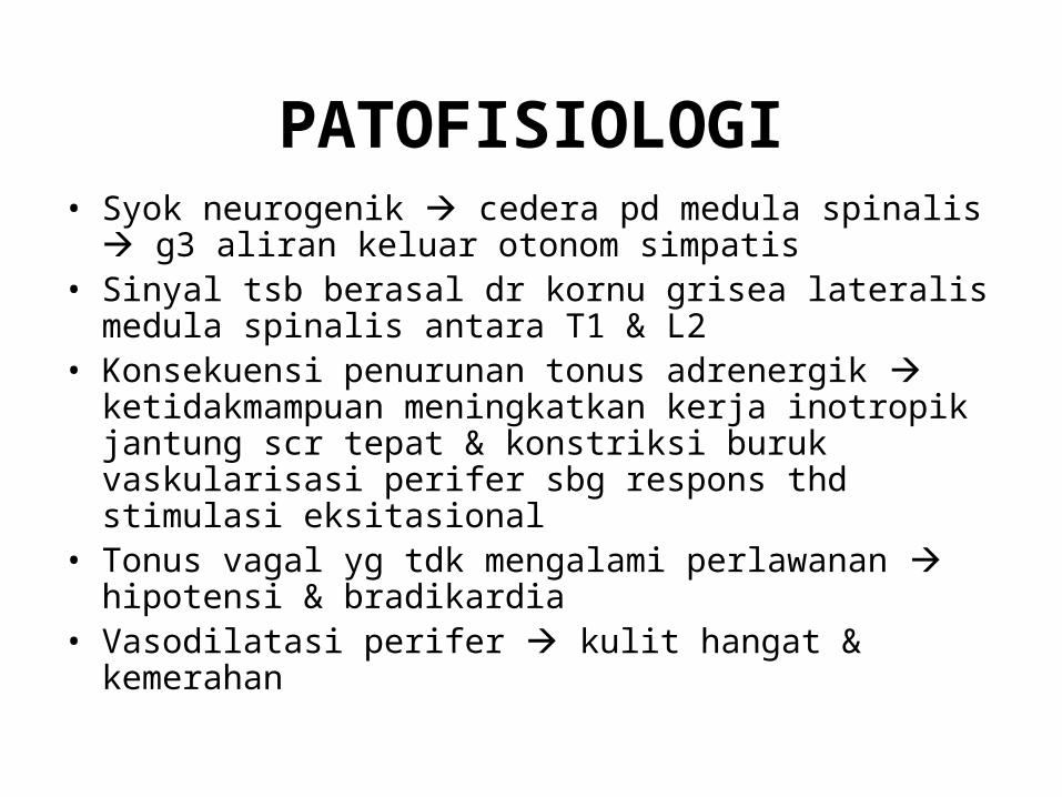
PATOFISIOLOGI• Syok neurogenik cedera pd medula spinalis g3 aliran
keluar otonom simpatis• Sinyal tsb berasal dr kornu grisea lateralis medula spinalis
antara T1 & L2• Konsekuensi penurunan tonus adrenergik
ketidakmampuan meningkatkan kerja inotropik jantung scr tepat & konstriksi buruk vaskularisasi perifer sbg respons thd stimulasi eksitasional
• Tonus vagal yg tdk mengalami perlawanan hipotensi & bradikardia
• Vasodilatasi perifer kulit hangat & kemerahan
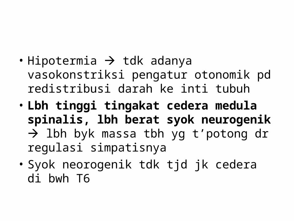
• Hipotermia tdk adanya vasokonstriksi pengatur otonomik pd redistribusi darah ke inti tubuh
• Lbh tinggi tingakat cedera medula spinalis, lbh berat syok neurogenik lbh byk massa tbh yg t’potong dr regulasi simpatisnya
• Syok neorogenik tdk tjd jk cedera di bwh T6
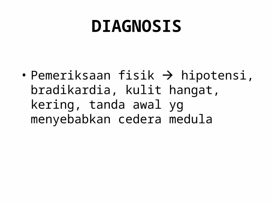
DIAGNOSIS
• Pemeriksaan fisik hipotensi, bradikardia, kulit hangat, kering, tanda awal yg menyebabkan cedera medula
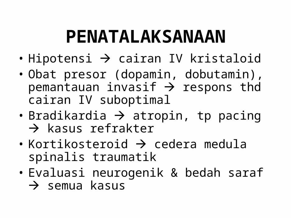
PENATALAKSANAAN• Hipotensi cairan IV kristaloid• Obat presor (dopamin, dobutamin), pemantauan
invasif respons thd cairan IV suboptimal• Bradikardia atropin, tp pacing kasus
refrakter• Kortikosteroid cedera medula spinalis
traumatik• Evaluasi neurogenik & bedah saraf semua
kasus
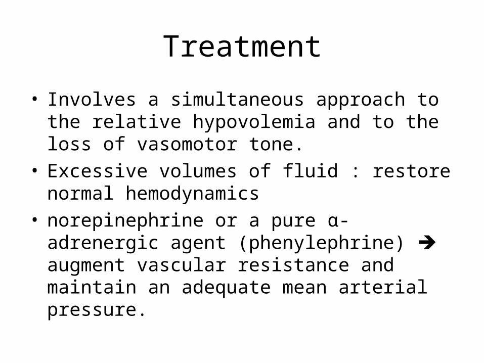
Treatment
• Involves a simultaneous approach to the relative hypovolemia and to the loss of vasomotor tone.
• Excessive volumes of fluid : restore normal hemodynamics
• norepinephrine or a pure α-adrenergic agent (phenylephrine) augment vascular resistance and maintain an adequate mean arterial pressure.
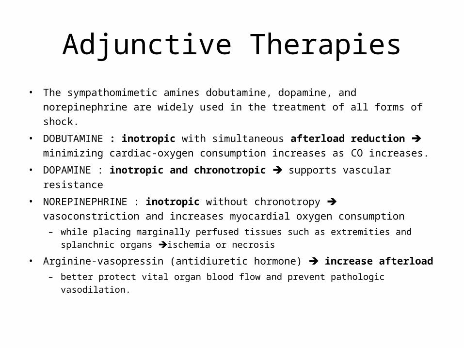
Adjunctive Therapies• The sympathomimetic amines dobutamine, dopamine, and norepinephrine are
widely used in the treatment of all forms of shock.
• DOBUTAMINE : inotropic with simultaneous afterload reduction minimizing cardiac-oxygen consumption increases as CO increases.
• DOPAMINE : inotropic and chronotropic supports vascular resistance
• NOREPINEPHRINE : inotropic without chronotropy vasoconstriction and increases myocardial oxygen consumption
– while placing marginally perfused tissues such as extremities and splanchnic organs ischemia or necrosis
• Arginine-vasopressin (antidiuretic hormone) increase afterload – better protect vital organ blood flow and prevent pathologic vasodilation.
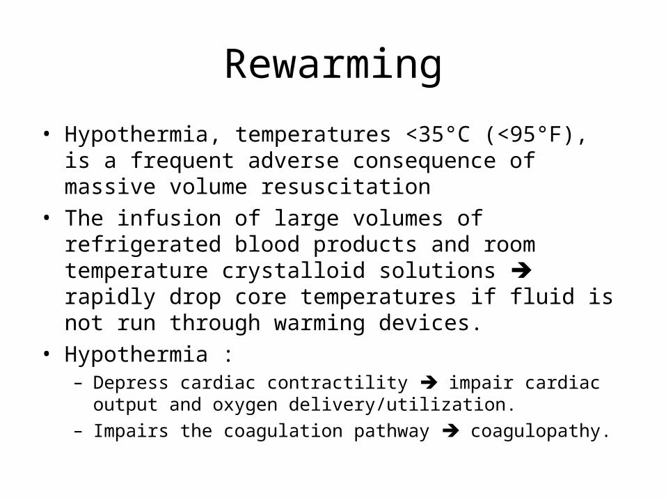
Rewarming
• Hypothermia, temperatures <35°C (<95°F), is a frequent adverse consequence of massive volume resuscitation
• The infusion of large volumes of refrigerated blood products and room temperature crystalloid solutions rapidly drop core temperatures if fluid is not run through warming devices.
• Hypothermia :– Depress cardiac contractility impair cardiac output and oxygen
delivery/utilization. – Impairs the coagulation pathway coagulopathy.
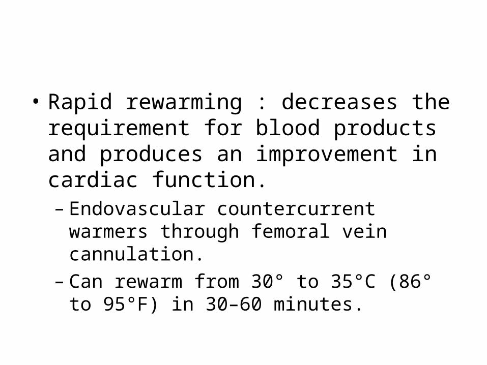
• Rapid rewarming : decreases the requirement for blood products and produces an improvement in cardiac function. – Endovascular countercurrent warmers through
femoral vein cannulation. – Can rewarm from 30° to 35°C (86° to 95°F) in 30–
60 minutes.
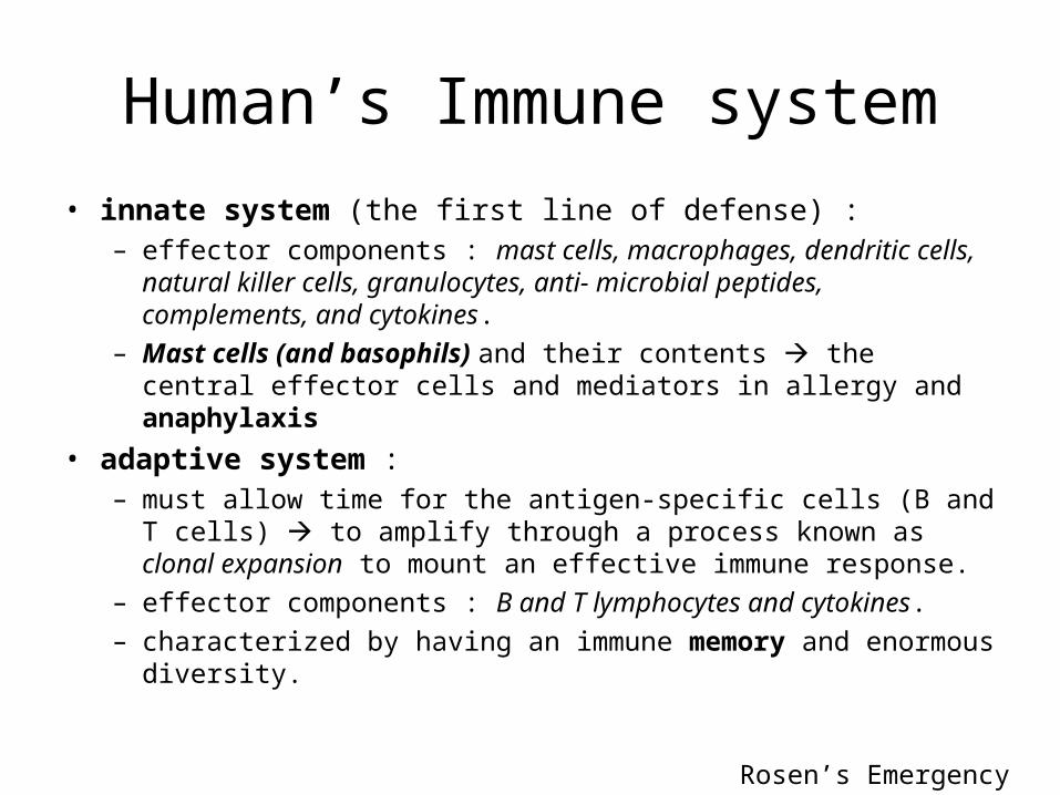
Human’s Immune system• innate system (the first line of defense) :
– effector components : mast cells, macrophages, dendritic cells, natural killer cells, granulocytes, anti- microbial peptides, complements, and cytokines.
– Mast cells (and basophils) and their contents the central effector cells and mediators in allergy and anaphylaxis
• adaptive system :– must allow time for the antigen-specific cells (B and T cells) to
amplify through a process known as clonal expansion to mount an effective immune response.
– effector components : B and T lymphocytes and cytokines. – characterized by having an immune memory and enormous diversity.
Rosen’s Emergency Medicine
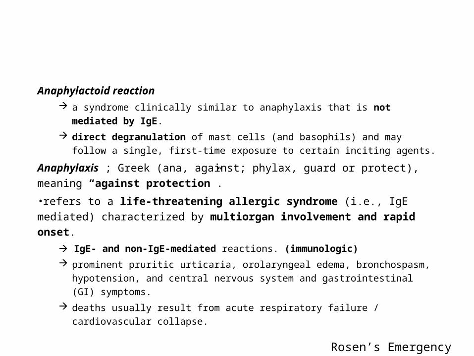
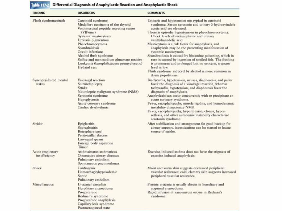
Anaphylactoid reaction a syndrome clinically similar to anaphylaxis that is not mediated by IgE. direct degranulation of mast cells (and basophils) and may follow a single, first-time
exposure to certain inciting agents.
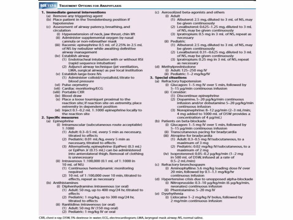
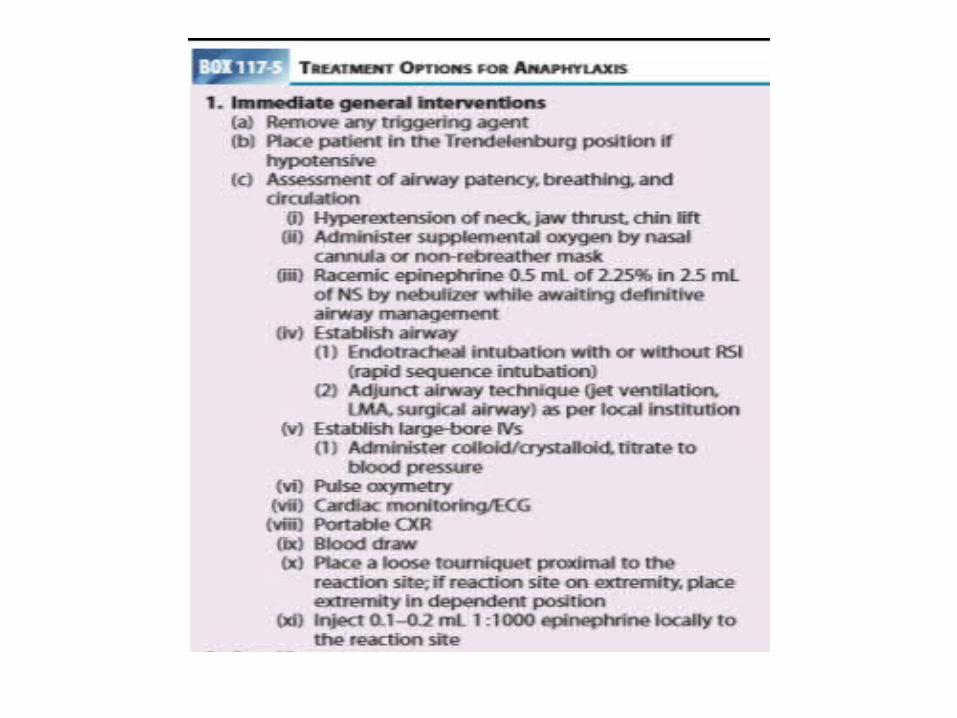
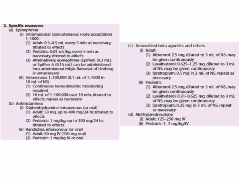
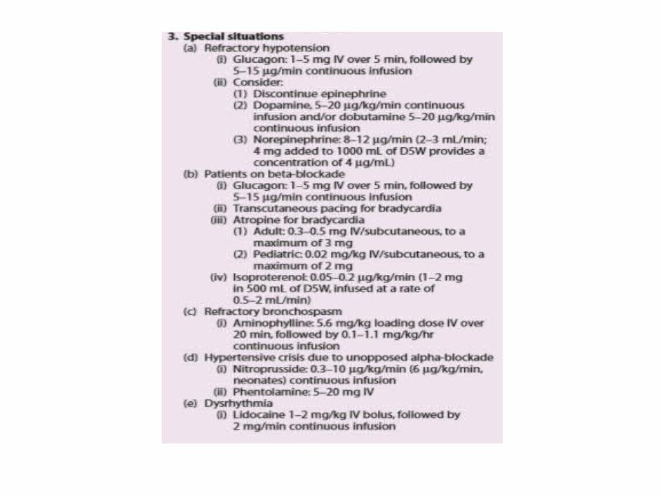
Anaphylaxis ; Greek (ana, against; phylax, guard or protect), meaning “against protection”.
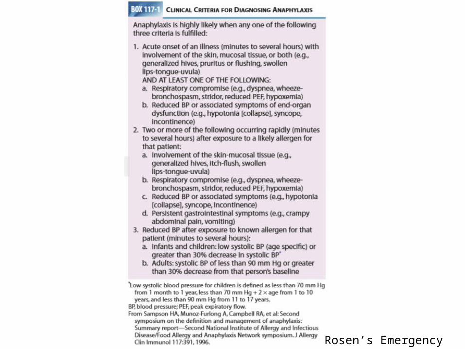
•refers to a life-threatening allergic syndrome (i.e., IgE mediated) characterized by multiorgan involvement and rapid onset.
IgE- and non-IgE-mediated reactions. (immunologic) prominent pruritic urticaria, orolaryngeal edema, bronchospasm, hypotension, and
central nervous system and gastrointestinal (GI) symptoms. deaths usually result from acute respiratory failure / cardiovascular collapse.
Rosen’s Emergency Medicine
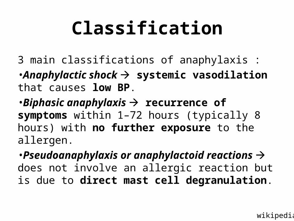
Classification
3 main classifications of anaphylaxis :•Anaphylactic shock systemic vasodilation that causes low BP. •Biphasic anaphylaxis recurrence of symptoms within 1–72 hours (typically 8 hours) with no further exposure to the allergen.
•Pseudoanaphylaxis or anaphylactoid reactions does not involve an allergic reaction but is due to direct mast cell degranulation.
wikipedia
Rosen’s Emergency Medicine
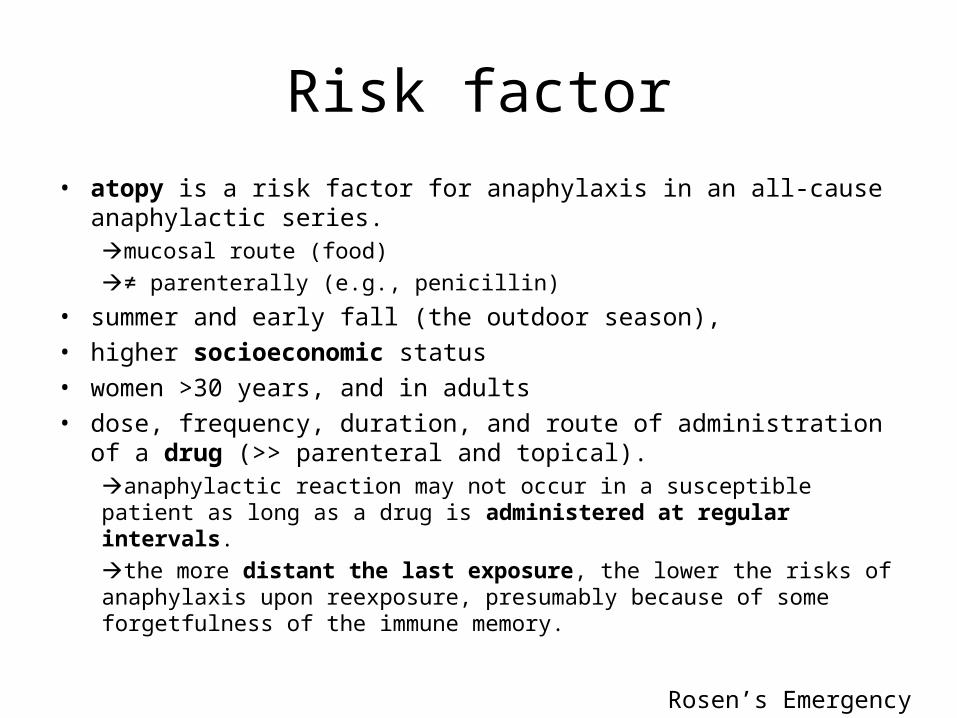
Risk factor• atopy is a risk factor for anaphylaxis in an all-cause anaphylactic series.
mucosal route (food)≠ parenterally (e.g., penicillin)
• summer and early fall (the outdoor season), • higher socioeconomic status• women >30 years, and in adults• dose, frequency, duration, and route of administration of a drug (>>
parenteral and topical).anaphylactic reaction may not occur in a susceptible patient as long as a drug is administered at regular intervals.the more distant the last exposure, the lower the risks of anaphylaxis upon reexposure, presumably because of some forgetfulness of the immune memory.
Rosen’s Emergency Medicine
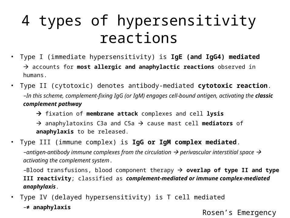
4 types of hypersensitivity reactions
• Type I (immediate hypersensitivity) is IgE (and IgG4) mediated accounts for most allergic and anaphylactic reactions observed in humans.
• Type II (cytotoxic) denotes antibody-mediated cytotoxic reaction. –In this scheme, complement-fixing IgG (or IgM) engages cell-bound antigen, activating the classic complement pathway
fixation of membrane attack complexes and cell lysis
anaphylatoxins C3a and C5a cause mast cell mediators of anaphylaxis to be released.
• Type III (immune complex) is IgG or IgM complex mediated. –antigen-antibody immune complexes from the circulation perivascular interstitial space activating the complement system.
–Blood transfusions, blood component therapy overlap of type II and type III reactivity; classified as complement-mediated or immune complex-mediated anaphylaxis.
• Type IV (delayed hypersensitivity) is T cell mediated –≠ anaphylaxis
Rosen’s Emergency Medicine
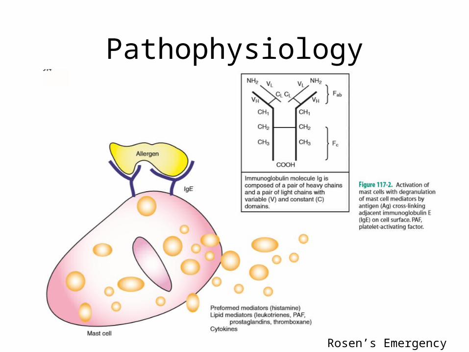
Pathophysiology
Rosen’s Emergency Medicine
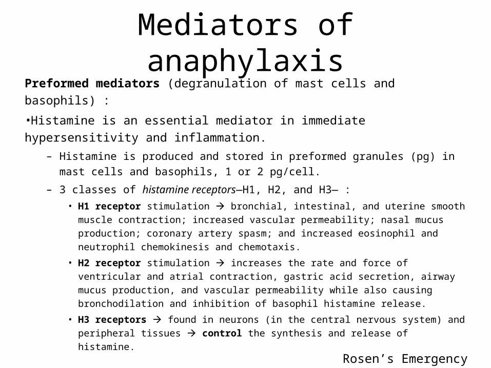
Mediators of anaphylaxisPreformed mediators (degranulation of mast cells and basophils) :
•Histamine is an essential mediator in immediate hypersensitivity and inflammation.
– Histamine is produced and stored in preformed granules (pg) in mast cells and basophils, 1 or 2 pg/cell.
– 3 classes of histamine receptors—H1, H2, and H3— :• H1 receptor stimulation bronchial, intestinal, and uterine smooth muscle
contraction; increased vascular permeability; nasal mucus production; coronary artery spasm; and increased eosinophil and neutrophil chemokinesis and chemotaxis.
• H2 receptor stimulation increases the rate and force of ventricular and atrial contraction, gastric acid secretion, airway mucus production, and vascular permeability while also causing bronchodilation and inhibition of basophil histamine release.
• H3 receptors found in neurons (in the central nervous system) and peripheral tissues control the synthesis and release of histamine.
Rosen’s Emergency Medicine
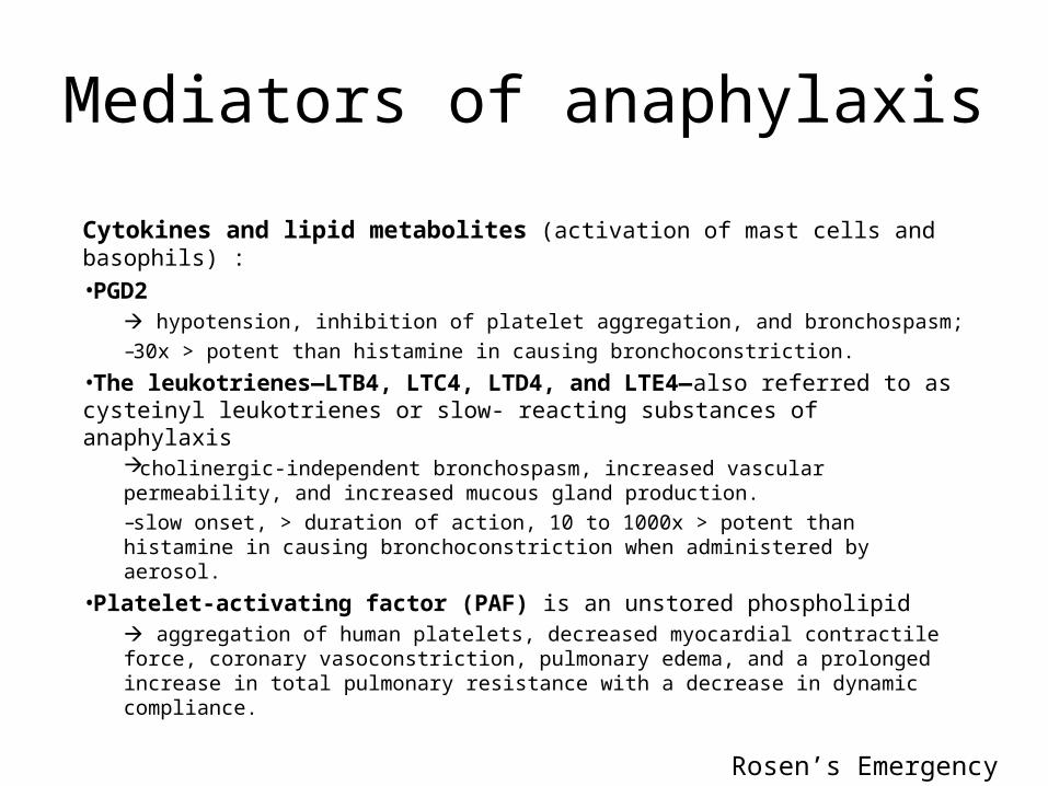
Mediators of anaphylaxis
Cytokines and lipid metabolites (activation of mast cells and basophils) :•PGD2
hypotension, inhibition of platelet aggregation, and bronchospasm; –30x > potent than histamine in causing bronchoconstriction.
•The leukotrienes—LTB4, LTC4, LTD4, and LTE4—also referred to as cysteinyl leukotrienes or slow- reacting substances of anaphylaxis
cholinergic-independent bronchospasm, increased vascular permeability, and increased mucous gland production. –slow onset, > duration of action, 10 to 1000x > potent than histamine in causing bronchoconstriction when administered by aerosol.
•Platelet-activating factor (PAF) is an unstored phospholipid aggregation of human platelets, decreased myocardial contractile force, coronary vasoconstriction, pulmonary edema, and a prolonged increase in total pulmonary resistance with a decrease in dynamic compliance.
Rosen’s Emergency Medicine
Rosen’s Emergency Medicine
References
• http://circ.ahajournals.org/content/117/5/686.full
• IPD/ harrison’s • Rosen’s emergency
Conclusion & Sugestion
Kesimpulan problem 2a• Kami telah mempelajari
– MM. Shock cardiogenic– MM. Shock hypovolemic– MM. Shock obstructive– MM. Shock distributive
• Shock neurogenic• Shock anaphylactic• Shock septic
Kesimpulan dan saran kelompok