Embed Size (px)
Citation preview
Guidelines for the Assessment and Management of Pain in Rodents and Rabbits Preface The assessment and management of pain in humans and animals has generated unprecedented discussion in the last 20 years. In the veterinary field, heightened awareness to pain has prompted several professional organizations to make formal position statements, which unequivocally mandate pain relief (McMillan, 2003). Regulatory bodies primarily concerned with the use of animals in biomedical research and teaching have also had an increased interest in pain and on July 10, 2003 the USDA's Animal and Plant Health Inspection Service (APHIS) published a Federal Register notice calling for comments on the definition and reporting of pain and distress in animals under the Animal Welfare Act. While there is much discussion on the continuum of stress, distress and pain in animals, there is little agreement on the difficulty of delineating crossover points for these apparently overlapping sensations. In addition, there is no agreed objective measure, which can be used to reliably identify pain in animals and its severity. Further and more difficult, is the assessment of different types of pain. For example, while there is an intuitive predilection to consider “real” pain as physical pain, several studies suggest psychological distress can also adversely affect animal welfare (McMillan, 2003). Semantics aside, it is generally agreed that pain adversely impacts the welfare of animals and that in research protocols, pain, if not controlled, is a variable, which can confound the interpretation of experimental results. A position statement of the American College of Laboratory Animal Medicine (ACLAM) declares that “Procedures expected to cause more than slight or momentary pain (e.g., pain in excess of a needle prick or injection) require the appropriate use of pain-relieving measures unless scientifically justified in an approved animal care and use protocol.”(http://www.aclam.org/pub_pain_distress.html). The responsibility to relieve pain in animals used in research is consistent with the generally accepted view of the moral responsibility of humans towards animals; with the opinion of the public, whose support for the use of animals in research declines as the pain experienced by these animals increases (Institute for Laboratory Animal Research, 2000); with relevant legislation (AWA, PHS) and with the goals of scientific research. This paper presents an overview of current concepts of pain and provides recommendations for the assessment, prevention and treatment of pain in rodents and rabbits. Also provided are guidelines for developing pain management protocols, tables describing the potential physiologic effects of some analgesic classes and examples of efficacious analgesic strategies. Introduction A. Definitions “Pain” is derived from the Latin “poena” or penalty, and has been defined by the Committee
on Taxonomy for the International Association for the Study of Pain as “an unpleasant sensory and emotional experience associated with actual or potential tissue damage” http://www.iasp-pain.org/trms-p.html. The USDA takes a more anthropomorphic view and defines a painful procedure as “any procedure that would reasonably be expected to cause more than slight or momentary pain and/or distress in a human being to which that procedure is applied, that is, pain in excess of that caused by injections or other minor procedures. AWA,9, CFR, Subchapter A, 1. The advantage of this definition is that it requires only that there be an understanding of pain by the personnel involved. For the purposes of this paper, a definition which combines these two concepts appears to be most suitable.
1
Pain is an unpleasant sensory and emotional experience associated with actual or potential tissue damage, and should be expected in an animal subjected to any procedure or disease model that would be likely to cause pain in a human.
Definitions of other terms used in this paper are listed below: Allodynia: Pain due to a stimulus, which does not normally provoke pain. Analgesia: Absence of pain in response to stimulation, which would normally be painful. Hyperalgesia: An increased response to a stimulus, which is normally painful.
Neurogenic pain: Pain initiated or caused by a primary lesion, dysfunction, or transitory perturbation in the peripheral or central nervous system.
Neuropathic pain: Pain initiated or caused by a primary lesion or dysfunction in the nervous system.
Nociceptor: A receptor preferentially sensitive to a noxious stimulus or to a stimulus, which would become noxious if prolonged.
Noxious stimulus: A noxious stimulus is one, which is damaging to normal tissues. Pain threshold: The least experience of pain, which a subject can recognize.
Pain tolerance level: The greatest level of pain, which a subject is prepared to tolerate. Pain tolerance varies both within and between species and is dependent on many factors, such as general condition of the animal, motivation, previous painful experiences, anxiety, and fear levels.
Peripheral neurogenic pain: Pain initiated or caused by a primary lesion or dysfunction or transitory perturbation in the peripheral nervous system.
Peripheral neuropathic pain: Pain initiated or caused by a primary lesion or dysfunction in the peripheral nervous system.
B. Special Circumstances in Biomedical Research that Impact the Alleviation of Pain in
Rabbits and Rodents, Including the Minimization of Variables That Could Confound the Interpretation of Data (e.g., pain and/or analgesics).
Management of pain in animals requires that pain either be anticipated and prevented (pre-
emptive), or recognized and alleviated (post-inductive). Pre-emptive analgesia presumes that the pain will result from the procedure and that non-pharmacological and pharmacological protocols would be instituted prior to the induction of pain.
Post-inductive analgesia is the administration of pain relief after pain has already been induced
and observed. Here, one is not relying on knowledge of the procedure and its likelihood of causing pain, but one is inferring pain from the observed behaviors of the animals. Regardless of the pain management strategy used, animals must be evaluated post-surgically to ensure that pain has been alleviated.
Confirming pain in animals is difficult because of differences between and within species in the
behavioral response to noxious stimuli. Many behaviors are consistent with, but not invariably indicative of pain. For example, an animal separated from a long time companion may show immobility, lethargy, inappetence and indifference to its surroundings, yet not be in pain. However, interpretation of this behavior and its relationship with pain would change had this animal had surgery within the last 24 hours. Confirming the presence of pain in an animal is further complicated by the fact that normal behavior is not always indicative of a pain-free
2
state because, despite the pain, the animal may show “normal” behavior as an inherent response to avoid predation (“displacement behavior”).
Pain in an animal is often inferred from the absence of normal behaviors (alertness, mobility, groomed coat, good appetite and general condition). However, rabbits and rodents are often housed in small groups and subtle changes in the behavior of an individual animal may be difficult to observe. In addition, rodents housed in translucent or opaque plastic cages on large racks are difficult to evaluate easily without moving the cage and disturbing the inhabitants. Rabbits housed singly are easier to observe, but they often sit for long periods in a far corner of the cage, and thus closer examination would be required to differentiate this behavior from one that is pain induced. In both rabbits and rodents, there is as yet no agreed method that facilitates the accurate identification of the genesis of behaviors, which could be induced by either anxiety or pain.
The identification of pain and the selection of the optimal method of pain management require professional judgment, and not all scientists and technical staff are adequately trained for these tasks. The goal of the study should be considered when assessing analgesic options. For example, non-steroidal anti-inflammatory drugs are recommended for mild pain but they could significantly affect the inflammatory cascade, and confound the interpretation of a study focused on cytokine interaction.
ACLAM certified veterinarians are trained and experienced in the evaluation of animals, their behavior, and its relationship to pain and other factors. These professionals should be consulted to discuss the options, which will alleviate pain and concurrently preserve the integrity of the study.
Pain – Physiology and Perception An understanding of the physiology and pharmacology of pain is essential to the ability to prevent and/or alleviate pain. When tissue injury occurs, cellular changes are elicited and release biochemical substances into the local environment. These substances activate specialized nerve cell endings (nociceptors). When sufficiently stimulated, these nerves generate action potentials, which travel along afferent nerve fibers to the spinal cord and brain, generating an experience of pain. There are 2 major types of nerve fibers associated with nociceptive signals; thin myelinated Aδ fibers that detect noxious stimulation and make the animal aware of stimuli that could or actually do damage body tissues, and unmyelinated C fibers that respond to a wide range of stimuli and chemical signals. Myelinated Aβ nerve fibers, a third category, are usually associated with non-noxious stimulation and transmit information regarding the state of the animal’s body and its immediate environment e.g., by touch. Many of the receptors are polymodal, especially C fibers, and can respond to a variety of stimuli of varying intensities. Furthermore, “silent” nociceptors respond only after tissue has undergone inflammatory changes (Short et al., 1992). Nerve fibers from nociceptors reach the gray matter of the spinal cord mainly via the dorsal roots of spinal nerves. Some afferents enter the spinal cord via the ventral roots and often loop back from the ventral roots to ultimately enter via the dorsal roots (Raj, 2000; Loeser et al., 2001). Nociceptive relay neurons in the spinal cord contribute to local spinal reflex responses such as withdrawal from the stimulus. Afferent pathways from the spinal cord to the brain include the spinothalamic tract, spinoreticular tract, spinomesencephalic tract, and the postsynaptic dorsal column tract (Raj, 2000). The relative importance of these 4 pathways in communicating nociceptive information cephalad varies between species. Because pain involves cognition, emotion and behavior, many cortical areas of the brain as well as sub cortical areas are involved, in particular the thalamus. Sensitization and Hyperalgesia: Peripheral sensitization occurs subsequent to tissue injury, resulting in a lower stimulus threshold and increased sensitivity to pain stimuli. The state that results from this sensitization is called hyperalgesia. After repeated exposure to a noxious stimulus, pain sensations can occur spontaneously in the absence of stimuli. Primary hyperalgesia occurs at the original site of injury. Secondary hyperalgesia involves increased pain and allodynia from uninjured tissue surrounding the original site of injury. This response is primarily a result of central sensitization via activated N-methyl-D-aspartate (NMDA) receptors, elevated intracellular calcium, and increased
3

sensitivity to glutamate. Repetitive C fiber stimulation can cause “windup” or increased central sensitization (Loeser et al., 2001). Mediators of Inflammation and Pain: At the time of tissue injury, a host of substances are released locally and contribute to the process of inflammation and increased sensitivity of nociceptors. Tissue damage releases bradykinins, prostaglandins and serotonin which activate and sensitize nociceptors (Raj, 2000). This sensitization also leads to the release of substance P, a tachykinin, and other peptides. Local mast cells respond to increased substance P with degranulation and histamine release, which further excites nociceptors. Substance P also induces local vasodilatation with subsequent edema and further release of bradykinin. In the dorsal horn, spinal cord neuronal activity is mediated by excitatory amino acids, substance P, and neuropeptides. Normal anti-nociceptive mechanisms occur via inhibitory spinal cord neurons, which act to attenuate the excitatory level of afferent nociceptive neurons. Also at the level of the dorsal horn, two endogenous opioid peptides, enkephalin and dynorphin, are released after nociceptive stimulation from descending fibers originating in the brain stem. Opioid-containing neurons can have an excitatory or inhibitory effect, but primarily act to inhibit transmission of nociceptive activity. Morphine administration at the level of the dorsal horn (e.g., by epidural or intrathecal administration) decreases the perceived intensity of noxious stimuli. Another modulating mechanism, known as the “pain gate” phenomenon, occurs when stimulation of thick Aβ fibers of peripheral nerves decreases the sensitivity of post-synaptic cells in the dorsal horn to incoming signals from C and Aδ fibers. This property helps to explain how mechanical stimulation such as stroking or rubbing the skin can provide pain relief. Transcutaneous electrical nerve stimulation (TENS), a therapeutic modality used in humans for persistent pain, also may work through this mechanism (Loeser et al., 2001). Descending Control Pathways: In some circumstances, such as when an animal’s safety is under jeopardy, a lack of pain awareness and response is beneficial (a speedy flight response despite an injured leg). The descending pathways that inhibit pain perception are called antinociceptive, and include supraspinal-modulating loops. Central modulation of pain perception response involves in particular, parts of the cortex, the periaqueductal gray region of the midbrain, the lateral pons, and the ventromedial medulla. Stimulation of these sites can result in inhibition of dorsal horn neuronal activity. It also inhibits those behaviors and reflexes normally induced by a noxious stimulus. Endogenous opioids, including the enkephalins, the dynorphins, and the endorphins contribute to the endogenous analgesic response. Excitatory amino acids agonists, opioids, adrenergic drugs, cholinergic agonists, and GABAergic antagonists can all inhibit nociception by acting on one or several areas of the CNS involved in pain modulation (Short et al., 1992). Endogenous analgesia (antinociception) is tied integrally to the behavioral and emotional state of the animal and to the autonomic system. Thus, the experience of pain is the outcome of many complex processes and interrelationships. Possible sites of intervention: The pain response can be modified at the peripheral level, spinal cord, or the CNS. Anti-inflammatory agents may dramatically reduce tissue injury and thus decrease sensitization of nociceptive fibers. Methodologies such as massage may work through the pain gate mechanism. Administration of intrathecal and epidural agents, such as opioid agonists and local anesthetics, work at the level of the dorsal horn by reducing excitation of nociceptive neurons and stimulating inhibitory pathways. Centrally acting agents work on receptors in the various “pain centers” of the brainstem and cortex, to stimulate descending antinociceptive pathways. Postoperative pain: Surgical trauma and pain cause an endocrine stress response with release of cortisol, catecholamines, and other stress hormones. These may evoke a host of physiologic and metabolic changes, including tachycardia, hypertension, immune system alterations, hyperglycemia, lipolysis and a negative nitrogen balance (Loeser et al., 2001). Furthermore, pain itself is a stressor and thus may contribute to loss of homeostasis and lead to distress (Grandin et al., 2002). Pain treatment in humans recognizes the vast benefit of analgesia to the reduction of the stress response and improved recovery of patients. In addition to improved recovery of rodent and rabbit patients, analgesics reduce the effects that stress responses have on experimental data. Although laboratory animal medicine is significantly more restricted in its options for pain management in rodents, the
4
general concepts used in humans are still applicable and should help guide us in seeking improved methods of managing rodent patients. Pain Management A. Non Pharmacological Considerations
Management of pain in animals can be enhanced by providing appropriate housing, handling, and restraint and by utilizing appropriate experimental techniques. This is especially true when surgery is part of the protocol. A skilled surgeon who utilizes proper surgical techniques can minimize complications of surgery and tissue trauma, which contribute to postoperative comfort. Surgical complications such as infection, seromas, hemorrhage and inflammation induce painful and stressful sensations. Selection of appropriate suture materials and utilization of proper instrumentation both can help to alleviate postoperative trauma as well as perioperative care, which emphasizes maintenance of homeostasis. For instance, many animals have inflammatory reactions to surgical gut and silk sutures that can be avoided by use of newer synthetic suture materials, which are less likely to produce inflammatory responses (Flecknell, 1996; Swindle et al., 2002; Thurmon et al., 1996).
Housing appropriate for the species may reduce post-procedural discomfort. Animals housed in a stressful situation can be more vulnerable to pain. For example, animals generally require an increased environmental temperature to recover quickly from anesthesia. Wet bedding materials may also contribute to hypothermia and increase the chance of infection. Animals, which have been habituated to handling and husbandry routines, may experience less distress. Husbandry may have to be modified to provide animals with easier access to food and water if defects such as spinal cord trauma have been induced. Use of nesting materials, soft food, bandaging and other types of nursing care, such as expressing the bladder in animals with spinal cord dysfunction, may also be indicated as adjuncts to analgesics (Flecknell, 1996; Swindle et al., 2002).
Diet may contribute to post-procedural recovery. For example, soy-containing diets have been demonstrated to help alleviate pain in rats with chronic sensory disorders. Consuming a soy-containing diet prevents development of tactile and heat allodynia, but not mechanical hyperalgesia in rats with partial sciatic nerve ligation (Shir et al., 2001). Softened foods or foods with high caloric content may be helpful in assisting animals with oral lesions or debilitating procedures.
Essential to any program of post-procedural care is training to make investigators aware of species-specific requirements and appropriate experimental techniques that may reduce the discomfort level of the animal.
B. Pharmacology, Mode of Action, and Biomedical Considerations for Use of Drugs
The selection of an analgesic protocol for a particular procedure should include an assessment of the physiologic effects of the agents and potential complications to the goals of the research. Species-specific variations in the reactions to pharmacologic agents must be considered. Analgesic protocols that use combinations of agents may have different physiologic effects than those using a sole agent. Adjuncts to analgesia include tranquilizers and corticosteroids, which induce sleep, reduce anxiety and/or relieve inflammation (Flecknell, 1996; Heavner, 1997; Swindle et al., 2002; Thurmon et al., 1996). Antidepressants, such as venlafaxine, have been tested in rats and shown to be effective for reduction of neuropathic pain and other tricyclic antidepressants and/or anticonvulsants may be effective in animals, but their use has not been reported in the clinical laboratory animal literature (Marchand, et al., 2003).
Generally, preemptive analgesia provides for enhanced pain relief and a shorter recovery period than administering analgesics after a painful procedure (Danneman, 1997). However, telemetry studies in mice have not demonstrated any significant difference between using
5
preemptive vs. postoperative analgesics for mock ova transplant (Goecke et al., 2005). Preemptive techniques include parenteral administration of systemic analgesics, infiltration of a suture line with local anesthetics, and epidural administration of analgesics. Parenteral analgesia with an opioid or nonsteroidal anti-inflammatory drug (NSAID) should be administered prior to making a surgical incision. Incision lines can be infiltrated with a local anesthetic prior to making the incision. Local anesthetics can also be used to perform dorsal nerve root blocks for procedures such as a lateral thoracotomy or a flank incision. Epidural administration of analgesics may be of benefit for abdominal procedures. For major surgery in larger animals such as rabbits, all three of these techniques preemptively may be useful (Flecknell, 1996; Swindle et al., 2002; Thurmon et al., 1996). To maximize analgesia, it is sometimes beneficial to combine classes of agents such as opioids and NSAIDS. For example, an opioid may be given by injection as preemptive analgesia, and a post-procedural NSAID may be given orally. In general, the local anesthetics and NSAIDS have the most effect for blocking peripheral nociception and the opioids are more effective for blocking dorsal lamina neurons.
Opioids: Opioids, of which morphine is the standard for comparison, bind to mu, delta and kappa receptors to produce analgesia by blocking nociception. Mu receptors largely exist in the cerebrum, mesolimbic system, thalamus, hypothalamus and periaqueductal gray matter regions of the CNS. Kappa receptors are located in the dorsal horn of the spinal cord and provide spinal analgesia and sedation. Delta receptors are located in the brain cortex and are less important in analgesia than the other types. Opioids also affect the limbic system, which makes pain more tolerable. Opioids may be classified as agonists (morphine), agonist-antagonists (butorphanol) or partial agonists (buprenorphine) in their activity on these receptors. They are generally indicated for moderate to severe acute pain (Flecknell, 1996; Heavner, 1997; Swindle et al., 2002; Thurmon et al., 1996).
Opioids have a wide range of side effects (Table 1), which vary in significance across species, dose and agent. In general, opioids are associated with respiratory depression, bradycardia, nausea, hypotension, constipation and decreased mental capacity. Effects of over dosage may include seizures, ataxia and death by respiratory and cardiac depression. Chronic administra-tion may result in physical dependence and/or tolerance. For this reason, all of the opioids used clinically are controlled substances. Naloxone reverses the effects of opioids on the mu receptors (Flecknell, 1996; Heavner, 1997; Swindle et al., 2002; Thurmon et al., 1996).
Opioids may be administered per os (po), intravenously (iv), intramuscularly (im), subcutaneously (sc), transdermally or epidurally, although in rodents the sc route is nearly always employed. Most opioids are metabolized by glucuronidase in the liver with renal excretion. Buprenorphine is largely bound to plasma protein and is excreted via bile in feces (Heavner, 1997).
The morphine related drugs, such as oxymorphone, fentanyl, sufentanil, alfentanil and codeine, affect all three receptors, with primary agonist activity on the mu receptors. The agonist-antagonist agents, such as butorphanol, pentazocine and nalbuphine, have agonist effects at kappa receptors but antagonist effects on the mu receptors. Buprenorphine is a partial agonist of mu receptors with antagonism of pure mu agonists. Because of the potential of agonist-antagonists blocking activity of mu receptors they should not be mixed with pure agonist opioids. In general, the newer opioids and the synthetics have fewer side effects and more specific and potent analgesic activity than morphine (Flecknell, 1996; Harder and An, 2003; Swindle et al., 2002; Thurmon et al., 1996).
NSAIDS: Nonsteroidal anti-inflammatory drugs have traditionally been used more for mild pain and anti-inflammatory activities than for chronic pain. However, newer NSAIDS have significant analgesic properties and overlap with the activity of the opioids. Aspirin is the standard for comparison within this class of drugs (Flecknell, 1996; Harder and An, 2003; Heavner, 1997; Heavner, 1996; Swindle et al., 2002; Thurmon et al., 1996).
NSAIDS function by inhibiting inflammation and the production of kinins and prostaglandins. They have varying degrees of effectiveness as antipyretics, analgesics and anti-inflammatory
6
agents. Newer agents (ketoprofen, carprofen, ketorolac, meloxicam) can alleviate acute pain, such as that produced by surgery (Cooper et al, 2005). They are also longer acting than the older agents and COX-2 specific inhibiting agents may have fewer side effects. (Flecknell, 1996; Harder and An, 2003; Heavner, 1997; Heavner, 1996; Swindle et al., 2002; Thurmon et al., 1996).
NSAIDS are generally classified upon their chemical structure. These include salicylates (aspirin), pyrazolone derivatives (phenylbutazone), p-aminophenol derivatives (acetaminophen), acetic acid derivatives (indomethacin, ketorolac), fenamates (meclfenamate), propionic acid derivatives (ibuprofen, ketoprofen, carprofen), oxicams (peroxicam) and nicotinic acid derivatives (flunixin) (Harder and An, 2003; Heavner, 1997). NSAIDS may be administered im, sc or po and metabolism and excretion vary widely among agents and species. In general, the NSAIDS are metabolized by the liver and excreted by the kidneys. Side effects of NSAIDS (Table 2) include ulceration of the GI tract, impairment of platelet aggregation, nephrotoxicity, delayed parturition, fetal abnormalities, blood dyscrasias, bone healing impairment and hepatotoxicity. Problematic side effects are usually the result of chronic administration and are rarely seen with short-term administration (Flecknell, 1997; Harder and An, 2003).
Local Anesthetics: Local anesthetic agents can be utilized for topical, local, regional and spinal anesthesia to prevent or alleviate pain. They are generally given by sc injection and may contain epinephrine as a vasoconstrictor to retard absorption. Lipid solubility and protein binding in the axons determine the potency of these agents, which generally are secondary or tertiary amines that are ester or amide linked. Most of the agents are metabolized by the liver and excreted by the kidneys. Local anesthetics block the action potential of axons by preventing the influx of sodium ions (Flecknell, 1996; Heavner, 1996; Swindle et al., 2002; Thurmon et al., 1996)
Toxicity from injections of these drugs is rare and is usually associated with IV injections. CNS complications such as seizures and cardiac dysfunction of the electrical conduction system are possible as well as localized tissue reactions. Toxicity is more likely with the long duration and high potency agents such as bupivacaine (Heavner, 1996).
Injectable agents are classified as low (procaine), intermediate (prilocaine, lidocaine) or high (tetracaine, bupivacaine) potency and duration. Duration of action may be as short as 1 hour in the low potency agents and as long as 10 hours in the high potency group. Benzocaine and lidocaine may be administered as sprays to mucous membranes or wounds for a topical effect. A prilocaine, and lidocaine crème and patches have been developed to provide anesthesia to intact skin (Flecknell, 1996; Heavner, 1996; Swindle et al., 2002; Thurmon et al., 1996). Topical applications are always susceptible to ingestion by animals and they should be monitored for this behavior in order to prevent toxicity associated with ingestion.
Current Methods of Assessing Pain Much has been written and discussed regarding how to clearly and unambiguously identify an animal in pain that requires human intervention, be it analgesic administration, nonpharmacologic management, invoking humane endpoints or euthanasia. Despite the primarily behavioral and physiologic methods and instrumentation discussed below, recognition of pain in animals is as much an art as a science and ultimately relies on the skill, experience and professional judgment of both the research and veterinary staff. There is no foolproof table, machine or pain scoring rubric that will accurately and without fail identify every animal and circumstance that cause pain in a research animal as well as judge how effectively pain has been alleviated by various therapeutic modalities. To this end, the discussion below represents 1) criteria to be considered proactively when composing and critiquing research proposals, 2) commonly used techniques that frequently aid in pain identification and perhaps most importantly, and 3) how to assess and reassess the animals to determine whether our pain recognition criteria as well as modalities for managing pain are succeeding. The latter is
7

increasingly exemplified by use of pain scoring paradigms. In summary, pain is best recognized and managed in research animals by trained observers utilizing their professional judgments, often in a team setting, incorporating frequent assessments and reassessments of the animal with appropriate action. Identification and management of pain in rodents presents unique problems. Rodents are frequently group housed, thus alterations in behavior or mobility may be more difficult to discriminate than similar signs in a larger, individually housed animal. A group-housed animal in pain may also be less likely to outwardly manifest aberrant behavioral signs indicative of pain in order to maintain its social status and decrease the likelihood that it will be traumatized by cage mates. Increasingly, rabbits are also being maintained in a herd or grouped format. In addition, research requirements may dictate specialized caging of rodents (e.g., flexible film or rigid isolators, or ventilated caging) that not only limits observation of individual animals, but also makes physical examination, and thus efforts to evaluate painful behavior, difficult. Before ethical and legal requirements to provide relief from pain can be fulfilled, logical and indisputable criteria to define and recognize pain in rodents and rabbits must be established. Like humans, rodents and rabbits show individual variability in both their pain threshold and pain tolerance. As in people, threshold-intensity pain is perceived but can be tolerated. Thus, the animal often shows no overt signs of pain. In these cases, the need for and benefits of analgesic therapy are difficult to assess. When pain exceeds the intensity of the animal's pain tolerance obvious signs of discomfort become evident. In these cases, some modality of pain management (nonpharmaco-logic, administration of analgesics, invoking humane endpoints) is indicated, and successful pain management is much easier to monitor. Recognition of pain in laboratory animals requires cooperation among the research team, the IACUC, and the laboratory animal care and veterinary staff. From an IACUC standpoint, asking the appropriate questions during the protocol review process to assure that pain will be detected during the actual conduct of the study is crucial. Relevant questions include: 1. What will be the cause of pain? Different types of painful stimuli lead to different clinical
signs. For example, visceral pain in rodents will often lead to writhing-type movements, whereas neuropathic pain due to lesioning of a peripheral nerve may lead to self-mutilation of the affected limb.
2. What is the anticipated pain intensity and duration? Are the experimental manipulations
expected to cause acute, highly intense pain or mild but chronic pain? These two situations usually elicit different behavioral and physiological signs of pain. Will any analgesic drugs chosen for the study be appropriate for the pain intensity and duration? This should include the consideration that chronic pain is often much more difficult to effectively treat and often requires a combination of analgesic drugs and perhaps inclusion of husbandry, exercise and perhaps additional alternative medicine therapies, as well, to be truly effective.
3. Will more than one painful manipulation occur in the study that will manifest in different
clinical signs? 4. Will a manipulation make it difficult for the animal to show pain after subsequent
manipulations? 5. How will criteria for pain perception be used to define when appropriate analgesic therapy
should be instituted? 6. Is the primary investigator familiar with detecting pain in this animal model? Will the research
team recognize pain when they see it? The team must understand the known diversity in expression and sensitivity to pain in various rodent species and strains and sexes. If needed, pilot studies with significant participation of a clinical veterinarian can be helpful to identify
8
signs and behaviors deviating from normal that would suggest the animals are in pain or distress.
7. Are the criteria proposed for detecting pain in this model unambiguous such that all members
of the research team as well as the veterinary staff will recognize and acknowledge the occurrence of pain? Alternatively, are the criteria highly subjective such that disagreement will occur throughout the conduct of the study? Will ambiguity prevent staff members from taking an active role to mitigate pain?
8. When will the animals be observed for signs of pain perception? Does this schedule afford
appropriate timing for evaluating signs indicative of pain? Or will the signs most associated with pain be missed in, for example, sleeping animals? Because rodents are nocturnal and primarily active at night, can the research team expect to see signs of decreased ambulation and appetite, which may be used to indicate pain perception, during daylight hours? Will the temporal plan for observing signs of pain correlate with anticipated duration of analgesics administered to relieve pain?
9. Who will observe the study animals to decide if they are in pain? Are their contact numbers
(work, home telephone, cell pager numbers) posted so that they can be contacted if needed to treat animals thought to be in pain?
10. Are all members of the research team that are responsible for animals in the proposed study
skilled in the needed techniques and empowered to take action if they detect animals in pain? Alternatively, will they need to “call in” other team members with higher-level authority and skills to administer analgesic drugs, invoke humane endpoints, etc., risking added time that the animal(s) may suffer thus increasing the time period before appropriate strategies to obtund pain are implemented?
11. Should the IACUC observe the animals and judge for itself whether or not pain is occurring?
This role is often delegated to the veterinary staff and their clinical observations are discussed with the IACUC if there are procedural or compliance concerns. Occasionally, members of the IACUC will want to observe study methods and animals themselves to learn about the animal models as well as perform post-approval monitoring of compliance.
12. Will neuromuscular blockers be used in any component of the study such that behavioral signs
of pain would not be observed? 13. Is pain research, per se, the objective of the proposed study? In studies of this nature, some
pain will be expected (such as movement in regard to heat or pressure). Acute pain research studies often use methods that are uncomfortable (just above the pain threshold level) but not agonizing for the animal. How can intolerable pain, which cannot be escaped or terminated by the animal, be recognized?
14. Has the plan for pain recognition allowed for professional judgment? 15. How will the IACUC, veterinary staff, and research team manage animals that cannot be
given analgesics for scientific reasons but are showing signs clearly thought to be due to pain? In these cases, has an effort been made to consider all of the various classes of analgesic drugs, routes of administration and drug clearance metrics before concluding that no option exists for analgesic management short of euthanasia?
16. Is it appropriate in an individual protocol for the IACUC and research staff to
prospectively conclude that the planned procedure(s) carry such a high likelihood for pain that the research staff should deliver analgesics intraoperatively to prevent the animals from feeling pain upon awakening from anesthesia? In humans, classes of surgical manipulations are known to carry a high risk for intense postoperative pain, such that the great benefit of intraoperative (pre-emptive) analgesia overcomes the risk of needlessly administering analgesics to the patient. To this end, animal models of
9
these same procedures might benefit from a similar risk: benefit assessment for early analgesic administration. This is but one example illustrating the concept of pre-emptive analgesia in research animals (this concept is discussed elsewhere in this paper in depth) but is certainly not the only situation wherein professional judgment may well dictate that the animals should be given the “benefit of the doubt” and administered analgesia prior to showing signs of pain.
17. Does the IACUC have a mechanism in place to follow and track animals used in chronic studies that may move between the definitions of several pain Categories? 18. Are appropriate mechanisms in place for analgesic drug purchase and storage? (Often including procedures for drug procurement as well as facilities for Storage of controlled substances) and consistent record-keeping that will meet regulatory requirements? 19. Is there a mechanism in place to document the IACUC’s approved protocol for
pain management procedures is being followed? 20. Would there be any benefit to the IACUC by bringing in consultants with
particular expertise and training in a specific research discipline to assist in protocol review of an animal model new to the institution? For example,
complex and chronic animal models, such as those of Parkinson’s Disease, present dilemmas to the IACUC to determine how clinical signs such as debilitation, tremors and dysphagia are best managed to decrease stress and assist the animal with normal daily activities. Often a colleague experienced in successful management of these models can be an invaluable asset to help the IACUC complete a thorough protocol review with added insight to benchmark all aspects of post-approval monitoring and support of the animal model.
21. Would it benefit the IACUC to develop guidelines for procedures that are commonly performed and often are considered to be painful? For example, some institutions that use rabbits for polyclonal antibody production using
Freund’s Complete Adjuvant have developed guidelines to assist scientists and the IACUC in study design. These guidelines include methods to minimize the chance of contamination during injection, distribute the adjuvant between multiple sites and manage any infected injection sites that may occur.
A. Clinical Assessment of Pain in Unanesthetized Animals 1. Strategies for Recognizing and Alleviating Pain In Individual Rodents and Rabbits
("Patient Oriented Approach")
Detection of pain in individual rodents and rabbits is based upon subjective evaluation of behavioral and attitudinal changes as well as an objective analysis of physiologic parameters. Behavioral signs indicative of pain perception may include reluctance to move, abnormal posturing, teeth grinding, decreased appetite and vocalization. Attitudinal changes indicative of pain perception include anxiety, apprehension and hypersensitivity as well as depression or, uncommonly, overt aggression. Signs indicative of pain-induced distress that are particularly characteristic to rodents include polyphagia of bedding that may progress to self-mutilation. Recognition of attitudinal changes signifying pain in rodents and rabbits depends upon some pre-existing knowledge of the temperament and behavior of each animal. In this regard, research staff, veterinary and animal husbandry technicians are crucial members of the veterinary care team, for these are the individuals that work with the animals on a daily basis, allowing assessment of temperamental changes in individual animals.
10
Physiologic signs of pain perception include fluctuations in blood pressure, heart rate, respiratory rate and body temperature. These measures are crucial when studies require the use of neuromuscular blocking agents. Additional objective parameters used to infer pain include alternations from pre-study baseline levels of food and water consumption and body weight. The reliability and reproducibility of these latter methods to detect animals in pain and to judge the effectiveness of various analgesic agents are particularly advantageous when assessing chronic pain.
2. Strategies for Recognizing and Alleviating Pain In Groups of Rodents and Rabbits
(“Herd Approach”)
Rodents and rabbits are often used in a "herd" approach of anesthesia and surgery. Investigators may prospectively design an analgesic protocol that will make post-operative analgesic therapy uniformly available to each study participant. In these cases, investigators consider pain intensity of a given research manipulation, often based on that procedure's discomfort level in companion animal species or in humans. This prediction is often based upon the type of procedure and location of the lesion producing the noxious stimulus (e.g., spinal cord or peripheral nerves, producing neuropathic pain; thoracic or abdominal cavities, producing visceral pain; or peripheral skin, muscular, osseous, and subcutaneous tissue, producing somatic pain). Based upon the type and location of pain, analgesics can be chosen to act selectively at central or peripheral sites. Procedures that have historically produced a high pain score (based upon literature reviews or pilot study results) require a prospective plan for post-operative analgesic treatment of all study animals that will undergo this manipulation.
Behavioral, attitudinal and physiologic signs of pain perception are currently the best methods available to clinically detect pain and therapeutic efficacy in individual rodents and rabbits. Any one of these signs may not be sufficient to designate an animal in need of analgesic therapy. Rather, a compilation of parameters must be interpreted in the context of the overall condition of the animal and its departure from normal. The basis for this compilation of various indicators of pain with the objective to determine both when pain must be alleviated as well as to judge how existing study methods are succeeding is to include a pain scoring rubric within the study design.
B. Pain Scoring
Pain scoring is a method to convert subjective observations of pain into an objective scoring system. In human medicine, pain scoring has been used for over 25 years to identify and grade perceived pain (Quiding et al., 1981; Kremer and Atkinson, 1981). Today, various methods are in use, including the popular Visual Analog Pain Scale. Using these systems, adult and verbal pediatric patients are asked to report their pain intensity using scaled systems from 1—10, with 10 being intensive pain that cannot be tolerated and 1 representing a very mild ache to minor irritation (Tamburini et al., 1987; Grossman et al, 1991; Schechter et al., 1991). These verbal and cognitively based reports are considered so powerful and valid in humans as to often be the sole criteria used to determine the need for analgesic therapy. In babies, pain-scoring systems are used in which gestures, such as facial grimaces as well as unabated crying are used in summary to determine infants in pain and to thereby justify decisions on analgesic management.
Pain scoring systems have evolved and been customized for use in both companion and research animals (Grimm and Hardie, 2004; Hawkins, 2002; Petersen, 2004; Roughan, 2003). In animals, a panel of behaviors observed by the clinical and/or research staff is formulated to designate inferred animal pain and justify decisions on alleviation of the pain. Each behavior /criteria that is chosen by observers as indicative of pain perception is given a weighted value (often ranging from 1-5) and summed to create an overall pain score for the surgical intervention (companion animal medicine) or research manipulation. Scales of pain scores are then created,
11
with the higher total scores indicative of higher pain states and frequently used to justify the need for potent analgesic treatment, humane endpoints or euthanasia. Lower scores are frequently used to justify acute, lower intensity pain. Certain clinical criteria (e.g., whimpering/vocalization on palpation or autotomy in rodents with digital trauma) can be given a higher weight (e.g., maximum score of 10), thus strongly influencing decisions on analgesic treatment, study exclusion, euthanasia, etc.
Effective pain scoring in research animals requires training of observers to assure the scores are sensitive (i.e., will not miss animals in pain), specific (i.e., will not confuse nonpainful states, such as paralysis, with reluctance to move due to pain) and will be reproducible between different observers. In rodents in particular, it is also important that pain scoring take into account the normal nocturnal behavior of this species and not assume that inactivity during the day is solely due to pain. It is also best to formulate pain scoring rubrics in a proactive format, in which the criteria and pain scale is formulated during study design and often with IACUC input. A team approach is also valuable, including input from veterinarians trained in recognition and methods to detect pain in laboratory animals, as well as input from scientists and research technicians experienced in working with the specific animal model in question. Not to be forgotten is input from the animal care staff, who are accustomed to observing the animals during the acclimation period and are thus aware of how the animals manifest normal species-specific behaviors. It is quite challenging to design a pain-scoring scheme for a type of research study or animal model that is new to all study parti-cipants. In this case, an open-minded approach should be maintained and a pilot study format considered so that the pain scoring system may be updated to optimally benefit the animals as the initial studies progress.
Equipment needed for pain scoring may range from simplistic charting of scores by hand to recording using computer software applications such as capture in Excel spreadsheets. Portable hand-held data capture systems (e.g., Palm Pilots) have also been used (Hampshire, 2001).
Disadvantages to complex pain scoring rubrics are the time needed to train any and all possible observers as well as the potentially large investment in time needed to score large study populations (e.g., the time needed to do pain scoring every hour on 200 rodents). The pain scoring system should be designed thoughtfully and tailored to the specific project/animal model to ensure it is not so clumsy and overly complicated that staff will not use it properly. Buy-in should be obtained from staff that will be using the system to evaluate and record pain scores, preferably by seeking their input in designing the method.
Often pain scores are obtained at frequent intervals initially after a research manipulation (e.g., in the surgical post-operative period) and are tapered to more infrequent intervals over the following 24-48 hours as it is expected that any discomfort from the manipulation will be abating (e.g., following the initial postoperative period). Suffice it to say that the time spent in pain scoring should not detract from the time needed to seek and deliver adequate analgesic therapy to the animal, should the latter be needed! For example, if the study design is for pain scoring to be done on 100 rats every 30 minutes, then the pain scoring rubric must be designed and the team must be adequately resourced such that potentially all 100 rats could be scored and treated before the next pain scoring interval arrives. Otherwise, the system must be simplified to avoid risking animals in unmitigated pain until the pain scoring person/team can reach them to address their unmet analgesic needs.
Since the goal of pain scoring is to unequivocally identify animals in pain and in need of either analgesic treatment, humane endpoints, or euthanasia, this system works best in detecting acute, high intensity pain. It is particularly challenging to design pain-scoring rubrics that will adequately signal the transition from acute to chronic pain states as studies progress. Criteria useful to identify acute pain may not be the same ones appropriate to signal chronic pain. Summary scores that would justify, for
12
example, opioid analgesic treatment for acute postoperative pain may be misinter-preted as the pain scores fall to indicate healing and diminished need for analgesic therapy rather than be recognized to signal the presence of a more chronic, lower intensity pain state that may still warrant intervention, albeit with a longer acting analgesic of a different pharmacologic classification (e.g., NSAIDS). Vigilant and an open-minded approach to assessment of the animals is needed to assure that the pain scoring system is appropriate to predict and remediate the intensity and duration of pain in animals used in research studies.
In contrast to formulating decisions on relief of pain in individual animals, pain scoring can also be retrospectively used in a composite fashion to contribute to guidelines for pain categorization. In this format, pain scores for all animals used in a particular study are averaged and compared with a panel of average pain scores for other research manipulations with the goal of identifying research procedures that can be predicted to be most painful. This information may then be used by IACUCs to formulate institutional policies for management of painful procedures. These guide-lines or policies may benefit animal welfare by suggesting that the animals partici-pating in studies which can be predicted to have a high pain classification be observed by the research team with greater frequency.
Guidelines for Species-Specific Criteria and Considerations for Assessment and Management of Pain A. Perspectives
Pain management must reflect a holistic approach that encompasses selection of effective non-pharmaceutical methods and pharmaceutical agents, appropriate nursing and husbandry, and skilled procedural techniques by the surgeon. Sound professional judgment is key to successful pain management. The art and science associated with pain assessment and management in rodents is now evolving at a quicker pace, but much needs to be learned through well-controlled clinical studies. The authors have attempted to place within this document the latest published data on this topic and information based upon their own experiences in providing pain management for rodents and rabbits. However, the reader must understand the importance of remaining current with newly published information on assessment and management of pain in rodents and rabbits.
B. General Recommendations
There are several general recommendations that can be made concerning effective pain control in rabbits and rodents. It is more desirable to prevent pain by performing preemptive analgesic techniques if they are not contraindicated by the protocol. This can include preemptive usage of opioids, NSAIDs or local anesthetics. At this time, buprenorphine and carprofen are the most commonly administered analgesics for rodents and rabbits, and may be considered to be the default agents to be administered in most circumstances, if there are not any conflicts with the scientific goals of the protocol. Of equivalent importance is the planning and conduction of the protocol by persons with expertise in the technical procedures and consideration of the species and protocol specific husbandry options that may be associated with the procedure.
If preemptive analgesia cannot be performed, strong consideration should be given to use of anesthetics, which provide at least short-term postoperative analgesia. If unforeseen post-procedural complications occur, a cooperative approach between the investigators and veterinarians to resolve any issues by modifications of the methodologies and/or husbandry methods, which are employed.
The self-administration by rodents of an analgesic in food and/or water has some appeal because handling and related stress are reduced, and less technical skill and time are required compared to giving injections. Buprenorphine in flavored gelatin cubes has been
13
recommended (Flecknell et al., 1999) and widely used, but more recent evaluation has raised doubts about the analgesia achieved (Martin et al., 2001; Thompson et al., 2004). Acetaminophen in drinking water has also been used to ameliorate postoperative pain but its effectiveness is uncertain (Speth et al, 2001). Also, relying upon sufficient food or water intake postoperatively to achieve efficacious analgesic blood levels is problematic. Until the extent and reproducibility of analgesia provided by compounds delivered via food or drinking water are confirmed, it would seem prudent to rely only on parenteral administration of proven analgesics for relief of pain.
C. Special Considerations
Listed below are procedures that are frequently presented within rodent and rabbit protocol proposals for IACUC review. Not included in the table are disease models such as those of neoplasia and inflammation that are extremely variable in the degree of pain that may be induced. The list is not to be interpreted as exhaustive and the categorizations are only guides to assist in case-by-case assessment and professional judgment by veterinarians and investigators. The intensity and duration of post-surgical/-procedural pain will vary according to a number of factors; such as the invasiveness of procedure, degree of tissue trauma, surgical time, skill of the surgeon and the tissues or organs that will be involved. Thus the reader should consider these categories as movable sliding scales.
← PAIN POTENTIAL →
Minimal to Mild Pain
Mild to Moderate Pain
Moderate to Severe Pain
Catheter implantation Minor laparotomy
incisions Major laparotomy/organ incision
Tail clipping Thyroidectomy Thoracotomy
Ear notching Orchidectomy Heterotopic organ transplantation
Superficial tumor implantation
C-section Vertebral procedures
Orbital sinus venotomy
Embryo transfer Burn procedures
Superficial lymphadenectomy
Hypophysectomy Trauma models
Ocular procedures Thymectomy Orthopedic procedures
Multiple ID antigen injections
Intracerebral electrode implantation
Vasectomy Vascular access port implantation
14
D. Species Specific Criteria and Considerations
In this section the species-specific considerations in effective pain management are outlined. The categories listed in the chart above are used as a general guideline for this discussion.
1. Mice and Rats
General and species specific considerations: Selection of the appropriate analgesic depends upon (1) time until onset of effect, (2) magnitude of its effect, and (3) duration of its effect (Gades et al., 2001). The intensity and duration of pain subsequent to surgery depends not only upon the specific procedure, but also upon the skill of the operator, the anesthetic protocol, and details of perioperative care and housing. Age, gender and genetic background play an important role (Morgan et al., 1999; Semenova et al., 1995). The latter variable is particularly problematic for genetically modified mice with different background phenotypes. Accordingly, selection of a specific dosage on a species-basis may lead to over dosage or to ineffective analgesia in some strains. Therefore, effective treatment of pain in mice and rats depends upon clinical assessment and adjustment of drugs and dosages to meet the analgesic needs of the animal or specific strain. A pilot study may be one means to determine the most efficacious dosage for a specific line/strain.
a. Clinical assessment of post-procedural pain i. Mice
• reduced grooming • reduced level of spontaneous activity(Karas et al., 2001; Goecke
et al., 2005) • piloerection • hunched posture • squint-eyes • pale eyes (if albino) • increased aggressiveness when handled • distance themselves from cage mates • reduced food/water intake *
ii. Rats
• reduced level of spontaneous activity • increased back arching, horizontal stretching, abdominal writhing,
falling/staggering, poor gait and twitching (Roughan and Flecknell, 2000, 2003)
• decreased grooming • porphyrin secretions (ocular/nares) • squint-eyed • pale eyes (if albino) • piloerection • reduced food and water intake * • increased aggressiveness when handled
* (opioids may induce reduced food and water intake/loss of weight if multiple doses are given)
15
b. Pain management recommendations by pain assessment category
i. Non pharmacologic postoperative support methods for mice and rats
Minimal to Mild
Mild to Moderate
Moderate to Severe
Wound care
Wound care
Wound care
House singly until ambulatory
Soft, absorbent bedding, nest material
Soft, absorbent bedding, nest material
Modified food and water access
Modified food and water access
House singly until ambulatory
Increased food palatability
Supplemental heat Supplementary heat and hydration, sc or ip
House singly until ambulatory
ii. Suggested pharmacologic methods for mice (Dobromylskyj et al, 2000; Flecknell, 1996; Swindle et al, 2002; Wixson and Smiler, 1997). The ultimate decision on the appropriate use of an analgesic and its dosage must be made on site by the veterinary staff because of the many variables associated with pain management.
Minimal to Mild
Mild to Moderate
Moderate to Severe
Local anesthesia Lidocaine/Bupivacaine
Lidocaine/Bupivacaine (adjunct to systemic
analgesic)
Lidocaine/Bupivacaine (adjunct to systemic
analgesic) Butorphanol
1 - 5 mg/kg, s.c. q 4 hr
Buprenorphine 0.05 - 0.1 mg/kg, s.c.
q 8-12 hr
Buprenorphine *1 0.05 - 0.1 mg/kg, s.c.
q 8-12 hr
Carprofen 2.5 - 5.0 mg/kg, s.c.
once
Carprofen 2.5 – 5.0 mg/kg, s.c.
q 24 hr
Carprofen *1 2.5 – 5.0 mg/kg, s.c.
q 24 hr Morphine
2 - 5 mg/kg, s.c., q 2-4 hr
*1 Severe pain may be better addressed by the addition of a NSAID to an opioid. This multimodal approach allows for action at different points on the pain pathways, and will allow for a lower dosage of both components (Dobromylskyj et al., 2000). Buprenorphine, alone, is recommended for only moderate pain management.
16
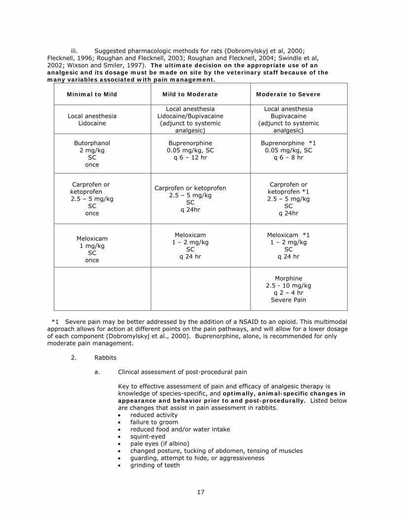
iii. Suggested pharmacologic methods for rats (Dobromylskyj et al, 2000; Flecknell, 1996; Roughan and Flecknell, 2003; Roughan and Flecknell, 2004; Swindle et al, 2002; Wixson and Smiler, 1997). The ultimate decision on the appropriate use of an analgesic and its dosage must be made on site by the veterinary staff because of the many variables associated with pain management.
Minimal to Mild
Mild to Moderate Moderate to Severe
Local anesthesia
Lidocaine
Local anesthesia Lidocaine/Bupivacaine (adjunct to systemic
analgesic)
Local anesthesia Bupivacaine
(adjunct to systemic analgesic)
Butorphanol 2 mg/kg
SC once
Buprenorphine 0.05 mg/kg, SC
q 6 – 12 hr
Buprenorphine *1 0.05 mg/kg, SC
q 6 – 8 hr
Carprofen or ketoprofen
2.5 – 5 mg/kg SC
once
Carprofen or ketoprofen
2.5 – 5 mg/kg SC
q 24hr
Carprofen or
ketoprofen *1 2.5 – 5 mg/kg
SC q 24hr
Meloxicam 1 mg/kg
SC once
Meloxicam
1 – 2 mg/kg SC
q 24 hr
Meloxicam *1 1 – 2 mg/kg
SC q 24 hr
Morphine
2.5 - 10 mg/kg q 2 – 4 hr
Severe Pain
*1 Severe pain may be better addressed by the addition of a NSAID to an opioid. This multimodal approach allows for action at different points on the pain pathways, and will allow for a lower dosage of each component (Dobromylskyj et al., 2000). Buprenorphine, alone, is recommended for only moderate pain management. 2. Rabbits a. Clinical assessment of post-procedural pain
Key to effective assessment of pain and efficacy of analgesic therapy is knowledge of species-specific, and optimally, animal-specific changes in appearance and behavior prior to and post-procedurally. Listed below are changes that assist in pain assessment in rabbits. • reduced activity • failure to groom • reduced food and/or water intake • squint-eyed • pale eyes (if albino) • changed posture, tucking of abdomen, tensing of muscles • guarding, attempt to hide, or aggressiveness • grinding of teeth
17
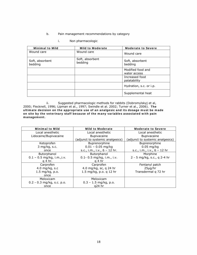
b. Pain management recommendations by category i. Non pharmacologic
Minimal to Mild Mild to Moderate Moderate to Severe Wound care
Wound care
Wound care
Soft, absorbent bedding
Soft, absorbent bedding
Soft, absorbent bedding
Modified food and water access
Increased food palatability
Hydration, s.c. or i.p.
Supplemental heat
ii. Suggested pharmacologic methods for rabbits (Dobromylskyj et al, 2000; Flecknell, 1996; Lipman et al., 1997; Swindle et al. 2002; Turner et al., 2006). The ultimate decision on the appropriate use of an analgesic and its dosage must be made on site by the veterinary staff because of the many variables associated with pain management.
Minimal to Mild Mild to Moderate Moderate to Severe Local anesthetic
Lidocaine/Bupivacaine
Local anesthetic Bupivacaine
(adjunct to systemic analgesics)
Local anesthetic Bupivacaine
(adjunct to systemic analgesics) Ketoprofen
3 mg/kg, s.c. once
Buprenorphine 0.01 – 0.05 mg/kg
s.c., i.m., i.v., 6 – 12 hr.
Buprenorphine 0.05 mg/kg
s.c., i.m., i.v., 6 – 12 hr Butorphanol
0.1 – 0.5 mg/kg, i.m.,i.v. q 4 hr.
Butorphanol 0.1- 0.5 mg/kg, i.m., i.v.
q 4 hr
Morphine 2 - 5 mg/kg, s.c., q 2-4 hr
Carprofen 4.0 mg/kg, s.c 1.5 mg/kg, p.o.
once
Carprofen 4.0 mg/kg, sc, q 24 hr 1.5 mg/kg, p.o. q 12 hr
Fentanyl patch 25µg/hr
Transdermal q 72 hr
Meloxicam 0.2 - 0.3 mg/kg, s.c. p.o.
once
Meloxicam 0.3 – 1.5 mg/kg, p.o.
q24 hr
18
3. Hamsters, Gerbils and Guinea pigs
There is scant information available on the efficacy of analgesic agents in hamsters, gerbils and guinea pigs. Information that has been published reflects authors’ personal experiences or limited clinical reports, but not well controlled clinical studies. The most frequently recommended analgesic in these 3 species is buprenorphine at 0.01 – 0.05 mg/kg, q 8 hr, s.c.
Similarly to pain assessment for rats and mice, persons responsible for surgical procedures and post-procedural care of hamsters, gerbils and guinea pigs should be familiar with the normal behavior and appearance of these species to enable an assessment of pain-induced modified behaviors and appearance.
19
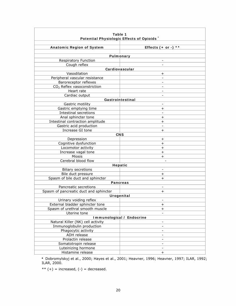
Table 1
Potential Physiologic Effects of Opioids *
Anatomic Region of System Effects (+ or -) **
Pulmonary
Respiratory Function - Cough reflex -
Cardiovascular Vasodilation +
Peripheral vascular resistance - Baroreceptor reflexes -
CO2 Reflex vasoconstriction - Heart rate -
Cardiac output - Gastrointestinal
Gastric motility - Gastric emptying time + Intestinal secretions - Anal sphincter tone +
Intestinal contraction amplitude + Gastric acid production -
Increase GI tone + CNS
Depression + Cognitive dysfunction +
Locomotor activity + Increase vagal tone +
Miosis + Cerebral blood flow -
Hepatic Biliary secretions - Bile duct pressure +
Spasm of bile duct and sphincter + Pancreas
Pancreatic secretions - Spasm of pancreatic duct and sphincter +
Urogenital Urinary voiding reflex -
External bladder sphincter tone + Spasm of urethral smooth muscle +
Uterine tone - Immunological / Endocrine
Natural Killer (NK) cell activity - Immunoglobulin production -
Phagocytic activity - ADH release -
Prolactin release - Somatotropin release - Luteinizing hormone - Histamine release +
* Dobromylskyj et al., 2000; Hayes et al., 2001; Heavner, 1996; Heavner, 1997; ILAR, 1992; ILAR, 2000.
** (+) = increased, (-) = decreased.
20
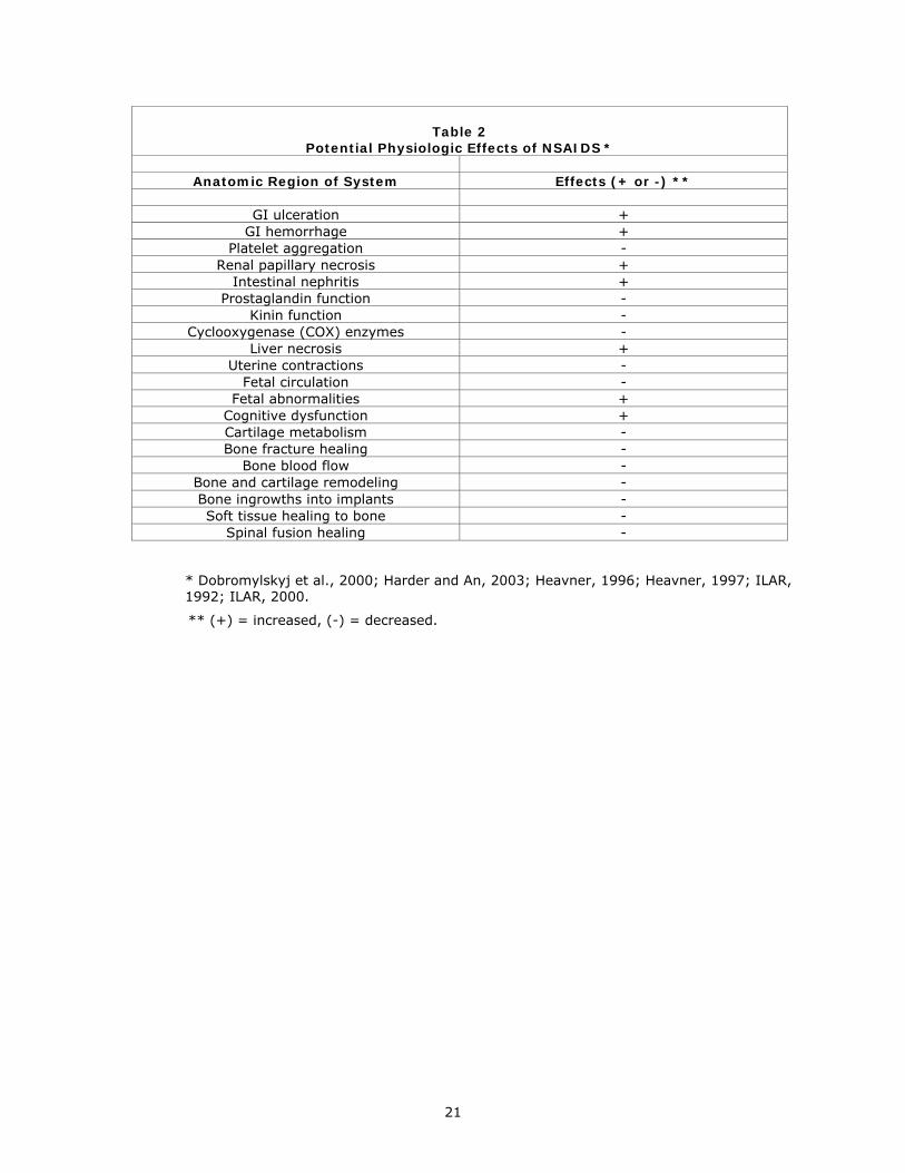
Table 2
Potential Physiologic Effects of NSAIDS *
Anatomic Region of System Effects (+ or -) **
GI ulceration + GI hemorrhage +
Platelet aggregation - Renal papillary necrosis +
Intestinal nephritis + Prostaglandin function -
Kinin function - Cyclooxygenase (COX) enzymes -
Liver necrosis + Uterine contractions -
Fetal circulation - Fetal abnormalities +
Cognitive dysfunction + Cartilage metabolism - Bone fracture healing -
Bone blood flow - Bone and cartilage remodeling - Bone ingrowths into implants - Soft tissue healing to bone -
Spinal fusion healing -
* Dobromylskyj et al., 2000; Harder and An, 2003; Heavner, 1996; Heavner, 1997; ILAR, 1992; ILAR, 2000.
** (+) = increased, (-) = decreased.
21
References Cooper, D.M., W. Hoffman, N. Wheat, and Hsiu-Yung Lee. 2005. Duration of effects on clinical parameters and referred hyperalgesia in rats after abdominal surgery and multiple doses of analgesic. Comp. Med. 55:344-352. Dobromylskyj, P., P.A.Flecknell, B.D. Lascelles, P.J. Pascoe, P. Taylor, A. Waterman-Pearson. 2000. Management of postoperative and other acute pain. In Pain Management in Animals, ed. P. Flecknell and A. Waterman-Pearson. W.B. Saunders, London. Flecknell, P.A., 1996. “Laboratory Animal Anesthesia”, 2nd Ed. Elsevier Academic Press, Inc., San Diego, CA. Flecknell P.A., J.V. Roughan, and R. Stewart. 1999. Use of oral buprenorphine (‘buprenorphine jello’) for postoperative analgesia in rats – a Clinical trial. Lab. Anim. 33:169-174. Gades, N.M., P.J. Danneman, S.K. Wixson, and E.A. Tolley. 2001. The magnitude and duration of the analgesic effect of morphine, butorphanol, and buprenorphine in rats and mice. Contemp. Topics of Lab. Anim. Sci. 39(2): 8-13. Gillingham, M.B., M.D. Clark, E.M. Dahly, L.A. Krugner-Higby, and D.M. Ney. 2001. A comparison of two opioid analgesics for relief of visceral pain induced by intestinal resection in rats. Contemp. Topics Lab. Anim. Sci. 40:21-26. Goecke, J.C., H. Awad, J.C. Lawson, and G.P. Boivin. 2005. Evaluating postoperative analgesics in mice using telemetry. Comp. Med. 55:37-44. Grandin T., and M. Deesing. 2002. Distress in animals: is it fear, pain or physical stress? American Board of Veterinary Practitioners Symposium. Manhattan Beach, CA. Grimm, K.A., and E.M. Hardie. 2004. Protocols for managing pain in elective procedures. Managing Medical, Surgical, Chronic and Traumatic pain. Protocols and Nondrug Approaches. Proceedings of a Symposium held at North American Veterinary Conference, Western Veterinary Conference, American Animal Hospital Association Annual Meeting and American Veterinary Medical Association Annual Convention. 49-60. Grossman, S.A., V.R. Sheidler, K. Swedeen, J. Mucenski, and S. Piantadosi. 1991. Correlation of patient and caregiver ratings of cancer pain. J. Pain Symptom. Manage. Feb; 6(2): 53-57. Hampshire, V. 2001. Handheld digital equipment for weight composite distress paradigms: new considerations and rapid documentation and intervention of rodent populations. Contemp. Top. Lab. Anim. Sci. 40: 11-7. Harder, A.T. and Y.H. An. 2003. The mechanism of the inhibitory effects of nonsteroidal anti-inflammatory drugs on bone healing: a concise review. J. Clin. Pharmacol. 43:807-815. Hawkins, P. 2002. Recognizing and assessing pain, suffering and distress in laboratory animals: a survey of current practices in the UK with recommendations. Lab. Anim. 36: 378-395. Hayes, K.E., J.A. Raucci, N.M. Gades, and L.A. Toth. 2001. An evaluation of analgesic regimens for abdominal surgery in mice. Contemp. Topics Lab. Anim. Sci. 39:18-23. Heavner, J.E., 1997. Pharmacology of analgesics, pp. 43-56. In Anesthesia and Analgesia in Laboratory Animals. D. Kohn, S. Wixson, W. White, and G. Benson (ed.), Academic Press, Inc., San Diego, CA. Heavner, J.E., 1996. Local anesthetics, pp. 330-336. In J.C. Thurmon, W.J. Tranquilli, and G.J. Benson (ed.), Lumb and Jones’ Veterinary Anesthesia, 3rd ed. Williams and Wilkins, Baltimore, MD.
22
Institute of Laboratory Animal Research (ILAR), Committee on Pain and Distress in Laboratory Animals, I.L.A.R., Recognition and Alleviation of Pain and Distress in Laboratory Animals. 1992, Washington DC, National Academy Press. 137. Institute of Laboratory Animal Research (ILAR), Definition of pain and distress and reporting requirements for laboratory animals. Proceedings of Workshop. 2000. Washington DC, National Academic Press. Karas, A.Z., K. Gostyla, M. Aronovitz, and R.H. Karas. 2001. Diminished body weight and activity patterns in mice following surgery: implications for control of post procedural pain/distress in laboratory animals. Contemp. Top. Lab. Anim. Sci. 40:83-87. Kremer, E. and J.H. Atkinson. 1981. Pain measurement: construct validity of the affective dimension of the McGill Pain Questionnaire with chronic benign pain patients. Pain 11(1): 93-100. Lipman, N.S., Marini, P.R., and Flecknell, P.A., Anesthesia and Analgesia in Rabbits. In Anesthesia and Analgesia in Laboratory Animals, D. Kohn, S. Wixson, W. White and G. Benson, (ed) 1997, Academic Press, San Diego. Loeser,J.D., Ed. In Bonica’s Management of Pain. 3rd ed. 2001, Lippincott Williams & Wilkins, Philadelphia. McMillan F.D. 2003. A world of hurts-JAVMA 223 (2):182-186. Marchand F., A. Alloui, T. Pelissier, A. Hernandez, N. Authier, P. Alvarez, A. Eschalier, D. Ardid. 2003. Evidence for an antihyperalgesic effect of venlafaxine in vincristine-induced neuropathy in rat. Brain Research. 980(1):117-20. Martin, L.B., A.C. Thompson, T. Martin, and M.B. Kristal. 2001. Analgesic efficacy of orally admin-istered buprenorphine. Comp. Med. 51:43-48. Merskey, H.M. and N. Bogduk, Eds. In Classification of Chronic Pain. 2nd ed. IASP Task Force on Taxonomy. 1994, IASP Press: Seattle. pp. 209-214. Morgan, D., C.D. Cook, M.J. Picker. 1999. Sensitivity to the discriminative stimulus and antinociceptive effects of mu opioids: role of strain of rat, stimulus intensity, and intrinsic efficacy at the mu opioid receptor. J. Pharmacol. Exp. Ther. 289:965-975. Peterson, N.C. 2004. Assessment of pain scoring. Contemp. Top. Lab. Anim. Sci. 43(1): 74-76. Quiding, H., E. Oksala, R.P. Happonen, K. Lehtimaki, and T. Ojala. 1981. The visual analog scale in multiple-dose evaluations of analgesics. J. Clin. Pharmacol. 21(10), 424-429. Raj, P.P.,In Practical Management of Pain. 3rd Ed. 2000, St. Louis. Mosby, Inc. Roughan, J.V., and P.A. Flecknell. 2000. Behavioral effects of laparotomy and analgesic effects of ketoprofen and carprofen in rats. Pain 90:65-74. Roughan J.V., P.A. Flecknell. 2002. Buprenorphine: a reappraisal of its antinociceptive effects and therapeutic use in alleviating post-operative pain in animals. Lab Anim 36 (3): 322-43. Roughan J.V., P.A. Flecknell. 2003. Evaluation of a short duration behaviour-based post-operative pain scoring system in rats. Eur. J. Pain. 7( 5): 397-406. Roughan, J.V. and P.A. Flecknell. 2004. Behaviour-based assessment of the duration of laparotomy-induced abdominal pain and the analgesic effects of carprofen and buprenorphine in rats. Behav. Pharmacol. 15:461-72. Schechter, N.L., B.A. Bernstein, A. Beck, L.Hart, and L. Scherzer. 1991. Individual differences in
23
children’s response to pain: role of temperament and parental characteristics. Pediatrics 87(2): 171-177. Semenova, S., A. Kuzmin, E. Zvartau. 1995. Strain differences in the analgesic and reinforcing action of morphine in mice. Pharmacol Biochem. Behav. 50:17-21. Shir, Y., R. Sheth, J.N. Campbell, S.N. Raja, Z. Seltzer. 2001. Soy-containing diet suppresses chronic neuropathic sensory disorders in rats. Anesth. Analg. 92(4): 1029-1034. Short, C.E. and A. van Poznak. Animal Pain. 1992, New York: Churchill Livingstone. 587. Speth, R.C., M.S. Smith, and R.S. Brogan. 2001. Regarding the inadvisability of administering postoperative analgesics in the drinking water of rats (Rattus Norvegicus). Contemp. Topics Lab. Anim. Sci. 40:15-17. Swindle, M.M., G.A. Vogler, L.K. Fulton, R.P. Marini, and S. Popilskis. 2002. Preanesthesia, anesthesia, analgesia, and euthanasia, pp. 956-1005. In Laboratory Animal Medicine, 2nd ed. J.G. Fox, L.C. Anderson, F.M. Loew, and F.W. Quimby (ed), Academic Press, Inc., San Diego, CA. Tamburini, M., S. Selmi, F. De Conno, and V. Ventafridda. 1987. Semantic descriptors of pain. Pain 29(2): 187-93. Thompson, A.C., M.B. Kristal, A. Sallaj, A. Acheson, L.B. Martin, and T. Martin. 2004. Analgesic efficacy of orally administered buprenorphine in rats: methodological considerations. Comp. Med. 54:2293-300. Thurmon, J.C., W.J. Tranquilli, and G.J. Benson. 1996. Perioperative pain and distress, pp. 40-61. In J.C. Thurmon, W.J. Tranquilli, and G.J. Benson (ed.), Lumb and Jones’ Veterinary Anesthesia, 3rd ed. Williams and Wilkins, Baltimore, MD. Turner, P.V., Chen, H.Cheng, and Taylor, W. M. 2006. Pharmacokinetics of meloxicam in rabbits after single and repeat oral dosing. Comp. Med. 56:63-67. Wixson, S.K. and Smiler, K.L., Anesthesia and Analgesia in Rodents. In Anesthesia and Analgesia in Laboratory Animals. D. Kohn, S. Wixson, W. White, and G. Benson. (ed), Academic Press, Inc., San Diego. Task Force Members Dennis F. Kohn Co-Chairman Thomas E. Martin Co-Chairman Patricia L. Foley Timothy H. Morris M. Michael Swindle George A. Vogler Sally K. Wixson July 2006
24