Embed Size (px)
Citation preview
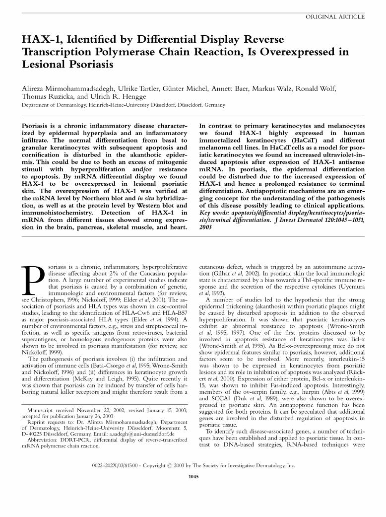
ORIGINAL ARTICLE
HAX-1, Identi¢ed by Di¡erential Display ReverseTranscription Polymerase Chain Reaction, Is Overexpressed inLesional Psoriasis
Alireza Mirmohammadsadegh, Ulrike Tartler, Gˇnter Michel, Annett Baer, MarkusWalz, RonaldWolf,Thomas Ruzicka, and Ulrich R. HenggeDepartment of Dermatology, Heinrich-Heine-University Dˇsseldorf, Dˇsseldorf, Germany
Psoriasis is a chronic in£ammatory disease character-ized by epidermal hyperplasia and an in£ammatoryin¢ltrate. The normal di¡erentiation from basal togranular keratinocytes with subsequent apoptosis andcorni¢cation is disturbed in the akanthotic epider-mis. This could be due to both an excess of mitogenicstimuli with hyperproliferation and/or resistanceto apoptosis. By mRNA di¡erential display we foundHAX-1 to be overexpressed in lesional psoriaticskin. The overexpression of HAX-1 was veri¢ed atthe mRNA level by Northern blot and in situ hybridiza-tion, as well as at the protein level by Western blot andimmunohistochemistry. Detection of HAX-1 inmRNA from di¡erent tissues showed strong expres-sion in the brain, pancreas, skeletal muscle, and heart.
In contrast to primary keratinocytes and melanocyteswe found HAX-1 highly expressed in humanimmortalized keratinocytes (HaCaT) and di¡erentmelanoma cell lines. In HaCaTcells as a model for psor-iatic keratinocytes we found an increased ultraviolet-in-duced apoptosis after expression of HAX-1 antisensemRNA. In psoriasis, the epidermal di¡erentiationcould be disturbed due to the increased expression ofHAX-1 and hence a prolonged resistance to terminaldi¡erentiation. Antiapoptotic mechanisms are an emer-ging concept for the understanding of the pathogenesisof this disease possibly leading to clinical applications.Key words: apoptosis/di¡erential display/keratinocytes/psoria-sis/terminal di¡erentiation. J Invest Dermatol 120:1045 ^1051,2003
Psoriasis is a chronic, in£ammatory, hyperproliferativedisease a¡ecting about 2% of the Caucasian popula-tion. A large number of experimental studies indicatethat psoriasis is caused by a combination of genetic,immunologic and environmental factors (for review,
see Christophers, 1996; Nickolo¡, 1999; Elder et al, 2001). The as-sociation of psoriasis and HLA types was shown in case-controlstudies, leading to the identi¢cation of HLA-Cw6 and HLA-B57as major psoriasis-associated HLA types (Elder et al, 1994). Anumber of environmental factors, e.g., stress and streptococcal in-fection, as well as speci¢c antigens from retroviruses, bacterialsuperantigens, or homologous endogenous proteins were alsoshown to be involved in psoriasis manifestation (for review, seeNickolo¡, 1999).The pathogenesis of psoriasis involves (i) the in¢ltration and
activation of immune cells (Bata-Csorgo et al, 1995;Wrone-Smithand Nickolo¡, 1996) and (ii) di¡erences in keratinocyte growthand di¡erentiation (McKay and Leigh, 1995). Quite recently itwas shown that psoriasis can be induced by transfer of cells har-boring natural killer receptors and might therefore result from a
cutaneous defect, which is triggered by an autoimmune activa-tion (Gilhar et al, 2002). In psoriatic skin the local immunologicstate is characterized by a bias towards aTh1-speci¢c immune re-sponse and the secretion of the respective cytokines (Uyemuraet al, 1993).A number of studies led to the hypothesis that the strong
epidermal thickening (akanthosis) within psoriatic plaques mightbe caused by disturbed apoptosis in addition to the observedhyperproliferation. It was shown that psoriatic keratinocytesexhibit an abnormal resistance to apoptosis (Wrone-Smithet al, 1995; 1997). One of the ¢rst proteins discussed to beinvolved in apoptosis resistance of keratinocytes was Bcl-x(Wrone-Smith et al, 1995). As Bcl-x-overexpressing mice do notshow epidermal features similar to psoriasis, however, additionalfactors seem to be involved. More recently, interleukin-15was shown to be expressed in keratinocytes from psoriaticlesions and its role in inhibition of apoptosis was analyzed (Rˇck-ert et al, 2000). Expression of either protein, Bcl-x or interleukin-15, was shown to inhibit Fas-induced apoptosis. Interestingly,members of the ov-serpin family, e.g., hurpin (Abts et al, 1999)and SCCA1 (Duk et al, 1989), were also shown to be overex-pressed in psoriatic skin. An antiapoptotic function has beensuggested for both proteins. It can be speculated that additionalgenes are involved in the disturbed regulation of apoptosis inpsoriatic tissue.To identify such disease-associated genes, a number of techni-
ques have been established and applied to psoriatic tissue. In con-trast to DNA-based strategies, RNA-based techniques were
Reprint requests to: Dr. Alireza Mirmohammadsadegh, Departmentof Dermatology, Heinrich-Heine-University Dˇsseldorf, Moorenstr. 5,D-40225 Dˇsseldorf, Germany, Email: [email protected]: DDRT-PCR, di¡erential display of reverse-transcribed
mRNA polymerase chain reaction.
Manuscript received November 22, 2002; revised January 15, 2003;accepted for publication January 26, 2003
0022-202X/03/$15.00 . Copyright r 2003 by The Society for Investigative Dermatology, Inc.
1045
shown to be more applicable to the isolation of psoriasis-speci¢cgenes. By di¡erential display of reverse-transcribed mRNApolymerase chain reaction (DDRT-PCR) (Liang and Pardee,1992) mRNA molecules are ampli¢ed using arbitrary PCR pri-mers and subsequently visualized by gel electrophoresis. A com-parison between two RNA populations, e.g., from psoriaticlesional and nonlesional skin, allows the identi¢cation of di¡er-entially expressed genes. Isolated PCR fragments can subse-quently be subcloned and sequenced.By using DDRT-PCRwe have searched for di¡erentially ex-
pressed genes in psoriatic skin by comparing RNA isolated fromlesional and nonlesional skin. Expression of the apoptosis-relatedgene HAX-1was further veri¢ed by Northern andWestern blotsusing skin biopsies, di¡erent melanoma cell lines, HaCaT cells,and keratinocytes. We further studied the antiapoptotic activityof HAX-1 in HaCaT cells, which exhibit proliferative activityand apoptosis resistance similar to psoriatic keratinocytes. A pos-sible role of HAX-1 in the process of apoptosis regulation inpsoriasis is discussed.
MATERIALS AND METHODS
Skin samples Split-thickness sections (0.3 mm), which werepredominantly composed of epidermis, were obtained from the backs ofpatients with untreated acute plaque-type psoriasis (Schulz et al, 1993). Forcontrol purposes, una¡ected skin specimens were obtained from clinicallynormal skin areas. Normal skin was donated by healthy volunteers. Allbiopsies were taken with the informed patient’s consent and approval ofthe local ethical committee.
Cell culture The spontaneously transformed human epidermal cell lineHaCaT and the human melanoma cell lines MV3, BLM, and SK-Mel-28were cultured in Dulbecco’s modi¢ed Eagle’s medium (Gibco BRL, LifeTechnologies, Eggenstein, Germany) supplemented with 10% fetal bovineserum, 100 U per ml penicillin, and 100 pg per ml streptomycin. Normalhuman keratinocytes were obtained by dispase treatment of breast skinspecimens (15�10 cm) and cultured under serum-free conditions inkeratinocyte basal medium (Biowhittaker, Vervier, Belgium). Primaryhuman melanocytes were obtained from PromoCell (Heidelberg,Germany) and cultured in phorbol-myristate-acetate-free medium(PromoCell). Cell culture was maintained in a 371C incubator in a moistatmosphere of 5% CO2.
RNA isolation Shave biopsies were snap frozen in liquid nitrogen.Biopsies were homogenized using a dismembrator (Braun-Melsungen,Melsungen, Germany). The powder was immediately transferred intoTrizolt reagent and total RNA was isolated according to themanufacturer’s instructions (Gibco BRL). Contaminating DNA wasremoved by treatment with DNase I (Boehringer Mannheim, Germany).RNA quality was controlled by agarose gel electrophoresis.
DDRT-PCR The DDRT-PCR technology was ¢rst described byLiang and Pardee (1992). The protocol used has been successfullyemployed for the identi¢cation of ultraviolet (UV) regulated genes(Abts et al, 1997) and details have been described earlier (Abts et al, 2000).Primers were obtained from GenHunter (RNAimaget, Kit 4, Cat.No. G504). Brie£y 1 mg each of total RNA prepared from lesionalpsoriatic and nonlesional skin specimens was split in three aliquotsand reverse transcribed in separate reactions by 200 U Superscript IIreverse transcriptase (Gibco BRL) using three HindIII-oligo-dT,one-base-anchored primers modi¢ed at their 30 ends to A, G, and C (H-T11N), and 20 mm of each dNTP according to Liang and Pardee (1992).Display PCR ampli¢cation of 2 ml of the cDNAs was carried outbetween the same anchored primer (each 0.2 mm) and one of 130merprimers with arbitrary sequence including an HindIII restriction site in atotal volume of 20 ml containing 2 U Taq polymerase and 0.4 ml [32p]-a-dCTP (4 mCi). The reactions were hot-started and cycled for 1min at 951C,1 min at 411C, and 2 min at 721C in the ¢rst PCR cycle, followed by 29cycles of 50 s at 941C, 1 min at 421C (þ temperature increment of 0.11Cper cycle), and 1 min at 721C. H-AP25 (GenHunter, RNAimaget, Kit 4,Cat. No. G504) was used as an upstream arbitrary primer. Radio-labeledPCR products corresponding to lesional and nonlesional skin wereloaded on a denaturing 6% polyacrylamide gel. The gels were run untilthe xylene cyanol dye reached the bottom, and then were blotted onto
Whatman 3M ¢lter paper and dried. Gels were exposed to X-ray ¢lm at�701C for 12 h.
Reampli¢cation After autoradiography, bands of at least 150 bpshowing di¡erent strength in corresponding lanes were cut out from thedried gel and eluted in 100 ml H2O. The cDNA was ethanol^glycogenprecipitated and the pellet was resuspended in 10 ml H2O. Using the sameprimer combination as for the display reaction 4 ml of the eluted materialwas reampli¢ed using a touch-down protocol in which the annealingtemperature was decreased from 501C to 451C during the ¢rst ¢ve cyclesand then kept constant at 451C for 34 additional cycles. An aliquot of thereaction was run on an agarose gel to con¢rm a reaction product.
Cloning Bluescript KS was digested with EcoRV and incubated withTaq polymerase (Amersham Biosciences, Freiburg, Germany) in thepresence of 1 mm TTP to add single base dT residues to the 30 endsmaking use of the non-template-dependent activity of the Taqpolymerase. Reampli¢ed cDNA fragments were gel-puri¢ed using thegel extraction kit (Qiagen, Hilden, Germany) and cloned into the above-described vector via the T/A cloning procedure.
Northern blot hybridization 20 mg of total RNA per lane frommultiple psoriasis patients as well as cultured normal keratinocytes andHaCaT cells were electrophoresed and blotted onto Hybond-Nþmembrane (Amersham Biosciences). In parallel, a commercially availablemultitissue Northern blot (Clontech, Heidelberg, Germany) washybridized. The probe was generated by random-primed labeling of thegel-puri¢ed insert. Hybridization was carried out in DIG Easy Hybsolution (Roche, Mannheim, Germany). The autoradiograph signals werequantitated by laser densitometry relative to the band intensities of thecorresponding 28S rRNA, which served as loading controls.
Preparation of protein extracts andWestern blot analysis The shavebiopsies were snap frozen in liquid nitrogen. Under constant supply ofliquid nitrogen biopsies were homogenized using a dismembrator (Braun-Melsungen). The powder was immediately transferred into ice-cold lysisbu¡er containing 25 mm hydroxyethylpiperazine ethanesulfonic acid pH7.9, 50 mM NaF, 15 mM Triton X-100, 5 mM ethylenediamine tetraaceticacid, 100 mM NaCl, and one tablet protease inhibitor cocktail per 10 mlbu¡er (Roche).Cells were washed twice with ice-cold phosphate-bu¡ered saline
(PBS) and lyzed in lysis bu¡er (see above). The samples were pretreatedwith ultrasound and cell extracts were centrifuged at 14,000g for 20 min at41C. The protein concentration of the cellular extracts was determinedusing the advanced protein assay reagent (TEBU, Frankfurt, Germany).Of the protein extract 20 mg were electrophoresed on 4%^12% NuPageBis-Tris-Glycin-Gels (Invitrogen, Karlsruhe, Germany) for 2 h at 120 V.Proteins were blotted onto polyvinylidene £uoride membranes (Bio-Rad,Munich, Germany) at 25 V for 90 min using a tank blot system.The membranes were blocked with 5% nonfat dry milk powder in 10mM Tris^HCl pH 7.5, 150 mM NaCl, and 0.05% Tween 20(TBST bu¡er) overnight at 41C, washed three times with TBST, andincubated with the primary monoclonal mouse antibody against HAX-1mouse IgG1 (BD Transduction Laboratories), dilution 1:5000 in TBSTwith 1% nonfat dry milk, overnight at 41C. The membranes were washedthree times with TBST and incubated with the respective secondaryantibody (anti mouse IgG, dilution 1:5000, Amersham-Pharmacia,Freiburg, Germany) and visualized using the enhanced chemilu-minescence detection system (Amersham-Pharmacia) following thesupplier’s instructions.
In situ hybridization Cryosections of lesional and nonlesional psoriaticskin on microscope slides were ¢xed in 4% paraformaldehyde/5%formaldehyde overnight at 41C. After washing in PBS the slides wereprehybridized in precoat solution, containing 5� sodium citrate/chloridebu¡er, 50% deionized formamide, 5% blocking reagent (Roche), 0.02%sodium dodecyl sulfate, 0.1% N-lauryl sarcosin, 50 mM Tris^HCl pH 7.4,at 451C for 1 h. The cRNA probes were synthesized using DIG-UTP andan in vitro transcription kit from Roche, following the manufacturer’sprotocol. As template for the reactions 1 mg of pCRII (Invitrogen) withthe HAX-1 fragment, linearized by XhoI, and SP6 polymerase forantisense and BamH I for T7 polymerase for sense orientation were used.25 ng per 50 ml of the labeled probes were denatured at 941C for 10 minand added to the prehybridized sections and hybridized overnight at 451Cunder a coverslip. Next, the sections were stringency washed with di¡erentsteps and temperatures. For detection, an alkaline-phosphatase-coupledanti-DIG-antibody and NTB/BCIP as substrate were used.
1046 MIRMOHAMMADSADEGH ETAL THE JOURNAL OF INVESTIGATIVE DERMATOLOGY
Sequence analysis Insert-containing plasmids were puri¢ed from 10 mlcultures using QIAprep-spin columns (Qiagen, Hilden, Germany),RNAse-treated, and sequenced using the Sequenase Kit from USB(Nebraska) and M13 sequencing primers. The sequences were compared tothe EMBL sequence databank entries by FASTA.
Immunohistochemistry Cryosections of psoriatic and healthy skinwere ¢xed in acetone for 20 min at ^201C and then incubated with apolyclonal rabbit anti human HAX-1 antibody (kind gift of Dr.Watanabe, Department of Molecular Immunology, Kyushu University,Japan) for 1 h. After three washing steps in PBS the bound primaryantibody was detected by sequential incubation with biotinylatedsecondary antibodies, peroxidase-coupled avidin, and peroxidase substrate(VectastainR ABC Kit, Burlingame, CA) as recommended by themanufacturer. To demonstrate the proliferative activity and to appreciatethe skin architecture parallel sections were stained with commerciallyavailable ready-to-use anti-Ki67 antibody (Dako, Hamburg, Germany)and counterstained with hemalaun.
Transfection and caspase assay Full-length HAX-1 cDNA (accessionno. U68566) was ampli¢ed with a speci¢c 50 primer and an oligo dTprimer for the 30 end and cloned into pCRII TA plasmid (Invitrogen).Subsequently, HAX-1 cDNA was subcloned in sense and antisenseorientation into pIRES-EGFP (Clontech, Palo Alto, CA).1�106 HaCaT cells were transfected with 1 mg of endotoxin-free
plasmid DNA of the HAX^EGFP constructs by electroporation (Amaxa,Cologne, Germany). The transfection e⁄ciency was determined after anadditional 16 h incubation by counting the number of green £uorescentcells relative to the total number of cells in three di¡erent microscopy¢elds.Caspase-3 activity was measured using the ApoAlert CPP32 protease
assay kit (Clontech). The transiently transfected cells were exposed to 280J per m2 UVB radiation as described earlier (Abts et al, 2000) and furtherincubated for 6 h in new standard culture medium with supplements/serum. Controls were mock-treated. 1.85�105 of each of the transfectedcell populations as well as nontransfected cells were lyzed. Afterincubation with the substrate (DEVD-AFC) for 1 h, caspase-3 activity wasobtained by measuring the £uorescence emission in a £uorometer(Fluoroscan Ascent Labsystems) (ex 400 nm, em 505 nm). The emissionvalues were corrected for the respective transfection e⁄ciencies: 30% forHAX-1 in sense orientation, 35% for the antisense construct, and 50% forpIRES without insert.
RESULTS
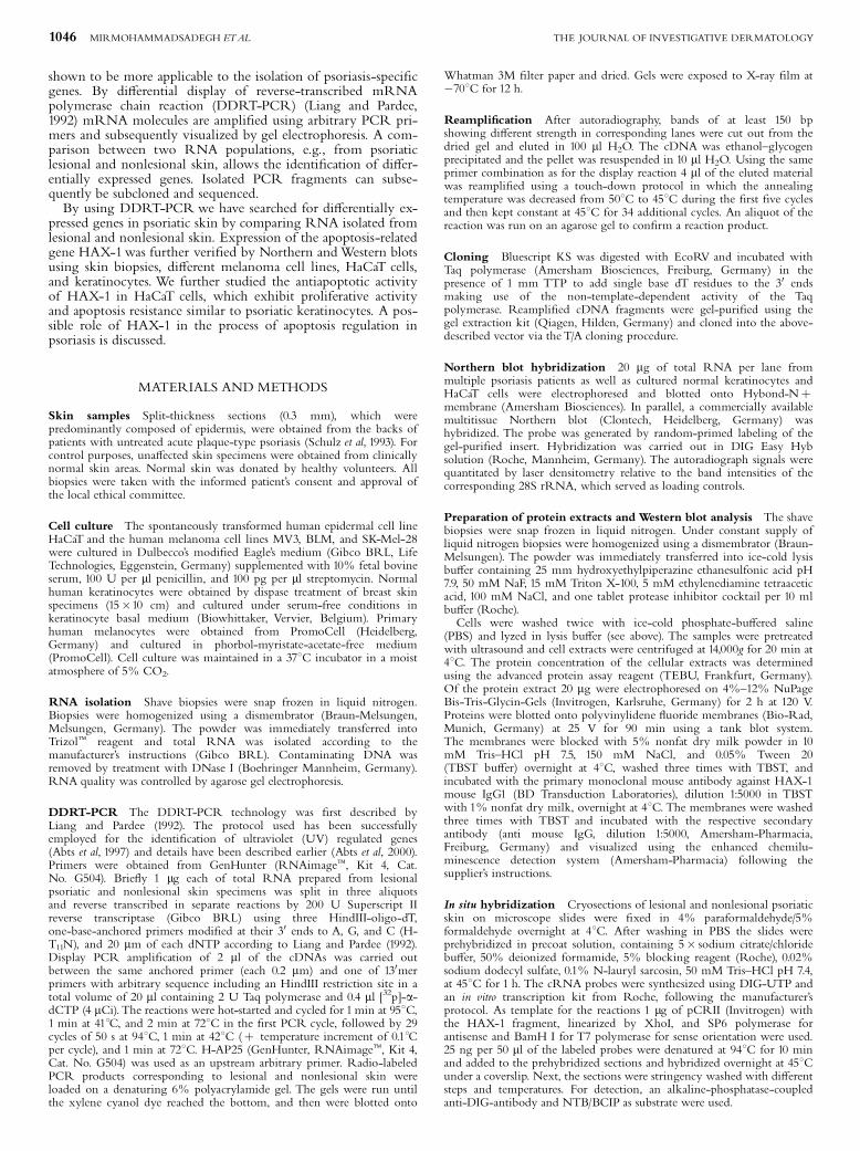
RNA ¢ngerprints were generated from psoriatic lesional andnonlesional skin using DDRT-PCR (Fig 1). For each primercombination more than 100 bands were displayed per lane. Useof individual primer combinations resulted in a large number ofidentical fragments in comparable RNA populations studied.Several products showed di¡erent band intensities re£ecting dif-ferent quantities of the corresponding mRNAs. The parallel ana-lysis of four to ¢ve related RNA populations helped todiscriminate bands that varied randomly between lanes, thus re-ducing the risk of isolating false positives. Independent DDRT-PCR analysis showed that the band pattern obtained for a givenprimer combination and cDNA preparation was reproducible(Fig 1). The complex pattern revealed several genes that wereupregulated or downregulated in lesional psoriatic skin(Fig 1). These bands were eluted from the gel, reampli¢ed,and cloned as described above.We further analyzed one particularclone (clone 2) that was di¡erentially expressed in all threepsoriasis patients in more detail. The product was generatedby ampli¢cation using HindIII-oligo-T(11)A and H-AP25 asprimers.The initial veri¢cation was performed using slot blot analysis. 1
mg plasmid DNA of the cloned cDNA fragments was immobi-lized onto nylon membranes under denaturing conditions. Hy-bridization was performed using radioactively labeled cDNA (1mg) from lesional and nonlesional skin (data not shown).Although the hybridization con¢rmed the overexpression ofclone 2, clones 8 and 12 were not di¡erentially expressed. Conse-quently, further experiments were performed with clone 2.
Identi¢cation of di¡erential cDNAs by sequenceanalysis The reampli¢ed and gel-puri¢ed cDNAs were clonedand sequenced. Sequence analysis of clone 2 and subsequentFASTA comparison in the EMBL sequence databank revealed98% homology over a 455 bp stretch of clone 2 to the 30translation region of the described 1196 bp HAX-1 codingsequence from lymphocytes (Suzuki et al, 1997). Sequencehomology of the clone 2 fragment included bases 717^1172 ofthe entire HAX-1 cDNA.
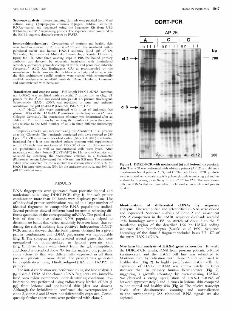
Northern blot analysis of HAX-1 gene expression To verifythe DDRT-PCR results, RNA from psoriasis patients, culturedkeratinocytes, and the HaCaT cell line was submitted toNorthern blot hybridization with clone 2 and compared tohealthy skin (Fig 2). In highly proliferative HaCaT cells theexpression of HAX-1 mRNA was approximately 13 timesstronger than in primary human keratinocytes (Fig 2),suggesting a growth advantage by overexpressing HAX-1.We observed a strong upregulation of HAX-1 mRNA ofbetween approximately 2 and 16 times in lesional skin comparedto nonlesional and healthy skin (Fig 2). The relative transcriptlevels after densitometric scanning and normalizationto the corresponding 28S ribosomal RNA signals are alsodepicted.
Figure1. DDRT-PCRwith nonlesional (n) and lesional (l) psoriaticskin.The PCRwas performed with arbitrary primer (AP) 25 and di¡erentone-base-anchored primers A, G, and C. The radiolabeled PCR productswere separated on a denaturing 6% polyacrylamide sequencing gel and vi-sualized by exposing to an X-ray ¢lm at ^701C for 12 h. The arrow showsdi¡erent cDNAs that are dysregulated in lesional versus nonlesional psoria-tic skin.
HAX-1 IN PSORIASIS 1047VOL. 120, NO. 6 JUNE 2003
Using multiple tissue blot, the distribution of HAX-1mRNAshowed a ubiquitous expression pattern with the strongest signalsin skeletal and heart muscle (Fig 3).
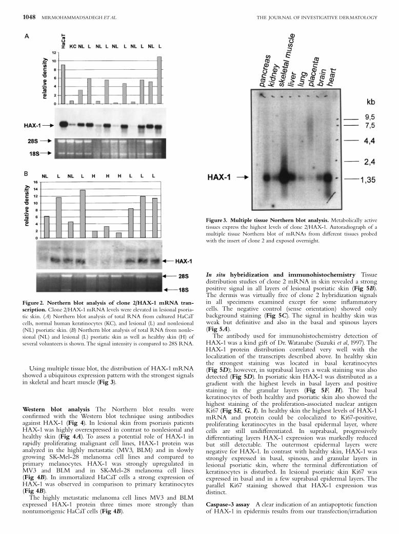
Western blot analysis The Northern blot results werecon¢rmed with the Western blot technique using antibodiesagainst HAX-1 (Fig 4). In lesional skin from psoriasis patientsHAX-1 was highly overexpressed in contrast to nonlesional andhealthy skin (Fig 4A). To assess a potential role of HAX-1 inrapidly proliferating malignant cell lines, HAX-1 protein wasanalyzed in the highly metastatic (MV3, BLM) and in slowlygrowing SK-Mel-28 melanoma cell lines and compared toprimary melanocytes. HAX-1 was strongly upregulated inMV3 and BLM and in SK-Mel-28 melanoma cell lines(Fig 4B). In immortalized HaCaT cells a strong expression ofHAX-1 was observed in comparison to primary keratinocytes(Fig 4B).The highly metastatic melanoma cell lines MV3 and BLM
expressed HAX-1 protein three times more strongly thannontumorigenic HaCaT cells (Fig 4B).
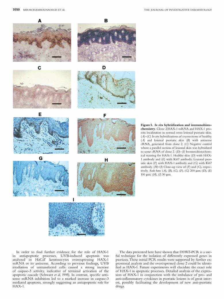
In situ hybridization and immunohistochemistry Tissuedistribution studies of clone 2 mRNA in skin revealed a strongpositive signal in all layers of lesional psoriatic skin (Fig 5B).The dermis was virtually free of clone 2 hybridization signalsin all specimens examined except for some in£ammatorycells. The negative control (sense orientation) showed onlybackground staining (Fig 5C). The signal in healthy skin wasweak but de¢nitive and also in the basal and spinous layers(Fig 5A).The antibody used for immunohistochemistry detection of
HAX-1was a kind gift of Dr.Watanabe (Suzuki et al, 1997). TheHAX-1 protein distribution correlated very well with thelocalization of the transcripts described above. In healthy skinthe strongest staining was located in basal keratinocytes(Fig 5D); however, in suprabasal layers a weak staining was alsodetected (Fig 5D). In psoriatic skin HAX-1 was distributed as agradient with the highest levels in basal layers and positivestaining in the granular layers (Fig 5F, H). The basalkeratinocytes of both healthy and psoriatic skin also showed thehighest staining of the proliferation-associated nuclear antigenKi67 (Fig 5E, G, I). In healthy skin the highest levels of HAX-1mRNA and protein could be colocalized to Ki67-positive,proliferating keratinocytes in the basal epidermal layer, wherecells are still undi¡erentiated. In suprabasal, progressivelydi¡erentiating layers HAX-1 expression was markedly reducedbut still detectable. The outermost epidermal layers werenegative for HAX-1. In contrast with healthy skin, HAX-1 wasstrongly expressed in basal, spinous, and granular layers inlesional psoriatic skin, where the terminal di¡erentiation ofkeratinocytes is disturbed. In lesional psoriatic skin Ki67 wasexpressed in basal and in a few suprabasal epidermal layers. Theparallel Ki67 staining showed that HAX-1 expression wasdistinct.
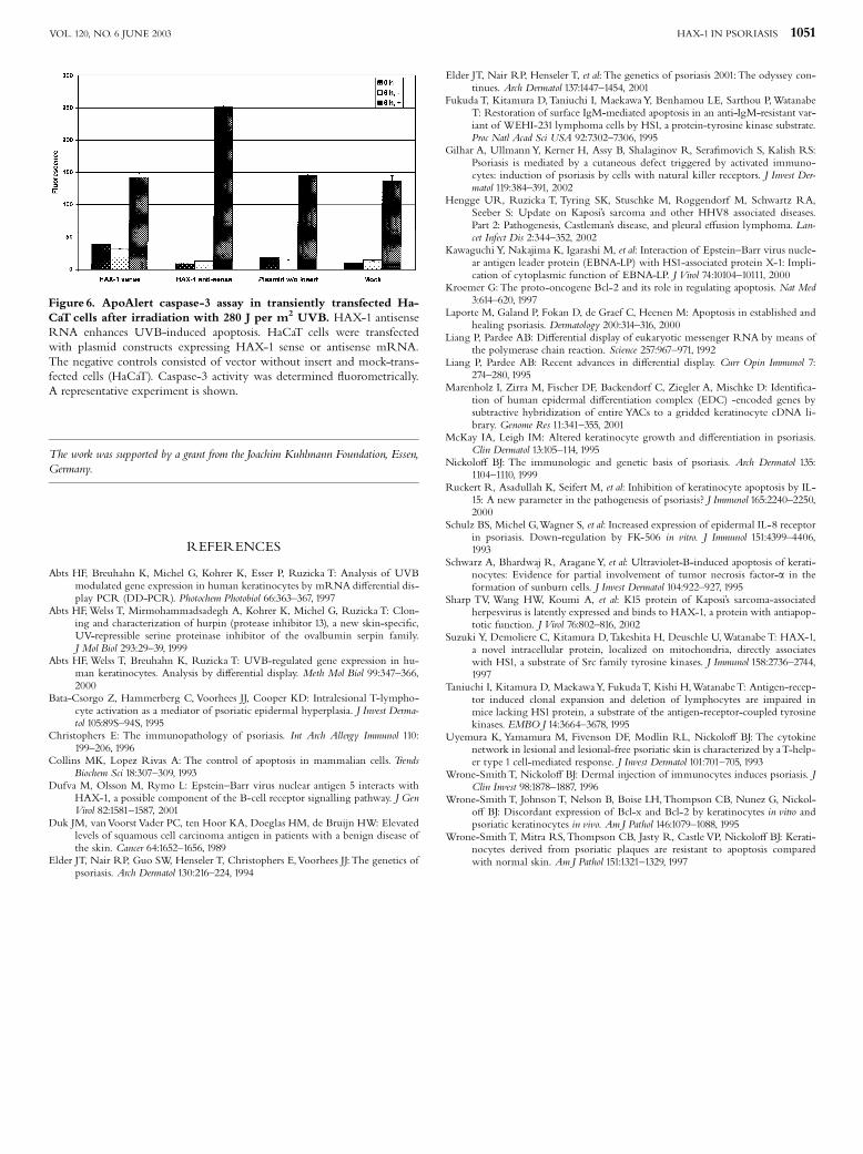
Caspase-3 assay A clear indication of an antiapoptotic functionof HAX-1 in epidermis results from our transfection/irradiation
Figure 2. Northern blot analysis of clone 2/HAX-1 mRNA tran-scription. Clone 2/HAX-1mRNA levels were elevated in lesional psoria-tic skin. (A) Northern blot analysis of total RNA from cultured HaCaTcells, normal human keratinocytes (KC), and lesional (L) and nonlesional(NL) psoriatic skin. (B) Northern blot analysis of total RNA from nonle-sional (NL) and lesional (L) psoriatic skin as well as healthy skin (H) ofseveral volunteers is shown.The signal intensity is compared to 28S RNA.
Figure 3. Multiple tissue Northern blot analysis. Metabolically activetissues express the highest levels of clone 2/HAX-1. Autoradiograph of amultiple tissue Northern blot of mRNAs from di¡erent tissues probedwith the insert of clone 2 and exposed overnight.
1048 MIRMOHAMMADSADEGH ETAL THE JOURNAL OF INVESTIGATIVE DERMATOLOGY
experiments. The induction of apoptosis by UV light wasdescribed earlier (Schwarz et al, 1995). UV irradiation caused anapproximately 10-fold induction of caspase-3 activity in HaCaTcells (Fig 6). Enzyme activity could be further increased to alevel of 20-fold compared to unirradiated cells when a plasmidconstruct expressing HAX-1 antisense mRNA was transfectedinto the cells. Transfection of both HAX-1 in sense orientationor a control vector without the insert led to an increase incaspase activity in irradiated cells of up to the level ofuntransfected cells.
DISCUSSION
Psoriasis is a chronic in£ammatory disease characterizedby increased proliferation and altered di¡erentiation of keratino-cytes, in the context of vascular alterations and epidermal in¢ltra-tion of activated T lymphocytes. In order to characterizegenes di¡erently expressed in psoriasis, gene expression withinpsoriatic lesions was compared to healthy skin using DDRT-PCR. Compared to cDNA arrays, the DDRT-PCR technique
allows identi¢cation and isolation of a broad variety of di¡erentlyregulated genes without preselection and independent of any ex-isting gene sequence. By using this technique, the di¡erent ex-pression of a number of clones was veri¢ed by slot blot analysisto prevent further analysis of false positives, a major drawback ofthe DDRT-PCR technique (Liang and Pardee, 1995). As a resultof this, the di¡erential expression of clone 2 was further substan-tiated on the transcription and protein level using Northern andWestern blot analysis, respectively.Sequence analysis in the gene bank found clone 2 to be identi-
cal to the cDNA sequence encoding for the HS1-binding proteinHAX 1. This 35 kDa protein was initially identi¢ed in lymphaticcells and shown to in£uence the B cell receptor (BCR) signalingpathway by interaction with HS1, potentially counteracting thecell-death function of HS-1 in lymphocytes (Fukuda et al, 1995;Taniuchi et al, 1995; Suzuki et al, 1997).As we found HAX-1 to be upregulated in psoriatic lesions, a
functional relevance in human skin can be assumed. Subse-quently, Northern andWestern blot analysis con¢rmed the ratherweak HAX-1 expression in healthy and nonlesional skin com-pared to hyperproliferative psoriatic lesions as initially shown byDDRT-PCR analysis. The genomic localization within the epi-dermal di¡erentiation complex on chromosome 1q21 suggests aregulating function during the epidermal maturation process(Marenholz et al, 2001). Analysis of the HAX-1 amino acidsequence revealed similarity to the functionally important do-mains BH1 and BH2 of the Bcl-2 subfamily of proteins, whichcounteract apoptosis (Kroemer, 1997). These proteins are, asHAX-1, mainly located in mitochondria, nuclear envelope, andendoplasmatic reticulum (Suzuki et al, 1997). According to thestructural and locational homologies to antiapoptotic proteins,the authors speculated that HAX-1 promotes cell survival by in-hibiting apoptosis.Indeed, tissues with high numbers of mitochondria, indicative
for high energy metabolism, such as brain, heart, skeletal muscles,and pancreas, show high HAX-1 levels (Fig 5). Assuming an anti-apoptotic activity, increased HAX-1 expression could be assumedto protect from cell death due to respiratory stress in tissues withhigh energy metabolism. In the opposite case, placental tissuepresenting a rather low level metabolism with a high turnoverrate shows only low HAX-1 levels.Further supportive ¢ndings for involvement of HAX-1 in
apoptotic processes reveal sequence homology of HAX-1 toNip3, an adenovirus E1B interacting protein that itself hasbeen shown to interact with several antiapoptotic viral andcellular proteins, including Bcl-2 and BHRF-1 (Sharp et al,2002). Consistent with the regulatory function of Nip3 inapoptosis, a T lymphoma cell line acquired resistance toapoptosis following various stimuli, including Fas treatment, g-irradiation, and serum deprivation, when HAX-1 was overex-pressed (Suzuki et al, 1997). Moreover, several proteins have re-cently been identi¢ed to interact with HAX-1, such as theEpstein^Barr virus nuclear antigen 5 and latent protein, whichmay be involved in immortalization of B lymphocytes (Kawagu-chi et al, 2000; Dufva et al, 2001), or the K15 protein of Kaposi’ssarcoma-associated herpesvirus, which may regulate viral latencyor promote growth of the infected cells (Hengge et al, 2002;Sharp et al, 2002). The expression of HAX-1 in several cell linesin contrast to their primary counterparts suggests a broader rolein immortalization.Beside an increased proliferation rate a diminished susceptibil-
ity to apoptosis is assumed heading to epidermal hyper-proliferation in psoriasis (Collins and Lopez Rivas, 1993;Laporte et al, 2000). Indeed, keratinocytes isolated frompsoriatic lesions showed an increased expression of theantiapoptotic Bcl-x that can be further induced by interferon-g,one of the key cytokines in the pathogenesis of psoriasis(Nickolo¡, 1999). The induction of antiapoptotic proteinstogether with antiproliferative e¡ects of interferon-g inpsoriasis indicate that proliferation and antiapoptosis areindependent.
Figure 4. Western blot analysis of clone 2/HAX-1 expression.To con-¢rm protein expressionWestern blot analysis was performed on protein ex-tracts of nonlesional (nl) versus lesional (l) psoriatic skin of three di¡erentpatients (A). (B) Expression of HAX-1 primary keratinocytes versus HaCaTcells, and in di¡erent melanoma cell lines versus primary melanocytes,respectively: 1, primary melanocytes; 2, MV3; 3, BLM; 4, SK-Mel-28; 5,HaCaT; 6, primary keratinocytes. The signal intensity is compared toa-tubulin.
HAX-1 IN PSORIASIS 1049VOL. 120, NO. 6 JUNE 2003
In order to ¢nd further evidence for the role of HAX-1in antiapoptotic processes, UVB-induced apoptosis wasanalyzed in HaCaT keratinocytes overexpressing HAX-1mRNA or its antisense. According to previous ¢ndings, UVBirradiation of untransfected cells caused a strong increaseof caspase-3 activity, indicative of terminal activation of theapoptotic cascade (Schwarz et al, 1995). In contrast, speci¢c anti-sense mRNA inhibition led to a marked increase in caspase-3mediated apoptosis, strongly suggesting an antiapoptotic role forHAX-1.
The data presented here have shown that DDRT-PCR is a use-ful technique for the isolation of di¡erently expressed genes inpsoriasis. These initial PCR results were supported by further ex-pressional analysis and the overexpressed clone 2 could be identi-¢ed as HAX-1. Future experiments will elucidate the exact roleof HAX-1 in apoptotic processes. Detailed analysis of the expres-sion of HAX-1 in conjunction with the imbalance of pro- andanti-in£ammatory cytokines in psoriatic lesions is of great inter-est, possibly facilitating the development of new anti-psoriaticdrugs.
Figure 5. In situ hybridization and immunohisto-chemistry. Clone 2/HAX-1 mRNA and HAX-1 pro-tein localization in normal versus lesional psoriatic skin.(A)^(C) In situ hybridizations of cryosections of healthy(A) and lesional psoriatic skin (B) with antisensecRNA, generated from clone 2. (C) Negative controlwhere a parallel section of lesional skin was hybridizedto sense cRNA of clone 2. (D)^(I) Immunohistochem-ical staining for HAX-1. Healthy skin (D) with HAX-1 antibody and (E) with Ki67 antibody. Lesional psor-iatic skin (F) with HAX-1 antibody and (G) with Ki67antibody. (H)^(I) Close-up view of (F) and (G), respec-tively. Scale bars: (A), (B), (C), (F), (G) 200 mm; (D), (E)100 mm; (H), (I) 50 mm.
1050 MIRMOHAMMADSADEGH ETAL THE JOURNAL OF INVESTIGATIVE DERMATOLOGY
The work was supported by a grant from the Joachim Kuhlmann Foundation, Essen,Germany.
REFERENCES
Abts HF, Breuhahn K, Michel G, Kohrer K, Esser P, Ruzicka T: Analysis of UVBmodulated gene expression in human keratinocytes by mRNA di¡erential dis-play PCR (DD-PCR). Photochem Photobiol 66:363^367, 1997
Abts HF,Welss T, Mirmohammadsadegh A, Kohrer K, Michel G, Ruzicka T: Clon-ing and characterization of hurpin (protease inhibitor 13), a new skin-speci¢c,UV-repressible serine proteinase inhibitor of the ovalbumin serpin family.J Mol Biol 293:29^39, 1999
Abts HF,Welss T, Breuhahn K, Ruzicka T: UVB-regulated gene expression in hu-man keratinocytes. Analysis by di¡erential display. Meth Mol Biol 99:347^366,2000
Bata-Csorgo Z, Hammerberg C, Voorhees JJ, Cooper KD: Intralesional T-lympho-cyte activation as a mediator of psoriatic epidermal hyperplasia. J Invest Derma-tol 105:89S^94S, 1995
Christophers E: The immunopathology of psoriasis. Int Arch Allergy Immunol 110:199^206, 1996
Collins MK, Lopez Rivas A: The control of apoptosis in mammalian cells. TrendsBiochem Sci 18:307^309, 1993
Dufva M, Olsson M, Rymo L: Epstein^Barr virus nuclear antigen 5 interacts withHAX-1, a possible component of the B-cell receptor signalling pathway. J GenVirol 82:1581^1587, 2001
Duk JM, vanVoorst Vader PC, ten Hoor KA, Doeglas HM, de Bruijn HW: Elevatedlevels of squamous cell carcinoma antigen in patients with a benign disease ofthe skin. Cancer 64:1652^1656, 1989
Elder JT, Nair RP, Guo SW, Henseler T, Christophers E,Voorhees JJ: The genetics ofpsoriasis. Arch Dermatol 130:216^224, 1994
Elder JT, Nair RP, Henseler T, et al: The genetics of psoriasis 2001: The odyssey con-tinues. Arch Dermatol 137:1447^1454, 2001
Fukuda T, Kitamura D,Taniuchi I, MaekawaY, Benhamou LE, Sarthou P,WatanabeT: Restoration of surface IgM-mediated apoptosis in an anti-IgM-resistant var-iant of WEHI-231 lymphoma cells by HS1, a protein-tyrosine kinase substrate.Proc Natl Acad Sci USA 92:7302^7306, 1995
Gilhar A, UllmannY, Kerner H, Assy B, Shalaginov R, Sera¢movich S, Kalish RS:Psoriasis is mediated by a cutaneous defect triggered by activated immuno-cytes: induction of psoriasis by cells with natural killer receptors. J Invest Der-matol 119:384^391, 2002
Hengge UR, Ruzicka T, Tyring SK, Stuschke M, Roggendorf M, Schwartz RA,Seeber S: Update on Kaposi’s sarcoma and other HHV8 associated diseases.Part 2: Pathogenesis, Castleman’s disease, and pleural e¡usion lymphoma. Lan-cet Infect Dis 2:344^352, 2002
Kawaguchi Y, Nakajima K, Igarashi M, et al: Interaction of Epstein^Barr virus nucle-ar antigen leader protein (EBNA-LP) with HS1-associated protein X-1: Impli-cation of cytoplasmic function of EBNA-LP. J Virol 74:10104^10111, 2000
Kroemer G: The proto-oncogene Bcl-2 and its role in regulating apoptosis. Nat Med3:614^620, 1997
Laporte M, Galand P, Fokan D, de Graef C, Heenen M: Apoptosis in established andhealing psoriasis. Dermatology 200:314^316, 2000
Liang P, Pardee AB: Di¡erential display of eukaryotic messenger RNA by means ofthe polymerase chain reaction. Science 257:967^971, 1992
Liang P, Pardee AB: Recent advances in di¡erential display. Curr Opin Immunol 7:274^280, 1995
Marenholz I, Zirra M, Fischer DF, Backendorf C, Ziegler A, Mischke D: Identi¢ca-tion of human epidermal di¡erentiation complex (EDC) -encoded genes bysubtractive hybridization of entire YACs to a gridded keratinocyte cDNA li-brary. Genome Res 11:341^355, 2001
McKay IA, Leigh IM: Altered keratinocyte growth and di¡erentiation in psoriasis.Clin Dermatol 13:105^114, 1995
Nickolo¡ BJ: The immunologic and genetic basis of psoriasis. Arch Dermatol 135:1104^1110, 1999
Ruckert R, Asadullah K, Seifert M, et al: Inhibition of keratinocyte apoptosis by IL-15: A new parameter in the pathogenesis of psoriasis? J Immunol 165:2240^2250,2000
Schulz BS, Michel G,Wagner S, et al: Increased expression of epidermal IL-8 receptorin psoriasis. Down-regulation by FK-506 in vitro. J Immunol 151:4399^4406,1993
Schwarz A, Bhardwaj R, AraganeY, et al: Ultraviolet-B-induced apoptosis of kerati-nocytes: Evidence for partial involvement of tumor necrosis factor-a in theformation of sunburn cells. J Invest Dermatol 104:922^927, 1995
Sharp TV, Wang HW, Koumi A, et al: K15 protein of Kaposi’s sarcoma-associatedherpesvirus is latently expressed and binds to HAX-1, a protein with antiapop-totic function. J Virol 76:802^816, 2002
Suzuki Y, Demoliere C, Kitamura D,Takeshita H, Deuschle U,Watanabe T: HAX-1,a novel intracellular protein, localized on mitochondria, directly associateswith HS1, a substrate of Src family tyrosine kinases. J Immunol 158:2736^2744,1997
Taniuchi I, Kitamura D, MaekawaY, FukudaT, Kishi H,WatanabeT: Antigen-recep-tor induced clonal expansion and deletion of lymphocytes are impaired inmice lacking HS1 protein, a substrate of the antigen-receptor-coupled tyrosinekinases. EMBO J 14:3664^3678, 1995
Uyemura K, Yamamura M, Fivenson DF, Modlin RL, Nickolo¡ BJ: The cytokinenetwork in lesional and lesional-free psoriatic skin is characterized by aT-help-er type 1 cell-mediated response. J Invest Dermatol 101:701^705, 1993
Wrone-SmithT, Nickolo¡ BJ: Dermal injection of immunocytes induces psoriasis. JClin Invest 98:1878^1887, 1996
Wrone-Smith T, Johnson T, Nelson B, Boise LH,Thompson CB, Nunez G, Nickol-o¡ BJ: Discordant expression of Bcl-x and Bcl-2 by keratinocytes in vitro andpsoriatic keratinocytes in vivo. AmJ Pathol 146:1079^1088, 1995
Wrone-Smith T, Mitra RS,Thompson CB, Jasty R, Castle VP, Nickolo¡ BJ: Kerati-nocytes derived from psoriatic plaques are resistant to apoptosis comparedwith normal skin. AmJ Pathol 151:1321^1329, 1997
Figure 6. ApoAlert caspase-3 assay in transiently transfected Ha-CaT cells after irradiation with 280 J per m2 UVB. HAX-1 antisenseRNA enhances UVB-induced apoptosis. HaCaT cells were transfectedwith plasmid constructs expressing HAX-1 sense or antisense mRNA.The negative controls consisted of vector without insert and mock-trans-fected cells (HaCaT). Caspase-3 activity was determined £uorometrically.A representative experiment is shown.
HAX-1 IN PSORIASIS 1051VOL. 120, NO. 6 JUNE 2003