Embed Size (px)
Citation preview
THE JOURNAL OF BIOLOGICAL CHEMISTRY Vol. 258 No. 24 Issue of December 25, pp. 15028-15036,1983 Printed in U.S.A.
The Hepatic Microsomal Formation of Bilirubin Diglucuronide* (Received for publication, October 13, 1982)
Ellen R. Gordon, Ursula Sommerer, and Carl A. Goresky$ From the MeGill University Medical Clinic, Montreal General Hospital and the Departments of Medicine and Physiology, McGill University, Montreal, Quebec H3G lA4, Canada
Although the formation of bilirubin monoglucuro- nide by hepatic microsomes has been easily demonstra- ble, that of bilirubin diglucuronide, the principal con- jugate of bile, has been more difficult. Therefore, an examination of the uridine diphosphate glucuronate- dependent microsomal formation of these two conju- gates has been made utilizing a high performance liq- uid chromatographic method which quantitates the iso- meric forms of the products. Initial studies indicated that at high starting bilirubin concentrations, only bil- irubin monoglucuronide was formed; whereas at lower concentrations (- 15 PM), bilirubin diglucuronide could be formed efficiently, but only under very specific conditions. Untreated microsomes and microsomes treated with Triton X-100 did not differ; each formed monoglucuronide efficiently, yet formed diglucuronide poorly. Digitonin or UDP-N-acetylglucosamine pre- treatment, in contrast, was found to facilitate bilirubin diglucuronide formation, the former much more than the latter. The activity of mannose 6-phosphatase, an enzyme located on the inner surface of the microsomal vesicles, did not correlate well with the bilirubin diglu- curonide formation. Time course studies with digitonin and UDP-N-acetylglucosamine indicated a precursor- product relation between bilirubin monoglucuronide and bilirubin diglucuronide, and product isomer com- position studies indicated that the bilirubin tetrapyr- roles were stable (no random dipyrrolic exchange had occurred). Temperature studies with the digitonin- treated preparation demonstrated an increase in mono- glucuronide-forming activity over the 0-25 “C range, whereas diglucuronide formation increased dramatic- ally over the range from 25 to 35 “C. The results indi- cate that microsomal diglucuronide-forming activity differs characteristically from monoglucuronide- forming activity, and that it is intensely sensitive to the manipulation of its microsomal membrane environ- ment.
The hepatic UDP-glucuronyltransferases (EC 2.4.1.17) con- sist of a series of microsomal membrane-bound enzymes (1, 2 ) whose properties are modulated by their immediate envi- ronment (3-10). In its reactions, this group of enzymes cata- lyzes the transfer of D-glucuronic acid from UDP-a-D-glucu- ronic acid to the substrate with the inversion of the C-1 atom
*This work was supported by the Medical Research Council of Canada and the Canadian Liver Foundation. The costs of publication of this article were defrayed in part by the payment of page charges. This article must therefore be hereby marked ‘‘advertisement” in accordance with 18 U.S.C. Section 1734 solely to indicate this fact.
4 Career Investigator of the Medical Research Council of Canada. To whom reprint requests should be addressed at, University Medical Clinic, Montreal General Hospital, 1650 Cedar Avenue, Montreal, Quebec H3G 1A4, Canada.
of the sugar to yield the P-glucuronide (1). One of the known endogenous substrates of this system is bilirubin, the end product of heme catabolism (11). The major polar compound of bilirubin detected in the bile of humans, dogs, rats, or cats is not bilirubin monoglucuronide but rather bilirubin diglu- curonide (12-16). The bilirubin glucuronidation mechanism is thus more complex than that for most other substrates. It would appear that either the microsomal glucuronyltransfer- ase system has the capacity to add 2 mol of glucuronide successively to 1 mol of bilirubin or that two successive enzyme reactions, potentially located in different sites, are involved in the formation of bilirubin diglucuronide. Earlier studies appeared to support the latter concept. I t was dem- onstrated by Jansen et al. (17) and by Chowdhury and col- leagues (18) that hepatic preparations enriched in plasma membranes and bile canaliculi converted 2 mol of bilirubin monoglucuronide to 1 mol of bilirubin diglucuronide and 1 mol of bilirubin by what appeared to be a transglucuronidation reaction, in the absence of uridine diphosphate glucuronate.
The idea that the second glucuronidation occurs at the cell membrane or canalicular level in vivo has recently been chal- lenged, however. It has been demonstrated by ourselves (19) and by Blanckaert et al. (20) that digitonin-treated hepatic microsomal preparations have the capacity to form bilirubin diglucuronide in the presence of uridine diphosphate glucu- ronate, if physiological levels of bilirubin (5-15 p ~ ) are uti- lized in the assay system. Moreover, it was also possible to demonstrate that at the higher concentrations of bilirubin (160-340 p ~ ) utilized in earlier studies, bilirubin monoglu- curonide was the only product formed (21-23). Systematic in vitro studies indicated that the relative proportions of biliru- bin monoglucuronide and bilirubin diglucuronide formed by these microsomal preparations depended on the concentration of bilirubin utilized in the assay system (19), the proportion of bilirubin diglucuronide formed decreasing with increasing bilirubin concentration. Sieg and colleagues (24) recently reinvestigated the formation of bilirubin diglucuronide by plasma membranes from rat liver. They demonstrated that the conversion of bilirubin monoglucuronide to bilirubin di- glucuronide by these preparations was independent of the amount of membrane present, that it was not prevented by heat denaturation, that it was characterized by the formation ofthe nonphysiological IIIa and XIIIa isomers in proportions indicating random dipyrrole exchange, and that it was pre- vented by small proportions of the free radical scavenger ascorbic acid. They concluded that this proposed second site reaction was a nonenzymic in vitro phenomenon, and that it does not represent a major in vivo metabolic pathway.
These findings reinforce the need to further characterize the UDP-glucuronate-dependent microsomal mechanism for the formation of both the mono- and diconjugates of bilirubin, since it now appears likely that this mechanism is responsible for the in vivo production of bilirubin diglucuronide. The
15028
by guest on Decem
ber 26, 2018http://w
ww
.jbc.org/D
ownloaded from
Hepatic Microsomal Bilirubin Diglucuronide Formation 15029
mechanism underlying the addition of the second glucuronide to bilirubin has been particularly difficult to unmask. Studies carried out with other hepatic UDP-glucuronyltransferase systems indicate that the in vitro activity of these membrane- associated enzymes can usually be enhanced if one can modify the structural organization of the membrane in an appropriate fashion (3-10). The focus of the present work is to character- ize in detail the microsomal systems catalyzing the formation of bilirubin diglucuronide. As part of this, the effects of a group of agents expected to affect membrane organization have been explored. The data are presented below.
EXPERIMENTAL PROCEDURES
Preparation and Treatment of Hepatic Microsomal Fractions Male Sprague-Dawley rats in the fed state were utilized in this
study. Under light anesthesia, the abdomen was opened, the portal vein cannulated, and the liver perfused with 20 ml of medium con- taining 0.25 M sucrose and I mM EDTA. The liver was then removed and homogenized, and the microsomal fraction was isolated by dif- ferential centrifugation (25). The microsomal pellet was resuspended in the isolation medium and used in the enzyme assay procedures. The activity of the fraction was examined both in the untreated state and after the treatments outlined below (in the case of the other glucuronyltransferases, these approaches have usually been found to "activate" the preparation).
Digitonin-The microsomal pellet was resuspended in isolation media containing varying concentrations of digitonin (0.05-0.9 mg/ mg of microsomal protein) and left a t 4 'C for 60 min.
Triton X-100"The microsomal pellet was resuspended in isolation media containing various final concentrations of Triton X-100 (0.125, 0.25, 0.50, and 0.625%, v/v) and left a t 4 "C for 1 h.
In some experiments, Triton X-100 was removed from the suspen- sion by adding Bio-Beads SM-2 (styrene-divinylbenzene copolymer, Bio-Rad). The preparation of the beads and the procedure followed was as described by Holloway and Katz (26). The samples were mixed for 2 h in the dark at 4 "C. The supernatant was then removed from the beads and centrifuged at 105,000 X g for 1 h. Aliquots of the supernatant were then analyzed for UDP-glucuronate-dependent glu- curonyltransferase activity with bilirubin as substrate.
UDP-N-acetylglucosamine-Microsomal pellets were suspended in isolation media (10 mg of microsomal protein/ml) which contained varying concentrations of UDP-N-acetylglucosamine (0.5-4.4 mg/mg of microsomal protein).
Thp UDP-glucuronate-dependent Glucuronyltransferase Assay At zero time, the reaction mixture contained untreated or perturbed
microsomes (1.3 mg of microsomal protein/ml), bilirubin (5.7-20 p ~ ) , 0.05 M triethanolamine buffer, pH 7.8,5.7 mM UDP-glucuronate, and 8.3 mM MgC12 (this provides divalent cation substantially in excess of the 1 mM EDTA in the microsomal isolation medium). The bilirubin was dissolved in 0.05 N NaOH, and appropriate aliquots were added to triethanolamine buffer, pH 7.8. In some of the experi- ments, glucurono-1,4-lactone (50 mM), an inhibitor of 8-glucuroni- dase, was added to the assay medium. The incubation times and the temperature at which the reactions were carried out were varied and are therefore specified in each table and in the legends of each figure. The reaction was stopped by immersing the samples in dry ice and acetone, and chloroform containing 10 mM tetraheptylammonium chloride was used to extract the bilirubin and its conjugates from the reaction mixture (19). The samples were mixed on a Vortex mixer and centrifuged, the chloroform phase was dried down under NZ, and the pigments were then solubilized in ch1oroform:acetonitrile (5:3, v/ v). The concentrations of bilirubin and its conjugates in this organic solvent mixture were then determined by high performance liquid chromatography. Bilirubin and its conjugates were protected from light; the samples were handled in a light-subdued environment during the experimental procedures.
Mannose 6-Phosphatase Activity (27) was determined in the microsomal preparations
treated with various agents using mannose &phosphate as substrate. The reaction mixture contained 0.01 M mannose 6-phosphate, 1 mM EDTA, 13 mM histidine, 50 mM Tris-HC1 buffer, pH 6.5, and varying
concentrations of microsomal protein (100-300 pg) in a total volume of 0.5 ml. Samples were incubated at 37 "C for 10 or 30 min. The reaction was stopped by adding 2.5 ml of ice-cold 8% trichloroacetic acid solution, and, following centrifugation of the precipitate, inor- ganic phosphate was determined on the supernatant (28). Results are expressed as nanomoles of Pi formed min" mg".
Analysis of Bile Pigments A recently developed and validated high performance liquid chro-
matographic assay (29) was utilized to measure the levels of bilirubin and its conjugates in the extracts. The procedure offers the advantages of being both direct and relatively quick. The tetrapyrrole products themselves are detected and quantitated (this contrasts with classical diazo methodology, in which diazotization is carried out under reac- tion conditions designed to distinguish bilirubin from i ts conjugates and in which the resulting azodipyrroles are then identified and measured) (24), and the procedure is completed in a fraction of an hour rather than the 2 days needed for thin layer chromatographic separation of the somewhat labile bilirubin tetrapyrroles (30).
The chromatographic procedure is, briefly, as follows. The assays were carried out on a Hewlett-Packard 1084 high performance liquid chromatograph with a variable wavelength detector. Separation of the bile pigments was achieved with an oven temperature at 37 "C using two Hewlett-Packard reverse phase RP-18 columns in series, each 200 mm in length and 4.6 mm in diameter, with a particle size of 10 bm. The variable wavelength detection was set at 440 nm. The bile pigments were separated by the system in the following manner. The flow rate of the mobile phase, which consisted of 5 mM heptane- sulfonic acid in 0.1 M acetate buffer, pH 4.8 (solvent A), and aceton- itrile (solvent B), was maintained at 2.0 ml/min for 22 min and then abruptly changed to 3.5 ml/min. At zero time, the proportion of solvent A was arranged to be 75% (v/v) and that of solvent B to be 25% (v/v). During the first 20 min of the run, solvent B was increased linearly from 25% (v/v) to 45% (v/v) and then in the next 7 min, from 45% (v/v) to 80% (v/v) of the mobile phase, following which this ratio of solvents was maintained for the next 7 min, that being the time required to complete the assay.
The standards utilized to calibrate the assay were as follows. Bilirubin obtained from Fisher (Montreal) was used both for calibra- tion and assay procedures. After proving by thin layer chromatogra- phy (30) that it was not contaminated with other bile pigments, it was used without further purification. Human, dog, and rat bile were used as sources for the conjugates of bilirubin; these were purified as outlined previously (19).
RESULTS
The Effect of Varying the Concentrations and the Duration of the Pretreatment with the Microsome-perturbing Agents on the Total Bilirubin Glucuronyltransferme Actiuity-In a first set of experiments, the microsomal preparations were pre- treated with varying concentrations of Triton X-100, digi- tonin, and UDP-N-acetylglucosamine for different periods of time and the formation of conjugates at the bilirubin concen- tration of 12.5 pm was measured. Varying the concentration of Triton X-100 from 0.025 to 0.25% (v/v) did not markedly affect the total activity of the system (that is, the conversion of bilirubin to conjugated species, over the incubation period), and removal of the Triton X-100 from the preparation prior to the reaction with bilirubin did not alter either the activity or the pattern of products formed. Exposure of the microsomal pellets to varying concentrations of Triton X-100 for periods longer than 60 min produced no enhancement of the activity of the enzymic system, nor were the relative proportions of the products altered. Varying the concentration of the digi- tonin from 0.05 to 1.0 mg/mg of microsomal protein did not markedly affect the total amount of the bilirubin conjugated. Similar studies were carried out utilizing UDP-N-acetylglu- cosamine (0.5-4.4 mg/mg of microsomal protein), and again no significant differences were noted in the total amount of bilirubin converted. Therefore, in the next experiment, the following concentrations of perturbing agents were used Tri- ton X-100, 0.1% (v/v); UDP-N-acetylglucosamine, 0.5 mg/
by guest on Decem
ber 26, 2018http://w
ww
.jbc.org/D
ownloaded from
15030 Hepatic Microsomal Bilirubin Diglucuronide Formation
mg of microsomal protein; and digitonin, 0.35 mg/mg of microsomal protein.
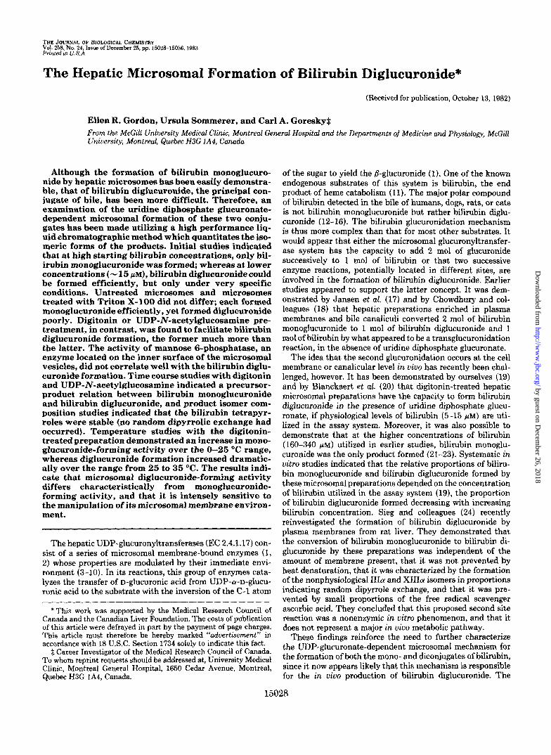
The Effect of Initial Bilirubin Concentrations on the Total Bilirubin Glucuronyltransferase Activity of Microsomal Prep- arations Treated with Different Perturbing Agents-The total activity of the bilirubin UDP-glucuronyltransferase systems in microsomal preparations treated with various agents known to alter microsomal membranes is presented in Fig. 1. The microsomal preparations were treated with digitonin (0.35 mg of digitonin/mg of microsomal protein), UDP-N- acetylglucosamine (0.5 mg/mg of microsomal protein), or Triton X-100 (0.1%). The concentration of bilirubin used varied from 5.7 to 30 p ~ . At the lowest concentration of bilirubin used (5.7 p ~ ) , no significant differences were de- tected in the rate of bilirubin conversion to polar compounds by the microsomal preparations treated with the various agents, and, as the concentration of bilirubin was increased, the activity of the UDP-glucuronyltransferase system in- creased in all cases, at relatively the same rate. A peak in activity was noted at bilirubin concentrations of the order of 25 ~ L M for both Triton X-100- and UDP-N-acetylglucosamine- treated microsomes. The activity of the digitonin-treated preparation, in contrast, exhibited a continuing slow rise in total activity over the higher bilirubin concentrations.
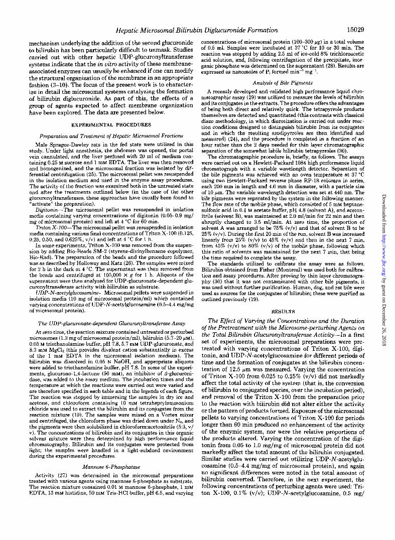
The Effect of Initial Bilirubin Concentration on the Conju- gates Formed-Experiments were carried out to define the level of initial bilirubin concentration at which bilirubin di- glucuronide formation occurred. The results of a set of studies in which microsomes were pretreated with digitonin (0.35 mg/ mg of microsomal protein) are illustrated in the top of Fig. 2. Bilirubin monoglucuronide is the major product formed by rat hepatic microsomal preparations at high concentrations of bilirubin (50-60 p ~ ) . As the initial concentration of bili- rubin is lowered, however, the proportion of bilirubin mono- glucuronide decreases and that of bilirubin diglucuronide in- creases until, at the lowest initial concentration, the latter becomes the major product. At an initial bilirubin concentra- tion of 12.5 p ~ , approximately equal amounts of bilirubin diglucuronide and bilirubin monoglucuronide were formed. This level of bilirubin was hereafter used in the assay system unless stated otherwise; it was felt that, with this initial
16
0 10 20 30
substrate ( p M )
FIG. 1. Changes in total conjugate formed at 30 min as a function of initial bilirubin concentrations. Incubations were carried out at 37 "C. The filled circles represent the conjugate formed by the Triton X-100-treated microsomes; the filled squares, that by the UDP-N-acetylglucosamine-treated microsomes; and the filled tri- angles, that by the digitonin-treated microsomes.
20
15
10
5
0
I I/
digitonin
digitonin
'b N-acetyl- UDP -
glucosamine
v E 0 5L 10 20 30 40 5 0 6 0
substrate ( p M ) FIG. 2. The effect of the initial bilirubin concentration on
the microsomal bilirubin-conjugating activity. Top, assays were carried out in a microsomal preparation pretreated with digitonin (0.35 mg/mg of microsomal protein); incubations were carried out for 30 min at 37 "C. The filled triangles represent the percentage of the bilirubin conjugate in the sample at 30 min which was bilirubin monoglucuronide; and the filled circles, the percentage of which was bilirubin diglucuronide. Middle, the conversion of bilirubin to conju- gates a t 30 min by microsomes pretreated with digitonin. The total conjugates are represented by the filled squares; bilirubin monoglu- curonide, by filled triangles; and bilirubin diglucuronide, by filled circles. Bottom, the conversion of bilirubin to conjugates at 30 min by microsomes pretreated with UDP-N-acetylglucosamine. The symbols are as in the middle of the figure.
concentration, either stimulation or inhibition of the mecha- nism resulting in the formation of bilirubin diglucuronide would be evident. The conjugate data correspond in form to those previously found by use of thin layer chromatographic analysis of the reaction products (19, 20).
The effect of the bilirubin concentrations becomes even more obvious when total activity of the digitonin-pretreated preparations is partitioned to reflect the bilirubin monoglu- curonide and bilirubin diglucuronide formed during the in- cubation period (Fig. 2, middle). The data indicate that bili- rubin diglucuronide was formed relatively more efficiently over the lower bilirubin concentration range, but that the diglucuronide-forming activity was inhibited as the higher initial bilirubin concentration range was explored. The opti- mum for this activity occurred with an initial bilirubin con- centration of the order of 15 p ~ . The monoglucuronide-
by guest on Decem
ber 26, 2018http://w
ww
.jbc.org/D
ownloaded from
Hepatic Microsomal Bilirubin Diglucuronide Formation 15031
forming activity, in contrast, continued to increase over the whole of the concentration range explored.
Similar studies were carried out with microsomes treated with UDP-N-acetylglucosamine (0.5 mg/mg of microsomal protein) or Triton X-100. The data obtained by use of micro- somes treated with UDP-N-acetylglucosamine are presented in the bottom of Fig. 2. An inhibiting effect of increasing concentrations of substrate on bilirubin diglucuronide for- mation is apparent. The total activity increased markedly (4.3-13.8 nmol of bilirubin converted per mg of microsomal protein) as the substrate levels increased from 5 to 25 pM. At low substrate concentrations (5 p ~ ) , bilirubin diglucuronide accounted for 28% of the total products formed. As the substrate level was increased, however, the total amount of bilirubin diglucuronide formed slowly decreased, and that of bilirubin monoglucuronide rapidly increased. When micro- somes were treated with Triton X-100, the major product detected was bilirubin monoglucuronide; this response was not affected by the concentration of substrate.
A summary of the relative proportions of bilirubin mono- glucuronide and bilirubin diglucuronide formed when the initial bilirubin concentration was 12.5 p~ and the micro- somes were treated by various agents is presented in Table I. Bilirubin monoglucuronide was the major conjugate formed when the assay was conducted on untreated microsomal prep- arations. It accounted for approximately 96% of the bilirubin conjugated (4% was converted to bilirubin diglucuronide). Triton X-100 did not alter this pattern. In contrast, after treatment of the microsomes with UDP-N-acetylglucosamine (0.5 mg/mg of microsomal protein), 25% of conjugate formed was bilirubin diglucuronide; and in the presence of digitonin (0.35 mg/mg of microsomal protein), 50% of the conjugate was bilirubin diglucuronide. These two substances selectively activated the mechanism underlying the addition of the sec- ond glucuronide to the bilirubin molecule.
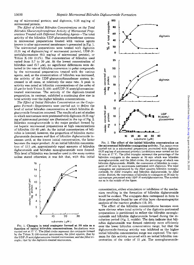
The Effect of Varying the Concentrations of the Microsomal Perturbing Agents on the Conjugates Formed-As stated be- fore, varying the concentration of digitonin from 0.05 to 1.0 mg/mg of microsomal protein did not markedly affect the total amount of bilirubin conjugated. On the other hand, the proportions of bilirubin monoglucuronide and diglucuronide formed were affected markedly (see Fig. 3). At digitonin levels of 0.05 mg/mg of microsomal protein, less than 5% of the total conjugates formed were accounted for by the formation of bilirubin diglucuronide. As the concentration of digitonin
TABLE I Effect of various forms of pretreatment on the proportion of bilirubin
diglwuronide formed by hepatic microsomes The initial bilirubin concentration was 12.5 b ~ . The values given
are means +- S.D. Conjugates"
Treatment of microsomal preparations
No. of Bilirubin Bilirubin experi-
mono- diglucu- merits glucuronide ronide
% Untreated 96.0 f 6.2 4.0 f 1.6 3 Triton X-100
0.125% 93.8 -+ 8.5 6.2 f 2.8 7 0.250% 97.1 f 9.4 2.9 rt 2.2 7
UDP-N-acetylglucosamine 74.9 f 11.0 25.1 f 5.9 6 (0.5 mg/mg microsomal
protein) Digitonin 49.9 f 5.5 50.1 rt 5.9 12
(0.35 rng/mg microsomal protein)
The conjugate formed is expressed in each case as a percentage of the total bilirubin converted. Standard deviations are given.
f loo r
digitonin (q/ mgof microsomal protein)
FIG. 3. The variation in the final proportions of bilirubin and its conjugates with change in digitonin concentration. In each assay, the initial bilirubin concentration was 12.5 FM, the duration of the incubation was 30 minutes, and the temperature of the incubation was 37 "C. The open squnres represent the remaining unconjugated bilirubin; the filled squares, bilirubin monoglucuronide; and the filled circles, bilirubin diglucuronide.
was increased, the proportion of the bilirubin converted to bilirubin diglucuronide increased until, at digitonin levels of 0.3-0.4 mg/mg of microsomal protein, 50% of the conjugates formed were accounted for by bilirubin diglucuronide. Increas- ing the concentration of digitonin even more suppressed the formation of bilirubin diglucuronide. At digitonin levels of 0.9 mg/mg of microsomal protein, diglucuronide accounted for only approximately one third of the bilirubin conjugates. Treatment of the microsomal preparations with varying con- centrations of digitonin for periods longer than 60 min did not markedly alter the observed pattern of response. A range of concentrations of added UDP-N-acetylglucosamine was also explored. Varying the concentration of this compound from 0.5 to 4.4 mg/mg of microsomal protein did not affect either the total conversion of biIirubin to conjugated bilirubin or the pattern of conjugates formed. Therefore, in subsequent experiments, 0.5 mg of UDP-N-acetylglucosamine/mg of mi- crosomal protein was utilized. With Triton X-100, no set of concentrations or conditions (presence or removal) was found which would promote more than the minimal diglucuronide formation recorded above.
The effect of perturbing the microsomal membranes with both Triton X-100 and digitonin is summarized in Table 11. The total glucuronide-forming activity was the same in mi- crosomal preparations treated with Triton X-100 or digitonin, or with both agents. However, the nature of the products formed differed. As noted before, bilirubin monoglucuronide was the major product formed when the microsomes were treated with Triton X-100, while bilirubin diglucuronide ac- counted for 49% of the product formed when microsomes were treated with digitonin. When the microsomes were treated with both agents, the formation of bilirubin diglucuronide was abolished. T k Latency of Mannose 6-Phosphatase Activity-The ef-
fect of the various agents on the leakiness of vesicles formed by the microsomal membranes was assessed by measuring the activity of mannose 6-phosphatase, a membrane-bound en- zyme considered to be located on the cisternal side of the microsomal membrane. The mannose 6-phosphatase activity of the microsomes changed with the various treatments (Fig. 4) . The untreated and Triton X-100-treated microsomes ex- hibited the same low level of activity, and UDP-N-acetylglu- cosamine-treated microsomes, a minor increase in activity. The digitonin-treated microsomes showed a marked increase in activity, indicating that the enzyme was now more acces- sible to substrate, and this accessibility was maintained when the microsomes were pretreated with Triton X-100 or UDP-
by guest on Decem
ber 26, 2018http://w
ww
.jbc.org/D
ownloaded from

15032 Hepatic Microsomal Bilirubin Diglucuronide Formation
digitonin
TABLE I1 Effect of Triton X-100 and digitonin on the nature of the products
formed by the microsomal UDP-glwuronyltransferase system Conjugates formed"
Treatment Bilirubin Bilirubin diglucu- monoglucu- ronide ronide
% Triton X-100 (0.1% v/v) 6.2 f 1.0 93.8 f 1.0 Digitonin (0.35 mg/mg microsomal 50.5 k 3.6 49.4 f 3.8
Triton X-100 (0.1% v/v) and 5.8 f 0.5 94.3 f 0.5 protein)
digitonin (0.35 mg/mg microsomal protein)
Mean values f S.D. are given. Four preparations were tested in
T T
FIG. 4. Bar graph illustrating the rate of release of phos- phate by the mannose 6-phosphatase activity of the various microsomal preparations.
N-acetylglucosamine, as well as digitonin. For the microsomes treated with single agents, there is a kind of parallelism between the mannose 6-phosphatase activity and the diglu- curonide-forming activity. This breaks down, however, when the combination digitonin:Triton X-100 pretreatment is uti- lized. The mannose 6-phosphatase continues to be accessible to its substrate, whereas the microsomal diglucuronide-form- ing activity is completely suppressed.
The Time Course of the Formation of Bilirubin Monoglucu- ronide and Bilirubin Diglucuronide-In our next series of experiments, the kinetic course of the formation of the con- jugates of bilirubin was characterized. These experiments were carried out with hepatic microsomal preparations treated either with 0.35 mg of digitonin or 0.5 mg of UDP-N-acetyl- glucosamine/mg of microsomal protein. The amounts of bili- rubin converted to bilirubin monoglucuronide and bilirubin diglucuronide were determined at 2- or 5-min intervals during a 30-min incubation. The results of these time course exper- iments are presented in Figure 5.
As illustrated in the top of Fig. 5, when the microsomal preparations were treated with digitonin, there was a very rapid formation of the conjugates of bilirubin. Over the first
IO 20 30
TIME (min)
FIG. 5. Time courses of conjugate formation. Top, the time course of the conversion of bilirubin to bilirubin monoglucuronide and bilirubin diglucuronide with microsomes pretreated with digi- tonin (0.35 mg/mg of microsomal protein). The initial bilirubin concentration was 12.5 PM, and the temperature of the incubation was 37 "C. The filled squures represent the remaining unconjugated bilirubin; the filled triangles, the bilirubin monoglucuronide; and the filled circles, the bilirubin diglucuronide. Bottom, the time course of the conversion of bilirubin to bilirubin monoglucuronide and bilirubin diglucuronide with microsomes pretreated with UDP-N-acetylgluco- samine (0.5 mg/mg of microsomal protein). The initial bilirubin concentration was 12.5 p ~ , and the temperature of the incubation was 37 "C. The filled squares represent unconjugated bilirubin; the filled triangles, bilirubin monoglucuronide; and the filled circles, bili- rubin diglucuronide.
10 min, 80% of the bilirubin disappeared, with a correspond- ingly rapid appearance of bilirubin monoglucuronide. Biliru- bin diglucuronide appearance was delayed; at 10 min, over 70% of the total bile pigment was still bilirubin monoglucu- ronide. The rate of formation of bilirubin diglucuronide did not become substantial until the succeeding time interval, between 10 and 20 min, when a large increment in bilirubin diglucuronide concentration was observed. With this, a marked decrease of bilirubin monoglucuronide concentration occurred. During this time period, there was no significant change in the concentration of unconjugated bilirubin. At 30 min, bilirubin diglucuronide accounted for approximately 55% of the total bile pigments, bilirubin monoglucuronide 30%, and bilirubin 15%. The forms of the concentration uersus time curves indicate that the reaction mode is sequential; bilirubin monoglucuronide is formed as a first step, and this then serves as the substrate for the second glucuronidation reaction.
Similar time course experiments were carried out with microsomal preparations activated with UDP-N-acetylgluco-
by guest on Decem
ber 26, 2018http://w
ww
.jbc.org/D
ownloaded from

Hepatic Microsomal Bilirubin Diglucuronide Formation 15033
samine (0.5 mg/mg of microsomal protein). The changes found in the proportions of bilirubin and its conjugates with time are illustrated in the bottom of Fig. 5. Following UDP- N-acetylglucosamine pretreatment, the rate of conversion of bilirubin to its conjugates was less than that found with digitonin pretreatment, and the amount of bilirubin converted to the bilirubin diglucuronide at 30 min was also decreased. Although the rates were lower, the pattern of formation of the conjugates was generally similar to that noted following digitonin treatment. Thirty-eight per cent of the bilirubin was converted in the first 10 min, thereafter the rate of conversion decreased, and by 30 min, only 70% of bilirubin had been converted. In the first 10 min, bilirubin monoglucuronide accounted for 90% of the conjugates; thereafter the amount of bilirubin diglucuronide increased fairly quickly, while that of bilirubin monoglucuronide increased only slowly. At the end of the incubation period, bilirubin diglucuronide ac- counted for 25%, bilirubin monoglucuronide for 45%, and bilirubin for 30% of the total bile pigments. The time course curves indicate that, in this instance, a larger conversion to bilirubin diglucuronide would have been achieved if the in- cubation had been carried on for a longer period. The form of the concentration-time course curves obtained with this prep- aration again indicate that bilirubin monoglucuronide is the precursor of bilirubin diglucuronide.
Experiments were carried out in which microsomal prepa- rations were treated with both digitonin (0.35 mg/mg of microsomal protein) and UDP-N-acetylglucosamine (0.5 mg/ mg of microsomal protein). The rate of formation of the conjugates was compared. The time courses and magnitudes of conjugate formation (initial bilirubin concentration, 15 pM; temperature, 37 "C) corresponded virtually exactly to that of digitonin alone. The digitonin effect completely superseded that of the N-acetylglucosamine. No additive effects were obtained.
In these experiments, the presence of glucurono-1,4-lactone made no difference to the compositions of the final product conjugate mixtures. There was no evidence of any substantial &glucuronidase activity in the microsomal preparations.
The high performance liquid chromatographic analysis (29) produces, as part of the run, data on the isomeric composition of bilirubin and its conjugates (the XIIIa, IXa, and IIIa isomers). The preponderant part of the starting material was, in each case, the IXa isomer. The proportion of the IXa isomer in the conjugates matched that in the added or parent bilirubin (that is, the proportion of the material which was IXa did not change during conjugate formation). The lack of change in the IXa content indicates that dipyrrole exchange can be effectively excluded (24) as part of the reaction mech- anism underlying the in vitro microsomal glucuronidation processes. In a dipyrrole exchange reaction, the product dis- tribution is, in terms of the XIIIa, IXa, and IIIa isomers, 1:2:1; the proportion of the I X a isomer in the mixture then necessarily decreases, so long as its initial proportion of the total is substantially above 0.5, the value correspondmg to total randomization.
The Effects of Temperature on the Relative Amounts of Bilirubin Monoglucuronide and Bilirubin Diglucuronide Formed by the Microsomal Glucuronyltransferase System-A series of experiments were carried out to determine the effect of temperature on the rate of formation of the conjugates of bilirubin. The data are presented in Fig. 6. With an initial bilirubin concentration of 12.5 PM, 35% of the bilirubin was converted at 10 "C; the proportion rose to 80% at 30 "C, and it remained approximately the same at 37 "C. An examination of the activity of the UDP-glucuronyltransferase system (as
100 R
60 n c
E .- 50 P 0 - a 5 40 - 0 c Y
30 * 20
10
I I
0 10 20 30 40
TEMPERATURE ( O C )
FIG. 6. The change in the final proportions of bilirubin and its conjugates with change in temperature. The reactions were carried out with microsomes pretreated with digitonin (0.35 mg/mg of microsomal protein). The initial bilirubin concentration was 12.5 p ~ , and the time of incubation was 30 min. The open squares represent unconjugated bilirubin; the filled squares, bilirubin mono- glucuronide; and the filled circles, bilirubin diglucuronide.
judged by the proportion of unconverted bilirubin) as a func- tion of temperature indicates that abrupt changes in enzyme activity occurred in two different temperature ranges (18-22 and 26-30 "C). The nature of the conjugates of bilirubin formed was also markedly affected by the temperature of the assay media. In the range from 0 to 25 "C, the amount of bilirubin converted to bilirubin monoglucuronide increased greatly, yet only a small proportion of this was converted to bilirubin diglucuronide. From 25 to 37 "C, increasing propor- tions of the bilirubin monoglucuronide were converted to bilirubin diglucuronide (even though, over this temperature range, there was no increase in the proportion of bilirubin conjugated) until finally, at 37 "C, approximately equal pro- portions of the conjugates were formed. The same pattern was observed if bilirubin monoglucuronide was used as substrate. Its conversion to bilirubin diglucuronide did not occur until the incubation temperature exceeded 20 "C. The formation of diglucuronide at the higher temperature appears to indicate an underlying steric change with temperature in either the enzyme or the membrane, or more likely both.
DISCUSSION
These studies indicate that the formation of bilirubin diglu- curonide by rat hepatic microsomal preparations is governed by both the concentration of bilirubin in the preparation and the structural state of the microsomal membranes. The time course studies indicate that the glucuronidation reactions take place in two steps in vitro, with bilirubin monoglucuronide serving as substrate for the formation of bilirubin diglucuro- nide.
It is not yet known if more than one uridine diphosphate
by guest on Decem
ber 26, 2018http://w
ww
.jbc.org/D
ownloaded from
15034 Hepatic Microsomal Bilirubin Diglucuronide Formation
glucuronyltransferase is involved in the microsomal glucuron- idation of bilirubin, or if a single membrane-bound enzyme regulates the addition of two glucuronides by virtue of changes in its conformational structure. The uridine diphosphate glu- curonyltransferase reactions convert a wide variety of com- pounds into biologically inactive products. The chemical di- versity of the compounds capable of being glucuronidated raises the question of the functional molecular basis for the heterogeneity of the system. Studies on differential induction (31, 32) and subcellular diversity of activity (33), as well as developmental studies (34), clearly demonstrate that the sub- strates of UDP-glucuronate-dependent glucuronyltransferase reactions fall into a series of distinct groups. Several investi- gators have attempted to elucidate this complex system fur- ther by purifying enzymic moieties with glucuronyltransferase activity from hepatic microsomal preparations (35-38). These studies indicate that there are various forms of UDP-glucu- ronyltransferase with different substrate affinities. As pointed out by Axelrod et al. in 1958 (39), all of the substrates possess a nucleophilic functional group and one enzyme could catalyze a nucleophilic substitution reaction between all acceptor com- pounds and UDP-glucuronic acid. Although three distinct forms of microsomal UDP-glucuronate-dependent glucuron- yltransferase appear to have been isolated, it is difficult to ascertain from the reconstituted systems whether these are distinct or are isoenzymes or parts thereof. Their functional differences, such as substrate specificity, could simply reflect the properties of the membranes regulating their activity (4, 39). A microsomal glucuronyltransferase which exhibits a high affinity for bilirubin but shows little activity with p-nitro- phenol or estrone has recently been partially purified (40). In a reconstituted system, the partially purified enzyme is found to convert bilirubin to bilirubin monoglucuronide but not to bilirubin diglucuronide (in the presence of UDP-glucuronate). The inability of this in u i t ~ o preparation to form bilirubin diglucuronide may simply indicate that the diglucuronide- forming enzyme has a separate identity or that in the recon- stituted system the required structural environment was not recreated.
A survey of the literature indicates that factors regulating the activity of membrane-bound enzymes are very complex (5, 41) and that the functional capacities of these enzymes will usually vary, depending on the assay conditions under which the membrane-bound enzyme is investigated (19, 20, 42,43). The data obtained from our experiments substantiate these findings. Thus, although the total activity of the UDP- glucuronate-dependent glucuronyltransferase system as measured by the conversion of bilirubin to total conjugates of bilirubin was independent of the manner in which the micro- somal preparation was treated, examination of the products indicated that the relative amounts of bilirubin monoglucu- ronide and bilirubin diglucuronide formed depended upon the manner in which the membranes had been perturbed.
The general effects of compounds with detergent-like prop- erties on the activities of membrane-bound enzyme have been well characterized. It has been shown that their effects on the activity of the enzyme depend on the concentration of the agent utilized. Small concentrations usually enhance the ac- tivity, and large concentrations inhibit it (5). It has been proposed that detergent-induced activation indicates that the activity is dependent on specific lipid-protein interactions. In our explorations of bilirubin monoglucuronide formation, none of our experimental manipulations had an activating effect. Under our experimental conditions, the rate of forma- tion of bilirubin monoglucuronide appeared not to be influ- enced by any of the perturbations which generally activate
other microsomal glucuronyltransferases. On the other hand, the formation of bilirubin diglucuronide was greatly influ- enced by the manner in which the membrane was altered. This suggests that the lipid-protein interactions in the mem- brane have a major regulating effect on the system which forms bilirubin diglucuronide. Treatment of the membrane with digitonin or UDP-N-acetylglucosamine appeared not to destroy the physical properties of the membrane required for the formation of bilirubin diglucuronide; rather, it uncovered the activity.
It is of interest to compare the effects on the membrane of those treatments that affect bilirubin diglucuronide formation with those that do not. Treatment of the membrane with Triton X-100 did not enhance the total glucuronidation of bilirubin. It has been demonstrated that Triton X-100 acts by selectively removing certain phospholipids from the mem- brane and that this, in turn, induces a change from a single membrane lamellar structure to that of mixed protein-lipid- detergent micelles (44). The &glucuronide-forming enzymic activity associated with these membrane vesicles was not enhanced; the access of mannose 6-phosphate to the intra- membranous mannose 6-phosphatase was not increased. The only product formed in substantial proportion was bilirubin monoglucuronide. Now consider those perturbations which enhance the formation of bilirubin diglucuronide. The most effective of these, treatment of the microsomal preparation with digitonin, has been found to remove cholesterol from the membrane. At the concentrations of digitonin found optimal for the formation of bilirubin diglucuronide, the membrane was more permeable to mannose 6-phosphate. This response suggests that the second enzymic activity may be on the cisternal side of the microsomal membrane. Thus, it would appear that treatment of the membrane with digitonin makes the active sites of the &glucuronide-forming enzyme more accessible but at the same time preserves the linkage of the enzyme to its milieu in the membrane in such a fashion as to preserve its activity. When Triton X-100 is present as well as digitonin, although the increased mannose 6-phosphatase ac- tivity is preserved, the diglucuronide-forming activity no longer persists. The reasons for this are not clear. The Triton X-100 may be inhibiting a cisternally located diglucuronide- forming activity, or it may be changing the local milieu of such an enzyme by virtue of its removal of the phospholipid from the membrane; or, alternately, the diglucuronide-form- ing activity may not be on the cisternal side of the membrane at all, but may simply undergo conformational changes, in a very sensitive fashion, in response to local changes in the membrane. The effect of UDP-N-acetylglucosamine on mem- brane structure is not known, but it has been inferred that the basis of UDP-N-acetylglucosamine-induced activation in other systems is an enhancement of the affinity of the enzyme for UDP-glucuronic acid (5). It is of interest to note that after the addition of both these agents to the microsomal prepara- tion, the digitonin effect dominated and was not modified. The qualitative nature of their actions on the enzyme may be similar. Both may simply make the active site on the enzyme more accessible to the UDP-glucuronate.
The mannose 6-phosphatase activity determinations have thus provided a new set of directions for exploration. They have, unfortunately, not provided crisp clear answers con- cerning the location and state of the &glucuronide-forming activity in the microsomal membrane.
Many investigators have noted that the activity of mem- brane-bound enzymes is governed in part by the structural state of the phospholipid environment. The glucuronidation of other nonpolar compounds by microsomal glucuronyltrans-
by guest on Decem
ber 26, 2018http://w
ww
.jbc.org/D
ownloaded from
Hepatic Microsomal Bilirubin Diglucuronide Formation 15035
ferase systems has been shown to be regulated in such a fashion. Zakim and Vessey (B) , for instance, have shown that the activity of UDP-glucuronyltransferase with p-nitrophenol as substrate is modulated in parallel with change in the physical state of the membrane phospholipid, as measured by a spin-labeled molecular probe. Arrhenius-type plots revealed a discontinuity in enzyme activity in the range of 13-16 "C. It was proposed that a phase change in the phospholipid of the membrane had occurred and that this led to a modification of the properties of this enzyme system. The change was shown to be associated with both an alteration in the speci- ficity of substrate binding at the UDP-glucuronic acid site and a change in the response of the enzyme to allosteric activators. Below 13 "C, UDP-glucose, UDP-mannose, and UDP-xylose all inhibited this UDP-glucuronyltransferase but not at 37 "C. A similar pattern was also observed with UDP- N-acetylglucosamine which had no effect below 13 "C but activated the p-nitrophenol glucuronyltransferase at temper- atures over 16 "C (5). Our experiments also indicated that the activity and functions of the second step of the uridine di- phosphate glucuronyltransferase system with bilirubin mono- glucuronide as a substrate are markedly dependent on tem- perature. Bilirubin diglucuronide was formed in appreciable amounts only at temperatures above 25 "C, and its rate of formation then increased with temperature. The formation of bilirubin monoglucuronide from bilirubin exhibited a quali- tatively different change in behavior over this range. This activity (as reflected by the amount of unconverted bilirubin remaining at the end of the period of incubation), rather than increasing, was constant over this temperature interval.
These studies in aggregate appeared to indicate that the function of UDP-glucuronyltransferase in uitro, with bilirubin monoglucuronide as substrate (the activity underlying the addition of the second glucuronide), is regulated in a partic- ular way by the physical state of the membrane. Perturbations of the membrane by treatments which disrupt the structure of the membrane do not alter the capability to produce bili- rubin monoglucuronide, nor do they enhance the formation of bilirubin diglucuronide. The uncovering of the diglucuro- nide-forming activity appears to require a less radical altera- tion, involving some preservation of the structure of the membrane but at the same time making the active site of the enzyme more accessible. The results of the temperature- dependent studies indicate that the immediate environment of the enzyme system must have a regulatory effect on this activity. Although thermal induced phase transitions are not expected to play a part in living systems which maintain a constant temperature, the results of our studies demonstrate that a specific structural state of the microsomal membrane, which is determined by the temperature, is integral to the formation of bilirubin diglucuronide.
It is clear that the present studies provide an unequivocal demonstration of the capability of liver microsomal prepara- tions to form bilirubin diglucuronide in uitro. They show that there is a sequential reaction mechanism, bilirubin monoglu- curonide formed during a first step, providing the substrate for diglucuronide formation; they provide some insight into the approaches needed to uncover the second glucuronidation activity; and they indicate that only when the integrity of the membrane is relatively maintained will bilirubin diglucuro- nide be formed in uitro. It is likely, in view of the findings of Sieg et al. (24), reviewed in the Introduction, that this micro- somal mechanism represents the major in uiuo mechanism for bilirubin diglucuronide formation in the liver.
Acknowledgments-We thank Janet Jan6 for her technical assist-
ance and Pamela Lilley and Margaret Mulherin for typing this manuscript.
REFERENCES
1. Dutton, G. J. (1966) in Glucuronic Acid-Free and Combined
2. Dutton, G. J., and Burchell, B. (1977) Prog. Drug Metab. 2 , 1-70 3. Vessey, D. A., and Zakim, D. (1971) J. Biol. Chem. 246 , 4649-
4656 4. Zakim, D., Goldenberg, J., and Vessey, D. A. (1973) Eur. J.
Biochem. 33.59-63 5. Zakim, D., and Vessey, D. A. (1976) in The Enzymes of Biological
Membranes (Martonosi, A., ed) Vol. 2, pp. 443-461, Plenum Press, New York
6. Leuders, K. K., and Kuff, E. L. (1967) Arch. Biochem. Biophys.
7. Zakim, D., and Vessey, D. A. (1975) Biochim. Biophys. Acta 410 ,
8. Zakim, D., and Vessey, D. A. (1975) J. Biol. Chem. 250,342-343 9. Graham, A. B.. and Wood. G. C. (1973) Biochim. Bionhvs. Acta
(Dutton, G. J., ed) pp. 185-299, Academic Press, New York
126,198-203
61-73
311,45-50 '
_ -
10. Hochman. Y.. Zakim. D.. and Vessev. D. A. (1981) J. Bwl. Chem. I~
256,4783-4788 11. Schmid, R., and McDonagh, A. F. (1978) in The Metabolic Basis
of Inherited Disease (Stanbury, J. B., Wyngaarden, J. D., and Frederickson, D. S., eds) pp. 1221-1257, McGraw Hill, New York
", . ,
12. Jansen, P. L. M. (1974) Biochim. Biophys. Acta 338 , 170-182 13. Gordon, E. R., Goresky, C. A., Chang, T.-H., and Perlin, A. S.
14. Fevery, J., Van de Vijver, M., Michiels, R., and Heirwegh, K. P.
15. Gordon, E. R., Shaffer, E. A., and Sass-Kortsak, A. (1976) Gas-
16. Blanckaert, N. (1980) Biochem. J . 185,115-128 17. Jansen, P. L. M., Chowdhury, J. R., Fischberg, E. B., and Arias,
18. Chowdhury, J. R., Chowdhury, N. R., Bhargava, M. M., and
19. Gordon, E. R., and Goresky, C. A. (1980) Can. J . Biochem. 58 ,
20. Blanckaert, N., Gollan, J. L., and Schmid, R. (1979) Proc. Natl.
21. Van Roy, F. P., and Heirwegh, K. P. M. (1968) Biochem. J . 107,
22. Heirwegh, K. P. M., Meuwissen, J. A. T. P., and Fevery, J. (1973) Adu. Clin. Chem. 16,239-289
23. Chowdhury, J. R., Jansen, P. L. M., Fischberg, E. B., Daneller, A., and Arias, I. M. (1978) J. Clin. Inuest. 62, 191-196
24. Sieg, A., van Hees, G. P., and Heirwegh, K. P. M. (1982) J . Clin. Invest. 69,347-357
25. Siekevitz, P. (1962) Methods Enzymol. 5 , 61-68 26. Holloway, P. W., and Katz, J. T. (1972) Biochemistry 11, 3689-
27. Lichenstein, A. H., and Brecher, P. (1980) J. Biol. Chem. 255 ,
28. Fiske, C. H., and SubbaRow, Y. (1925) J. Biol. Chem. 6 6 , 375-
29. Gordon, E. R., and Goresky, C. A. (1982) Can. J. Biochem. 6 0 ,
30. Gordon, E. R., Chan, T.-H., Samodai, K., and Goresky, C. A.
31. Bock, K. W., Frohling, W., Remmer, H., and Rexer, B. (1973)
32. Wishart, G. J. (1978) Biochem. J. 174,671-672 33. Gram, T. E., Hansen, A. R., and Fouts, J. R. (1968) Biochem. J.
34. Dutton, G. J. (1966) Biochem. Phurmacol. 15, 947-951 35. Gorski, J. P., and Kasper, C. B. (1977) J. Biol. Chem. 252,1336-
36. Vellar, D., Sanchez, E., Autor, A. P., and Tephly, T. R. (1975)
37. Tukey, R. M., and Tephly, T. R. (1981) Methods Enzymol. 77,
38. Bock, K. W., Josting, D., Lilienblum, W., and Pfeil, H. (1979)
(1976) Biochem. J . 155,477-486
M. (1977) Biochem. J. 164, 737-746
troenterology 7 0 , 761-765
I. M. (1977) J. Biol. Chem. 2 5 2 , 2710-2716
Arias, I. M. (1979) J. Biol. Chem. 254,8336-8339
1302-1310
Acad. Sci. U. S. A. 7 6 , 2037-2047
507-518
3696
9098-9104
400
1050-1057
(1977) Biochem. J . 1 6 7 , l - 8
Bwchim. Biophys. Acta 327 , 46-56
106,587-591
1343
Mol. Phurmucol. 11, 236-245
177-188
Eur. J. Biochem. 98 , 19-26
by guest on Decem
ber 26, 2018http://w
ww
.jbc.org/D
ownloaded from
15036 Hepatic Microsomal Bilirubin Diglucuronide Formation
39. Alexrod, J., Inscoe, J. K., and Tomkins, G. M. (1958) J. Biol. sterdam Chem. 232,835-841 42. Farias, R. N., Bloj, B., Morero, R., Sineriz, F., and Trucco, R. E.
40. Chowdhury, J. R., Chowdhury, N. R., Moscione, D., Shouval, R., (1975) Biochim. Biophys. Acta 416,231-251 Tukey, R., and Arias, I. M. (1981) Hepatology 1, 503 (abstr.) 43. Coleman, R. (1973) Biochim. Biophys. Acta 300 , 1-30
41. Wishart, G. J., Campbell, M. T., and Dutton, D. J. (1978) in 44. Durtubay, J. I. G., Gori, F. M., Gomez-Fernandez, J. C., Ota- Conjugation Reactions in Drug Biotransformation (Ahio, A., ed) men&, J. J., and Macarulla, J. M. (1980) J. Bioenerg. Bio- pp. 179-187, Elsevier/North-Holland Biomedical Press, Am- membr. 12,47-70
by guest on Decem
ber 26, 2018http://w
ww
.jbc.org/D
ownloaded from
E R Gordon, U Sommerer and C A GoreskyThe hepatic microsomal formation of bilirubin diglucuronide.
1983, 258:15028-15036.J. Biol. Chem.
http://www.jbc.org/content/258/24/15028Access the most updated version of this article at
Alerts:
When a correction for this article is posted•
When this article is cited•
to choose from all of JBC's e-mail alertsClick here
http://www.jbc.org/content/258/24/15028.full.html#ref-list-1
This article cites 0 references, 0 of which can be accessed free at
by guest on Decem
ber 26, 2018http://w
ww
.jbc.org/D
ownloaded from
![Effect of medications on Speaking, Hearing and [Autosaved] · 2/26/2015 3 Hepatic: Abnormal hepatic function tests, hepatic coma, increased serum bilirubin, increased serum transaminases](https://img.pdfslide.net/doc/110x75/5f5d825ed8f24413b242029c/effect-of-medications-on-speaking-hearing-and-autosaved-2262015-3-hepatic.jpg)