Embed Size (px)
DESCRIPTION
Summary
Citation preview

Histology of Bones
By: Mohammed Jabr Premed Summer Enrichment Program
Alfaisal University
Definition
Bone is a specialized form of connective tissue that, like other connective tissues, consists of cells and extracellular matrix. The feature that distinguishes bone from other connective tissues is the mineralization of its matrix. The mineral is calcium phosphate.
The organization of the bone
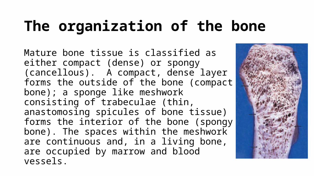
Mature bone tissue is classified as either compact (dense) or spongy (cancellous). A compact, dense layer forms the outside of the bone (compact bone); a sponge like meshwork consisting of trabeculae (thin, anastomosing spicules of bone tissue) forms the interior of the bone (spongy bone). The spaces within the meshwork are continuous and, in a living bone, are occupied by marrow and blood vessels.
General structure of bones
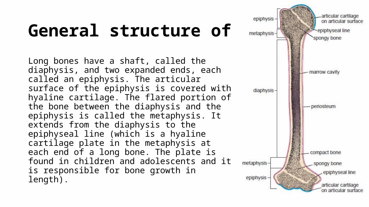
Long bones have a shaft, called the diaphysis, and two expanded ends, each called an epiphysis. The articular surface of the epiphysis is covered with hyaline cartilage. The flared portion of the bone between the diaphysis and the epiphysis is called the metaphysis. It extends from the diaphysis to the epiphyseal line (which is a hyaline cartilage plate in the metaphysis at each end of a long bone. The plate is found in children and adolescents and it is responsible for bone growth in length).
General structure of bones
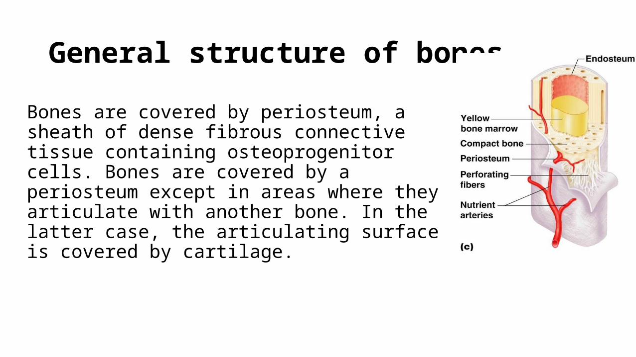
Bones are covered by periosteum, a sheath of dense fibrous connective tissue containing osteoprogenitor cells. Bones are covered by a periosteum except in areas where they articulate with another bone. In the latter case, the articulating surface is covered by cartilage.
General structure of bones
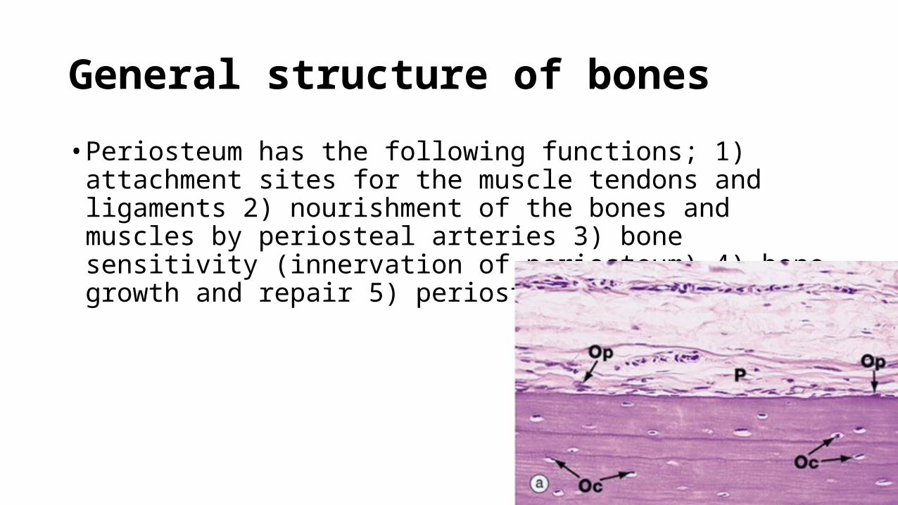
• Periosteum has the following functions; 1) attachment sites for the muscle tendons and ligaments 2) nourishment of the bones and muscles by periosteal arteries 3) bone sensitivity (innervation of periosteum) 4) bone growth and repair 5) periosteal hinge
General structure of bones
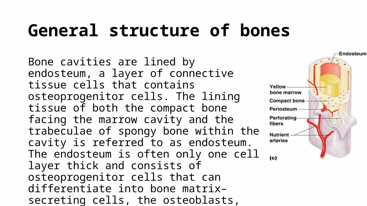
Bone cavities are lined by endosteum, a layer of connective tissue cells that contains osteoprogenitor cells. The lining tissue of both the compact bone facing the marrow cavity and the trabeculae of spongy bone within the cavity is referred to as endosteum. The endosteum is often only one cell layer thick and consists of osteoprogenitor cells that can differentiate into bone matrix–secreting cells, the osteoblasts, and bone-lining cells.
General structure of bones
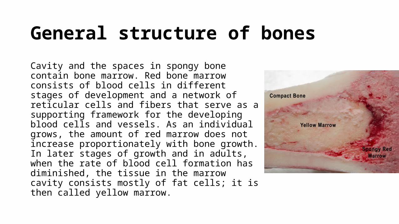
Cavity and the spaces in spongy bone contain bone marrow. Red bone marrow consists of blood cells in different stages of development and a network of reticular cells and fibers that serve as a supporting framework for the developing blood cells and vessels. As an individual grows, the amount of red marrow does not increase proportionately with bone growth. In later stages of growth and in adults, when the rate of blood cell formation has diminished, the tissue in the marrow cavity consists mostly of fat cells; it is then called yellow marrow.
Haversian system
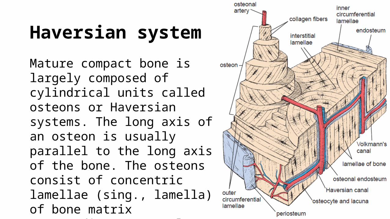
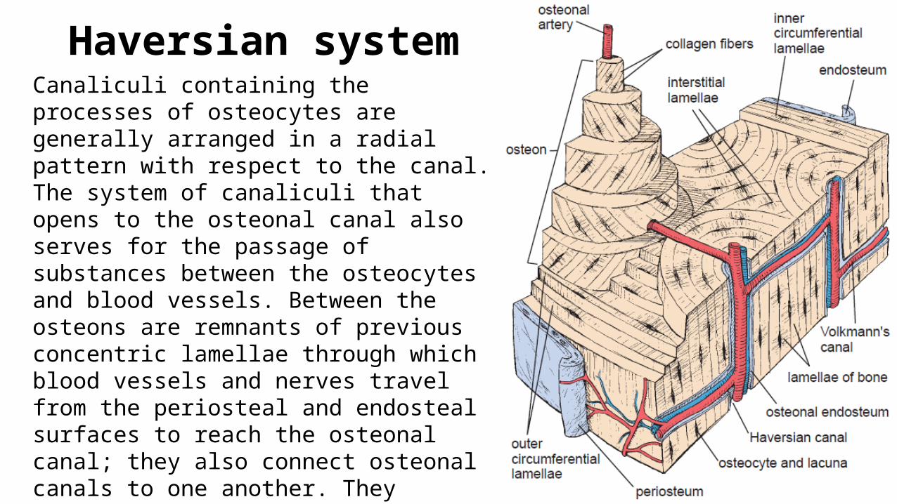
Mature compact bone is largely composed of cylindrical units called osteons or Haversian systems. The long axis of an osteon is usually parallel to the long axis of the bone. The osteons consist of concentric lamellae (sing., lamella) of bone matrix surrounding a central canal, the osteonal (Haversian) canal, which contains the vascular and nerve supply of the osteon.
Haversian system
Canaliculi containing the processes of osteocytes are generally arranged in a radial pattern with respect to the canal. The system of canaliculi that opens to the osteonal canal also serves for the passage of substances between the osteocytes and blood vessels. Between the osteons are remnants of previous concentric lamellae through which blood vessels and nerves travel from the periosteal and endosteal surfaces to reach the osteonal canal; they also connect osteonal canals to one another. They usually run at approximately right angles to the long axis of the osteons and of the bone. Volkmann’s canals are not surrounded by concentric lamellae, a key feature in their histologic identification.
Bone Matrix
Bone matrix contains collagenous and non-collagenous proteins• Collagenous .. All collagen molecules constitute about 90% of the total
weight of the bone matrix proteins. The major structural component of bone matrix is type I collagen and, to a lesser extent, type V collagen. Trace amounts of other types such as type III, XI, and XIII collagens have also been found in the matrix.• Non-collagenous .. Which constitute the ground substance of bone that
is only 10% of the total weight of bone matrix proteins.
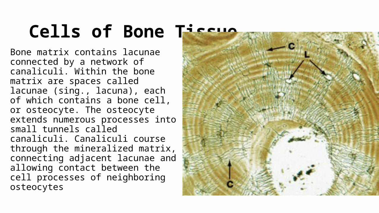
Cells of Bone TissueBone matrix contains lacunae connected by a network of canaliculi. Within the bone matrix are spaces called lacunae (sing., lacuna), each of which contains a bone cell, or osteocyte. The osteocyte extends numerous processes into small tunnels called canaliculi. Canaliculi course through the mineralized matrix, connecting adjacent lacunae and allowing contact between the cell processes of neighboring osteocytes
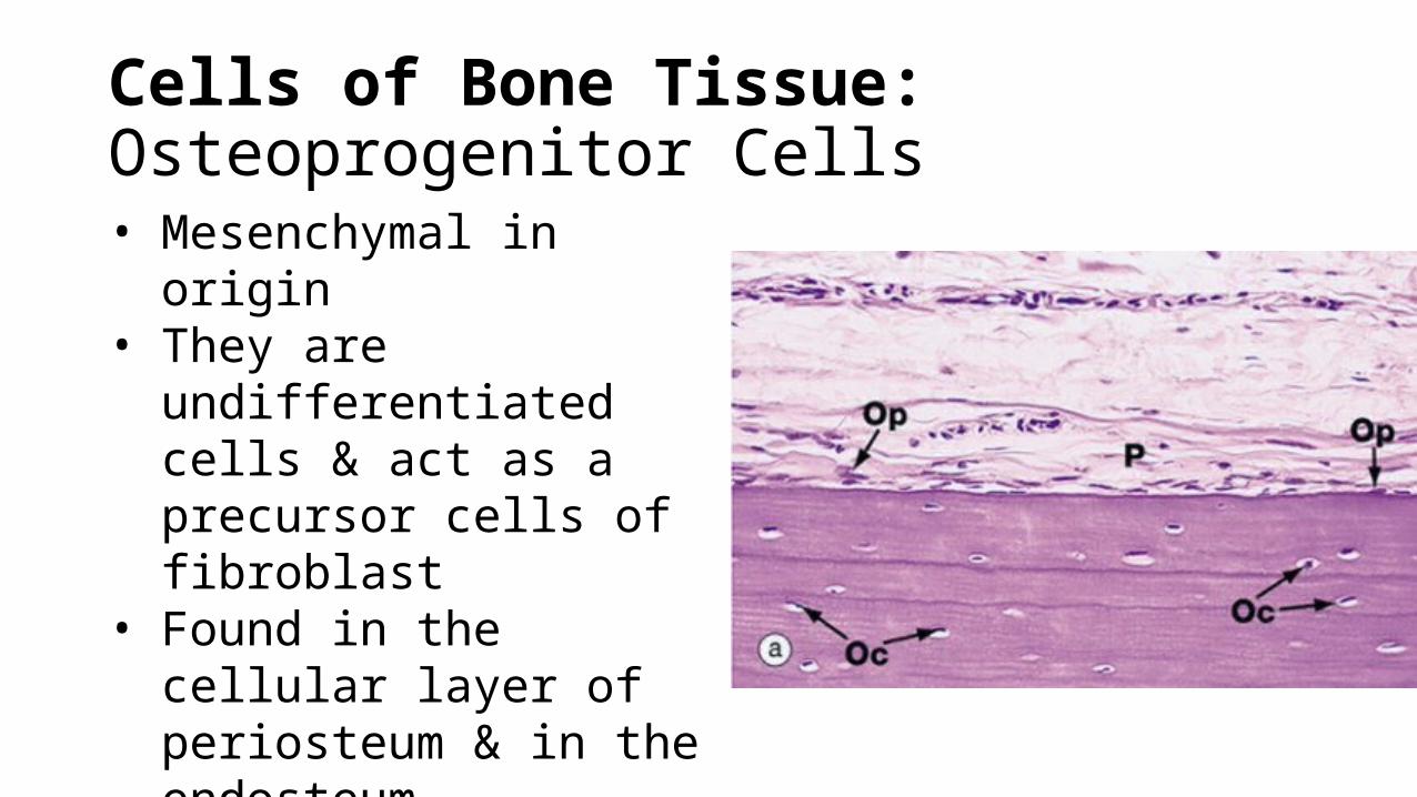
Cells of Bone Tissue: Osteoprogenitor Cells• Mesenchymal in origin• They are undifferentiated
cells & act as a precursor cells of fibroblast
• Found in the cellular layer of periosteum & in the endosteum
• They are found scattered & do not form a continuous lining
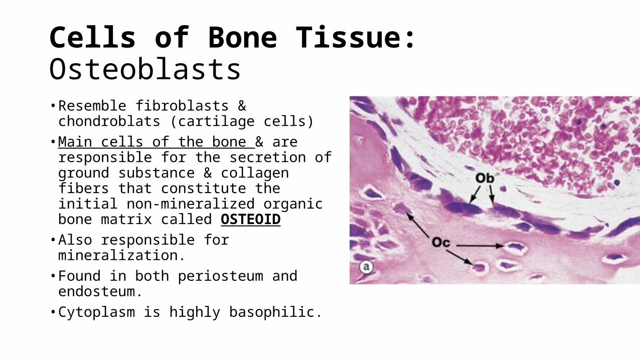
Cells of Bone Tissue: Osteoblasts
• Resemble fibroblasts & chondroblats (cartilage cells)• Main cells of the bone & are
responsible for the secretion of ground substance & collagen fibers that constitute the initial non-mineralized organic bone matrix called OSTEOID• Also responsible for mineralization.• Found in both periosteum and
endosteum.• Cytoplasm is highly basophilic.
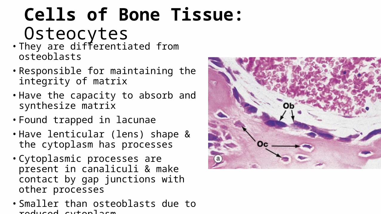
Cells of Bone Tissue: Osteocytes• They are differentiated from osteoblasts• Responsible for maintaining the integrity of
matrix• Have the capacity to absorb and synthesize
matrix• Found trapped in lacunae• Have lenticular (lens) shape & the cytoplasm
has processes• Cytoplasmic processes are present in canaliculi
& make contact by gap junctions with other processes• Smaller than osteoblasts due to reduced
cytoplasm• During life, do not fully occupy the lacunae
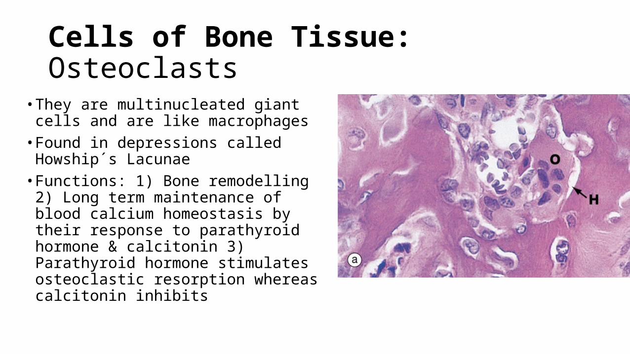
Cells of Bone Tissue: Osteoclasts
• They are multinucleated giant cells and are like macrophages• Found in depressions called Howship s ˊ
Lacunae• Functions: 1) Bone remodelling 2) Long
term maintenance of blood calcium homeostasis by their response to parathyroid hormone & calcitonin 3) Parathyroid hormone stimulates osteoclastic resorption whereas calcitonin inhibits
Cells of Bone Tissue: Bone-lining cellsIn sites where remodeling is not occurring, the bone surface is covered by a layer of flat cells with attenuated cytoplasm and a paucity of organelles beyond the perinuclear region. These cells are designated simply as bone lining cells. Bone-lining cells on external bone surfaces are called periosteal cells, and those lining internal bone surfaces are often called endosteal cells. Bone-lining cells represent a population of cells that are derived from osteoblasts. They are thought to function in the maintenance and nutritional support of the osteocytes embedded in the underlying bone matrix and regulate the movement of calcium and phosphate into and out of the bone. In these respects, bone-lining cells are somewhat comparable to osteocytes.