Embed Size (px)
Citation preview
Received on: 05-02-2014 Accepted on: 25-02-2014 Published on: 15-03-2014
Arup Kalita* Department of Veterinary Anatomy and Histology, College of Veterinary Sciences and A.H. Central Agricultural University, Selesih, Aizawl, Mizoram, India Email: [email protected]
Histomorphological Study of the Respiratory System of Mizo Local Pig (zo vawk)
Arup Kalita*
Department of Veterinary Anatomy and Histology, College of Veterinary Sciences and A.H. Central Agricultural University, Selesih, Aizawl, Mizoram, India
Abstract The present study provides a baseline data on histology of respiratory system of Mizo local pig (Zo Vawk). The Occurrence of nasal glands was high in the respiratory region of nasal cavity which might enhance the efficiency of humidifying the inhaled air. The visceral pleura and interlobar and interlobular septa were thick containing elastic fibres which might increase the elasticity of the lung. The respiratory bronchioles were well developed in Mizo local pig which might enhance their respiratory efficiency in higher altitude and lesser atmospheric oxygen tension in Mizoram. Frequent occurrence of alveolar macrophages might be attributed to the better resistance to respiratory diseases of the indigenous pigs of Mizoram (Zo Vawk). Keywords: Histomorphology, Respiratory system, Mizo local pig
QR Code for Mobile users
Cite this article as:
Arup Kalita. Histomorphological Study of the Respiratory System of Mizo Local Pig (zo vawk). Asian Journal of Biomedical and
Pharmaceutical Sciences; 04 (29); 2014; 50-54.
A., Kalita. et al.: Asian Journal of Biomedical and Pharmaceutical Sciences; 4(29) 2014, 50-54.
© Asian Journal of Biomedical and Pharmaceutical Sciences, all rights reserved. Volume 4, Issue 29, 2014. 51
INTRODUCTIONRespiratory system performs the vital functions of conduction and exchange of oxygen, olfaction, phonation and thermoregulation of the body. Anatomical study on the respiratory system has been done extensively in domestic mammals. However, studies on this system in indigenous varieties of pigs especially that of Mizoram are still lacking. Animals living in different zones of altitude have to cope-up structurally and functionally with different levels of atmospheric pressure and oxygen tension for optimum respiratory activities. The indigenous pigs of Mizoram are distributed within the range of 500-1000 meters high altitude. Anatomical study on the respiratory system in this variety of pig is, therefore, essential to elucidate the structural peculiarities in connection with their functional status at such high altitude. METHODS The Mizo local pigs are reared in semi intensive system in the pig farm of the College of Veterinary Sciences and Animal Husbandry, Central Agricultural University, Selesih, Aizawl, Mizoram, India, following the standard management procedures. Adequate feed, drinking water and health care are provided to the animals. Excess numbers of animals than the parent stock are slaughtered by using captive bolt pistol for commercial purpose. Organs of respiratory system of 10 (ten) apparently healthy adult indigenous pigs of Mizoram (Zo vowk) of either sex were utilized for the research project. The respiratory systems were exposed in-situ by fine dissection. The lung was than fixed by intratracheal instillation of 10% neutral buffered formalin until it was of life size; ligated on the trachea and immersed in the same fixative in a container for proper fixation as per the technique described by Drury and Wallington [1]. Median and cross sections of the head were made to expose the nasal cavity and biometrical measurements were recorded. All experimental materials were fixed and preserved in 10% neutral buffered formalin. For histomorphological study, small pieces of tissue were collected from all the respiratory organs at different representative locations. These tissues were then processed for paraffin sectioning following standard procedures. The paraffin blocks were sectioned at 5 (five) µ thickness by Leica semi motorized rotary microtome. The sections were stained following routine staining techniques as per Luna [2]. The histomorphological characteristics of the respiratory organs were observed and interpreted for cellular details mainly on H & E stained sections along with co project investigators with the help of Olympus
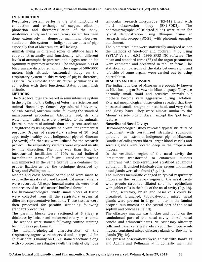
trinocular research microscope (BX-41) fitted with multi observation body (BX2-SOD2). The photomicrographs of selected slides were taken for typical demonstration using Olympus trinocular research microscope (BX-51) with photomicrography attachment. The biometrical data were statistically analyzed as per the methods of Snedecor and Cochran [3] by using SYSTAT Version 6.0.1., 1996 SPSS INC software. The mean and standard error (SE) of the organ parameters were estimated and presented in tabular forms. The statistical comparisons of the data between right and left side of some organs were carried out by using paired‘t’ test. RESULTS AND DISCUSSION The indigenous pigs of Mizoram are popularly known as Mizo local pig or Zo vawk in Mizo language. They are normally small, timid and sensitive animals but mothers become very aggressive during lactation. External morphological observation revealed that they possessed small, straight, pointed head, and very thick and glossy hairs. They were almost similar to the “doom” variety pigs of Assam except the “pot belly” feature. Nostrils and Nasal Cavity: Histomorphological study revealed typical structure of integument with keratinized stratified squamous epithelium at nostrils of indigenous pigs of Mizoram. Bundles of collagenous fibres, larger blood vessels and serous glands were located deep in the propria-sub mucosa. In the vestibular region of the nasal cavity the integument transformed to cutaneous mucous membrane with non-keratinized stratified squamous epithelium. Branched tubuloalveolar, serous and mixed nasal glands were also found (Fig. 1a). The mucous membrane changed to typical respiratory mucosa in the respiratory region of the nasal cavity with pseudo stratified ciliated columnar epithelium with goblet cells in the bulk of the nasal cavity (Fig. 1b). Ciliated, secretory, brush and basal cells could be visualized. Branched, tubuloalveolar, mixed nasal glands were present in large number in the lamina propria- sub mucosa on the rostral part of the nasal septum and conchae (Fig. 1d). The olfactory mucosa was thicker and found on the caudodorsal part of the nasal cavity, dorsal nasal concha and ethmoturbinates. Neurosensory olfactory cells and basal cells were observed. The propria-sub mucosa contained mixed olfactory glands or Bowman’s glands (Fig. 1c). The present observations were at par with Banks [4] and Adams and Dellmann [5] in domestic mammals
A., Kalita. et al.: Asian Journal of Biomedical and Pharmaceutical Sciences; 4(29) 2014, 50-54.
© Asian Journal of Biomedical and Pharmaceutical Sciences, all rights reserved. Volume 4, Issue 29, 2014. 52
including common large breeds of pigs. However the occurrence of nasal glands was found to be high in respiratory region in Mizo local pig, even on the nasal conchae in the present investigation. This might enhance the efficiency of humidifying the air in these animals [4]. Higher occurrence of nasal glands in pigs was also recorded by Hare [6]. However, Bacha and Bacha [7] did not find nasal glands in the nasal conchae in dog.
Figure 1: Histology of the respiratory system of Zo Vawk. (a)
Nasal mucosa: CM cutaneous mucosa and NG nasal gland, H&E,
X400. (b) Nasal septum: RM respiratory mucosa and NG nasal
gland, H&E, X400. (c) Dorsal nasal concha: OM olfactory
mucosa, O olfactory cell and BG bowman’s gland, H&E, X400.
(d) Ventral nasal concha: RM respiratory mucosa, B spongy
bone and NG nasal gland, H&E, X40. (e) Laryngeal wall: NE
nonkeratinized stratified epithelium and G mixed glands, H&E,
X400. (f) Epiglottis: NE nonkeratinized stratified epithelium,
EC elastic cartilage and A adipose tissue, H&E, X400.
Nasopharynx, larynx, trachea and extra pulmonary bronchi: The lining epithelium of the nasopharynx was pseudo stratified ciliated columnar epithelium with goblet cells. Laryngeal wall cranial to the vocal fold was lined by nonkeratinized stratified squamous epithelium (Fig. 1e). It changed to typical respiratory epithelium caudal
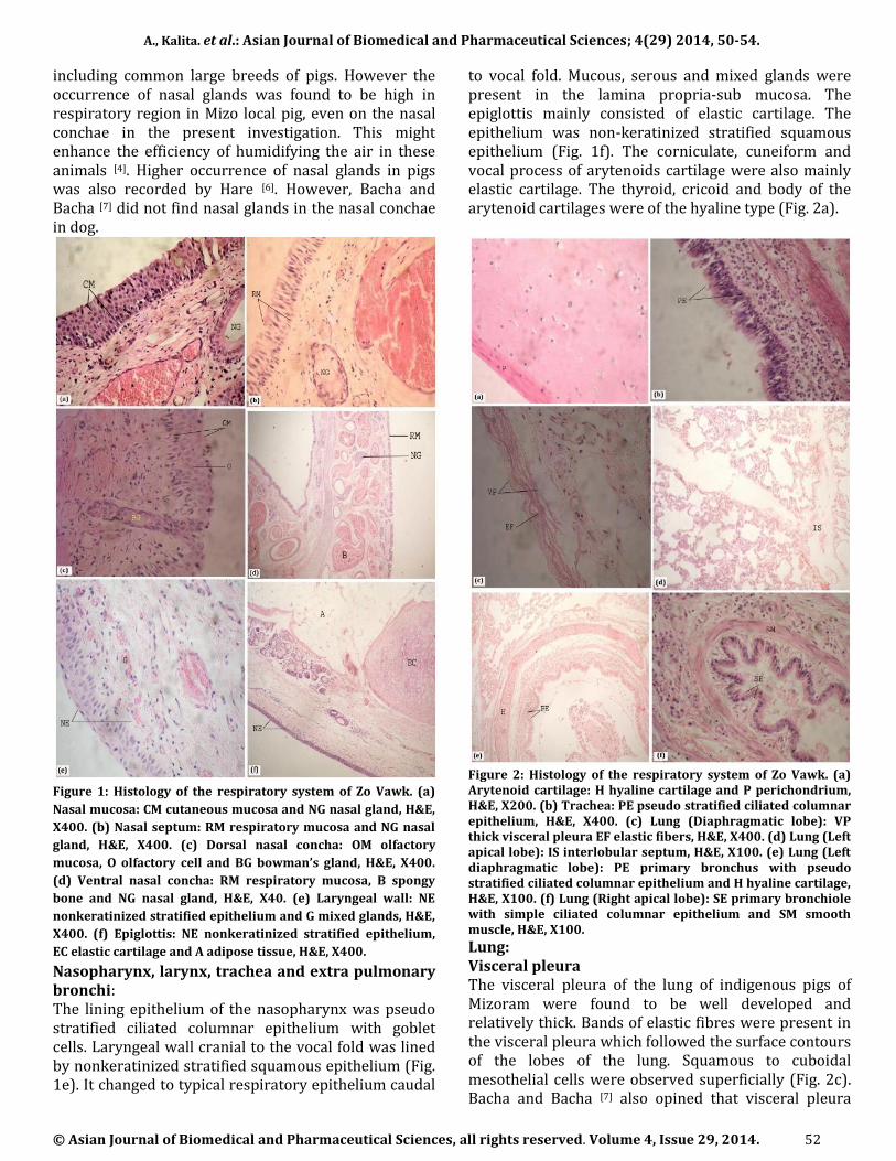
to vocal fold. Mucous, serous and mixed glands were present in the lamina propria-sub mucosa. The epiglottis mainly consisted of elastic cartilage. The epithelium was non-keratinized stratified squamous epithelium (Fig. 1f). The corniculate, cuneiform and vocal process of arytenoids cartilage were also mainly elastic cartilage. The thyroid, cricoid and body of the arytenoid cartilages were of the hyaline type (Fig. 2a).
Figure 2: Histology of the respiratory system of Zo Vawk. (a) Arytenoid cartilage: H hyaline cartilage and P perichondrium, H&E, X200. (b) Trachea: PE pseudo stratified ciliated columnar epithelium, H&E, X400. (c) Lung (Diaphragmatic lobe): VP thick visceral pleura EF elastic fibers, H&E, X400. (d) Lung (Left apical lobe): IS interlobular septum, H&E, X100. (e) Lung (Left diaphragmatic lobe): PE primary bronchus with pseudo stratified ciliated columnar epithelium and H hyaline cartilage, H&E, X100. (f) Lung (Right apical lobe): SE primary bronchiole with simple ciliated columnar epithelium and SM smooth muscle, H&E, X100. Lung: Visceral pleura The visceral pleura of the lung of indigenous pigs of Mizoram were found to be well developed and relatively thick. Bands of elastic fibres were present in the visceral pleura which followed the surface contours of the lobes of the lung. Squamous to cuboidal mesothelial cells were observed superficially (Fig. 2c). Bacha and Bacha [7] also opined that visceral pleura
A., Kalita. et al.: Asian Journal of Biomedical and Pharmaceutical Sciences; 4(29) 2014, 50-54.
© Asian Journal of Biomedical and Pharmaceutical Sciences, all rights reserved. Volume 4, Issue 29, 2014. 53
were thick in domestic mammals including pig, but thinner in carnivores. The interlobar and interlobular connective tissue septa were well developed, relatively thick and continued to interalveolar septa (Fig. 2d) which resulted in the distinct surface lobulation. The septa were composed of loose connective tissue rich in elastic fibres. Banks [4] also mentioned distinct lobular subdivision on the surface of lung in pigs and ruminants. The high amount of loose connective tissue with elastic fibers might be attributed to the elastic nature of lung of Mizo local pigs.
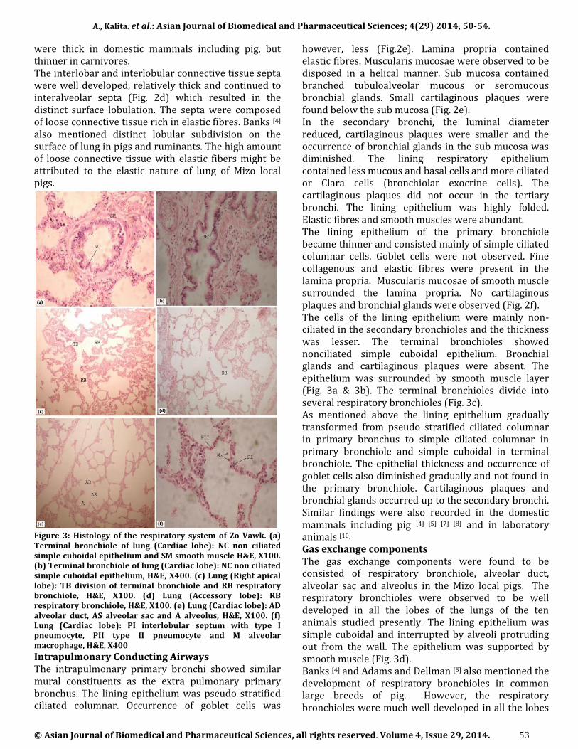
Figure 3: Histology of the respiratory system of Zo Vawk. (a) Terminal bronchiole of lung (Cardiac lobe): NC non ciliated simple cuboidal epithelium and SM smooth muscle H&E, X100. (b) Terminal bronchiole of lung (Cardiac lobe): NC non ciliated simple cuboidal epithelium, H&E, X400. (c) Lung (Right apical lobe): TB division of terminal bronchiole and RB respiratory bronchiole, H&E, X100. (d) Lung (Accessory lobe): RB respiratory bronchiole, H&E, X100. (e) Lung (Cardiac lobe): AD alveolar duct, AS alveolar sac and A alveolus, H&E, X100. (f) Lung (Cardiac lobe): PI interlobular septum with type I pneumocyte, PII type II pneumocyte and M alveolar macrophage, H&E, X400 Intrapulmonary Conducting Airways The intrapulmonary primary bronchi showed similar mural constituents as the extra pulmonary primary bronchus. The lining epithelium was pseudo stratified ciliated columnar. Occurrence of goblet cells was
however, less (Fig.2e). Lamina propria contained elastic fibres. Muscularis mucosae were observed to be disposed in a helical manner. Sub mucosa contained branched tubuloalveolar mucous or seromucous bronchial glands. Small cartilaginous plaques were found below the sub mucosa (Fig. 2e). In the secondary bronchi, the luminal diameter reduced, cartilaginous plaques were smaller and the occurrence of bronchial glands in the sub mucosa was diminished. The lining respiratory epithelium contained less mucous and basal cells and more ciliated or Clara cells (bronchiolar exocrine cells). The cartilaginous plaques did not occur in the tertiary bronchi. The lining epithelium was highly folded. Elastic fibres and smooth muscles were abundant. The lining epithelium of the primary bronchiole became thinner and consisted mainly of simple ciliated columnar cells. Goblet cells were not observed. Fine collagenous and elastic fibres were present in the lamina propria. Muscularis mucosae of smooth muscle surrounded the lamina propria. No cartilaginous plaques and bronchial glands were observed (Fig. 2f). The cells of the lining epithelium were mainly non-ciliated in the secondary bronchioles and the thickness was lesser. The terminal bronchioles showed nonciliated simple cuboidal epithelium. Bronchial glands and cartilaginous plaques were absent. The epithelium was surrounded by smooth muscle layer (Fig. 3a & 3b). The terminal bronchioles divide into several respiratory bronchioles (Fig. 3c). As mentioned above the lining epithelium gradually transformed from pseudo stratified ciliated columnar in primary bronchus to simple ciliated columnar in primary bronchiole and simple cuboidal in terminal bronchiole. The epithelial thickness and occurrence of goblet cells also diminished gradually and not found in the primary bronchiole. Cartilaginous plaques and bronchial glands occurred up to the secondary bronchi. Similar findings were also recorded in the domestic mammals including pig [4] [5] [7] [8] and in laboratory animals [10] Gas exchange components The gas exchange components were found to be consisted of respiratory bronchiole, alveolar duct, alveolar sac and alveolus in the Mizo local pigs. The respiratory bronchioles were observed to be well developed in all the lobes of the lungs of the ten animals studied presently. The lining epithelium was simple cuboidal and interrupted by alveoli protruding out from the wall. The epithelium was supported by smooth muscle (Fig. 3d). Banks [4] and Adams and Dellman [5] also mentioned the development of respiratory bronchioles in common large breeds of pig. However, the respiratory bronchioles were much well developed in all the lobes
A., Kalita. et al.: Asian Journal of Biomedical and Pharmaceutical Sciences; 4(29) 2014, 50-54.
© Asian Journal of Biomedical and Pharmaceutical Sciences, all rights reserved. Volume 4, Issue 29, 2014. 54
of the lungs of the animals under present study. Bal and Gloshal [10], on the other hand, could not detect respiratory bronchioles in laboratory animals like mouse, rat, hamster, guinea pig, gerbil and rabbit, which was in accord with Banks [4]. The well developed respiratory bronchioles might enhance the respiratory efficiency of the Mizo local pigs and rendered them more suitable to thrive in higher altitude and lesser atmospheric oxygen tension in Mizoram. This finding could not be seconded and compared due to lack of anatomical study on the lungs of local Mizo pigs in the available literature. Typical alveolar ducts and alveolar sacs were observed in all the lungs of the indigenous pigs of Mizoram investigated presently. Alveolar ducts were lined by alveoli with smooth muscle along the luminal border at the apices between adjacent alveoli. Clusters of alveoli forming alveolar sacs and atrium were also well developed in the lungs of the animals under study (Fig. 3e). Presently the structure of alveolar duct and alveolar sac were recorded in accordance with other domestic mammals including common large breeds of pigs [4] [5] [7] [8]. In the wall of the alveoli both Type I pneumocyte or squamous alveolar epithelial cells and Type II pneumocytes or granular (great) alveolar epithelial cells (Fig. 3f) were distinguished. The Type I pneumocytes showed flat nucleus and attenuated cytoplasm forming the interface between air and blood to allow the passage of gases. The Type II pneumocytes were cuboidal with spherical nucleus and foamy cytoplasm projecting into the alveolar lumen (Fig. 3f). Large alveolar macrophages (Fig. 3f) were also detected frequently in the alveolar lumen. The Type I pneumocytes formed the main lining epithelium of the alveoli and the Type II pneumocytes were found occasionally among the Type I cells, as also observed in goat [11]. Although could not be seconded and compared due to scarcity of anatomical study on the indigenous variety of pigs, the occurrence of alveolar macrophage was found significantly frequent which might be attributed to the better resistance to respiratory diseases in the local Mizo pig (Zo vawk). ACKNOWLEDGMENT The authors are grateful to The Honourable Vice Chancellor and The Director of Research, Central Agricultural University, Imphal, India for sanctioning and funding the research project under intra mural research programme. Special thanks go to The Dean, C.V.Sc. & A.H. Selesih, Aizawl for providing all the facilities to carry out the research work. REFERENCES 1. [1] Drury RAB, Wallington EA. Carleton’s Histological Technique. 5th edn. Oxford, Oxford University Press. 1980.
[2] Luna LG. Manual of histological staining methods of Armed Forces Institute of Pathology. 3rd edn. Mc Graw Hill Book Company, New York. 1968. [3] Snedecor GW, Cochran WG. Statistical methods. 9th edn. Iowa State University Press, Ames. 1994; 124-130. [4] Banks WJ, Applied Veterinary Histology. 3rd edn. Williams and Wilkins, Baltimore. 1993. [5] Adams DR, Dellmann HD. Respiratory System. In Textbook of Veterinary Histology. 5th edn. Lippincott Williams & Wilkins, Philadelphia. 1998. [6] Hare WCD. Respiratory System. In, Sisson and Grossman’s The Anatomy of the Domestic Animals , Vol. 2. 5th edn. W.B. Saunders Co. Philadelphia. 1975. [7] Bacha WJ, Bacha LM. Color Atlas of Veterinary Histology. 2nd edn. Philadelphia, Baltimore. Lippincott Williams & Wilkins. 2000; 69-84. [8] Aughey E, Frye FL. Comparative Veterinary Histology with Clinical Correlates. 1st edn. Manson Publishing Ltd. London. 2001. [9] Verlindin K, Ginneken CV, Weyns A, Meir FV. Distribution of T and B cells in the pig lung. J. Anat. 2002; 200(2):208. [10] Bal HS, Ghoshal NG. Morphology of the terminal bronchiolar region of Common Laboratory mammals. Laboratory Animals. 1988; 22:76-82. [11] Baba MA, Choudhary AR. 2008 Histomorphology of the pulmonary alveoli of goat (Capra hircus). Veterinary World. 2008; 1(10): 312-3.