Embed Size (px)
Citation preview
of April 6, 2018.This information is current as
Hyperplasia and AngiogenesisReceptor-Like 1 Induces Synovial Serum Amyloid A Binding to Formyl Peptide
Suh, Wan-Uk Kim and Sung Ho RyuMi-Sook Lee, Seung-Ah Yoo, Chul-Soo Cho, Pann-Ghill
http://www.jimmunol.org/content/177/8/5585doi: 10.4049/jimmunol.177.8.5585
2006; 177:5585-5594; ;J Immunol
Referenceshttp://www.jimmunol.org/content/177/8/5585.full#ref-list-1
, 30 of which you can access for free at: cites 57 articlesThis article
average*
4 weeks from acceptance to publicationFast Publication! •
Every submission reviewed by practicing scientistsNo Triage! •
from submission to initial decisionRapid Reviews! 30 days* •
Submit online. ?The JIWhy
Subscriptionhttp://jimmunol.org/subscription
is online at: The Journal of ImmunologyInformation about subscribing to
Permissionshttp://www.aai.org/About/Publications/JI/copyright.htmlSubmit copyright permission requests at:
Email Alertshttp://jimmunol.org/alertsReceive free email-alerts when new articles cite this article. Sign up at:
Print ISSN: 0022-1767 Online ISSN: 1550-6606. Immunologists All rights reserved.Copyright © 2006 by The American Association of1451 Rockville Pike, Suite 650, Rockville, MD 20852The American Association of Immunologists, Inc.,
is published twice each month byThe Journal of Immunology
by guest on April 6, 2018
http://ww
w.jim
munol.org/
Dow
nloaded from
by guest on April 6, 2018
http://ww
w.jim
munol.org/
Dow
nloaded from
Serum Amyloid A Binding to Formyl Peptide Receptor-Like 1Induces Synovial Hyperplasia and Angiogenesis1
Mi-Sook Lee,* Seung-Ah Yoo,† Chul-Soo Cho,† Pann-Ghill Suh,* Wan-Uk Kim,2† andSung Ho Ryu2*
Serum amyloid A (SAA) is a major acute-phase reactant, and has been demonstrated to mediate proinflammatory cellularresponses. Although SAA has been used as an indicator for a variety of inflammatory diseases, the role of SAA in synovialhyperplasia and proliferation of endothelial cells, a pathological hallmark of rheumatoid arthritis (RA), has yet to be elucidated.In this study, we have demonstrated that SAA promotes the proliferation of human fibroblast-like synoviocytes (FLS). In addition,SAA protects RA FLS against the apoptotic death induced by serum starvation, anti-Fas IgM, and sodium nitroprusside. Theactivity of SAA appears to be mediated by the formyl peptide receptor-like 1 (FPRL1) receptor, as it was mimicked by theWKYMVm peptide, a specific ligand for FPRL1, but completely abrogated by down-regulating the FPRL1 transcripts with shortinterfering RNA. The effect of SAA on FLS hyperplasia was shown to be caused by an increase in the levels of intracellularcalcium, as well as the activation of ERK and Akt, which resulted in an elevation in the expression of cyclin D1 and Bcl-2.Moreover, SAA stimulated the proliferation, migration, and tube formation of endothelial cells in vitro, and enhanced the sprout-ing activity of endothelial cells ex vivo and neovascularization in vivo. These observations indicate that the binding of SAA toFPRL1 may contribute to the destruction of bone and cartilage via the promotion of synoviocyte hyperplasia and angiogenesis,thus providing a potential target for the control of RA. The Journal of Immunology, 2006, 177: 5585–5594.
R heumatoid arthritis (RA)3 is a multisystem autoimmunedisease, which is characterized by chronic joint inflam-mation (1). The hallmark characteristics of RA pathology
include the infiltration of inflammatory leukocytes, the prolifera-tion of synovial cells, and the presence of extensive angiogenesis,which is also commonly referred to as rheumatoid pannus (2–4).Rheumatoid pannus is sometimes considered to be a local tumor.For example, synovial fibroblasts, the principal components of in-vading pannus, proliferate abnormally, resist apoptosis, and invadethe local environment (5, 6). Synovial fibroblasts obtained fromRA patients exhibited several oncogenes, including H-ras and p53,harboring somatic mutations (7, 8). They also abundantly expressantiapoptotic proteins, including the FLICE inhibitory protein (9)and Bcl-2 (10), both of which exert protective effects against theapoptosis initiated via death receptor- or mitochondria-dependentpathways. Moreover, in a fashion similar to that of carcinogenesis,
angiogenesis is considered to be a critical step in the progressionof RA (4, 11–13).
Serum amyloid A (SAA) is a multifunctional apolipoprotein,12- to 14-kDa in size. This protein is normally present in thebloodstream at a concentration of �0.1 �M, but the concentrationof SAA can increase up to 1000-fold within the first 24–36 h inresponse to a variety of injuries, including trauma, infection, in-flammation, and neoplasia (14, 15). As with other acute-phase re-actants, the liver is the primary site at which SAA productionoccurs, but the overproduction of SAA in extrahepatic areas hasalso been implicated in the pathogenesis of several chronic inflam-matory diseases, including atherosclerosis, Alzheimer’s disease,inflammatory arthritis, and several cancer variants (16, 17). More-over, elevated SAA levels appear to be an important indicator forboth the diagnosis and prognosis of chronic inflammatory diseases.For example, increased levels of SAA are frequently observed inthe sera, synovial fluid, and inflamed synovium of RA patients, andthese levels have been commonly used as highly sensitive markersfor the disease activity of RA (18–20).
There are two known SAA receptors, including CD36 andLIMPII analogous-1 (CLA-1) (21) and lipoxin A4 receptor/formylpeptide receptor-like 1 (FPRL1) (22, 23). FPRL1 is one of theclassic chemoattractant receptors encompassing G protein-coupledseven transmembrane domains. Previous reports have pointed to arole for FPRL1 in the regulation of a variety of cellular responsesin several cell types, including astrocytoma cell lines (24), neutro-phils, monocytes, and T cells (25), as well as HUVECs (26). Re-cently, O’Hara et al. (27) showed that overexpressed SAA andFPRL1 in inflamed synovial tissue can be associated with the pro-duction of matrix metalloproteinase. However, it remains to bedetermined whether SAA and FPRL1 in the RA synovium areinvolved directly in the synovial proliferation and formation of aninvading pannus. Furthermore, very little information is currentlyavailable regarding the intracellular pathway relevant to SAA sig-naling in RA synoviocytes.
*Division of Molecular Life Sciences, Pohang University of Science and Technology,Pohang, Korea; †Department of Internal Medicine, Catholic University of Korea,Seoul, Korea
Received for publication February 28, 2006. Accepted for publication August 2, 2006.
The costs of publication of this article were defrayed in part by the payment of pagecharges. This article must therefore be hereby marked advertisement in accordancewith 18 U.S.C. Section 1734 solely to indicate this fact.1 This work was supported by a grant from the Ministry of Science and Technology/Korea Science and Engineering Foundation to the National Core Research Center forSystems Bio-Dynamics and the 21st Frontier Proteome Research of the Ministry ofScience and Technology in the Republic of Korea.2 Address correspondence and reprint requests to Dr. Wan-Uk Kim, Department ofInternal Medicine, Division of Rheumatology, School of Medicine, Catholic Univer-sity of Korea, St. Vincent’s Hospital, 93 Chi-Dong, Suwon 442-723, South Korea;E-mail address: [email protected] or Dr. Sung Ho Ryu, Division of MolecularLife Sciences, Pohang University of Science and Technology, Pohang 790-784, SouthKorea. E-mail address: [email protected] Abbreviations used in this paper: RA, rheumatoid arthritis; SAA, serum amyloid A;FPRL1, formyl peptide receptor-like 1; OA, osteoarthritis; FLS, fibroblast-like syno-viocyte; SNP, sodium nitroprusside; VEGF, vascular endothelial growth factor;siRNA, short interfering RNA; [Ca2�]i, intracellular Ca2� concentration.
The Journal of Immunology
Copyright © 2006 by The American Association of Immunologists, Inc. 0022-1767/06/$02.00
by guest on April 6, 2018
http://ww
w.jim
munol.org/
Dow
nloaded from

In this study, we evaluated the role of SAA in synoviocyte hy-perplasia and angiogenesis, as both processes are crucial and mu-tually complementary with regard to RA pathogenesis. We deter-mined that SAA enhances the proliferation and survival ofsynovial fibroblasts via binding to its receptor, FPRL1, and thatthis effect is mediated by the activation of intracellular calcium,ERK, and Akt. Moreover, SAA appears to participate in neovas-cularization by increasing the proliferation, migration, tube forma-tion, and sprouting activity of endothelial cells. Taken together,our data suggest that SAA may be directly involved in the destruc-tion of bone and cartilage via the promotion of synoviocyte hy-perplasia and angiogenesis, and thus may constitute a potentialtarget for the treatment of RA.
Materials and MethodsMaterials
Recombinant human SAA (endotoxin level � 0.1 ng/�g) was purchasedfrom PeproTech. The amino acid sequence of recombinant human SAAcorresponds to the sequence of human SAA 1� isotype except for additionof an N-terminal Met and substitution of Asp for Asn at position 60 andsubstitution of His to Arg at position 71. The synthetic peptide, WKYMVmwas synthesized from Anygen A & Pep. All Abs used in this study werepurchased from Cell Signaling Technology.
Isolation and culture of synovial fibroblasts and HUVECs
Fibroblast-like synoviocytes (FLS) were prepared from synovial samplesobtained from patients with RA and osteoarthritis (OA), all of whom werealso undergoing total joint replacement surgery. The FLS were isolatedfrom the synovial tissues in accordance with a previously described pro-cedure (28). In brief, fresh synovial tissues were minced into 2- to 3-mmpieces, then treated for 4 h with 4 mg/ml type I collagenase (WorthingtonBiochemical), and maintained in DMEM containing 10% FBS at 37°C inan atmosphere containing 5% CO2. The cells were used at three to eightpassages, during which time they evidenced a homogenous fibroblast pop-ulation, and also exhibited a typical bipolar configuration, as observedunder inverse microscopy. HUVECs were isolated from fresh human um-bilical cords via collagenase (Worthington Biochemical) digestion, andthen maintained in 20% FBS-containing M-199 medium, as previouslydescribed. All HUVECs were used after no more than five passages.
Cell proliferation assay
The RA FLS, OA FLS, and HUVECs were plated onto 24-well culturedishes at a density of 2 � 104 cells/well, and then permitted to attachovernight. After 24 h of serum starvation, the cells were treated for 72 hwith a variety of mitogens. [3H]Thymidine (1 �Ci) was added to each ofthe wells before the final 6 h of incubation (29). Cell growth was alsoevaluated by counting the viable cells. Control and mitogen-treated cellswere harvested by trypsinization, and the number of cells was determinedwith a hemocytometer, under �100 magnification.
Apoptosis assay
Synoviocyte apoptosis was induced by 3 days of serum deprivation, or bytreating the cells for 12 h with either sodium nitroprusside (SNP, 0.7 mM)or anti-Fas IgM (0.7 �g/ml) plus cyclohexamide (1 �g/ml). The degree ofapoptosis was then evaluated by MTT assay and ELISA for DNA frag-mentation. In the MTT assay, FLS were seeded in 24-well culture plates ata density of 2 � 104 cells/well. After 72 h of incubation with SAA or mediaalone, MTT solution was added to each of the wells, and then incubated for2 h. The reaction was halted via the removal of MTT. Thereafter, DMSO(200 �l) was added to solubilize the formazan crystals. The plates werethen subjected to 5 min of gentle shaking to ensure that the crystals haddissolved completely, and the absorbance was read at 540 nm with a mi-croplate reader. The cellular DNA fragmentation assay was conducted us-ing an ELISA kit (Roche Applied Science), based on the quantitative sand-wich ELISA principle, using two mouse mAbs targeted against DNA andBrdU, as previously described (30). In brief, the BrdU-labeled DNA frag-ments of the samples were bound to the immobilized anti-DNA Ab, fixingit within the wells of a microtiter plate. The immune-complexed BrdU-labeled DNA fragments were then denatured and fixed to the surfaces ofthe plates via the application of microwave irradiation. In the final step, theanti-BrdU peroxidase conjugate was allowed to react with the BrdU thathad been incorporated into the DNA. After the removal of the unboundperoxidase conjugates, the quantity of peroxidase bound within the im-
mune complex was determined photometrically, using tetramethylbenzi-dine as a substrate.
Generation and transfection of short interfering RNA (siRNA)for FPRL1 transcripts
To down-regulate the FPRL1 transcripts using siRNA, the following targetsequence was used: 300AAU UCA CAU CGU GGU GGA CAU320. Theresults of a BLAST search of siRNA sequence revealed no significanthomology to any other sequences stored in the database. This oligonucle-otide yielded comparable results. RA FLS were used in the siRNA trans-fection procedure (31). These cells were transfected with a final concen-tration of 20 nM FPRL1 siRNA or luciferase siRNA, as a control, usingLipofectamine reagent (Invitrogen Life Technologies) in accordance withthe manufacturer’s instructions. The cells were washed with serum-freemedium and then incubated with transfection mixture for 4 h and 30 min,after which medium containing 10% FBS was added. The cells were col-lected after 24, 48, and 72 h of incubation, after which the levels of FPRL1expression were determined via Northern blot analysis and ligand bindingassay.
Northern blot analysis
The total RNA from the transfected RA FLS cells was isolated using acommercially available TRI reagent (Molecular Research Center), in ac-cordance with the manufacturer’s instructions. RNA samples (20 �g/lane)were electrophoresed on 1% agarose gels containing formaldehyde, trans-ferred to Hybond-N membrane (Amersham Biosciences), immobilizedwith UV light and then hybridized with FPRL1 or actin probes labeled withbiotin using the PCR DNA biotinylation kit (Kirkegaard & Perry Labora-tories). The membranes were washed in prehybridization buffer with de-natured 10 mg/ml carrier DNA (Sigma-Aldrich) at 42°C for 1 h. The de-natured probe was then added into the buffer and incubated at 42°Covernight. The membranes were washed, incubated with detector blocksolution for 45 min, and then incubated with alkaline phosphatase-labeledstreptavidin for 30 min. The membranes were washed again, and incubatedwith CDP-Star Chemiluminescent (Kirkegaard & Perry Laboratories). X-ray film was exposed to the membranes with intensifying screens and thendeveloped.
Ligand binding assay
Ligand binding assay was performed as previously described (32). Briefly,RA FLS and OA FLS were seeded at 1 � 105 cells/well onto a 24-wellplate and cultured overnight. After blocking them with blocking buffer (33mM HEPES (pH 7.5), 0.1% BSA in DMEM) for 2 h, 500 pM of 125I-labeled WKYMVm (Amersham Biosciences) was added to the cells inbinding buffer (PBS containing 0.1% BSA), in the presence or absence ofunlabeled WRWWWW (WKYMVm competitor for FPRL1 binding) (32),and incubated for 3 h at 4°C with continuous shaking. The samples werethen washed five times with ice-cold binding buffer, and 200 �l of lysisbuffer (20 mM Tris (pH 7.5) and 1% Triton X-100) was added to each well.After 20 min at room temperature, the lysates were collected and countedusing a gamma ray counter.
ELISA for SAA
Synovial fluid of 10 RA patients with joint effusions was collected byarthrocentesis, as previously described (33). The concentration of humanSAA in RA synovial fluid was measured by ELISA kit (BioSource Inter-national), according to the manufacturer’s instruction.
Intracellular Ca2� measurement
The isolated FLS were incubated with fluo-3-AM working solution (Mo-lecular Probes) (34), containing 0.03% plutonic F-127 (the final concen-tration of fluo-3-AM was 20 �M/L) for 1 h at 37°C. After incubation,fluo-3-AM fluorescence in the cells was elicited at 488 nm with a high-power Ar� laser, and the emission bands were detected at 530 nm with aphotomultiplier. The fluorescence signal was detected using a confocallaser scanning system (Lasersharp MRA2; Bio-Rad), equipped with a Ni-kon E-600 Eclipse microscope. The fluorescence intensity was measuredboth before (F0) and after (F) the addition of SAA or PMA. The change inintracellular Ca2� concentration [Ca2�]i was expressed in terms of the F/F0
ratio. A total of 50–120 images were scanned in each cell.
Western blot analysis
RA FLS were incubated for 24 h in DMEM without FBS, and then SAA(3 �M) was added to the cells for the indicated times. The treated RA FLSwere then washed twice in PBS, dissolved in sample buffer (50 mM Tris-HCl, 100 mM NaCl, 0.1% SDS, 1% Nonidet P-40, 50 mM NaF, 1 mM
5586 ROLE OF SAA IN RHEUMATOID ARTHRITIS
by guest on April 6, 2018
http://ww
w.jim
munol.org/
Dow
nloaded from

Na3VO4, 1 �g/ml aprotinin, 1 �g/ml pepstatin, and 1 �g/ml leupeptin),boiled, separated via SDS-PAGE, and transferred to nitrocellulose mem-branes. After immunoblot analysis with phospho-ERK1/2 (Thr202/Tyr204),phospho-Akt (Ser473), phospho-STAT3 (Tyr705), cyclin D1, or Bcl-2 Abs,the membranes were stripped and reincubated with anti-ERK, Akt, STAT3,or �-actin Ab, respectively, to detect total protein amounts.
Wounding migration and tube formation assay
The wounding migration and tube formation activity of the HUVECs weremeasured as previously described (35, 36). In brief, HUVECs plated atconfluence on 60-mm culture dishes were wounded with pipette tips, thentreated with SAA (0–5 �g/ml), WKYMVm (10 nM), or vascular endo-thelial growth factor (VEGF, 20 ng/ml) in M-199 medium, supplementedwith 1% serum and 1 mM thymidine. After 12 h of incubation, migrationwas quantitated by counting the cells that moved beyond the reference line.For the tube formation assay, the HUVECs were seeded on a layer ofpreviously polymerized Matrigel (BD Biosciences) with SAA (5 �g/ml),WKYMVm peptide (10 nM), a specific ligand for FPRL1 (37, 38) orVEGF (20 ng/ml). After 18 h of incubation, the cell morphology wasvisualized via phase-contrast microscopy and photographed. The degree oftube formation was quantified by measuring the length of tubes in fiverandomly chosen low-power fields (magnification, �40) from each wellusing the image-Pro Plus v4.5 (Media Cybernetics).
Rat aorta ring assay
Aortas from male Sprague-Dawley rats were cross-sectioned into rings,and mounted onto polymerized Matrigel dishes (39). Matrigel (150 �l) wasthen positioned on top and allowed to gel. After 7 days, the aortic rings,incubated with PBS, SAA (3 and 5 �g/ml), WKYMVm (10 nM), VEGF(20 ng/ml), or FBS (10%) were analyzed under an inverted microscope.
Mouse Matrigel plug assay
C57BL/6 mice (7 wk of age) were given s.c. injections of 500 �l of Ma-trigel (40) containing PBS, SAA (80 �g), or WKYMVm (1 �g). After 7days, the skins of the mice were pulled back to expose the Matrigel plugs,which remained intact. After the noting and photographing of any quanti-tative differences, hemoglobin levels were measured by the Drabkin
method, using a Drabkin reagent kit 525 (Sigma-Aldrich) for the quanti-tative assessment of blood vessel formation. The hemoglobin concentrationwas calculated from the parallel assay of a known amount of hemoglobin.The Matrigel plugs were fixed in 4% formalin, embedded with paraffin, andstained using H&E.
Statistical analysis
All data are expressed as the mean � SD from several separate experi-ments. Statistical comparisons were conducted via Student’s t test, and avalue for p � 0.05 was considered statistically significant.
ResultsSAA stimulates synoviocyte proliferation
Synovial hyperplasia is one of the hallmarks of RA pathology (3,41). Several studies have shown that RA FLS tend to divide at amore rapid rate than do synoviocytes obtained from normal orosteoarthritic joints (42). Therefore, we have attempted to deter-mine whether SAA accelerates the proliferation of FLS acquiredfrom both RA and OA patients by [3H]thymidine incorporationassays. When the FLS were stimulated with 5 �M SAA for 1–5days, the DNA synthesis activities of RA FLS and OA FLS werepeaked at 3 days (Fig. 1A). Therefore, we treated 0.1–5 �M SAAfor 3 days to RA FLS and OA FLS to check the dose dependencyof SAA on FLS proliferation. As a result, the incorporation rate of[3H]thymidine was increased in a dose-dependent fashion, with themaximal effect being detected at a SAA concentration of 5 �M(Fig. 1B). The numbers of RA FLS and OA FLS were also dose-dependently increased as the result of SAA treatment (Fig. 1, Cand D), and this effect was greater for the RA FLS than for the OAFLS (Fig. 1C). Moreover, when the synovial fluid of different RApatients (n � 10) was added to RA FLS, the FLS proliferation wascorrelated well with the concentration of SAA in the synovial fluid(r � 0.733, p � 0.016). These results suggest that SAA is capable
FIGURE 1. Proliferative effect of SAA on FLS.Primary cultured RA FLS and OA FLS were plated intriplicate, and [3H]thymidine incorporation was usedin the measurement of DNA synthesis activity in thepresence of human recombinant SAA (5 �M) for 1, 3,or 5 days (A) and SAA (0, 0.1, 1, 3, or 5 �M) for 72 h(B). C, After 72 h of incubation with increasing dosesof SAA (0, 0.1, 1, 3, or 5 �M), the RA FLS and OAFLS were trypsinized, and the cell numbers per wellwere determined under a microscope. The results arepresented as the mean � SD of three independentexperiments using different cells from three differentRA and OA patients. �, p � 0.05 vs the [3H]thymi-dine incorporation (B) and cell number (C) of notreatment of SAA. D, RA FLS incubated in the pres-ence or absence of 5 �M SAA for 72 h were photo-graphed. Original magnification, �50. E, SAA con-centration of synovial fluid from 10 different RApatients was measured using SAA ELISA and theproliferative activity of RA FLS was evaluated after72 h treatment of RA synovial fluid using [3H]thymi-dine incorporation assay. Correlation between twovariables was performed using Spearman’s rank cor-relation coefficient.
5587The Journal of Immunology
by guest on April 6, 2018
http://ww
w.jim
munol.org/
Dow
nloaded from
of stimulating the abnormal proliferation of FLS, particularly inRA joints.
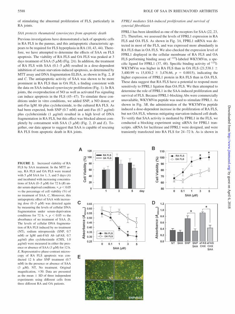
SAA protects rheumatoid synoviocytes from apoptotic death
Previous investigations have demonstrated a lack of apoptotic cellsin RA FLS in the pannus, and this antiapoptotic characteristic ap-pears to be required for FLS hyperplasia in RA (10, 43, 44). There-fore, we have attempted to determine the effects of SAA on FLSapoptosis. The viability of RA FLS and OA FLS was peaked at 3days treatment of SAA (5 �M) (Fig. 2A). In addition, the treatmentof RA FLS with SAA (0.1–5 �M) resulted in a dose-dependentinhibition of serum starvation-induced apoptosis, as determined byMTT assay and DNA fragmentation ELISA, as shown in Fig. 2, Band C. The antiapoptotic activity of SAA was shown to be moreprominent in RA FLS than in OA FLS, a finding consistent withthe data on SAA-induced synoviocyte proliferation (Fig. 1). In RAjoints, the overproduction of NO as well as activated Fas signalingcan induce apoptosis in the FLS (45–47). To simulate these con-ditions under in vitro conditions, we added SNP, a NO donor, oranti-Fas IgM Ab plus cycloheximide, to the cultured RA FLS. Ashad been expected, both SNP (0.7 mM) and anti-Fas (0.7 �g/ml)plus cycloheximide (1 �g/ml) resulted in a high level of DNAfragmentation in RA FLS, but this effect was blocked almost com-pletely by cotreatment with SAA (3 �M) (Fig. 2, D and E). To-gether, our data appear to suggest that SAA is capable of rescuingRA FLS from apoptotic death in RA joints.
FPRL1 mediates SAA-induced proliferation and survival ofsynovial fibroblasts
FPRL1 has been identified as one of the receptors for SAA (22, 23,27). Therefore, we assessed the levels of FPRL1 expression in RAFLS and OA FLS. As shown in Fig. 3A, FPRL1 mRNA was de-tected in most of the FLS, and was expressed more abundantly inRA FLS than in OA FLS. We also checked the expression level ofFPRL1 displayed in the cellular membrane of RA FLS and OAFLS performing binding assay of 125I-labeled WKYMVm, a spe-cific ligand for FPRL1 (37, 48). Specific binding activity of 125I-WKYMVm was higher in RA FLS than in OA FLS (21,538.1 �3,400.99 vs 15,830.2 � 3,476.66, p � 0.0015), indicating thehigher expression of FPRL1 protein in RA FLS than in OA FLS.These data suggest that RA FLS have a potential to respond moresensitively to FPRL1 ligation than OA FLS. We then attempted todetermine the role of FPRL1 in the SAA-induced proliferation andsurvival of FLS. Because FPRL1-blocking Abs were commerciallyunavailable, WKYMVm peptide was used to stimulate FPRL1. Asshown in Fig. 3B, the administration of the WKYMVm peptideinduced a dose-dependent increase in the proliferation of RA FLS,but not OA FLS, whereas mitigating starvation-induced cell death.To verify that SAA activity is mediated by FPRL1 in the FLS, weconducted a blocking experiment using siRNA for FPRL1 tran-scripts. siRNA for luciferase and FPRL1 were designed, and weretransiently transfected into RA FLS for 24–72 h. As is shown in
FIGURE 2. Increased viability of RAFLS by SAA treatment. In the MTT as-say, RA FLS and OA FLS were treatedwith 5 �M SAA for 1, 3, and 5 days (A)and incubated with increasing concentra-tions of SAA (0–5 �M) for 72 h (B) un-der serum-deprived conditions. �, p � 0.05vs the percentage of cell viability (%) ofno treatment of SAA. C, Moreover, thisantiapoptotic effect of SAA with increas-ing dose (0–5 �M) was detected againby measuring the levels of cellular DNAfragmentation under serum-deprivationconditions for 72 h. �, p � 0.05 vs theabsorbance of no treatment of SAA. D,The levels of cellular DNA fragmenta-tion of RA FLS induced by no treatment(NT), sodium nitroprusside (SNP, 0.7mM) or IgM anti-FAS Ab (aFAS; 0.7�g/ml) plus cycloheximide (CHX, 1.0�g/ml) were measured in either the pres-ence or absence of SAA (3 �M) for 12 h.E, Representative phase-contrast micros-copy of RA FLS apoptosis was con-ducted 12 h after SNP treatment (0.7mM) in the presence or absence of SAA(3 �M). NT, No treatment. Originalmagnification, �50. Data are presentedas the mean � SD of three independentexperiments using different cells fromthree different RA and OA patients.
5588 ROLE OF SAA IN RHEUMATOID ARTHRITIS
by guest on April 6, 2018
http://ww
w.jim
munol.org/
Dow
nloaded from
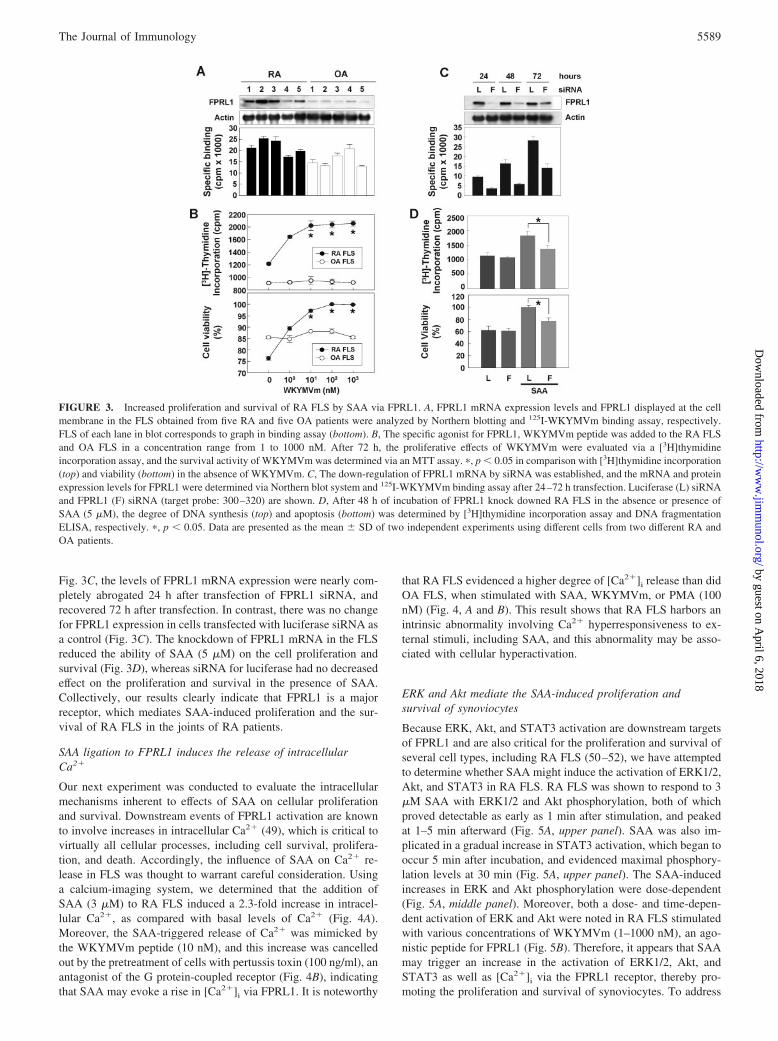
Fig. 3C, the levels of FPRL1 mRNA expression were nearly com-pletely abrogated 24 h after transfection of FPRL1 siRNA, andrecovered 72 h after transfection. In contrast, there was no changefor FPRL1 expression in cells transfected with luciferase siRNA asa control (Fig. 3C). The knockdown of FPRL1 mRNA in the FLSreduced the ability of SAA (5 �M) on the cell proliferation andsurvival (Fig. 3D), whereas siRNA for luciferase had no decreasedeffect on the proliferation and survival in the presence of SAA.Collectively, our results clearly indicate that FPRL1 is a majorreceptor, which mediates SAA-induced proliferation and the sur-vival of RA FLS in the joints of RA patients.
SAA ligation to FPRL1 induces the release of intracellularCa2�
Our next experiment was conducted to evaluate the intracellularmechanisms inherent to effects of SAA on cellular proliferationand survival. Downstream events of FPRL1 activation are knownto involve increases in intracellular Ca2� (49), which is critical tovirtually all cellular processes, including cell survival, prolifera-tion, and death. Accordingly, the influence of SAA on Ca2� re-lease in FLS was thought to warrant careful consideration. Usinga calcium-imaging system, we determined that the addition ofSAA (3 �M) to RA FLS induced a 2.3-fold increase in intracel-lular Ca2�, as compared with basal levels of Ca2� (Fig. 4A).Moreover, the SAA-triggered release of Ca2� was mimicked bythe WKYMVm peptide (10 nM), and this increase was cancelledout by the pretreatment of cells with pertussis toxin (100 ng/ml), anantagonist of the G protein-coupled receptor (Fig. 4B), indicatingthat SAA may evoke a rise in [Ca2�]i via FPRL1. It is noteworthy
that RA FLS evidenced a higher degree of [Ca2�]i release than didOA FLS, when stimulated with SAA, WKYMVm, or PMA (100nM) (Fig. 4, A and B). This result shows that RA FLS harbors anintrinsic abnormality involving Ca2� hyperresponsiveness to ex-ternal stimuli, including SAA, and this abnormality may be asso-ciated with cellular hyperactivation.
ERK and Akt mediate the SAA-induced proliferation andsurvival of synoviocytes
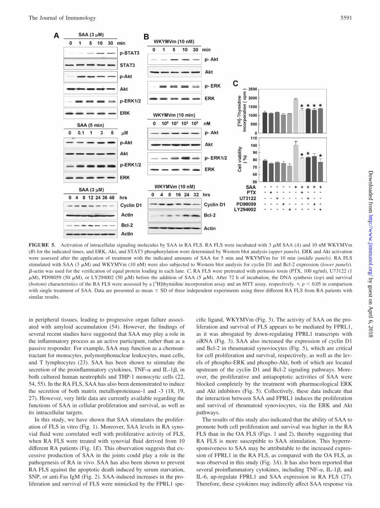
Because ERK, Akt, and STAT3 activation are downstream targetsof FPRL1 and are also critical for the proliferation and survival ofseveral cell types, including RA FLS (50–52), we have attemptedto determine whether SAA might induce the activation of ERK1/2,Akt, and STAT3 in RA FLS. RA FLS was shown to respond to 3�M SAA with ERK1/2 and Akt phosphorylation, both of whichproved detectable as early as 1 min after stimulation, and peakedat 1–5 min afterward (Fig. 5A, upper panel). SAA was also im-plicated in a gradual increase in STAT3 activation, which began tooccur 5 min after incubation, and evidenced maximal phosphory-lation levels at 30 min (Fig. 5A, upper panel). The SAA-inducedincreases in ERK and Akt phosphorylation were dose-dependent(Fig. 5A, middle panel). Moreover, both a dose- and time-depen-dent activation of ERK and Akt were noted in RA FLS stimulatedwith various concentrations of WKYMVm (1–1000 nM), an ago-nistic peptide for FPRL1 (Fig. 5B). Therefore, it appears that SAAmay trigger an increase in the activation of ERK1/2, Akt, andSTAT3 as well as [Ca2�]i via the FPRL1 receptor, thereby pro-moting the proliferation and survival of synoviocytes. To address
FIGURE 3. Increased proliferation and survival of RA FLS by SAA via FPRL1. A, FPRL1 mRNA expression levels and FPRL1 displayed at the cellmembrane in the FLS obtained from five RA and five OA patients were analyzed by Northern blotting and 125I-WKYMVm binding assay, respectively.FLS of each lane in blot corresponds to graph in binding assay (bottom). B, The specific agonist for FPRL1, WKYMVm peptide was added to the RA FLSand OA FLS in a concentration range from 1 to 1000 nM. After 72 h, the proliferative effects of WKYMVm were evaluated via a [3H]thymidineincorporation assay, and the survival activity of WKYMVm was determined via an MTT assay. �, p � 0.05 in comparison with [3H]thymidine incorporation(top) and viability (bottom) in the absence of WKYMVm. C, The down-regulation of FPRL1 mRNA by siRNA was established, and the mRNA and proteinexpression levels for FPRL1 were determined via Northern blot system and 125I-WKYMVm binding assay after 24–72 h transfection. Luciferase (L) siRNAand FPRL1 (F) siRNA (target probe: 300–320) are shown. D, After 48 h of incubation of FPRL1 knock downed RA FLS in the absence or presence ofSAA (5 �M), the degree of DNA synthesis (top) and apoptosis (bottom) was determined by [3H]thymidine incorporation assay and DNA fragmentationELISA, respectively. �, p � 0.05. Data are presented as the mean � SD of two independent experiments using different cells from two different RA andOA patients.
5589The Journal of Immunology
by guest on April 6, 2018
http://ww
w.jim
munol.org/
Dow
nloaded from

this hypothesis, we have conducted a series of blocking experi-ments using some pharmacological inhibitors of the described sig-naling molecules. As is shown in Fig. 5C, pretreatment of RA FLSwith the G protein-coupled receptor inhibitor, PTX (100 ng/ml),the phospholipase C inhibitor U73122 (1 �M), the MEK inhibitorPD98059 (50 �M), or the PI3K inhibitor LY294002 (50 �M) for1 h (6 h for pertussis toxin) resulted in the almost complete block-age of the proliferative and antiapoptotic activities of SAA. Col-lectively, our results show that the binding of SAA to FPRL1facilitates the proliferation and survival of RA FLS via an increasein [Ca2�]i, as well as an enhancement of the activation of the ERKand Akt pathways.
The activation of the MAPK, ERK, and Akt, contributes to themaintenance of mitochondrial integrity, via the up-regulation ofBcl-2 expression (53). Based on our data regarding the survivaladvantage driven by SAA, we also examined the effects of SAA oncyclin D1 expression, which induces the transition of cells from G1
arrest to the S phase, thereby leading to cell proliferation, as wellas the expression of Bcl-2, a representative antiapoptotic molecule.When the RA FLS were treated with SAA (3 �M) or WKYMVm(10 nM) for various times, cyclin D1 expression increased signif-icantly, exhibiting peak values as early as 4 h after treatment (Fig.5, A and B, lower panels). The expression of Bcl-2 was also grad-ually elevated 8 h after stimulation with SAA or WKYMVm, andachieved peak levels between 12 and 24 h after stimulation (Fig. 5,A and B, lower panels). Collectively, our results suggest that SAAtriggers the proliferation and survival of RA FLS, via the promo-tion of cyclin D1 and Bcl-2 expression.
SAA increases angiogenesis via the induction of endothelialproliferation, migration, tube formation, and sprouting activity
We finally attempted to determine whether SAA stimulates theproliferation of other types of FPRL1-harboring cells. As angio-genesis is considered to be a critical step in the progression of RA,and because HUVECs express FPRL1 on the surfaces of the cells,we assessed the proliferation activity of SAA in culturedHUVECs. As expected, SAA (0.1–5 �M) induced DNA synthesisin the HUVECs in a dose-dependent manner, with the maximumeffects occurring at 5 �M. These effects were comparable to thosegenerated in conjunction with the administration of 10 nMWKYMVm peptide and 20 ng/ml VEGF, a known mitogen in
endothelial cells (Fig. 6A). Furthermore, the HUVECs treated withSAA (5 �M) evidenced concentration-dependent increases in mi-gration from the edge of the wound into the open area (Fig. 6B).The migratory activity of the HUVECs stimulated with SAA (5�M), WKYMVm (10 nM), or VEGF (20 ng/ml) was approxi-mately three times higher than that of the control cells. We alsoexamined the effects of SAA on the morphological differentiationof endothelial cells in the tube formation assay. Our findings in-dicated that the formation of elongated and robust tube-like struc-tures was organized in a far superior fashion in the HUVECstreated with SAA (5 �M) than in the control HUVECs (Fig. 6C).To confirm the angiogenic potential of the SAA, the sprouting ofendothelial cells from aortic rings, ex vivo and in vivo Matrigelplug angiogenesis trials were investigated in the presence of SAA.As can be seen in Fig. 6D, the sprouting of endothelial cells wasincreased as the result of SAA treatment, in a dose-dependent man-ner, whereas it was rarely observed in the absence of SAA. More-over, the in vivo exposed Matrigel mixtures containing SAA (80�g) or WKYMVm (1 �g) evidenced orange to red coloring,whereas the gels containing PBS retained their original white toamber coloring (Fig. 6E). In an attempt to quantify the angiogen-esis in these samples, we measured the hemoglobin contents of theMatrigel mixture gels. The mean hemoglobin content of the SAA-treated Matrigels was 4.90 � 0.66 g/dL, whereas the hemoglobincontent of the PBS-contained gels was 0.53 � 0.16 g/dL ( p �0.05). The stained sections indicated that Matrigels containing theSAA or WKYMVm peptide had produced more vessels in the gelsthan had the Matrigel containing the PBS (Fig. 6, F–H). These newvessels were filled with an abundance of intact RBC, indicating theformation of a functional vasculature within the Matrigels, andblood circulation in the newly formed vessels resulting from theangiogenesis induced by treatment with SAA or the WKYMVmpeptide. Collectively, our results appear to suggest that SAA haspotent angiogenic activity, under in vitro, ex vivo, and in vivoconditions.
DiscussionSAA has been implicated in a variety of chronic inflammatorydiseases, including atherosclerosis, Alzheimer’s disease, cancer,and RA. Prolonged or repeated inflammatory conditions, resultingin elevated SAA levels, can induce a reactive form of amyloidosis
FIGURE 4. SAA-induced increasein [Ca2�]i levels. Fluo-3-AM-loadedRA FLS and OA FLS were stimulatedwith SAA (3 �M) (A) and WKYMVm(10 nM) (B), an agonistic peptide forFPRL1, after which the relative levelsof [Ca2�]i were monitored with acalcium-imaging system. B, Pertussistoxin (PTX, 100 ng/ml) was adminis-tered to FLS for 12 h before the addi-tion of WKYMVm. The results arepresented as the mean � SD of threeindependent experiments using differ-ent cells isolated from three differentRA patients.
5590 ROLE OF SAA IN RHEUMATOID ARTHRITIS
by guest on April 6, 2018
http://ww
w.jim
munol.org/
Dow
nloaded from
in peripheral tissues, leading to progressive organ failure associ-ated with amyloid accumulation (54). However, the findings ofseveral recent studies have suggested that SAA may play a role inthe inflammatory process as an active participant, rather than as apassive responder. For example, SAA may function as a chemoat-tractant for monocytes, polymorphonuclear leukocytes, mast cells,and T lymphocytes (23). SAA has been shown to stimulate thesecretion of the proinflammatory cytokines, TNF-� and IL-1�, inboth cultured human neutrophils and THP-1 monocytic cells (22,54, 55). In the RA FLS, SAA has also been demonstrated to inducethe secretion of both matrix metalloproteinase-1 and -3 (18, 19,27). However, very little data are currently available regarding thefunctions of SAA in cellular proliferation and survival, as well asits intracellular targets.
In this study, we have shown that SAA stimulates the prolifer-ation of FLS in vitro (Fig. 1). Moreover, SAA levels in RA syno-vial fluid were correlated well with proliferative activity of FLS,when RA FLS were treated with synovial fluid derived from 10different RA patients (Fig. 1E). This observation suggests that ex-cessive production of SAA in the joints could play a role in thepathogenesis of RA in vivo. SAA has also been shown to preventRA FLS against the apoptotic death induced by serum starvation,SNP, or anti-Fas IgM (Fig. 2). SAA-induced increases in the pro-liferation and survival of FLS were mimicked by the FPRL1 spe-
cific ligand, WKYMVm (Fig. 3). The activity of SAA on the pro-liferation and survival of FLS appears to be mediated by FPRL1,as it was abrogated by down-regulating FPRL1 transcripts withsiRNA (Fig. 3). SAA also increased the expression of cyclin D1and Bcl-2 in rheumatoid synoviocytes (Fig. 5), which are criticalfor cell proliferation and survival, respectively, as well as the lev-els of phospho-ERK and phospho-Akt, both of which are locatedupstream of the cyclin D1 and Bcl-2 signaling pathways. More-over, the proliferative and antiapoptotic activities of SAA wereblocked completely by the treatment with pharmacological ERKand Akt inhibitors (Fig. 5). Collectively, these data indicate thatthe interaction between SAA and FPRL1 induces the proliferationand survival of rheumatoid synoviocytes, via the ERK and Aktpathways.
The results of this study also indicated that the ability of SAA topromote both cell proliferation and survival was higher in the RAFLS than in the OA FLS (Figs. 1 and 2), thereby suggesting thatRA FLS is more susceptible to SAA stimulation. This hyperre-sponsiveness to SAA may be attributable to the increased expres-sion of FPRL1 in the RA FLS, as compared with the OA FLS, aswas observed in this study (Fig. 3A). It has also been reported thatseveral proinflammatory cytokines, including TNF-�, IL-1�, andIL-6, up-regulate FPRL1 and SAA expression in RA FLS (27).Therefore, these cytokines may indirectly affect SAA response via
FIGURE 5. Activation of intracellular signaling molecules by SAA in RA FLS. RA FLS were incubated with 3 �M SAA (A) and 10 nM WKYMVm(B) for the indicated times, and ERK, Akt, and STAT3 phosphorylation were determined by Western blot analysis (upper panels). ERK and Akt activationwere assessed after the application of treatment with the indicated amounts of SAA for 5 min and WKYMVm for 10 min (middle panels). RA FLSstimulated with SAA (3 �M) and WKYMVm (10 nM) were also subjected to Western blot analysis for cyclin D1 and Bcl-2 expression (lower panels).�-actin was used for the verification of equal protein loading in each lane. C, RA FLS were pretreated with pertussis toxin (PTX, 100 ng/ml), U73122 (1�M), PD98059 (50 �M), or LY294002 (50 �M) before the addition of SAA (5 �M). After 72 h of incubation, the DNA synthesis (top) and survival(bottom) characteristics of the RA FLS were assessed by a [3H]thymidine incorporation assay and an MTT assay, respectively. �, p � 0.05 in comparisonwith single treatment of SAA. Data are presented as mean � SD of three independent experiments using three different RA FLS from RA patients withsimilar results.
5591The Journal of Immunology
by guest on April 6, 2018
http://ww
w.jim
munol.org/
Dow
nloaded from

FIGURE 6. Effects of SAA on in vitro, ex vivo, and in vivo angiogenesis. The angiogenesis assays were conducted as described in Materials andMethods. A, The HUVECs were plated on M-199 supplemented with 20% serum. After 12 h of culture, different doses of SAA (0–5 �M), WKYMVm(10 nM), or VEGF (20 ng/ml) were added to M-199 medium supplemented with 1% serum. At 48 h, the amounts of DNA amount were determined viaquantitation of the incorporated thymidine. Wm, WKYMVm; VE, VEGF. B, Confluent HUVECs were wounded with the tip of a micropipette, andincubated further in M-199 containing 1% serum with SAA (0–5 �M), WKYMVm (10 nM), or VEGF (20 ng/ml). After 12 h, the cells migrating beyondthe reference line were photographed (magnification, �50) and counted. C, The HUVECs were seeded on 48-wells precoated with Matrigel, and incubatedin the presence of SAA (5 �M), WKYMVm (10 nM), or VEGF (20 ng/ml) for 18 h (original magnification, �40). The degree of tube formation wasquantified by measuring the length of tubes in five randomly chosen low-power fields from each well using the image-Pro Plus v4.5. NT, Not treated. �,p � 0.05 in comparison with no treatment of SAA (A–C). D, Rat aortic explants were incubated in M-199 harboring different dosages of SAA (3 and 5�M), WKYMVm (Wm, 100 nM), VEGF (VE, 20 ng/ml), or 10% FBS. After 7 days, the ECs sprouting from the explants were photographed. Threeindependent experiments were then conducted, each in duplicate. NT, No treatment. E–H, C57BL/6 mice were injected s.c. with 0.5 ml of Matrigelsupplemented with PBS, SAA (80 �g), or WKYMVm (Wm, 1 �g). After 7 days, the mice were sacrificed and the Matrigel plugs were excised and fixed.E, Representative Matrigel plugs containing PBS, SAA (80 �g), or WKYMVm (Wm, 1 �g) and the quantification of new vessel formation via measure-ments of the hemoglobin within the Matrigels are shown. Five mice were used for each group. Statistical comparisons were conducted by Student’s t test.�, p � 0.05 vs the hemoglobin contents of the Matrigel containing PBS. F–H, Representative photograph shown of the gels shown in cross-section andstained with H&E. Original magnification, �100. Data represent the mean � SD, and similar results were obtained with two different experiments.
5592 ROLE OF SAA IN RHEUMATOID ARTHRITIS
by guest on April 6, 2018
http://ww
w.jim
munol.org/
Dow
nloaded from
the up-regulation of FPRL1. Another possible explanation for thehyperresponsiveness might involve differences in the SAA-evokedsignal transduction pathway between the RA FLS and OA FLS(Fig. 4). These increases in intracellular Ca2� levels, as well as theactivation of ERK and Akt, may more potently stimulate the ex-pression of cyclin D1 and Bcl-2, resulting in enhanced prolifera-tion and survival. Given the elevated SAA and FPRL1 expressionlevels in RA joints as compared with OA joints, the Ca2� responseand the activation of signaling molecules, most notably ERK andAkt, might be accentuated or further prolonged under in vivo ar-thritic conditions.
The supply of sufficient oxygen and nutrients via neovascular-ization is required for the perpetuation of synovial hyperplasia (4,11). Furthermore, the newly formed blood vessels provide a sur-face to which leukocytes can adhere and through which they canmigrate, delivering more inflammatory cells and molecules to ar-thritic lesions (12). Therefore, angiogenesis is essential to the pro-gression of chronic arthritis, and also constitutes an early determi-nant of RA. Recently, Mullan et al. (56) demonstrated that SAAstimulates the migration of endothelial cells, leukocyte recruit-ment, and matrix degradation in RA. However, the functions ofSAA in endothelial proliferation, as well as its in vivo effects onangiogenesis, remain to be clearly elucidated. In the present study,we determined that SAA stimulated proliferation, migration, andthe formation of capillary tubes in vitro (Fig. 6). Moreover, thesprouting of endothelial cells was found to be up-regulated bySAA treatment in an ex vivo rat aorta sprouting assay (Fig. 6). Theangiogenic activity of SAA was confirmed by the results of an invivo mouse Matrigel plug assay (Fig. 6). Collectively, our find-ings, coupled with the findings of an earlier report (56), suggestthat, in RA patients, SAA may facilitate the destruction of jointsvia the promotion of angiogenesis.
There are several potential mechanisms whereby SAA mightexert positive effects on the survival characteristics of synovio-cytes. First, as was suggested in this study, SAA, which is gener-ated primarily by macrophages, endothelial cells, and synovio-cytes, can exert an inhibitory effect on the apoptotic death of FLS,while inducing heightened cellular proliferation. Second, SAAmay participate indirectly in the survival characteristics of syno-viocytes, via the activation of inflammatory cascades. For exam-ple, SAA may recruit leukocytes in the synovial membrane (22,55, 57), in which newly used leukocytes might induce the prolif-eration of synoviocytes via cell-to-cell contact. Thirdly, SAA pro-motes angiogenesis, which may diminish the growing burden ofthe synoviocytes, via the supply of oxygen and nutrients for tissuemetabolism. As a result, expanded FLS might secrete elevatedquantities of SAA, which would then further stimulate the prolif-eration of FLS in an autocrine or paracrine manner, thereby con-structing a positive feedback loop. Taking these possibilities intoaccount, SAA can be considered to be a critical mediator of pannusformation, and thus the development of an antagonist that wouldblock the activity of SAA or FPRL1 might eventually prove usefulwith regard to the development of a treatment for RA. Such apossibility is currently under study and consideration.
In conclusion, SAA was shown to induce the proliferation ofboth FLS and endothelial cells, via its binding to its receptor,FPRL1. SAA was also shown to exert a protective effect againstsynoviocyte apoptosis. The cytoprotective and proliferative activ-ity of SAA is achieved via the stimulation of intracellular Ca2�,ERK, and Akt activity in the FLS. Our findings suggest that theinteraction between SAA and FPRL1 may be critical to the hy-perplasia of rheumatoid synoviocytes, and may also have impor-tant implications in terms of abnormal synoviocyte growth andtherapeutic intervention in RA.
DisclosuresThe authors have no financial conflict of interest.
References1. Cornelis, F., S. Faure, M. Martinez, J. F. Prud’homme, P. Fritz, C. Dib, H. Alves,
P. Barrera, N. de Vries, A. Balsa, et al. 1998. New susceptibility locus for rheu-matoid arthritis suggested by a genome-wide linkage study. Proc. Natl. Acad. Sci.USA 95: 10746–10750.
2. Kinne, R. W., R. Brauer, B. Stuhlmuller, E. Palombo-Kinne, andG. R. Burmester. 2000. Macrophages in rheumatoid arthritis. Arthritis Res. 2:189–202.
3. Pap, T., U. Muller-Ladner, R. E. Gay, and S. Gay. 2000. Fibroblast biology: roleof synovial fibroblasts in the pathogenesis of rheumatoid arthritis. Arthritis Res.5: 361–367.
4. Koch, A. E. 2003. Angiogenesis as a target in rheumatoid arthritis. Ann. Rheum.Dis. 62: ii60-ii67.
5. Feldmann, M., F. M. Brennan, and R. N. Maini. 1996. Rheumatoid arthritis. Cell85: 307–310.
6. Firestein, G. S. 1996. Invasive fibroblast-like synoviocytes in rheumatoid arthri-tis. Arthritis Rheum. 39: 1781–1790.
7. Roivainen, A., J. Jalava, L. Pirila, T. Yli-Jama, H. Tiusanen, and P. Toivanen.1997. H-ras oncogene point mutations in arthritic synovium. Arthritis Rheum. 40:1636–1643.
8. Firestein, G. S., F. Echeverri, M. Yeo, N. J. Zvaifler, and D. R. Green. 1997.Somatic mutations in the p53 tumor suppressor gene in rheumatoid arthritis sy-novium. Proc. Natl. Acad. Sci. USA 94: 10895–10900.
9. Schedel, J., R. E. Gay, P. Kuenzler, C. Seemayer, B. Simmen, B. A. Michel, andS. Gay. 2002. FLICE-inhibitory protein expression in synovial fibroblasts and atsites of cartilage and bone erosion in rheumatoid arthritis. Arthritis Rheum. 46:1512–1518.
10. Perlman, H., C. Georganas, L. J. Pagliari, A. E. Koch, K. Haines III, andR. M. Pope. 2000. Bcl-2 expression in synovial fibroblasts is essential for main-taining mitochondrial homeostasis and cell viability. J. Immunol. 164:5227–5235.
11. Koch, A. E. 1998. Angiogenesis: implications for rheumatoid arthritis. ArthritisRheum. 41: 951–962.
12. Kimball, E. S., and J. L. Gross. 1991. Angiogenesis in pannus formation. AgentsActions 34: 329–331.
13. Colville-Nash, P. R., and D. L. Scott. 1992. Angiogenesis and rheumatoid ar-thritis: pathogenic and therapeutic implications. Ann. Rheum. Dis. 51: 919–925.
14. Meek, R. L., and E. P. Benditt. 1986. Amyloid A gene family expression indifferent mouse tissues. J. Exp. Med. 164: 2006–2017.
15. Meek, R. L., S. Urieli-Shoval, and E. P. Benditt. 1994. Expression of apolipopro-tein serum amyloid A mRNA in human atherosclerotic lesions and cultured vas-cular cells: implications for serum amyloid A function. Proc. Natl. Acad. Sci.USA 91: 3186–3190.
16. Jensen, L. E., and A. S. Whitehead. 1998. Regulation of serum amyloid A proteinexpression during the acute-phase response. Biochem. J. 334: 489–503.
17. Uhlar, C. M., and A. S. Whitehead. 1999. Serum amyloid A, the major vertebrateacute-phase reactant. Eur. J. Biochem. 265: 501–523.
18. Mitchell, T. I., C. I. Coon, and C. E. Brinckerhoff. 1991. Serum amyloid A(SAA3) produced by rabbit synovial fibroblasts treated with phorbol esters orinterleukin 1 induces synthesis of collagenase and is neutralized with specificantiserum. J. Clin. Invest. 87: 1177–1185.
19. Vallon, R., F. Freuler, N. Desta-Tsedu, A. Robeva, J. Dawson, P. Wenner,P. Engelhardt, L. Boes, J. Schnyder, C. Tschopp, et al. 2001. Serum amyloid A(apoSAA) expression is up-regulated in rheumatoid arthritis and induces tran-scription of matrix metalloproteinases. J. Immunol. 166: 2801–2807.
20. Cunnane, G., S. Grehan, S. Geoghegan, C. McCormack, D. Shields,A. S. Whitehead, B. Bresnihan, and O. Fitzgerald. 2000. Serum amyloid A in theassessment of early inflammatory arthritis. J. Rheumatol. 27: 58–63.
21. Baranova, I. N., T. G. Vishnyakova, A. V. Bocharov, R. Kurlander, Z. Chen,M. L. Kimelman, A. T. Remaley, G. Csako, F. Thomas, T. L. Eggerman, andA. P. Patterson. 2005. Serum amyloid A binding to CLA-1 (CD36 and LIMPIIanalogous-1) mediates serum amyloid A protein-induced activation of ERK1/2and p38 mitogen-activated protein kinases. J. Biol. Chem. 280: 8031–8040.
22. He, R., H. Sang, and R. D. Ye. 2003. Serum amyloid A induces IL-8 secretionthrough a G protein-coupled receptor, FPRL1/LXA4R. Blood 101: 1572–1581.
23. Su, S. B., W. Gong, J. L. Gao, W. Shen, P. M. Murphy, J. J. Oppenheim, andJ. M. Wang. 1999. A seven-transmembrane, G protein-coupled receptor, FPRL1,mediates the chemotactic activity of serum amyloid A for human phagocyticcells. J. Exp. Med. 189: 395–402.
24. Le, Y., J. Hu, W. Gong, W. Shen, B. Li, N. M. Dunlop, D. O. Halverson,D. G. Blair, and J. M. Wang. 2000. Expression of functional formyl peptidereceptors by human astrocytoma cell lines. J. Neuroimmunol. 111: 102–108.
25. De, Yang, Q. Chen, A. P. Schmidt, G. M. Anderson, J. M. Wang, J. Wooters,J. J. Oppenheim, and O. Chertov. 2000. LL-37, the neutrophil granule- and ep-ithelial cell-derived cathelicidin, utilizes formyl peptide receptor-like 1 (FPRL1)as a receptor to chemoattract human peripheral blood neutrophils, monocytes, andT cells. J. Exp. Med. 192: 1069–1074.
26. Koczulla, R., G. von Degenfeld, C. Kupatt, F. Krotz, S. Zahler, T. Gloe,K. Issbrucker, P. Unterberger, M. Zaiou, C. Lebherz, et al. 2003. An angiogenicrole for the human peptide antibiotic LL-37/hCAP-18. J. Clin. Invest. 111:1665–1672.
27. O’Hara, R., E. P. Murphy, A. S. Whitehead, O. FitzGerald, and B. Bresnihan.2004. Local expression of the serum amyloid A and formyl peptide receptor-like
5593The Journal of Immunology
by guest on April 6, 2018
http://ww
w.jim
munol.org/
Dow
nloaded from
1 genes in synovial tissue is associated with matrix metalloproteinase productionin patients with inflammatory arthritis. Arthritis Rheum. 50: 1788–1799.
28. Yoo, S. A., D. G. Bae, J. W. Ryoo, H. R. Kim, G. S. Park, C. S. Cho, C. B. Chae,and W. U. Kim. 2005. Arginine-rich anti-vascular endothelial growth factor (anti-VEGF) hexapeptide inhibits collagen-induced arthritis and VEGF-stimulated pro-ductions of TNF-� and IL-6 by human monocytes. J. Immunol. 174: 5846–5855.
29. Guidoboni, M., P. Zancai, R. Cariati, S. Rizzo, C. J. Dal, A. Pavan, A. Gloghini,M. Spina, A. Cuneo, F. Pomponi, et al. 2005. Retinoic acid Inhibits the prolif-erative response induced by CD40 activation and Interleukin-4 in mantle celllymphoma. Cancer Res. 65: 587–595.
30. Bauer, S., and P. H. Patterson. 2005. The cell cycle-apoptosis connection revis-ited in the adult brain. J. Cell Biol. 171: 641–650.
31. Sen, M., J. Reifert, L. Kevin, V. Wolf, J. S. Rubin, M. Corr, and D. A. Carson.2002. Regulation of fibronectin and metalloproteinase expression by Wnt signal-ing in rheumatoid arthritis synoviocytes. Arthritis Rheum. 46: 2867–2877.
32. Bae, Y. S., H. Y. Lee, E. J. Jo, I. Kim, H. K. Kang, R. D. Ye, J. Y. Kwak, andS. H. Ryu. 2004. Identification of peptides that antagonize formyl peptide recep-tor-like 1-mediated signaling. J. Immunol. 173: 607–614.
33. Kim, W. U., S. Y. Min, M. L. Cho, J. Youn, J. K. Min, S. H. Lee, S. H. Park,C. S. Cho, and H. Y. Kim H. 2000. The role of IL-12 in inflammatory activity ofpatients with rheumatoid arthritis (RA). Clin. Exp. Immunol. 119: 175–181.
34. Harhun, M. I., D. V. Gordienko, O. V. Povstyan, R. F. Moss, and T. B. Bolton.2004. Function of interstitial cells of Cajal in the rabbit portal vein. Circ. Res. 95:619–626.
35. Lee, M. S., E. J. Moon, S. W. Lee, M. S. Kim, K. W. Kim, and Y. J. Kim. 2001.Angiogenic activity of pyruvic acid in in vivo and in vitro angiogenesis models.Cancer Res. 61: 3290–3293.
36. Kim, M. S., H. J. Kwon, Y. M. Lee, J. H. Baek, J. E. Jang, S. W. Lee, E. J. Moon,H. S. Kim, S. K. Lee, H. Y. Chung, et al. 2001. Histone deacetylases induceangiogenesis by negative regulation of tumor suppressor genes. Nat. Med. 7:437–443.
37. Le, Y., W. Gong, B. Li, N. M. Dunlop, W. Shen, S. B. Su, R. D. Ye, andJ. M. Wang. 1999. Utilization of two seven-transmembrane, G protein-coupledreceptors, formyl peptide receptor-like 1 and formyl peptide receptor, by thesynthetic hexapeptide WKYMVm for human phagocyte activation. J. Immunol.163: 6777–6784.
38. Dahlgren, C., T. Christophe, F. Boulay, P. Madianos, M. J. Rabiet, andA. Karlson. 2000. The synthetic chemoattractant Trp-Lys-Tyr-Met-Val-DMet ac-tivates neutrophils preferentially through the lipoxin A4 receptor. Blood 95:1810–1818.
39. Sounni, N. E., L. Devy, A. Hajitou, F. Frankenne, C. Munaut, C. Gilles,C. Deroanne, E. W. Thompson, J. M. Foidart, and A. Noel. 2002. MT1-MMPexpression promotes tumor growth and angiogenesis through an up-regulation ofvascular endothelial growth factor expression. FASEB J. 16: 555–564.
40. Ferrari, N., U. Pfeffer, R. Dell’Eva, C. Ambrosini, D. M. Noonan, and A. Albini.2005. The transforming growth factor-� family members bone morphogeneticprotein-2 and macrophage inhibitory cytokine-1 as mediators of the antiangio-genic activity of N-(4-hydroxyphenyl)retinamide. Clin. Cancer Res. 11:4610–4619.
41. Kramer, I., A. Wilulswas, K. Croft, and E. Genot. 2003. Rheumatoid arthritis:targeting the proliferative fibroblast. Prog. Cell Cycle Res. 5: 59–70.
42. Mohr, W., G. Beneke, and W. Mohing. 1975. Proliferation of synovial lining cellsand fibroblasts. Ann. Rheum. Dis. 34: 219–224.
43. Matsmoto, S., U. Muller-Ladner, R. E. Gay, K. Nishioka, and S. Gay. 1996.Ultrastructural demonstration of apoptosis, Fas and Bcl-2 expression of rheuma-toid synovial fibroblasts. J. Rheumatol. 23: 1345–1352.
44. Ceponis, A., J. Hietanen, M. Tamulaitiene, G. Partsch, H. Patiala, andY. T. Konttinen. 1999. A comparative morphometric study of cell apoptosis insynovial membranes in psoriatic, reactive and rheumatoid arthritis. Rheumtology38: 431–440.
45. Aupperle, K. R., D. L. Boyle, M. Hendrix, E. A. Seftor, N. J. Zvaifler,M. Barbosa, and G. S. Firestein. 1998. Regulation of synoviocytes proliferation,apoptosis, and invasion by the p53 tumor suppressor gene. Am. J. Pathol. 152:1091–1098.
46. Borderie, D., P. Hilliquin, A. Hernvann, H. Lemarechal, C. J. Menkes, andO. G. Ekindjian. 1999. Apoptosis induced by nitric oxide is associated withnuclear p53 protein expression in cultured osteoarthritic synoviocytes. Osteoar-thritis Cartilage 7: 203–213.
47. Santiago, B., M. Galindo, G. Palao, and J. L. Pablos. 2004. Intracellular regula-tion of Fas-induced apoptosis in human fibroblasts by extracellular factors andcycloheximide. J. Immunol. 172: 560–566.
48. Christophe, T., A. Karlsson, C. Dugave, M. J. Rabiet, F. Boulay, andC. Dahlgren. 2001. The synthetic peptide Trp-Lys-Tyr-Met-Val-Met-NH2 spe-cifically activates neutrophils through FPRL1/lipoxin A4 receptors and is an ag-onist for the orphan monocyte-expressed chemoattractant receptor FPRL2.J. Biol. Chem. 276: 21585–21593.
49. Hu, J. Y., Y. Le, W. Gong, N. M. Dunlop, J. L. Gao, P. M. Murphy, andJ. M. Wang. 2001. Synthetic peptide MMK-1 is a highly specific chemotacticagonist for leukocyte FPRL1. J. Leukocyte Biol. 70: 155–161.
50. Rubinfeld, H., and R. Seger. 2005. The ERK cascade: a prototype of MAPKsignaling. Mol. Biotechnol. 31: 151–174.
51. Krause, A., N. Scaletta, J. D. Ji, and L. B. Ivashkiv. 2002. Rheumatoid arthritissynoviocyte survival is dependent on Stat3. J. Immunol. 169: 6610–6616.
52. Morita, Y., N. Kashihara, M. Yamamura, H. Okamoto, S. Harada, andM. Kawashima. 1998. Antisense oligonucleotides targeting c-Fos m RNA inhibitrheumatoid synovial fibroblast proliferation. Ann. Rheum. Dis. 57: 122–124.
53. Pugazhenthi, S., A, Nesterova, C. Sable, K. A. Heidenreich, L. M. Boxer,L. E. Heasley, and J. E. Reusch. 2000. Akt/protein kinase B up-regulates Bcl-2expression through cAMP-response element-binding protein. J. Biol. Chem. 275:10761–10766.
54. Piirainen, H. I., A. T. Helve, T. Tornroth, and T. E. Pettersson. 1989. Amyloid-osis in mixed connective tissue disease. Scand. J. Rheumatol. 18: 165–168.
55. Furlaneto, C. J., and A. Campa. 2000. A novel function of serum amyloid A: apotent stimulus for the release of tumor necrosis factor-�, interleukin-1�, andinterleukin-8 by human blood neutrophil. Biochem. Biophys. Res. Commun. 268:405–408.
56. Mullan, R. H., B. Bresnihan, L. Golden-Mason, T. Markham, R. O’hara,O. Fitzgerald, D. J. Veale, and U. Fearon. 2006. Acute-phase serum amyloid Astimulation of angiogenesis, leukocyte recruitment, and matrix degradation inrheumatoid arthritis through an NF-�B-dependent signal transduction pathway.Arthritis Rheum. 54: 105–114.
57. Patel, H., R. Fellowes, S. Coade, and P. Woo. 1998. Human serum amyloid A hascytokine-like properties. Scand. J. Immunol. 48: 410–418.
5594 ROLE OF SAA IN RHEUMATOID ARTHRITIS
by guest on April 6, 2018
http://ww
w.jim
munol.org/
Dow
nloaded from