Embed Size (px)
Citation preview

Arch. Dis. Childhl., 1963, 38, 397.
HYPO-HYPERPARATHYROIDISMBY
J. M. COSTELLO and C. E. DENTFrom the Paediatric Department and Medical Unit, University College Hospital, London
(RECEIVED FOR PUBLICATION JANUARY 23, 1963)
Many interesting problems still require explana-tion concerning the role played by the parathyroidgland. The recent isolation (Rasmussen andCraig, 1961) of a seemingly pure hormone from thegland, which has both plasma calcium raising andplasma phosphorus lowering activity settles onepoint, namely that these two activities can be shownby the one substance. We still do not know,however, if the pure hormone can also produceosteitis fibrosa. Indeed there is some clinicalevidence based on the study of primary hyper-parathyroidism, which suggests that the osteitisfibrosa when present is not produced by the samefactor that causes the plasma Ca and P changes(Dent, 1962). Even more difficult to consider isthe problem of secondary hyperparathyroidismsince its presence as a complication, say, in a patientwith steatorrhoea or renal failure, can only besurmised from the indirect evidence provided by astudy of calcium and phosphorus metabolism orof the bone radiographs or biopsy, or by a study(usually post mortem) of the parathyroid glands.We will not solve many of these problems till wecan assay directly in plasma for the one or moreparathyroid hormones which may be produced.Till such time we will remain largely dependent onclinical studies of patients selected because of someparticularly significant feature.
In this paper we describe a child with what webelieve to be a new syndrome in which apparentlycontradictory findings with respect to parathyroidfunction are simultaneously present, namely, bio-chemical hypoparathyroidism as shown by plasmacalcium and phosphorus levels, and radiologicaland biochemical hyperparathyroidism as shown bythe pathognomonic x-ray changes in the boneswith a raised plasma alkaline phosphatase level.Both states have, as expected, reverted to normalwith a suitable large oral dose of vitamin D. Thepatient shows no other physical or metabolic
* Now at Auckland Hospital, Auckland, New Zealand.
abnormalities, indeed she has enjoyed quite normalhealth while under treatment over the past six years.She is becoming a champion netball player in herlocality, and is of above average intelligence.
Metabolic StudiesBiochemical determinations andmetabolic balances
were done as described previously (Dent, Harperand Philpot, 1961).
Case HistoryBefore Admission to University College Hospital. The
patient (Gail P.) was born on April 16, 1948, followinga normal delivery. Her birth weight was 8 lb. (3 * 48 kg.).She was breast fed. She received cod liver oil and orangejuice supplements until the age of 2 years. She walkedat 11 months. She was well until March 1955 when(at the age of 6 years 11 months) she began to walk withwaddling gait and soon afterwards complained of fatigue.On examination at that time she had almost completelimitation of abduction of both hip joints and tendernessover the left hip. She was admitted to the Mount GoldHospital in Plymouth under the care of Mr. F. T.Wheeldon from May 31, 1955 to August 13, 1955.
Investigations at this time included radiographs,which were considered to show the changes of renalrickets (Fig. la and b). Serum calcium estimation8-4 mg./100 ml.; serum alkaline phosphatase 19-2 King-Armstrong units; blood urea 26 mg./100 ml.
Treatment consisted of administration of 7,800 unitsof vitamin D (in the form of cod liver oil) daily throughouther stay in hospital and for one month following this.Orthopaedic traction of both legs was carried out and atthe time of discharge from hospital she was able to walkwith the aid of crutches.She was admitted to hospital a second time under the
care of Dr. Hugh Jolly at the South Devon and EastCornwall Hospital, Plymouth, from October 21, 1955 toDecember 14, 1955. At this time she was free of painbut only able to get about with crutches. Repeatedfurther investigations at this time confirmed her lowserum calcium values (8-4-8-7 mg./100 ml.) and raisedserum phosphorus values (5 6-7-8 mg./100 ml.). Theserum alkaline phosphatase varied from between 19 2units and 39*6 units. The 24-hour urinary calcium
397
copyright. on 25 June 2018 by guest. P
rotected byhttp://adc.bm
j.com/
Arch D
is Child: first published as 10.1136/adc.38.200.397 on 1 A
ugust 1963. Dow
nloaded from
ARCHIVES OF DISEASE IN CHILDHOOD
FIG. la.-Pelvis and hips of Gail P. (July 26, 1955). Note the metaphysial rarefaction and deformity. The femoral shafts are well formedand straight as if the bone disorder is of relatively recent origin.
excretion was 30 4 mg./24 hr.; CO2 combining power25 mEq/l.; serum cholesterol 137 mg./100 ml. Theamino acid excretion in her urine was considered notto be above normal. She was also found to have anormal fat balance, normal glucose tolerance test, anda normal intravenous pyelogram. The urea clearancewas 79% of average normal. Radiographs of the handsshowed subperiosteal resorption of the phalanges(Fig. Ic). She was treated with a 1,000-calorie diet andas a result lost weight. She continued to have no pain,was able to dispense with her crutches, and to walkwithout a waddle.
On Admission to University College Hospital. Thepatient was referred by Dr. Jolly to one of us (C.E.D.)at University College Hospital at the suggestion ofDr. Alex Russell, who also thought her bone radio-graphs showed hyperparathyroid changes. She wasadmitted in April 1956, at the age of 8 years. She hadbeen well recently apart from an episode of pins andneedles in both hands and feet which she had noticed onwaking in the morning three weeks before admission.Her thumbs were held across the palms and could bepulled out only with difficulty. This attack passed offwithin an hour. She had had no other definite attacksof carpal spasm, but she was prone to 'pins and needles'.
Her appetite was good. Bowels were regular and shehad no urinary symptoms.Apart from her illness which had caused her admissions
to hospital in Plymouth, the only feature of interest wasthat she had fractured her left wrist after falling off ascooter two years previously. This fracture had healednormally.The child's parents are healthy and unrelated. Both
have normal body proportions. The father is 6 ft. 1 in.(185 cm.) in stature and has plasma Ca 9-2 and P 3 5mg./100 ml., phosphatase 10 K.-A. units. The motheris 5 ft. 8 in. tall (173 cm.) and has plasma Ca 10 7 andP 3-6 mg./100 ml. and phosphatase 13-5 K.-A. units.One brother of 6 years who was well and of normalbody proportion was tall (4 ft. 8j in. (143 cm.)) for hisage (plasma Ca 9 7, P 4-9 mg./100 ml., phosphatase8 K.-A. units).
Examination at the time of admission on April 17,1956, revealed a healthy-looking plump girl of averageintelligence (I.Q. 102). Her height lay just above the90th percentile (531 in. (136 cm)); span 54 in. (137 cm.);pubis to heel 27 in. (68-5 cm.). There was 1 in. (2-54cm.) shortening of the left leg above the knee. No bonytenderness was present. The Chvostek sign was positiveon two occasions only. The cardiovascular system wasnormal, the blood pressure being 95/55 mm. Hg. The
398
copyright. on 25 June 2018 by guest. P
rotected byhttp://adc.bm
j.com/
Arch D
is Child: first published as 10.1136/adc.38.200.397 on 1 A
ugust 1963. Dow
nloaded from
H YPO-HYPERPARATHYROIDISM
FIG. l b. FIG. I C.
FIG. lb.-Left knee (July 5, 1955). There is density of the metaphysis and no trace whatever of a rachitic change. This means that the changesin I a cannot be due to rickets but are fully consistent in their distribution with hyperparathyroidism. This appearance of the knees remained
in all later films.FIG. Ic.-Hands (November 24, 1955). Typical subperiosteal erosions pathognomonic of hyperparathyroidism. No rachitic changes.
respiratory, alimentary and central nervous systemsappeared normal.
Investigations revealed a low plasma calcium (7 6mg./100 ml.), raised plasma phosphorus (8 8 mg./100 ml.),raised serum alkaline phosphatase (40 King-Armstrongunits). Plasma Na 135 mEq/l., K 4-4 mEq/l., HCO317 9 mEq/l., urea 24 mg./100 ml. Total serum protein7-1 g./l00 ml. (albumin 4-4, globulin 2-7). Urine: acidreaction, no protein, normal on microscopy. Renalfunction on routine ward testing: maximum urea con-centration 2 10,% maximum urine concentration SG 1,024,maximum urine dilution SG 1,003 (with normal excretionof water load), maximum urea clearance 74% (uncor-rected for age), standard urea clearance 57% (uncor-rected for age); fat balance: 96-7% of fat was absorbedon an intake of 60 g./day. Amino acid chromatogramof urine was normal. Radiographs revealed subperio-steal erosions of the phalanges of the hands, spottinessof the skull, and absence of the lamina dura of the teeth.The femoral necks showed compression deformities.Changes at the growing ends of the wrists and knees wereonly slight by comparison with those of the hips (Figs.2a, 3a, 4a and 5). In view of the pathognomonicradiological changes we decided that a bone biopsyexamination was not justifiable.
Treatmenit During First University College HospitalAdmission. The preliminary biochemical investigationssuggested that she had hypoparathyroidism. Thisdiagnosis, however, did not explain her raised alkalinephosphatase. On the other hand her radiologicalfindings were those of hyperparathyroidism, a diagnosisconsistent with the raised alkaline phosphatase. Thesefindings appeared contradictory and at that time unpre-cedented. It was decided to treat her hypocalcaemic tetanyand to see if this also had an effect on her bone lesions.The metabolic data obtained while in hospital are
summarized in Fig. 6. She was given a constant dietwith normal content ofcalcium, phosphorus and nitrogen,and after the usual equilibration period, six-day calciumand phosphorus balances were collected. During thetwo control periods she was shown to be in a positivecalcium balance of about 170 mg. per day, nearly all hercalcium excretion being in her stools. This was con-sidered to be a little less than it should be for her ageand to resemble that found in mild vitamin D deficiencyfor which, however, there was not a shred of otherevidence. She was then given 0 25 mg. of vitamin D3and two further six-day periods collected, as a similarsmall dose had appeared to produce clinical improve-ment in the summer of 1955. The balance did not
399
copyright. on 25 June 2018 by guest. P
rotected byhttp://adc.bm
j.com/
Arch D
is Child: first published as 10.1136/adc.38.200.397 on 1 A
ugust 1963. Dow
nloaded from

ARCHIVES OF DISEASE IN CHILDHOOD
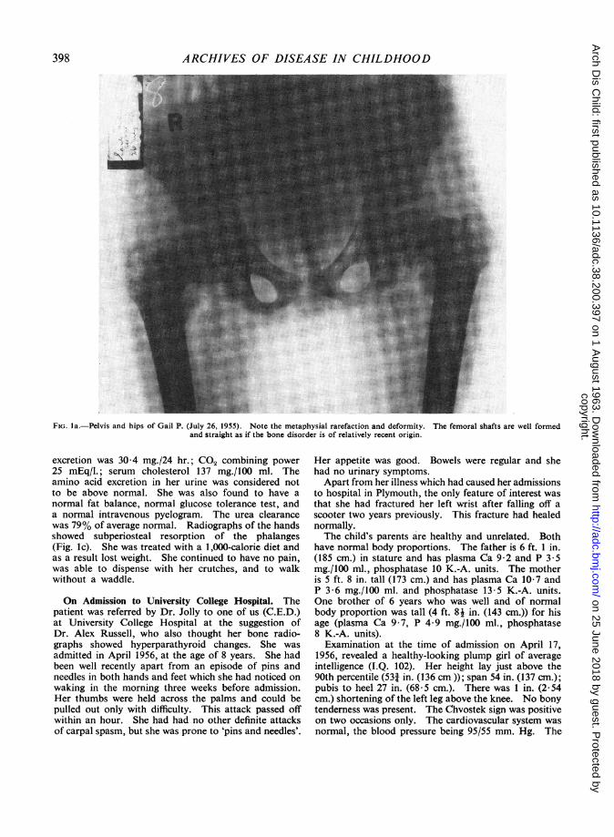
Fici. 2a. FiG. 2b. FIG. 2c.FiG. 2a.-The fingers (April 18, 1956). Subperiosteal erosions, terminal tuft erosions and early cyst formation, all typical of hyperparathyroidism.
FIG. 2b.-Fingers (September 17, 1956) showing a considerable degree of healing.FIG. 2c.-Fingers (January 5, 1960). These are quite normal and typical of all her later films.
change on this dose, nor were there any changes in theplasma levels of calcium and phosphorus. The dose ofvitamin D3 was, therefore, raised to 5 mg. a day. Onthis dose for four six-day periods she slowly went intoa positive calcium balance of about 400 mg. daily, theexcretion still being almost entirely in the stools. Duringthis latter treatment her plasma calcium rose to normaland there was a fall of about 1 mg./100 ml. in the phos-phorus level. Phosphatase was slowly falling from40 to 30 units during this time. Phosphorus balancethroughout was about half the calcium balance indicatingthat new bone formation was occurring. There wasno radiological change visible during the whole of herstay in hospital. She was sent home on the same doseof vitamin D3. From the fourth balance period andduring all her follow-up the vitamin D3 given wasprepared from pure crystalline cholecalciferol in capsulesregularly assayed for potency, as we had evidence thatthe tablet form previously given was not fully stable.
Subsequent Follow-up. Her subsequent progress issummarized in Fig. 7.
She was readmitted to U.C.H. for a second time fromSeptember 15 to 26, 1956. At that time she was welland had been getting around normally at school. Inspite of her previous period in hospital her schoolmistressreported that she had quickly caught up with the othergirls in her lessons. Investigations at this time revealed:plasma Ca 9 5 mg./100 ml., plasma P 6-7 mg./100 ml.,alkaline phosphatase 28 units/100 ml.; plasma HCO321-4 mEq/l. She was able to concentrate her urineto SG 1,022 overnight and to dilute to SG 1,001 afteringestion of water. The radiographs showed a definitelessening of the hyperparathyroid changes (Fig. 2b).It was decided that she had shown a response to thevitamin D3 in the hoped-for direction, both hypo- andhyperparathyroidism signs simultaneously improvingwith the one treatment. She was changed from a doseof 5 mg. vitamin D3 daily to a dose of 3 mg. daily.
400
copyright. on 25 June 2018 by guest. P
rotected byhttp://adc.bm
j.com/
Arch D
is Child: first published as 10.1136/adc.38.200.397 on 1 A
ugust 1963. Dow
nloaded from

H YPO-HYPERPARATHYROIDISM
FIG. 3a. FIG. 3b.
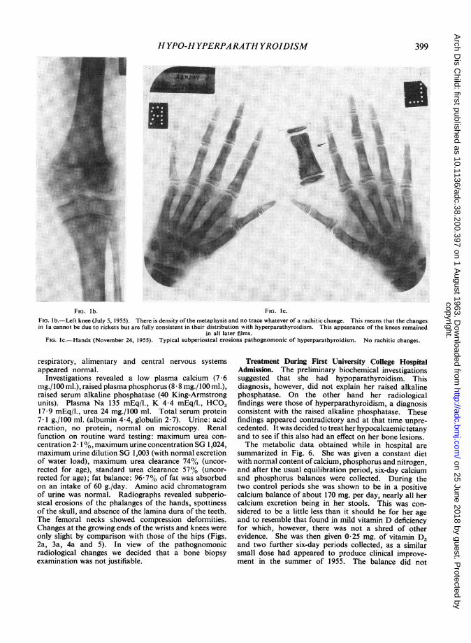
FIG. 3a.-Wrist (January 4, 1956). There are parathyroid erosions on both outer edges of the ends of the ulna and radius. There is no rickets.FIG. 3b.-Wrist (January 5, 1960). Typical appearance of healed bones during later follow-up.
We were prepared to take the risk of overdosage toensure as rapid healing of her hips as possible. Shewas followed subsequently with short admissions tohospital every three to six months. In March 1957(10 months after starting treatment), although asympto-matic, she was found to have a high normal plasmacalcium (11- 1 mg./100 ml.), high urinary calcium(325 mg./24 hrs) and high normal plasma urea (42 mg./100 ml.). A repeat plasma calcium estimation showed:total calcium 10-7 mg./100 ml., ionized calcium 5 9mg./100 ml., un-ionized ultrafiltrable calcium 1-2 mg./100 ml. As her ionized calcium was not as dangerouslyhigh as might have been implied from her total plasmacalcium level, it was decided not to change her treatment.We were still anxious to secure as rapid healing as possibleof the femoral neck lesions.
In July 1957 (14 months after starting treatment) shedeveloped slight thirst and nocturia. Investigations atthis time showed that she was suffering from vitamin Dintoxication. Plasma calcium (1) 14 7 mg./100 ml.;plasma calcium (2) 14-2 mg./100 ml. (total ultrafiltrablecalcium 8-9 mg./100 ml., ionized calcium 7-8 mg./100 ml.); plasma phosphorus 5 1 mg./100 ml.; alkaline
phosphatase 14 King-Armstrong units; 24-hour urinarycalcium 404 mg.; plasma urea 78 mg./100 ml., HCO324-5 mEq/l.; Hb 68%; absolute indices normal. A cleanspecimen of urine showed a trace of protein, numerouspus cells and fairly numerous hyaline casts. Radio-graphs of her hands showed almost complete disappear-ance of the parathyroid changes; the hips, however,were not yet fully reformed.Her vitamin D was stopped for one month and then
resumed at a dose of 1 mg./day. Her symptoms whichwere minimal soon subsided.
She was readmitted three months later (i.e. 17i monthsafter starting treatment) at which time she was symptomfree and had no abnormal physical signs. Her plasmacalcium was still raised (11-4 mg./100 ml.) suggestinga mild vitamin D intoxication, but a normal plasmaurea (33 mg./100 ml.) and a slightly raised alkalinephosphatase level (25 K.-A. units) did not support this.It was decided not to change her dose of vitamin Dbut to watch her carefully.
In February 1958 (21 months after starting treatment)she was well with no abnormal signs. Her heightcontinued to follow the 90 percentile. She had recovered
401
copyright. on 25 June 2018 by guest. P
rotected byhttp://adc.bm
j.com/
Arch D
is Child: first published as 10.1136/adc.38.200.397 on 1 A
ugust 1963. Dow
nloaded from
ARCHIVES OF DISEASE IN CHILDHOOD
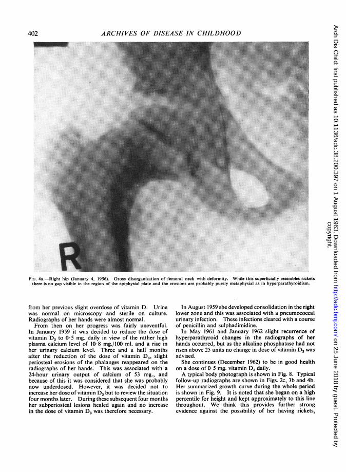
FIG. 4a.-Right hip (January 4, 1956). Gross disorganization of femoral neck with deformity. While this superficially resembles ricketsthere is no gap visible in the region of the epiphysial plate and the erosions are probably purely metaphysial as in hyperparathyroidism.
from her previous slight overdose of vitamin D. Urinewas normal on microscopy and sterile on culture.Radiographs of her hands were almost normal.From then on her progress was fairly uneventful.
In January 1959 it was decided to reduce the dose ofvitamin D3 to 05 mg. daily in view of the rather highplasma calcium level of 10-8 mg./100 ml. and a rise inher urinary calcium level. Three and a half monthsafter the reduction of the dose of vitamin D3, slightperiosteal erosions of the phalanges reappeared on theradiographs of her hands. This was associated with a24-hour urinary output of calcium of 53 mg., andbecause of this it was considered that she was probablynow underdosed. However, it was decided not toincrease her dose of vitamin D3 but to review the situationfour months later. During these subsequent four monthsher subperiosteal lesions healed again and no increasein the dose of vitamin D3 was therefore necessary.
In August 1959 she developed consolidation in the rightlower zone and this was associated with a pneumococcalurinary infection. These infections cleared with a courseof penicillin and sulphadimidine.
In May 1961 and January 1962 slight recurrence ofhyperparathyroid changes in the radiographs of herhands occurred, but as the alkaline phosphatase had notrisen above 25 units no change in dose of vitamin D. wasadvised.She continues (December 1962) to be in good health
on a dose of 0- 5 mg. vitamin D3 daily.A typical body photograph is shown in Fig. 8. Typical
follow-up radiographs are shown in Figs. 2c, 3b and 4b.Her summarized growth curve during the whole periodis shown in Fig. 9. It is noted that she began on a highpercentile for height and kept approximately to this linethroughout. We think this provides further strongevidence against the possibility of her having rickets,
402
copyright. on 25 June 2018 by guest. P
rotected byhttp://adc.bm
j.com/
Arch D
is Child: first published as 10.1136/adc.38.200.397 on 1 A
ugust 1963. Dow
nloaded from
HYPO-HYPERPARATHYROIDISM
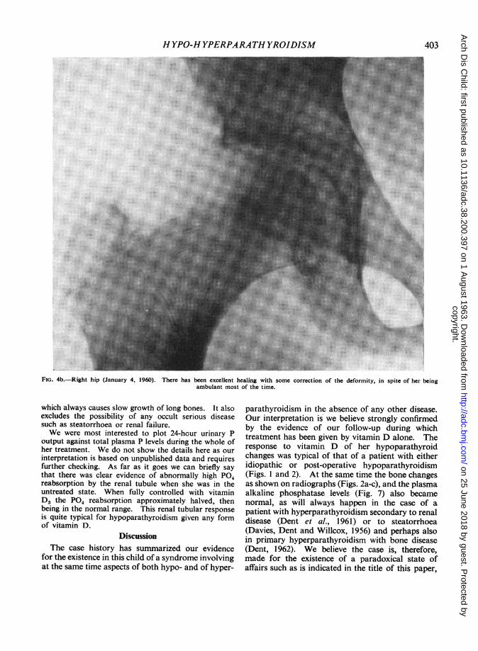
FIG. 4b.-Right hip (January 4, 1960). There has been excellent healing with some correction of the deformity, in spite of her beingambulant most of the time.
which always causes slow growth of long bones. It alsoexcludes the possibility of any occult serious diseasesuch as steatorrhoea or renal failure.We were most interested to plot 24-hour urinary P
output against total plasma P levels during the whole ofher treatment. We do not show the details here as ourinterpretation is based on unpublished data and requiresfurther checking. As far as it goes we can briefly saythat there was clear evidence of abnormally high P04reabsorption by the renal tubule when she was in theuntreated state. When fully controlled with vitaminD3 the P04 reabsorption approximately halved, thenbeing in the normal range. This renal tubular responseis quite typical for hypoparathyroidism given any formof vitamin D.
DiscussionThe case history has summarized our evidence
for the existence in this child of a syndrome involvingat the same time aspects of both hypo- and of hyper-
parathyroidism in the absence of any other disease.Our interpretation is- we believe strongly confirmedby the evidence of our follow-up during whichtreatment has been given by vitamin D alone. Theresponse to vitamin D -of her hypoparathyroidchanges was typical of that of a patient with eitheridiopathic or post-operative hypoparathyroidism(Figs. 1 and 2). At the same time the bone changesas shown on radiographs (Figs. 2a-c), and the plasmaalkaline phosphatase levels (Fig. 7) also becamenormal, as will always happen in the case of apatient with hyperparathyroidism secondary to renaldisease (Dent et al., 1961) or to steatorrhoea(Davies, Dent and Willcox, 1956) and perhaps alsoin primary hyperparathyroidism with bone disease(Dent, 1962). We believe the case is, therefore,made for the existence of a paradoxical state ofaffairs such as is indicated in the title of this paper,
403
copyright. on 25 June 2018 by guest. P
rotected byhttp://adc.bm
j.com/
Arch D
is Child: first published as 10.1136/adc.38.200.397 on 1 A
ugust 1963. Dow
nloaded from
ARCHIVES OF DISEASE IN CHILDHOOD
VitO02M-d-5Tabtets f25[
Plasma 9
Ca. E .
Levels 7j-mg.70 6L
AlkalinePhosphtase 40K-A units I-
02
Urine CaM 01.Facal Ca
g./da. 0-608
02
Urine P 0s.Facal P
g./d;j 06
0OB
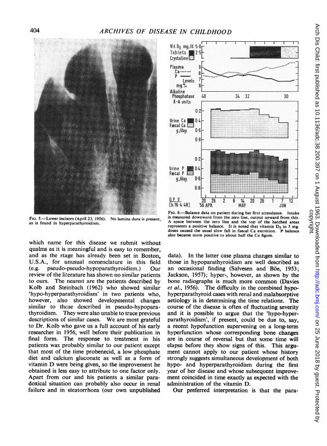
FIG. 5.-Lower incisors (April 23, 1956). No lamina dura is present,as is found in hyperparathyroidism.
which name for this disease we submit withoutqualms as it is meaningful and is easy to remember,and as the stage has already been set in Boston,U.S.A., for unusual nomenclature in this field(e.g. pseudo-pseudo-hypoparathyroidism.) Ourreview of the literature has shown no similar patientsto ours. The nearest are the patients described byKolb and Steinbach (1962) who showed similar'hypo-hyperparathyroidism' in two patients who,however, also showed developmental changessimilar to thcse described in pseudo-hypopara-thyroidism. They were also unable to trace previousdescriptions of similar cases. We are most gratefulto Dr. Kolb who gave us a full account of his earlyresearches in 1956, well before their publication infinal form. The response to treatment in hispatients was probably similar to our patient exceptthat most of the time probenecid, a low phosphatediet and calcium gluconate as well as a form ofvitamin D were being given, so the improvement heobtained is less easy to attribute to one factor only.Apart from our and his patients a similar para-doxical situation can probably also occur in renalfailure and in steatorrhoea (our own unpublished
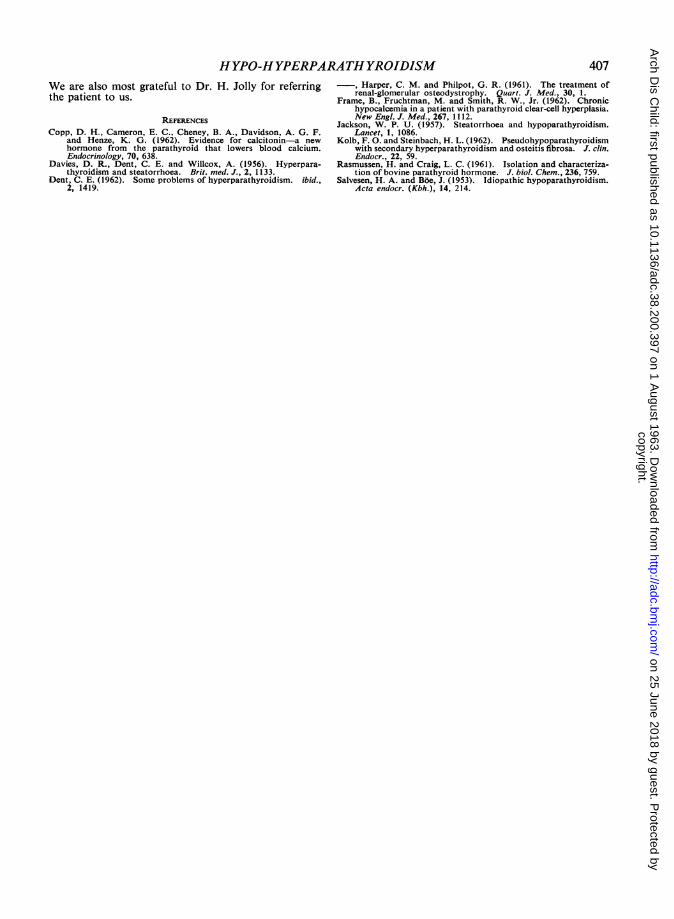
G.P. 9 20 26 2 6 1 20 26 1 7'13i[b.16 1N9l '56 APR MAY JUNFIG. 6.-Balance data on patient during her first attendance. Intakeis measured downward from the zero line, output upward from this.A space between the zero line and the top of the hatched areasrepresents a positive balance. It is noted that vitamin D3 in 5 mg.doses caused the usual slow fall in faecal Ca excretion. P balancealso became more positive to about half the Ca figure.
data). In the latter case plasma changes similar tothose in hypoparathyroidism are well described asan occasional finding (Salvesen and Boe, 1953;Jackson, 1957); hyper-, however, as shown by thebone radiographs is much more common (Davieset al., 1956). The difficulty in the combined hypo-hyperparathyroid cases with renal and malabsorptiveaetiology is in determining the time relations. Thecourse of the disease is often of fluctuating severityand it is possible to argue that the 'hypo-hyper-parathyroidism', if present, could be due to, say,a recent hypofunction supervening on a long-termhyperfunction whose corresponding bone changesare in course of reversal but that some time willelapse before they show signs of this. This argu-ment cannot apply to our patient whose historystrongly suggests simultaneous development of bothhypo- and hyperparathyroidism during the firstyear of her disease and whose subsequent improve-ment coincided in time exactly as expected with theadministration of the vitamin D.Our preferred interpretation is that the para-
.. . . . . . f f . .
404
I
I I I I I I I i
copyright. on 25 June 2018 by guest. P
rotected byhttp://adc.bm
j.com/
Arch D
is Child: first published as 10.1136/adc.38.200.397 on 1 A
ugust 1963. Dow
nloaded from

HYPO-HYPERPARATHYROIDISM
Cod Liver Oil m
Vit 03 Mg.Id. 2_5
I.,r
I I Ir I 1
12[Plasma
Ca 9p
Levels 6mg.%
3
30AlkalinePhosphatase 20K-A units.
10
Blood Urea 5mg./lOOml. 5
25
Urinary 600Ca P*I 400
mg./d. 200G.P.? I[born 16448.] 1 '56 57 58 '59 '60 61 '62
FIG. 7.-Summarized biochemical data on patient, explained more fully in the text.
thyroid gland normally produces at least twohormones: one, that recently isolated by Ras-mussen and Craig (1961), with an action on plasmacalcium and phosphorus levels, and another, whoseexcess production manifests itself by the productionof osteitis fibrosa and a rise in plasma alkalinephosphatase. In our patient it is supposed that no'Rasmussen and Craig hormone' is being producedwhile excess production occurs of 'osteitis fibrosahormone'. There are other possible interpretations.For instance, there could be an excess productionof one hormone having all these actions in thepresence of a circulating antagonist only of theplasma Ca and P actions, by whatever mechanismthis may act. The theory preferred by Kolb andSteinbach (1962) to explain the changes found intheir two rather similar cases was more along thelines of the Albright theory of renal tubular unres-
ponsiveness to the action of the one parathyroidhormone produced. This would produce a chroni-cally raised plasma phosphorus level, and then pos-sibly lower plasma calcium and stimulate the para-thyroids to produce an excess of hormone leading toosteitis fibrosa, the bones, unlike the kidney, beingnormally responsive to the hormone. Kolb andSteinbach did indeed treat one of their patientsfor a while with probenecid and high calcium andlow phosphorus diet only, so as to increase renalphosphorus excretion and to counteract such apossible vicious sequence at its origin. Plasmacalcium and phosphorus values became normalunder this regime, and they further claimed someimprovement in the bone radiographs. They didnot continue the treatment for more than threemonths, but then added vitamin D (as calciferolor AT 10), this combined therapy lasting the next
_~~~~~* ~X ~ '' .
J i............................................
t.:~~~~~~~~~~~JJ:
I
.. .. - - =. - .- M. - - Y. - -
405
1955
copyright. on 25 June 2018 by guest. P
rotected byhttp://adc.bm
j.com/
Arch D
is Child: first published as 10.1136/adc.38.200.397 on 1 A
ugust 1963. Dow
nloaded from

ARCHIVES OF DISEASE IN CHILDHOOD
Heightin. _,
FIG. 8.-Gail P (May 23, 1961) age 13 years.
five years. Naturally most of the healing of theosteitis fibrosa occurred during the latter period.We clearly need further studies along these lines
to elucidate the theoretical challenge posed bythese various patients. Recent work by Copp,Cameron, Cheney, Davidson and Henze (1962),suggests that the parathyroid gland may producea hypocalcaemic hormone as well as its betterknown one(s). They have not yet quoted studiesof the effect if any of their new hormone on renalphosphorus handling, so it is still difficult to applythis new knowledge to our current clinical problems.Nor do we yet know if the pure Rasmussen-Craigparathyroid hormone produces osteitis fibrosa.The recent paper by Frame, Fruchtman and
Smith (1962), in which a patient is described withlong-lasting hypocalcaemia and clear-cell hyper-plasia of her parathyroids, also poses problems ofsimilar perplexity to those in our patient.
SummaryA child is described who presented symptoms from
Gail P b.1948. a IU IL I uyearsFIG. 9.-Growth curve: she showed no stunting at any stage nor wasa spurt of growth induced by treatment. This is against her havingeven a mild rickets. The earliest height recorded was obtained fromher school record. The chart was obtained from the Department ofChild Health, Guy's Hospital, London. Her growth correspondsapproximately to the 90 percentile.
hyperparathyroid bone disease (osteitis fibrosageneralisata) simultaneous with those of mildtetany shown to be due to idiopathic hypopara-thyroidism.On treatment with vitamin D alone the bone
lesions healed and her plasma calcium and phos-phorus levels became normal. This improvementhas been maintained till the present time (six years).The patient is normal in height and in mental
and physical development. Steatorrhoea and renaldysfunction were not present, nor were the physicalfeatures of pseudohypoparathyroidism. In theselatter respects she is unlike some other patientsdescribed recently.The coincidence of signs of hypo- and of hyper-
parathyroidism in the same patient who has nosigns of disease in any other system presents us witha considerable theoretical problem.
We wish to thank the nurses, dietitians and bio-chemists in the Metabolic Ward for the full study of hercalcium and phosphorus balances during her admission,and for their work during her subsequent follow-up.
406
copyright. on 25 June 2018 by guest. P
rotected byhttp://adc.bm
j.com/
Arch D
is Child: first published as 10.1136/adc.38.200.397 on 1 A
ugust 1963. Dow
nloaded from
HYPO-HYPERPARATHYROIDISM 407We are also most grateful to Dr. H. Jolly for referringthe patient to us.
REFERENCES
Copp, D. H., Cameron, E. C., Cheney, B. A., Davidson, A. G. F.and Henze, K. G. (1962). Evidence for calcitonin-a newhormone from the parathyroid that lowers blood calcium.Endocrinology, 70, 638.
Davies, D. R., Dent, C. E. and Willcox, A. (1956). Hyperpara-thyroidism and steatorrhoea. Brit. med. J., 2, 1133.
Dent, C. E. (1962). Some problems of hyperparathyroidism. ibid.,2, 1419.
-, Harper, C. M. and Philpot, G. R. (1961). The treatment ofrenal-glomerular osteodystrophy. Quart. J. Med., 30, 1.
Frame, B., Fruchtman, M. and Smith, R. W., Jr. (1962). Chronichypocalcemia in a patient with parathyroid clear-cell hyperplasia.New Engl. J. Med., 267, 1112.
Jackson, W. P. U. (1957). Steatorrhoea and hypoparathyroidism.Lancet, 1, 1086.
Kolb, F. 0. and Steinbach, H. L. (1962). Pseudohypoparathyroidismwith secondary hyperparathyroidism and osteitis fibrosa. J. clin.Endocr., 22, 59.
Rasmussen, H. and Craig, L. C. (1961). Isolation and characteriza-tion of bovine parathyroid hormone. J. biol. Chem., 236, 759.
Salvesen, H. A. and Boe, J. (1953). Idiopathic hypoparathyroidism.Acta endocr. (Kbh.), 14, 214.
copyright. on 25 June 2018 by guest. P
rotected byhttp://adc.bm
j.com/
Arch D
is Child: first published as 10.1136/adc.38.200.397 on 1 A
ugust 1963. Dow
nloaded from