Embed Size (px)
Citation preview

Kidney International, Vol. 58 (2000), pp. 2351–2366
Hypoxia promotes fibrogenesis in human renal fibroblasts
JILL T. NORMAN, IAN M. CLARK, and PATRICIA L. GARCIA
Department of Medicine, Royal Free and University College Medical School, London, and School of Biological Sciences,University of East Anglia, Norwich, England, United Kingdom
Hypoxia promotes fibrogenesis in human renal fibroblasts. indicator of progression [3]. Tubulointersititial fibrosisBackground. The mechanisms underlying progressive renal is characterized by tubular dilation and atrophy, an in-
fibrosis are unknown, but the common association of fibrosis crease in interstitial cell number, activation of fibroblastsand microvascular loss suggests that hypoxia per se may be ato myofibroblasts with increased expression of a-smoothfibrogenic stimulus.muscle actin (a-SMA), obliteration of the microvascula-Methods. To determine whether human renal fibroblasts
(HRFs), the primary matrix-producing cells in the tubulointers- ture, and accumulation of extracellular matrix (ECM)titium, possess oxygen-sensitive responses relevant to fibro- [1–5]. Despite the large number of studies documentinggenesis, cells were exposed to 1% O2 in vitro. changes in a variety of ECM proteins and growth factors,Results. Hypoxia simultaneously stimulated extracellular
most notably transforming growth factor-b1 (TGF-b1)matrix synthesis and suppressed turnover with increased pro-[5, 6], in models of PRD and in human pathologies, theduction of collagen a1(I) (Coll-I), decreased expression of col-
lagenase, and increased tissue inhibitor of metalloproteinase mechanisms underlying accumulation of ECM remain(TIMP)-1. These effects are time dependent, require new RNA obscure. In trying to understand the association of glo-and protein synthesis, and are specific to hypoxia. The changes
merular damage, interstitial fibrosis, and tubular atrophyin Coll-I and TIMP-1 gene expression involve a heme-proteinin PRD, we have proposed a sequence of events thatO2 sensor and protein kinase- and tyrosine kinase-mediated sig-
naling. Although hypoxia induced transforming growth factor-b1 ascribes a central role to microvascular compromise and(TGF-b1), neutralizing anti–TGF-b1-antibody did not block hypoxia in the pathogenesis of interstitial fibrosis [7, 8].hypoxia-induced Coll-I and TIMP-1 mRNA expression. Fur- Hypoxia can regulate expression of a wide varietythermore, hypoxic-cell conditioned-medium had no effect on
of genes that may be induced or suppressed by eitherthe expression of these mRNAs in naive fibroblasts, suggestingtranscriptional and post-transcriptional mechanisms [re-direct effects on gene transcription. Transient transfections
identified a hypoxia response element (HRE) in the TIMP-1 viewed in 9–11]. Hypoxia response elements (HREs)promoter and demonstrated HIF-1–dependent promoter acti- have been identified in the regulatory regions of a num-vation by decreased ambient pO2. ber of genes and contain consensus binding sites for theConclusions. These data suggest that hypoxia co-ordinately
transcription factor hypoxia-inducible factor-1 (HIF-1)up-regulates matrix production and decreases turnover in renal[12–14]. A variety of other transcription factors are in-fibroblasts. The results support a role for hypoxia in the patho-
genesis of fibrosis and provide evidence for novel, direct hyp- duced by low O2, including hepatocyte nuclear factor-4oxic effects on the expression of genes involved in fibrogenesis. (HNF-4), nuclear factor–interleukin-6 (NF-IL-6), nu-
clear factor-kB (NF-kB), and members of the fos andjun (AP-1) families [9–11, 15, 16], many of which have
Progressive renal disease (PRD), characterized by in- been shown to be involved in the regulation of expressionterstitial fibrosis, represents a significant clinical problem of genes relevant to fibrogenesis. The question of how thewith poor outcome and limited therapeutic options [1, 2]. signal of decreased oxygen is transduced to changes inAlthough many progressive diseases in the kidney are gene expression remains open, but studies on a numberglomerular in origin, it is now well established that tubu- of hypoxia-regulated genes have implicated a cellularlointerstitial involvement provides the best prognostic heme-protein oxygen sensor [9–11, 17] and have shown
that hypoxia activates a variety of signal transductionpathways, including protein kinase C (PKC)-, protein
Key words: oxygen deprivation, fibrosis, collagen, matrix metallopro-kinase A (PKA), and tyrosine kinase (TK)-mediatedteinases, tissue inhibitor of metalloproteinases, extracellular matrix
turnover. pathways [9–14].Several studies have demonstrated changes in collagenReceived for publication March 10, 1999
gene expression in various mesenchymal cell types inand in revised form June 20, 2000Accepted for publication June 22, 2000 response to low O2 [18–21], and our previous study
showed that hypoxia increased Coll-I gene expression 2000 by the International Society of Nephrology
2351
Norman et al: Hypoxia stimulates fibrosis in fibroblasts2352
and collagen production in human proximal tubular epi- were passaged using trypsin:ethylenediaminetetraaceticacid (0.5:0.2 mg/mL; LTI). Every 5 to 10 passages, tsHRFthelial cells (PTEs) [20]. In fibrosis, both increased pro-
duction and decreased turnover contribute to ECM accu- were reselected in G418 to check for reversion and lossof antibiotic resistance. Comparison of morphologicalmulation [5]. The matrix metalloproteinases (MMPs)
[reviewed in 22, 23] and their inhibitors, the tissue inhibi- and immunocytochemical markers in tsHRF at the non-permissive temperature (378C) with nontransformed pa-tors of metalloproteinases (TIMPs) [reviewed in 24, 25],
are important regulators of ECM turnover. TIMP-1 rental cells showed that tsHRF retained parental charac-teristics. Cells displayed a typical fibroblastic morphologymRNA and protein are consistently increased in renal
fibroses in vivo, suggesting an important role for this and formed confluent monolayers with no evidence ofthe “hill and valley” growth characteristic of mesangialmolecule in disease pathogenesis [5]. Relatively little is
known about the effects of hypoxia on matrix turnover, cells. Immunocytochemistry for selected cytoskeletaland cell surface proteins showed that both parental HRFalthough our recent study in PTE showed that low oxy-
gen suppressed activity of at least one MMP (MMP-2) and tsHRF were positive for vimentin, fibroblast surfaceantigen (FSA), and SMA, with occasional desmin-posi-via a post-transcriptional mechanism(s) [20].
Interstitial fibroblasts are the major matrix-producing tive cells. Cells were negative for cytokeratin and myosinand for alkaline phosphatase activity, a marker of PTEcells in the tubulointerstitium and as such are likely to
be the major effector cells in fibrosis. The present study [20]. At 338C, all tsHRF showed positive nuclear stainingfor T antigen, which decreased with time at 378C and wasexamined the effect of hypoxia on growth, activation, and
ECM metabolism in human renal fibroblasts (HRFs). In further suppressed by incubation in serum-free medium.Nontransformed cells were uniformly negative. For ex-this cell type, hypoxia promotes a fibrogenic phenotype
increasing production of interstitial collagens and de- periments, confluent tsHRF were made quiescent by in-cubation in serum-free medium (quiescence medium)creasing turnover via TGF-b1–independent mechanisms
involving a heme-protein O2 sensor and activation of for 48 hours at 378C. All experiments were performedat 378C.PKC- and TK-mediated signaling pathways. Regulation
of at least one of the genes, TIMP-1, is dependent onImmunocytochemistrybinding of HIF-1 to a HRE in the 59 promoter, revealing
a novel regulatory mechanism for this family of mole- Immunocytochemistry was performed as described pre-viously [20] using the Vectastain-Elite biotin-streptavi-cules and identifying TIMP-1 as a new hypoxia-inducible
gene. Together, these data support the hypothesis that din-immunoperoxidase system (Vector Labs., Peterbor-ough, UK) with aminoethylcarbazole as a chromogenichypoxia plays an important role in the pathogenesis
of PRD. substrate giving a red-brown deposit. Cells were counter-stained with hematoxylin for nuclear identification.
Antibodies (dilution). These included: monoclonalMETHODS
anti-T antigen (pAb412, undiluted; a gift from Dr. P. Jat);Human renal cortical fibroblasts monoclonal anti-desmin (1:1000; Sigma); polyclonal rabbit
anti-bovine myosin (1:10; Sigma); monoclonal anti-panTo circumvent the limited in vitro lifespan of renalfibroblasts, cells with a fibroblastic morphology (passage cytokeratin (1:100; Sigma); monoclonal anti-vimentin
(Vim 3B4, 1:250; Roche Molecular Biologicals, Lewes,2), obtained as an outgrowth from an explanted biopsyof histologically normal renal cortex, were conditionally UK); monoclonal anti-human FSA (1:500; Sigma); and
monoclonal anti-SMA (1:1000; Sigma). SMA staining wasimmortalized by stable integration of amphoBAGg.U19retroviral construct encoding a thermolabile Simian Vi- quantitated by counting the number of stained compared
with unstained cells in five random high-power fields.rus 40 (SV40) T-antigen plus a neomycin-resistance gene(a gift from Dr. P. Jat, Ludwig Institute, London, UK)
Hypoxia[26]. Transfectants (tsHRF) were selected in Dulbecco’smodified Eagle’s:Ham’s F12 (1:1) medium (DME:F12; Confluent quiescent cells were exposed to 1% O2/5%
CO2/94% N2 (British Oxygen Co. Ltd., Luton, UK) in aLTI, Paisley, Scotland, UK) containing 10% fetal calfserum (FCS; LTI), 100U/mL penicillin, 100 mg/mL strep- Billups-Rothenburge Chamber (ICN-Flow, High Wy-
combe, UK) in open dishes for up to 72 hours. Cellstomycin, 0.25 mg/mL amphotericin-B (1% antibiotic-antimycotic; Sigma Chemical Co., Poole, UK), and 0.5 were overlaid with medium to a depth of 3 mm sufficient
to prevent dehydration. To examine the effects of reoxy-mg/mL G418 (Geneticin; LTI) at the permissive temper-ature, 338C, in a humidified atmosphere of 5% CO2. genation, cells were exposed to hypoxia (24 to 48 hours)
and then returned to normoxia (21%O2; up to 48 hours).Integration of the vector into cellular DNA was con-firmed by Southern blotting. tsHRFs were grown in me- The degree and duration of hypoxia used was based on
previous studies in PTE [20, 27]. Measurement of thedium containing G418 for several passages and thenmaintained in the absence of G418. At confluence, cells pO2 of the medium indicated that O2 levels fell rapidly

Norman et al: Hypoxia stimulates fibrosis in fibroblasts 2353
as the incubator was flushed with 1% O2. At the end South San Francisco, CA, USA). HIF-1a cDNA was agift from Dr. G. Bell (University of Chicago, Chicago,of the incubation, O2 concentration in the chamber was
checked using a Radiometer ABL4 (Radiometer, Copen- IL, USA).hagen, Denmark), and medium pH was also measured
Effect of cycloheximide (Cyc) and actinomycin-D[without cells, normoxia pH 7.05; hypoxia (48 hours),(Act-D) on gene expressionpH 6.75]. Control cells were maintained under normoxic
conditions (21% O2) for equivalent time periods. Condi- To examine the effect of inhibition of mRNA andprotein synthesis on hypoxia-induced changes, 0.25 totioned medium (CM; serum free) was collected at each
time point, clarified by centrifugation at 1000 3 g for 15 1.0 mg/mL Act-D (Sigma) or 0.25 to 1.0 mg/mL Cyc(Sigma) was added to the cells immediately prior tominutes at 48C and stored at 2808C. Protein concentra-
tion was measured using a modified Bradford assay [20]. exposure to hypoxia. Under normoxic conditions, 0.1to 1 mg/mL Cyc caused a dose-dependent inhibition ofAll experiments were repeated at least three times with
reproducible results. protein synthesis (3H-Phe-incorporation). One mg/mLinhibited synthesis by .90%, and concentrations .1
Cell viability, proliferation, and protein synthesis mg/mL (3.55 nmol/L) were cytotoxic. After 48 hours,CM was collected, and cells were harvested for RNACell viability was assessed by qualitative evaluation
of detached cells, trypan blue exclusion, and LDH re- extraction and Northern analysis.lease (LDH Assay Kit; Sigma). DNA synthesis was mea-
ELISA for TIMP-1 and MMP-1sured by incorporation of tritiated thymidine (3H-Thy)into DNA [20]. Total protein synthesis was measured Levels of TIMP-1 and MMP-1 protein in CM were
measured using the Biotrak ELISA systems (Amer-by incorporation of tritiated phenylalanine, L-[4-3H]-Phe (specific activity 28Ci/mmol; Amersham, Arlington sham) according to the manufacturer’s instructions using
100 mL aliquots of undiluted CM (MMP-1) or CM di-Heights, IL, USA). 3H-Phe, 2 mCi/well was added 24hours prior to harvesting the cells. Protein was precipi- luted 1:5 in assay buffer (TIMP-1). Absorbance was read
on a Multiskan MCC/340plate reader (Titertek Labsys-tated with ice-cold 10% trichloroacetic acid (TCA) andsolubilized, and radioactivity was counted. Data are pre- tems, Huntsville, IL, USA), and the amount was calcu-
lated per microgram of secreted protein.sented as 3H-Phe dpm/well.
Collagen production Gelatin-substrate gel zymography forgelatinase expressionTotal collagen production was measured by reverse-
phase high-performance liquid chromatography (RP- To measure secreted gelatinase activity, equal amountsof CM protein were electrophoresed through 10% so-HPLC; System Gold; Beckman Instruments Ltd., Palo
Alto, CA, USA) according to the method of Campa, dium dodecyl sulfate-polyacrylamide gels (SDS-PAGE)impregnated with 1 mg/mL gelatin and zymography per-McAnulty, and Laurent [28], as described previously
[20]. Procollagen production is expressed as nmol/L hy- formed as described previously [20]. Gelatinase activitywas visualized as cleared zones in Coomassie Blue G250-droxyproline/well.stained gels. Equal loading of protein was confirmed by
Northern blot analysis of mRNA expression silver staining of parallel gels. Zymograms were scannedand quantified by densitometry [20].Total RNA was extracted using Trizole (LTI), and
Northern blot analysis was performed as described inWestern blot analysisOrphanides, Fine, and Norman [20]. Photographs of
ethidium bromide-stained gels and autoradiograms were HIF-1a protein. Nuclear protein was prepared fromcells exposed to normoxia or hypoxia for two, four, six,scanned and quantified using ImageMaster Software
(Pharmacia, St. Albans, UK). Variations in loading were or eight hours. Cells were harvested onto ice, washedtwice with cold phosphate-buffered saline (PBS) and lysednormalized to the ethidium bromide staining of rRNA,
and changes in mRNAs were calculated by comparison in hypotonic buffer (10 mmol/L Tris, pH 7.8, 1.5 mmol/LMgCl2, 10 mmol/L KCl), and the nuclei were pelleted.to normoxic values assigned an arbitrary value of 1 [20].
Probes. cDNAs for human MMP-2, MMP-9, TIMP-1, Nuclear protein was extracted by incubation in hyper-tonic buffer (20 mmol/L Tris, pH 7.8, 1.5 mmol/L MgCl2,and TIMP-2 were a gift from Dr. G. Murphy (University
of East Anglia, Norwich, UK). Human TIMP-3 cDNA 420 mmol/L KCl, 20% glycerol), and the lysate was dia-lyzed against 20 mmol/L Tris, pH 7.8, 100 mmol/L KCl,was obtained from Dr. D. Edwards (University of East
Anglia). cDNAs for interstitial collagenase (MMP-1), 0.2 mmol/L ethylenediaminetetraacetic acid (EDTA), and20% glycerol. All buffers contained protease inhibitorscollagen a1(I) (Coll-I; Hf677), and a2(III) (Coll-III;
Hf934) were from ATCC (Rockville, MD, USA). cDNA (Complete inhibitor cocktail; Roche) and 1 mmol/L so-dium vanadate (Sigma). Protein concentration was mea-for TGF-b1 was a gift from Dr. R. Derynk (Genentech,

Norman et al: Hypoxia stimulates fibrosis in fibroblasts2354
sured using the bicinchoninic acid (BCA) assay reagent for three days, and then made quiescent in serum-free,low-glucose medium for 48 hours. mRNA expression(Pierce, Rockford, IL, USA). Proteins (10 mg/sample)was compared with cells in 17 mmol/L glucose medium.were electrophoresed through a 12% SDS-PAGE under
Hyperlactemia. Fifteen mmol/L lactic acid was addeddenaturing conditions. Proteins were transferred to ni-for 24 to 48 hours.trocellulose membrane (0.2 mm pore size; Schleicher &
Schuell, Dassel, Germany) for 1.5 hours at 180 mA andEffect of TGF-b1 on proliferation andstained with Ponceau-S (Sigma) to confirm equal loadingexpression of SMA, TIMP-1, Coll-I,and transfer. Membranes were blocked in 5% dried milkColl-III mRNA and secreted MMP-1 and MMP-2(Marvel) in 10 mmol/L Tris, pH 7.6, 150 mmol/L NaCl
To examine the effect of TGF-b1 on cell proliferation,containing 0.1% Tween 20 (TBS-Tween) for 30 minutesconfluent, quiescent tsHRF were exposed to 0 to 10 ng/mLat ambient temperature, followed by incubation in anti–TGF-b1 (Genzyme, West Malling, Kent, UK) for 24HIF-1a monoclonal antibody (Ha28b; a generous gifthours, and DNA synthesis was measured as describedfrom Dr. P. Ratcliffe, Institute of Molecular Medicine,previously in this article. For studies of gene expression,Oxford, UK) [29] diluted 1:100 in TBS-Tween overnightcells were treated with 0 to 10 ng/mL TGF-b1 for 24 hoursat 48C with gentle agitation. Control lanes were incubatedunder normoxic conditions. CM was collected and cellswithout primary antibody. Membranes were washed threeharvested for RNA. Cells treated with TGF-b1 for 24times, for five minutes each, in TBS-Tween followed byhours were stained for SMA.incubation in horseradish peroxidase-conjugated rabbit
anti-mouse secondary antibody (Dako) diluted 1:2000 Measurement of TGF-b productionin TBS-Tween for 30 minutes at ambient temperature.
The mink lung epithelial cell, CCL64, bioassay wasMembranes were washed three times three to four min-used to measure the concentration of TGF-b (all iso-utes each, and positive signals were visualized by ECLforms) in CM [20]. CM (with or without heat activation)(Amersham).was diluted 1:1 in medium, and the concentration ofColl-I protein. Secreted and cellular protein from nor-growth factor was determined by comparison with themoxic and hypoxic (24 and 48 h) cells was precipitatedinhibition of 3H-Thy induced by 0 to 10 ng/mL exogenouswith 67% ethanol overnight at 48C. The dishes wereTGF-b1.scraped, and the precipitate was collected by centrifuga-
tion and solubilized in SDS-PAGE sample buffer. Equal Effect of neutralizing antibody to TGF-b1 onloading of the samples was confirmed by Coomassie hypoxia-induced Coll-I and TIMP-1 mRNA expressionstaining of parallel lanes and by Ponceau-S staining of To establish whether the effects of hypoxia are me-the membrane after transfer. Western blot analysis was diated by autocrine TGF-b1, neutralizing antibody toperformed as described previously in this article using a TGF-b1 (1 to 2 mg/mL; R&D Systems Europe Ltd.,goat anti-human collagen type I polyclonal antibody (1: Abingdon, UK) was added to the cells immediately prior1000; Southern Biotechnology Associates Inc., Birming- to hypoxia. Nonimmune IgG was added to parallel cul-ham, AL, USA) and a horseradish peroxidase-conjugated tures to confirm the specificity of the antibody. The effi-rabbit anti-goat secondary antibody. The anti-Coll-I anti- cacy of the antibody was confirmed by the effect of thebody detects a doublet at approximately 200 kD. simultaneous addition of antibody and exogenous TGF-b1
on Coll-I and TIMP-1 gene expression in normoxic cells.Effect of cellular stress on TIMP-1 andColl-I mRNA expression Effect of hypoxic fibroblast conditioned medium on
To examine the specificity of the effects of hypoxia on gene expression in naive fibroblastsgene expression, cells were exposed to a variety of cellular To establish whether hypoxia induces secreted factorsstressors under normoxic conditions, and TIMP-1 and that stimulate TIMP-1 and Coll-I mRNA expression,Coll-I mRNAs were examined by Northern blotting. confluent quiescent tsHRF were treated with CM (con-
Oxidant stress. Cells were treated with 0 to 200 mmol/L taining equivalent amounts of protein) from either nor-H2O2 for 24 to 48 hours, with H2O2 replenished every 8 moxic tsHRF or from tsHRF exposed to hypoxia forhours. up to 72 or 48 hours of hypoxia plus 24 to 48 hours of
Heat shock. Cells were exposed to either heat shock reoxygenation. Naive fibroblasts were incubated withalone, 42 to 448C for 15 minutes, 30 minutes, and 1, 2, CM from cells for 48 hours under normoxic conditions,6, 8, and 16 hours or heat shock followed by up to 48 and mRNA levels were examined.hours at 378C.
Effect of desferrioxamine or CoCl2 on Coll-I andHypoglycemia. Cells were grown to near confluenceTIMP-1 gene expressionin growth medium (DME:F12 basal glucose concentra-
tion 17 mmol/L d-glucose), transferred to growth me- To investigate the effect of iron chelators and a transi-tion metal ion on TIMP-1 and Coll-I mRNA expression,dium containing 5.5 mmol/L d-glucose (Glutamax; LTI)

Norman et al: Hypoxia stimulates fibrosis in fibroblasts 2355
cells were exposed to 0 to 200 mmol/L desferrioxamine concentrations used, both PKC inhibitors blocked PMA(DFO) or cobalt chloride (CoCl2) for 24 or 48 hours under (10 ng/mL)-induced changes in gene expression (datanormoxic conditions, and mRNA expression was ana- not shown).lyzed by Northern blotting.
StatisticsTransient transfection with TIMP-1 promoter-CAT All values are expressed as mean 6 SD. Statisticalreporter constructs
significance (P , 0.05) was evaluated using Student’s t-test.The human TIMP-1 promoter-reporter constructs in
pBLCAT3 were as described previously [30]. DNAsRESULTSwere prepared using Qiagen columns (QIAGEN Ltd.,
Crawley, UK). Cells (either tsHRF or NRK-49F rat kid- Effect of hypoxia/reoxygenation on cell morphology,ney fibroblasts; ATCC) were plated at 5 3 104 cells per viability, proliferation, and protein synthesiswell in six-well plates, grown overnight in complete me- No differences in cell morphology or numbers of float-dium, and transfected with 2 mg DNA per well using ing cells were observed between normoxic and hypoxicLipofectin (LTI) 6 mL/well in Opti-Mem-I medium (LTI)
cultures, suggesting that up to 48 hours at 1% O2 followedfor 7 hours at 338C (tsHRF) or 378C (NRK). Controlby up to 48 hours of reoxygenation had no marked effectwells were transfected with the empty vector pBLCAT3.on cell viability. LDH release was similar in normoxicDNA-containing medium was replaced with DME:F12,(N) and 48-hour hypoxic (H) cells (N, 31.22 6 4.24%;0.5% FCS for one hour. Transfected cells were exposedH, 35.28 6 9.43%). Hypoxia (24 h) stimulated a moreto either normoxia or hypoxia for up to 48 hours andthan threefold increase in DNA synthesis (N, 2.09 6were harvested either immediately after hypoxia or after0.17; H, 6.47 6 1.1 3 102 3H-Thy dpm/mg protein; P ,24 hours of reoxygenation. Cells were washed three times0.005) which remained elevated after 48 hours of hy-with cold PBS. Lysates were prepared according to thepoxia, (N, 1.75 6 0.44; H, 2.77 6 0.82 3 102 3H-Thymanufacturer’s instructions, and CAT activity was mea-dpm/mg protein). During reoxygenation (24 to 48 h),sured by CAT-ELISA (Roche). Reporter gene activityDNA synthesis was similar to normoxic controls (N,was corrected for total cell protein and for efficiency of0.43 6 0.08; H/R, 0.41 6 0.06). Total protein synthesistransfection as monitored by the Hirt’s assay [30]. Datadecreased after 24 hours of hypoxia (to 63% of control),are presented as the % increase in CAT activity abovebut by 48 hours, hypoxia returned to normoxic valuesnormoxic cells.and increased slightly (37% above normoxia) during re-
Mutation of HIF binding site oxygenation.The HIF binding-site core sequence 59-RCGTG-39 at
Effect of hypoxia on SMA expression226/–23 in the 2102/195 bp TIMP-1 promoter constructwas replaced by the sequence 59-RAAAC-39 using oligo- Smooth muscle actin expression is a marker of fibro-nucleotide-based polymerase chain reaction (PCR) meth- blast activation to a myofibroblastic phenotype, and in-odology (QuikChange Site-directed Mutagenesis Kit; terstitial cell SMA expression is increased in fibrotic kid-Stratagene, La Jolla, CA, USA), and the mutation was neys in vivo [5, 31]. Quiescent tsHRF showed variableconfirmed by sequencing. Activity of the original and basal levels of SMA expression (37 to 68% cells) withmutated constructs was compared in transfected cells 9ominent intracellular fibrillar staining. Hypoxia (48 h)after 48 hours of hypoxia. stimulated SMA up to 1.8-fold with a more marked in-
crease in cultures with lower basal expression (Fig. 1).Effect of PKC and TK inhibitors on TIMP-1 andImmunostaining suggested that hypoxia increases bothColl-I gene expressionthe number of myofibroblastic cells and the intensity ofTo establish whether PKC activation mimicked the ef-staining.fects of hypoxia on mRNA expression, cells were treated
with phorbyl 12-myristate 13-acetate (PMA; 0 to 25 ng/mL) Effect of hypoxia on collagen productionfor 24 and 48 hours to activate PKC and mRNAs exam-
The mRNA for a1(I) collagen (Coll-I) appears as ained by Northern blotting. PKC inhibitors (Calbiochem,doublet of 5.2 and 4.8 kb. Hypoxia increased steady-San Diego, CA, USA), calphostin-C (Cal-C; 0.05, 0.1state Coll-I mRNA levels, which remained elevated aftermmol/L), and bisindolylmaleimide (BI; 5, 20 mmol/L) or24 hours of reoxygenation (Fig. 2A, B). mRNA expres-the TK inhibitors (Calbiochem) genistein (Gen; 10, 30sion initially increased between 6 and 12 hours of hypoxiammol/L) and lavendustin-A (Lav-A; 1, 10 mmol/L) wereand was increased further at 48 hours, the longest timeadded under subdued lighting conditions immediatelypoint examined (Fig. 2B). There was some variation inprior to hypoxia. Cells were incubated for 24 to 48 hours,the magnitude of the increase in expression, but hypoxicand TIMP-1 and Coll-I mRNA expression was ana-cells consistently showed higher levels of expression thanlyzed by Northern blot. These concentrations of inhibi-
tors had no qualitative effect on cell viability. At the normoxic controls (N 5 16 experiments). The addition

Norman et al: Hypoxia stimulates fibrosis in fibroblasts2356
Fig. 1. Effect of hypoxia on smooth muscleactin (SMA) expression in tsHRF. SMA ap-pears as prominent intracellular fibrillar stain-ing (arrowheads). (A) Normoxia. (B) Hypoxia(48 hours; 3400).
of either Cyc or Act-D immediately prior to hypoxia sup- TIMP-2 (,1, 3.5 kb) and TIMP-3 (,2.4, 4.5 kb), withTIMP-1 as the most abundant inhibitor. Hypoxia in-pressed the hypoxia-induced increase in Coll-I mRNAcreased TIMP-1 mRNA levels, which declined slightly(Fig. 2C), which was completely blocked at inhibitoron reoxygenation, although expression remained aboveconcentrations .0.5 mg/mL, indicating a requirement fornormoxic controls (Fig. 4A, B). The increase in mRNAboth de novo protein and new RNA synthesis. In parallelwas first apparent between 6 and 12 hours of hypoxiawith the increase in gene expression, total collagen pro-(Fig. 4B), following a similar time course to the hypoxia-duction (by HPLC) and Coll-I protein (Western blotting)induced increase in Coll-I mRNA. Likewise the hypoxia-increased (Fig. 2D). Coll-III mRNA was expressed atinduced increase in TIMP-1 mRNA was blocked by Act-Dlower levels than Coll-I mRNA in normoxic cells butor Cyc (Fig. 4C), indicating a requirement for de novowas induced by hypoxia with a similar time course (dataRNA and protein synthesis. The increase in TIMP-1 oc-not shown).curred in cells exposed to hypoxia in serum-free mediumor medium containing 0.5 to 2% FCS and was independentEffect of hypoxia on MMP and TIMP expressionof passage number. Hypoxia also increased TIMP-1 pro-Zymography of CM showed that tsHRF secrete pre-tein (after 48 hours; N, 50 ng/mL; H, 110 ng/mL), whichdominantly latent MMP-2 (72 kD Gelatinase-A) withremained slightly elevated during reoxygenation. Hyp-only low levels of active enzyme (68 kD). MMP-2 wasoxia differentially affected expression of the mRNAs forexpressed at relatively high levels in confluent quiescentthe three TIMPs, increasing expression of TIMP-1 and -3cells. No concentration of CM was required prior tomRNA, while TIMP-2 mRNA levels were unaffected,zymography. Neither hypoxia (24 to 48 h) nor hypoxia/although TIMP-2 mRNA could be induced by other
reoxygenation had any effect on the amount or activation stimuli, for example, 10% FCS (twofold after 48 hours).of secreted MMP-2 (Fig. 3A) or MMP-2 gene expression For quantitation, all transcripts for TIMP-2 or -3 were(Fig. 3B). tsHRF also secreted lower and more variable combined (Fig. 4D). To investigate the mechanisms ofamounts of MMP-9 (92 kD Gelatinase-B), which was hypoxia-induced changes in ECM metabolism, changesunaffected by hypoxia. MMP-1 was produced at very low in Coll-I and TIMP-1 mRNA were used as indicatorslevels by confluent quiescent tsHRF, precluding accurate of increased matrix production and decreased turnovercomparison of levels of secreted protein (even after con- respectively.centration of the medium), although slightly lower values
Effect of cellular stress on TIMP-1 andwere obtained in medium from hypoxic cells comparedColl-I mRNA expressionwith normoxic controls (24 hours N, 3.44 ng/mL; H, 3.13
ng/mL). By Northern blot analysis, MMP-1 mRNA lev- As shown in Figure 5, the effect of hypoxia on theels were decreased by low O2 (Fig. 3B). expression of either Coll-I or TIMP-1 in tsHRF was not
mimicked by a variety of cellular stresses. The concentra-tsHRF expresses mRNAs for TIMP-1 (,0.9 kb),

Norman et al: Hypoxia stimulates fibrosis in fibroblasts 2357
Fig. 2. Effect of hypoxia on collagen a1(I). (A) Northern blot of Coll-I mRNA expression after 48 hours of hypoxia (H) and 48 hours of hypoxia/24hours of reoxygenation (H/R). Ethidium bromide staining of the 28S rRNA is shown to demonstrate equal loading. (B) Time course of hypoxia-induced Coll-I mRNA expression. Signals were normalized to the 28S rRNA, normoxic cells (N; h). Each time point was assigned an arbitraryvalue of 1, and the fold increase with hypoxia (H; ) or hypoxia followed by reoxygenation (H/R) was calculated. N 5 4 experiments. *P , 0.05vs. N. (C ) Effect of Act-D to 1 mg/mL ( ) or Cyc 0 to 1 mg/mL ( ) on hypoxia-induced Coll-I mRNA expression. Inhibitors were addedimmediately prior to hypoxia, and cells were harvested after 48 hours. Other symbols are: (h) normoxia, N; ( ) hypoxia, H. *P , 0.05 vs. N. N 53 experiments. (D) Total collagen production measured by high-performance liquid chromatography. Symbols are: (h) normoxia; ( ) 48 hoursof hypoxia. Data are from a representative experiment with triplicate wells/sample. *P , 0.05 versus N. (Insert) Western blot of Coll-I protein innormoxic (N) and hypoxic (H) cells after 48 hours of hypoxia.
tions of H2O2 were selected to impose oxidative stress be expected to mimic the hypoxia induced-changes ingene expression. However, 15 mmol/L lactic acid for upbut not induce apoptosis. Lower concentrations of H2O2
(0 to 50 mmol/L) had no effect on either TIMP-1 or Coll-I to 48 hours had no marked effect on either TIMP-1 orColl-I mRNA levels.mRNA levels, but higher concentrations (100 to 200
mmol/L) suppressed both mRNAs in a concentration-TGF-b1 as a mediator of hypoxia-induceddependent manner with a more marked effect on Coll-Ichanges in ECM metabolismmRNA. The various periods of heat shock alone or fol-
lowed by normothermia (378C) had no effect on the Exogenous TGF-b1 (5 ng/mL, 48 h) stimulated expres-sion of mRNAs for TIMP-1 (1.2-fold), Coll-I (2.1-fold),expression of Coll-I and TIMP-1 mRNA. Coll-I mRNA
levels appeared slightly decreased by 16 hours of heat and Coll-III (2-fold) in quiescent tsHRF. TGF-b1 alsoincreased secreted MMP-2 levels and stimulated SMAshock followed by incubation at 378C. Reduced glucose
concentration (48 hours) had no effect on Coll-I and expression (,15%) but had no effect on cell number(data not shown).TIMP-1 mRNA, suggesting that the profibrogenic effects
of hypoxia are not an artifact promoted by the in vitro tsHRF constitutively expresses TGF-b1 mRNA andsecretes TGF-b protein, predominantly the latent form.bias to anaerobic respiration. Since lactate accumulates
with anaerobic respiration, exogenous lactic acid might Hypoxia (48 hours) increased the expression of TGF-b1

Norman et al: Hypoxia stimulates fibrosis in fibroblasts2358
CM appeared to suppress mRNA levels (Fig. 7). Thisdecrease did not appear to be a nonspecific effect ofincreased protein in the medium, since incubation of cellsin quiescence medium containing equivalent amounts ofbovine serum albumin (BSA) did not alter mRNA levels(data not shown).
Effect of DFO and CoCl2 on MMP, TIMP-1,and Coll-I
Iron chelators and transition metal ions have beenshown to mimic the effect of hypoxia on the expressionof a number of genes, suggesting that the cellular oxygensensor may be a heme protein [9–11, 17]. In tsHRF,neither compound (0 to 100 mmol/L) had any effect onMMP-2 activity, and both increased the expression ofTIMP-1 and Coll-I mRNA (Fig. 8), mimicking the effectof hypoxia on these two genes. At 50 mmol/L, DFOappeared to be a more potent inducer of gene expressionthan CoCl2. However, CoCl2 induced a concentration-dependent increase in the two mRNAs expression, whilethe effect of DFO appeared maximal at 50 mmol/L.Fig. 3. Effect of hypoxia on matrix metalloproteinases (MMPs). (A)
Zymography of conditioned medium (CM) shows tsHRF produce pre-Expression of HIF-1a mRNA and proteindominantly MMP-2 (inactive ,72 kD, filled arrow; active ,68 kD, open
arrow). Abbreviations are: N, normoxia; H, 48 hours of hypoxia; H/R, Hypoxia can directly regulate gene transcription via48 hours of hypoxia/24 hours of reoxygenation. (B) Northern blot ofbinding of HIF-1, an ab heterodimer, to cis-acting DNAMMP-2 (3.1 kb) and MMP-1 (2.5 kb) mRNA in normoxia (N) compared
with 48 hours of hypoxia (H). sequences. Since the a subunit of HIF-1 confers hypoxiainducibility [32], HIF-1a expression was examined in thisstudy. The cDNA probe for HIF-1a hybridized to asingle mRNA transcript approximately 3.2 kb, which
mRNA, which declined slightly during reoxygenation was constitutively expressed by tsHRF. Hypoxia rapidly(24 h) but remained above normoxic levels (Fig. 6A). increased HIF-1a mRNA, apparent by one hour (20 toThe amount of TGF-b, measured by the CCL64-bio- 25% above control), which peaked between 1 and 2assay, also increased slightly after hypoxia (48 h, 3.57 6 hours and then declined to below control levels by 61.05 ng/mL vs. 2.83 6 1.32 ng/mL in normoxic cells). hours, reached a nadir at around 18 hours, and remainedIn normoxia, neutralizing anti–TGF-b1 antibody (1 to suppressed up to 72 hours of hypoxia, although there2 mg/mL, sufficient to block the effect of 5 to 10 ng/mL was a trend back toward normoxic levels (Fig. 9A). Inexogenous TGF-b1 on TIMP-1 and Coll-I mRNAs by reoxygenated cells, HIF-1a mRNA remained below con-.50%) induced a small increase in expression of TIMP-1 trol for up to 48 hours of reoxygenation. Exposure ofmRNA and slightly suppressed Coll-I mRNA levels (Fig. tsHRF to a short period (6 hours) of hypoxia, encom-6B), suggesting a possible role for TGF-b1 in the basal passing the peak of HIF-1a mRNA expression, followedregulation of these genes. The antibody had no effect on by 42 hours normoxia, did not mimic the effect of hypoxiathe hypoxia-induced increase in TIMP-1 mRNA either to increase TIMP-1 or Coll-I mRNA expression, whichimmediately after hypoxia (48 hours; Fig. 6B) or during remained at nomoxic levels (data not shown). Localiza-reoxygenation. Although the antibody caused a slight tion of HIF-1a protein was analyzed by Western blotting.decrease (maximum 20%) in Coll-I mRNA levels in- The monoclonal antibody detects several proteins be-duced by 48 hours of hypoxia (Fig. 6B) or hypoxia/reoxy- tween 120 and 130 kD with a predominant band at ap-genation, mRNA levels remained elevated above nor- proximately 120 kD [29]. HIF-1a protein accumulatedmoxic controls. in the nucleus of hypoxic cells with a peak around four
hours and declining thereafter (Fig. 9B). No HIF-1aEffect of hypoxic cell-conditioned medium on protein was detected prior to exposure to hypoxia or ingene expression normoxic cell nuclei.
Incubation of confluent, quiescent tsHRF for 48 hours inRegulation of the TIMP-1 promoter by hypoxiaCM from normoxic cells exposed to 12, 24, 48, 72 hours
of hypoxia, 48 hours of hypoxia/24 hours or 48 hours In tsHRF transiently transfected with a TIMP-1 pro-of reoxygenation did not induce expression of either moter-CAT reporter construct [2738/195, containing
738 bp of 59 flanking sequence, exon 1 (48 bp) plus 47 bp ofTIMP-1 or Coll-I mRNA; rather, 48 hours’ hypoxic-cell

Norman et al: Hypoxia stimulates fibrosis in fibroblasts 2359
Fig. 4. Effect of hypoxia on tissue inhibitor of metalloproteinases (TIMPs). (A) Northern blot of TIMP-1 mRNA expression after 48 hours ofhypoxia (H) or 48 hours of hypoxia/24 hours of reoxygenation (H/R) compared with normoxic cells (N). TIMP-1 probe detects a single transcriptof 0.9 kb. Ethidium bromide stained 18S rRNA is also shown. (B) Time course of hypoxia-induced TIMP-1 mRNA expression. Signals werenormalized to 18S rRNA; normoxic cells ( ) at each time point were assigned a value of 1, and the fold change in hypoxia ( ) was calculated.H/R indicates 48 hours of hypoxia followed by 24 hours of reoxygenation. *P , 0.05 versus N. (C) Effect of actinomycin-D (Act-D) 0 to 1 mg/mL( ) or cycloheximide (Cyc) 0 to 1 mg/mL ( ) on the hypoxia-induced increase in TIMP-1 mRNA. Inhibitors were added prior to hypoxia andcells harvested after 48 hours. Symbols are: ( ) normoxia; ( ) hypoxia. *P , 0.05 vs. N. (D) Differential effect of hypoxia (48 h) on TIMP-1( ), TIMP-2 ( ), and TIMP-3 ( ) mRNA expression compared with normoxic cells (h).
intron 1] [30] hypoxia increased CAT activity (Fig. 10A), tion of this site, to 59-RAAAC-39, in a randomly-selectedconstruct (2102/195) suppressed hypoxia-induced re-which remained elevated during reoxygenation, suggesting
that increased TIMP-1 mRNA levels are due, at least in porter gene activity (Fig. 10C).part, to increased gene transcription. A more detailed
Effect of PKC and TK inhibitors on hypoxia-inducedpromoter analysis to identify putative HREs used thechanges in gene expressionrat renal fibroblast cell line, NRK-49F, in order to pro-
vide the large numbers of cells required. NRK fibroblasts Both PKC- and TK-mediated signal transduction path-ways have been implicated in hypoxic-regulation of vari-display a similar hypoxia-induced increase in TIMP-1
mRNA expression (data not shown). All of the promoter ous genes in different cell types [9–11]. In tsHRF, stimula-tion of PKC by PMA (5 to 25 ng/mL, 24 to 48 hours) doseconstructs tested were active in normoxic fibroblasts and
were induced by hypoxia with some differences in the dependently stimulated expression of TIMP-1 mRNA butsuppressed Coll-I mRNA levels (data not shown), sug-magnitude of the response (Fig. 10B). A comparison of
the various deletion constructs localized the hypoxia- gesting reciprocal effects of PKC activation on these twogenes. Two PKC inhibitors, Cal-C and BI (at concentra-inducible region to between 259/18 bp. Sequence analy-
sis of this region revealed a putative HIF-binding-site tions at least twice the IC50 which also blocked PMA-induced changes in gene expression), were tested forconsensus sequence (59-RCGTG-39 at 227/223). Muta-

Norman et al: Hypoxia stimulates fibrosis in fibroblasts2360
Fig. 5. Effect of cellular stress on Coll-I (A) and TIMP-1 (B) mRNAexpression in tsHRF. Cells were exposed to hypoxia (H; ); H2O2
(HPO, 200 mmol/L; ); heat shock (HS; 16 h at 42 to 458C/32h at378C; ); low glucose (Gl; 5.5 mmol/L; ); or lactate (L; 15 mmol/L;
) for 48 hours. Gene expression was analyzed by Northern blottingFig. 6. (A) Effect of hypoxia on expression of transforming growthand was quantitated by densitometry. *P , 0.05 versus N.factor-b1 (TGF-b1) mRNA (2.5 kb, arrow). tsHRF were exposed tohypoxia for 48 hours (H) or 48 hours of hypoxia followed by 24 hoursof normoxia (H/R) compared with normoxic controls (N). A representa-tive autoradiogram is shown, including the signals for Coll-I and TIMP-1
their effect on the hypoxia-induced increase in TIMP-1 relative to the 28S rRNA. (B) Effect of neutralizing anti–TGF-b1 anti-body on the hypoxia-induced increase in Coll-I (left panel) and TIMP-1and Coll-I mRNA. Cal-C blocked the hypoxia-induced(right panel) mRNA levels. Antibody was added immediately beforeincrease in both TIMP-1 and Coll-I mRNA (Fig. 11). BIexposure to hypoxia or normoxia, and cells were incubated for 48 hours.
(up to 10 mmol/L) slightly decreased Coll-I mRNA, but mRNA expression was measured by Northern blotting and quantitatedby densitometry: (2) nonspecific IgG (j, normoxia; h hypoxia);levels remained significantly increased above normoxic(1)2 mg/mL anti-TGF-b1 antibody ( , normoxia; , hypoxia). N 5controls, while the increase in TIMP-1 mRNA in the3 experiments. *P , 0.05 versus N. Insert shows Northern blot analysis.
presence of inhibitor was no longer significant (Fig. 11).Both TK inhibitors, Gen and Lav-A, blocked the in-crease in TIMP-1 mRNA induced by 48 hours of hypoxia,
disease and tubulointerstitial fibrosis has highlighted thewhile only Lav-A suppressed the expression of Coll-Iimportance of interstitial fibroblasts, the major matrix-mRNA (Fig. 11).producing cells, in this disease [5]. Relatively little is knownabout the biology of this cell type; however, a subpop-
DISCUSSION ulation of interstitial fibroblasts produces erythropoietinThe mechanisms underlying PRD have not been es- (EPO) and thus possess an O2-sensing mechanism [33].
tablished. We have previously suggested that hypoxia, a To determine whether hypoxia induces changes relevantresult of microvascular compromise, may play an impor- to fibrogenesis in renal fibroblasts, the in vitro effect oftant role in the pathogenesis of fibrosis [7, 8, 20, 27]. The 1% O2 on cell proliferation, myofibroblastic differentia-
tion, and ECM metabolism was evaluated.strong correlation between progression of end-stage renal

Norman et al: Hypoxia stimulates fibrosis in fibroblasts 2361
Fig. 7. Effect of conditioned medium (CM) from tsHRF exposed to48 hours of hypoxia (H48-CM) or 48 hours of normoxia (N48-CM) onColl-I ( ) and TIMP-1 ( ) mRNA in naive tsHRF after 48 hours.mRNA levels in cells under normoxic conditions in basal medium (h)were assigned a value of 1, and the fold change in response to CM wascalculated. N 5 4 experiments.
Fig. 9. Effect of hypoxia on hypoxia inducible factor-1 (HIF-1) expres-sion. (A) Time course of expression of HIF-1a mRNA in tsHRF exposedto hypoxia compared with normoxic cells (assigned a value of 1, indi-cated by the horizontal line). *P , 0.05 vs. N. N 5 3 experiments. (B)Western blot analysis of HIF-1a protein accumulation in the nuclei ofhypoxic cells with time (h).
increase in Coll-I mRNA and collagen production intsHRF. However, none of the previous studies have ad-dressed the mechanisms of hypoxic regulation of colla-Fig. 8. Effect of hypoxia (H), DFO (50 mmol/L), or CoCl2 (50 mmol/L)gen gene expression.on Coll-I ( ) and TIMP-1 ( ) mRNA expression in tsHRF after 48
hours. Fold change in mRNA calculated relative to normoxic controls In parallel with changes in ECM production, hypoxia(N; ) was assigned an arbitrary value of 1. *P , 0.05 vs. N. N 5 3 also altered ECM turnover suppressing expression ofexperiments.
MMP-1, the collagenase primarily responsible for degra-dation of fibrillar collagens [22, 23]. In contrast to PTEin which hypoxia suppressed MMP-2 [20], there was no
In vivo interstitial fibrosis is marked by an increase in change in MMP-2 expression in hypoxic tsHRF demon-interstitial cell number caused, at least in part, by an strating cell type-specific regulation of this enzyme byincrease in cell proliferation [5], together with an in- low O2. MMP-9 was also unaffected by hypoxia in tsHRF,crease in cells expressing a myofibroblastic phenotype consistent with a recent report on expression of this[5, 31]. Consistent with a role as a fibrogenic stimulus, enzyme in hypoxic tumor cell lines [35]. The decreasehypoxia stimulated fibroblast proliferation and induced in MMP-1 mRNA in hypoxic tsHRF suggests negativeSMA expression. The cardinal feature of interstitial fi- regulation of this gene via either decreased transcriptionbrosis is ECM accumulation, which occurs as a result of or decreased mRNA stability; this is in contrast to theboth increased synthesis and decreased turnover [5]. In translational or post-translational suppression of MMP-2the present study, Coll-I and -III were examined as repre- in hypoxic PTE [20]. Although there are examples ofsentative ECM components. Coll-I is the predominant genes that are suppressed by low O2 [20, 36], relativelycollagen representing 80% of collagen produced by fi- little is known about negative regulation of genes bybroblasts, with Coll-III comprising 15 to 20%, and these hypoxia and the mechanisms of hypoxic suppression ofare the predominant components of the fibrotic matrix MMP-1 gene expression remain to be investigated.[34]. Consistent with reports in other mesenchymal cells tsHRFs express three of the four known TIMPs, two[18, 19, 21] and with our previous study in renal PTE of which, TIMP-1 and -3, were induced by hypoxia, while
TIMP-2 mRNA expression was unaffected, demonstra-[20], hypoxia stimulated an early and time-dependent

Norman et al: Hypoxia stimulates fibrosis in fibroblasts2362
Fig. 10. Identification of a hypoxia-response element (HRE) in theTIMP-1 promoter. (A) Effect of hypoxia (H, 48h) or hypoxia/reoxygen-ation (HR, 48/24h) on activity of the 2738/195 TIMP-1 promoter-CATreporter construct in tsHRF compared with transfected cells undernormoxia (N). Data are corrected for cell protein and presented as the% increase in CAT activity relative to normoxic cells. The figure showsa representative experiment, triplicate wells/sample. *P , 0.05 vs. N.(B) Effect of hypoxia on TIMP-1 promoter activity. NRK-49F fibro-blasts were transfected with 59- and 39-deletion constructs and exposedto hypoxia (48 h). Control cells were transfected with the empty vectorpBLCAT3. Data are corrected for transfection efficiency and for cellprotein, and the percentage of increase in CAT activity was calculatedcompared with normoxic cells transfected with the same construct. Datashown are a representative experiment of four repeats. (C ) Effect ofmutation of the HIF consensus binding site hypoxia-induced TIMP-1promoter activity: The 59-RCGTG-39 sequence at 227/223 in the 2102/195 construct was mutated to 59-RAAAC-39. Cells were transfectedwith the original, and mutated construct and the % change in CATactivity with 48 hours of hypoxia (H and Hmut, respectively) comparedwith normoxic cells transfected with the parent construct (N). Datashown are the mean of three experiments. *P , 0.05 vs. N.
ting specific effects of hypoxia on different members of fibrosis. For example, TIMP-1 inhibits proliferation ofsome epithelial cells and inhibits angiogenesis, which maythe same family of inhibitors. Hypoxic induction of
TIMP-1 mRNA followed a similar time course to that be important in tubular regeneration and capillary renova-tion and replacement after injury. In terms of potentialof Coll-I mRNA, suggesting coordinate regulation of
these two genes. An increase in TIMP-1 mRNA appears autocrine effects, exogenous recombinant TIMP-1 hadno effect on tsHRF proliferation or myofibroblast differ-to be a widespread response to hypoxia in that up-regula-
tion of mRNA levels was observed in a variety of mesen- entiation (unpublished observations). TIMPs also haveboth pro- and anti-apoptotic effects [38, 39], which ifchymal cells including hepatic stellate cells, skin fibro-
blasts, and lung fibroblasts (unpublished observations) they occur in the kidney in vivo, would lead to inappro-priate cell accumulation or removal. Indeed, changes inpossibly pointing to a common, hypoxia-induced fibro-
genic pathway(s) in different tissues that succumb to apoptosis have been implicated in the pathogenesis offibrosis of a number of organs [40].fibrosis [37]. Although the primary consequence of in-
creased TIMP-1 would be decreased matrix degradation, To investigate the mechanisms underlying the fibro-genic response of tsHRF to hypoxia, Coll-I and TIMP-1TIMPs are multifunctional molecules with effects of cell
growth, differentiation, and apoptosis [24, 25, 38, 39] mRNA were used as indicators of ECM synthesis andturnover, respectively. The effects of hypoxia on theseindependent of their MMP-inhibitory activity and so may
have other autocrine or paracrine effects relevant to two genes appeared to be specific to low O2 rather than

Norman et al: Hypoxia stimulates fibrosis in fibroblasts 2363
CM transfer experiments both mitigate against a hyp-oxia-inducible autocrine mediator and suggest direct ef-fects on TIMP-1 and Coll-I gene expression.
To address this question, regulation of TIMP-1 wasexamined in more detail. Transient transfection assaysshowed that the TIMP-1 promoter contains cis-actingsequences between 259/18 that respond to O2. The hyp-oxia-induced increase in reporter activity was of similarmagnitude to the increase in endogenous steady-statemRNA levels, suggesting that increased transcription(rather than increased stability) accounts for the hyp-oxia-induced increase in mRNA consistent with tran-scriptional regulation of TIMP-1 [24, 25, 30].
Binding of HIF-1 to cis-acting HREs is a major regula-tory mechanism by which hypoxia alters gene transcrip-
Fig. 11. Effect of protein kinase C (PKC) and tyrosine kinase (TK) tion. HIF-1a mRNA is constitutively expressed in tsHRFinhibitors on hypoxia-induced (H) Coll-I ( ) TIMP-1 ( ) mRNAin vitro with a rapid transient increase in response tolevels. Inhibitors Cal-C (0.1 mmol/L), BI (10 mmol/L), Gen (10 mmol/L),
or Lav-A (1 mmol/L) were added prior to hypoxia and the cells incu- hypoxia, following a time course similar to that reportedbated under hypoxic conditions for 48 hours. The change in mRNA in in Hep3B cells [12] and preceding the increase in Coll-Iresponse to hypoxia was calculated relative to untreated, normoxic cells
and TIMP-1 mRNAs. The increase in HIF-1 mRNA is(N; ) assigned a value of 1. *P , 0.05 vs. N. N 5 3 experiments.followed by nuclear localization of HIF-1 protein. ThehTIMP-1 promoter sequence [30] contains several po-tential HIF binding-sites (59-RCGTG-39) [9–11, 32–34],
a general stress response in that a variety of cellular one of which (226/223) lies within the putative HRE.stresses failed to elicit a similar response. Although as Mutation of this site abrogated the hypoxia-induced in-increasing literature supports a contributory role for oxi- crease in promoter activity implicating HIF-1 in activa-dative stress in the pathogenesis of fibrosis [41, 42], the tion of the TIMP-1 promoter. HIF-1 binding is necessarylack of effect of H2O2 is consistent with one model of but not sufficient for transcription of hypoxia-induciblehypoxic regulation of gene expression in which specific genes, and other factors are required [9–11, 13–15]. Thegenes are normally suppressed by H2O2; decreased O2 259/18 fragment of the TIMP-1 promoter containsreleases this suppression and induces the genes [9], in binding sites for a number of other transcription factorswhich case exposure of cells to H2O2 would not be ex- that can be activated by hypoxia, including AP-2, AP-4,pected to induce a similar pattern of gene expression to Sp-1, NF-1, and Elk-1, but the accessory factors forthat induced by low O2. With regard to other cell stresses, TIMP-1 promoter activation remain to be identified. Fur-the lack of effect of heat shock on Coll-I or TIMP-1 thermore, other factors or indirect mechanisms must bemRNA expression in tsHRF supports the suggestion invoked in increased TIMP-1 mRNA levels during re-from studies on the EPO gene that hypoxia and heat oxygenation, since HIF binding declines rapidly on re-shock act via independent pathways [9–11, 33]. The lack turn to normoxia [12–14]. It is of interest that the TIMP-3of response to hypoglycemia and hyperlactemia, both of promoter also contains HIF-1 binding motifs, which arewhich have been reported to stimulate collagen produc- absent from the TIMP-2 promoter [46, 47], consistenttion in some mesenchymal cells [43–45], also suggests with the hypoxic inducibility of TIMP-3 but not TIMP-2.hypoxia-specific regulation of Coll-I and TIMP-1. Regulation of gene expression by O2 requires an O2
Hypoxia can alter gene expression via a variety of sensor that detects changes in pO2 and activates intracel-mechanisms: by increasing gene transcription via binding lular signal transduction pathways [9–11, 33]. Many ofof inducible transcription factors to HREs in 59- or 39- the actions of molecular oxygen are mediated by heme-regulatory sequences, increasing mRNA stability [9–14], containing proteins, and a number of lines of evidenceor by indirectly inducing autocrine/paracrine mediators. suggest the cellular O2 sensor is a heme-protein, althoughOne of the growth factors induced by hypoxia is TGF-b1 the protein(s) remains to be isolated and characterized.[9–11], perhaps the premier renal “fibrokine” [5, 6], and In tsHRF, the mimetic effect an iron chelator and athe stimulatory effects of hypoxia on collagen production transition metal on Coll-I and TIMP-1 gene expressionin mesangial cells and in dermal fibroblasts have been implicates a heme-protein O2 sensor similar to that in-attributed to the autocrine actions of this factor (abstract, volved in EPO gene regulation [9–11, 33, 48] in theSahai et al, J Am Soc Nephrol 13:910, 1995) [18]. Al- hypoxic-regulation of these two genes. One model of thethough TGF-b1 mRNA increased in response to hypoxia sensor proposes that the heme-protein converts O2 to
H2O2 altering the redox status of the cell and activatingin HRF, neutralizing anti–TGF-b1 antibody studies and

Norman et al: Hypoxia stimulates fibrosis in fibroblasts2364
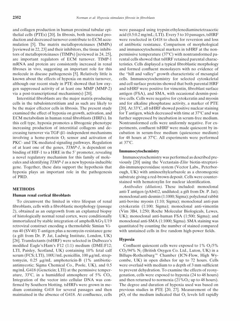
Fig. 12. Schematic summary of the mecha-nisms of hypoxia-induced fibrogenesis.
intracellular signaling pathways [9]; however, the fact considerable cell type- and gene-dependent differences[9–11]. The use of protein kinase inhibitors providesthat H2O2 does not mimic the effect of hypoxia on TIMP-1
and Coll-I mRNA argues against a redox-sensitive path- information on the signaling pathways involved in geneactivation, and data in this study implicate both PKC-way in the hypoxic induction of these genes.
Although numerous signaling pathways have been im- and TK-mediated pathways in the hypoxic induction ofTIMP-1. Of the PKC inhibitors tested, BI is consideredplicated in hypoxic signal transduction, direct evidence
linking specific pathways to transcriptional activation of more specific than Cal-C, which targets all cysteine-richproteins [49]. The lack of inhibition of hypoxic-inductionparticular hypoxia-induced genes is sparse, and there are

Norman et al: Hypoxia stimulates fibrosis in fibroblasts 2365
in the text, who generously provided reagents. Portions of the dataof Coll-I mRNA by BI implies that induction of Coll-Ipresented herein have been presented at the American Society of
mRNA does not require activation of PKC. Indeed, acti- Nephrology Meetings 1996, 1997, 1998; the XIVth International Con-gress of Nephrology 1997; and the New York Academy of Sciencevation of PKC with PMA down-regulates Coll-I mRNA.Conference on “Inhibition of matrix metalloproteinases: TherapeuticThe differential response of TIMP-1 and Coll-I mRNAapplications” and published in abstract form [J Am Soc Nephrol 7:1762,
expression to TK inhibitors also suggests that alternate 1996; 8:523A, 1997; 9:524A, 1998; Nephrology 3(Suppl 1):S41, 1997;Ann NY Acad Sci 878:503–505, 1999].intracellular pathways act to regulate these genes in re-
sponse to a common stimulus. In other cell types, PKCReprint requests to Jill Norman, Ph.D., Department of Medicine,
signaling increases HIF, and several protein kinase inhib- Royal Free and University College Medical School, 7th Floor, Sir JulesThorn Institute for Clinical Sciences, Middlesex Hospital, Mortimeritors, including genistein, staurosporine and 2-aminopu-Street, London, England W1T 3AA, United Kingdom.rine, have been reported to inhibit HIF-1–dependentE-mail: [email protected]
gene expression or HIF-1 DNA binding [32–34]. If hyp-oxic induction TIMP-1 mRNA is HIF-1–dependent and
REFERENCESHIF-1 is blocked by these inhibitors, the same inhibitors
1. Jacobson H: Chronic renal failure: Pathophysiology. Lancetwould be expected to block TIMP-1 mRNA expression,338:419–427, 1991
which is, indeed, the case. In contrast, Coll-I mRNA 2. Bohle A, Kressel G, Muller C, Muller G: The pathogenesisof chronic renal failure. Pathol Res Pract 185:421–440, 1989induction is not blocked by genistein, suggesting HIF-
3. Bohle A, Strutz F, Muller G: On the pathogenesis of chronicindependent regulation.renal failure in primary glomerulopathies: A view from the intersti-
In summary, our study demonstrates that hypoxia in- tium. Exp Nephrol 2:205–210, 19944. Bohle A, Gise HV, Mackensen-Haen S, Stark-Jacob B: Theduces a fibrogenic response in renal fibroblasts, simulta-
obliteration of the post-glomerular capillaries and its influenceneously stimulating production of ECM and decreasingupon the function of both glomeruli and tubuli. Klin Wochenschr
turnover (Fig. 12). These effects appear to independent 59:1043–1051, 19815. Eddy A: Molecular insights into renal fibrosis. J Am Soc Nephrolof secreted autocrine mediators and to be mediated via
7:2495–2508, 1996a heme-protein O2 sensor and PKC- and TK-signaling6. Border WA, Noble N: TGF-b in kidney fibrosis: A target for
pathways. The hypoxic-induction of TIMP-1 is depen- gene therapy. Kidney Int 51:1388–1396, 19977. Fine LG, Ong ACM, Norman JT: Mechanisms of tubulo-intersti-dent on binding HIF-1 to a 59 HRE, revealing a novel
tial injury in progressive renal diseases. Eur J Clin Invest 23:259–regulatory mechanism for this inhibitor and expanding265, 1993
the spectrum of genes regulated by changes in low O2 8. Fine LG, Orphanides C, Norman JT: Progressive renal disease:The chronic hypoxia hypothesis. Kidney Int 53(Suppl 65):S74–S78,to include those involved in ECM turnover. Taken to-1998gether, the data suggest an important role for hypoxia
9. Bunn HF, Poynton R: Oxygen sensing and molecular adaptationin the initiation and progression of fibrosis. Understand- to hypoxia. Physiol Rev 76:839–885, 1996
10. Bauer C, Kurtz A (eds): Forefronts in Nephrology: Oxygen sens-ing the molecular mechanisms by which decreased oxy-ing on the cellular and molecular levels Kidney Int 51:371–608,gen induces a fibrogenic program may open new avenues1997
to therapy for what are, currently, intractable diseases. 11. Pugh CW: Oxygen and genes in health and disease. Q J Med 90:307–310, 1997
12. Wood SM, Ratcliffe PJ: Mammalian oxygen sensing and hypoxia-inducible factor-1. Int J Biochem Cell Biol 29:1419–1432, 1997APPENDIX
13. Semenza GL: Hypoxia-inducible factor-1 and the molecular physi-Abbreviations used in this article are: Act-D, actinomycin-D; CM, ology of oxygen homeostasis. J Lab Clin Med 131:207–214, 1998
conditioned medium; CoCl2, cobalt chloride; Coll-I, collagen a1(I); 14. Wenger RH, Gassman M: Oxygen(es) and the hypoxia-inducibleColl-III, collagen a1(III); Cyc, cycloheximide; DFO, desferrioxamine; factor-1. Biol Chem 378:609–616, 1997ECM, extracellular matrix; EPO, erythropoietin; FSA, fibroblast sur- 15. Rupec RA, Baeuerle PA: The genomic response of tumor cells toface antigen; H, hypoxia; HIF, hypoxia-inducible factor; HPLC, high- hypoxia and reoxygenation: Differential activation of transcriptionpressure liquid chromatography; HRE, hypoxia response element; factors AP-1 and NF-kB. Eur J Biochem 234:632–640, 1995HRF, human renal fibroblasts; 3H-Phe, tritiated phenalaline; 3H-Thy, 16. Muller JM, Krauss B, Kaltschmidt C, Baeuerle PA, Rupectritiated thymidine; LDH, lactate dehydrogenase; MMP, matrix metal- RA: Hypoxia induces c-fos transcription via a mitogen-activatedloproteinase; N, normoxia; PKA, protein kinase A; PKC, protein kinase protein kinase-dependent pathway. J Biol Chem 272:23435–23439,C; PRD, progressive renal disease; PTE, proximal tubular epithelial 1997cells; SMA, a-smooth muscle actin; TGF-b, transforming growth 17. Ratcliffe PJ, Maxwell PH, Pugh CW: Beyond erythropoietin:factor-b; TIMP-1, tissue inhibitor of metalloproteinases; TK, tyrosine The oxygen sensor. Nephrol Dial Transplant 12:1842–1848, 1997kinase; tsHRF, conditionally immortalized human renal fibroblasts. 18. Falanga V, Matrin TA, Tagaki H, Kirsner RB, Helfman T,
Pardes KR: Low oxygen tension increases mRNA levels ofalpha1(I) procollagen in human dermal fibroblasts. J Cell PhysiolACKNOWLEDGMENTS157:408–412, 1994
19. Durmowicz AG, Parks WC, Hyde DM, Mecham RP, StenmarkThis work was supported by a British Heart Foundation ProjectGrant #PG/96045 to J.T.N. and an Arthritis Research Campaign Post- KR: Persistence, re-expression and induction of pulmonary arterial
fibronectin, tropoelastin and type I procollagen mRNA expressiondoctoral Fellowship to I.M.C. The authors would like to thank Dr.M. O’Hare (Department Surgery, Royal Free and University College in neonatal hypoxic pulmonary hypertension. Am J Pathol 145:
1411–1420, 1994Medical School, London, UK) for the immortalization and initial selec-tion of tsHRF. We thank C. Orphanides for technical assistance with 20. Orphanides C, Fine LG, Norman JT: Hypoxia stimulates proximal
tubular cell matrix production via a TGF-b1-independent mecha-cell characterization and J. Palmen for DNA sequencing. We are gratefulto Dr. J. Iredale (Department of Medicine, University of Southampton) nism. Kidney Int 52:637–647, 1997
21. Tamamori M, Ito H, Hiroe M, Marumo F, Hata R-I: Stimulation offor helpful discussions. We are indebted to numerous individuals, cited

Norman et al: Hypoxia stimulates fibrosis in fibroblasts2366
collagen synthesis in rat cardiac fibroblasts by exposure to hypoxic 35. Himelstein BP, Koch CJ: Studies of type IV collagenase regulationby hypoxia. Cancer Lett 124:127–133, 1998culture conditions and suppression of the effect by natriuretic pep-
36. McQuillan LP, Leung GK, Anderson PA, Kostyk SK, Kourem-tides. Cell Biol Int 21:175–180, 1997bas S: Hypoxia inhibits expression of eNOS via transcriptional and22. Birkedal-Hansen H, Moore WGI, Bodden MK, Windsor LJ,post-transcriptional mechanisms. Am J Physiol 267:H1921–H1927,Birkedal-Hansen B, Decarlo A, Engler JA: Matrix metallopro-1994teinases: A review. Crit Rev Oral Biol Med 4:197–250, 1993
37. El Nahas AM (ed): Renal scarring: A multi-organ approach to23. Stetler-Stevenson WG: Dynamics of matrix turnover duringfibrosis. Exp Nephrol 3:65–148, 1995pathologic remodeling of the extracellular matrix. Am J Pathol
38. Baker AH, Zaltsman AB, George SJ, Newby AC: Divergent148:1345–1350, 1996effects of TIMP-1-2 or -3 overexpression in rat vascular smooth24. Denhardt DT, Feng B, Edwards DR, Cocuzzi ET, Malyankar muscle cell invasion, proliferation and death in vitro. TIMP-3 pro-
UM: Tissue inhibitor of metalloproteinases (TIMP, aka ERP): motes apoptosis. J Clin Invest 101:1478–1487, 1998Structure, control of expression and biological functions. Pharma- 39. Alexander CM, Howard EW, Bissell MJ, Werb Z: Rescue ofcol Ther 59:329–341, 1993 mammary epithelial cell apoptosis and entactin degradation by
25. Gomez DE, Alonso DF, Yoshiji H, Thorgiersson UP: Tissue tissue inhibitor of metalloproteinases-1 transgene. J Cell Biolinhibitors of metalloproteinases: Structure, regulation and biologi- 135:1669–1677, 1996cal functions. Eur J Cell Biol 74:111–122, 1997 40. Iredale JP, Benyon RC, Pickering J, McCullen M, Northrop
M, Pawlet S, Hovell C, Arthur MJ: Mechanisms of spontaneous26. Jat PS, Sharp PA: Cell lines established by a temperature-sensitiveresolution of rat liver fibrosis: Hepatic stellate cell apoptosis andSimian Virus 40 large T-antigen gene are growth restricted at thereduced hepatic expression of metalloproteinase inhibitors. J Clinnon-permissive temperature. Mol Cell Biol 9:1672–1681, 1989Invest 102:538–549, 199827. Ong ACM, Jowett TP, Firth JD, Burton S, Karet FE, Fine
41. Poli G, Parola M: Oxidative damage and fibrogenesis. Free RadicLG: An endothelin-1 mediated autocrine growth loop involved inBiol Med 22:287–330, 1997human renal tubular regeneration. Kidney Int 48:390–401, 1995
42. Hernandez-Munoz R, Diaz-Munoz M, Chagoya de Sanchez28. Campa JS, McAnulty RJ, Laurent GJ: Application of high pres-V: Possible role of cell redox state on collagen metabolism insure liquid chromatography to studies of collagen production by carbon tetrachloride-induced cirrhosis as evidenced by adenosine
isolated cells in culture. Anal Biochem 186:257–263, 1990 administration to rats. Biochim Biophys Acta 1200:93–99, 199429. Weisener MS, Turley H, Allen WE, Willam C, Eckhardt KU, 43. Breborowicz A, Martis L, Oreopoulos DG: In vitro influence
Talks KL, Wood SM, Gatter KC, Harris AL, Pugh CW, Rat- of lactate on function of peritoneal fibroblasts. Adv Perit Dialcliffe PJ, Maxwell PH: Induction of endothelial PAS domain 10:225–229, 1994protein-1 by hypoxia: Characterisation and comparison with hyp- 44. Cerbon-Ambriz J, Cerbon-Solorzano J, Rojkind M: Regulationoxia inducible factor-1 alpha. Blood 92:2260–2268, 1998 of collagen production in freshly isolated cell populations from
30. Clark IM, Rowan AD, Edwards DR, Bech-Hansen T, Mann normal and cirrhotic rat liver: Effect of lactate. Hepatology 13:551–556, 1991DA, Bahr MJ, Cawston TE: Transcriptional regulation of the
45. Hunt TK, Banda MJ, Silver IA: Cell interactions in post-trau-human tissue inhibitor of metalloproteinases-1 (TIMP-1) gene inmatic fibrosis, in Fibrosis, Ciba Foundation Symposium, London,fibroblasts involves elements in the promoter, exon 1 and intronPitman, 1985, pp 127–1491. Biochem J 324:611–617, 1997
46. Declerk YA, Darville MI, Eeckhout Y, Rousseau GG: Charac-31. Boukhalfa G, Desmouliere A, Rondeau E, Gabbiani G, Sraerterisation of the promoter of the gene encoding human tissueJ: Relationship between alpha-smooth muscle actin expression andinhibitor of metalloproteinase-2 (TIMP-2). Gene 139:185–191, 1994fibrotic changes in the human kidney. Exp Nephrol 4:241–247, 1996
47. Wick M, Haronen R, Mumberg D, Burger C, Olsen BR, Budarf32. Pugh CW, O’Rourke JF, Nagao M, Gleadle JM, Ratcliffe PJ: M, Apte SS, Muller R: Structure of the human TIMP-3 gene andActivation of hypoxia-inducible transcription factor-1: Definition its cell cycle-regulated promoter. Biochem J 311:549–554, 1995of regulatory domains within the a subunit. J Biol Chem 272:11205– 48. Gleadle JM, Ebert BL, Firth JD, Ratcliffe P: Regulation of11214, 1997 angiogenic growth factor expression by hypoxia, transition metals
33. Ratcliffe P: Molecular biology of erythropoietin. Kidney Int and chelating agents. Am J Physiol 268:C1362–C1368, 199544:887–904, 1994 49. Nixon JS: The biology of protein kinase C inhibitors, in Protein
34. Kovacs EJ, Dipietro LA: Fibrogenic cytokines and connective Kinase C, edited by Parker PJ, Dekker LV, London, RG LandesCompany, 1997, pp 205–236tissue production. FASEB J 8:854–861, 1994