Embed Size (px)
Citation preview

Wnt Signaling
A subject collection from Cold Spring Harbor Perspectives in Biology
Copyright 2013 Cold Spring Harbor Laboratory Press. Not for distribution. Do not copy without written permission from Cold Spring Harbor Laboratory Press.
OTHER SUBJECT COLLECTIONS FROM COLD SPRING HARBORPERSPECTIVES IN BIOLOGY
Protein Synthesis and Translational Control
The Synapse
Extracellular Matrix Biology
Protein Homeostasis
Calcium Signaling
The Golgi
Germ Cells
The Mammary Gland as an Experimental Model
The Biology of Lipids: Trafficking, Regulation, and Function
Auxin Signaling: From Synthesis to Systems Biology
The Nucleus
Neuronal Guidance: The Biology of Brain Wiring
Cell Biology of Bacteria
Cell–Cell Junctions
Generation and Interpretation of Morphogen Gradients
Immunoreceptor Signaling
NF-kB: A Network Hub Controlling Immunity, Inflammation, and Cancer
Symmetry Breaking in Biology
The Origins of Life
The p53 Family
SUBJECT COLLECTIONS FROM COLD SPRING HARBORPERSPECTIVES IN MEDICINE
Parkinson’s Disease
Type 1 Diabetes
Angiogenesis: Biology and Pathology
HIV: From Biology to Prevention and Treatment
The Biology of Alzheimer Disease
Copyright 2013 Cold Spring Harbor Laboratory Press. Not for distribution. Do not copy without written permission from Cold Spring Harbor Laboratory Press.
Wnt SignalingA subject collection from Cold Spring Harbor Perspectives in Biology
EDITED BY
Roel Nusse Xi He
Stanford University Harvard Medical School
Renee van Amerongen
Netherlands Cancer Institute
COLD SPRING HARBOR LABORATORY PRESS
Cold Spring Harbor, New York † www.cshlpress.org
Copyright 2013 Cold Spring Harbor Laboratory Press. Not for distribution. Do not copy without written permission from Cold Spring Harbor Laboratory Press.
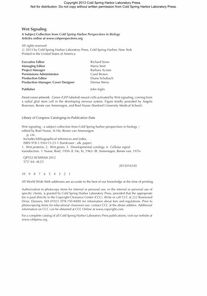
Wnt SignalingA Subject Collection from Cold Spring Harbor Perspectives in BiologyArticles online at www.cshperspectives.org
All rights reserved# 2013 by Cold Spring Harbor Laboratory Press, Cold Spring Harbor, New YorkPrinted in the United States of America
Executive Editor Richard SeverManaging Editor Maria SmitProject Manager Barbara AcostaPermissions Administrator Carol BrownProduction Editor Diane SchubachProduction Manager/Cover Designer Denise Weiss
Publisher John Inglis
Front cover artwork: Green (GFP-labeled) neural cells activated by Wnt signaling, coming froma radial glial stem cell in the developing nervous system. Figure kindly provided by AngelaBowman, Renee van Amerongen, and Roel Nusse (Stanford University Medical School).
Library of Congress Cataloging-in-Publication Data
Wnt signaling : a subject collection from Cold Spring harbor perspectives in biology /edited by Roel Nusse, Xi He, Renee van Amerongen.
p. cm.Includes bibliographical references and index.ISBN 978-1-936113-23-1 (hardcover : alk. paper)
1. Wnt proteins. 2. Wnt genes. 3. Developmental cytology. 4. Cellular signaltransduction. I. Nusse, Roel, 1950- II. He, Xi, 1962- III. Amerongen, Renee van, 1976-
QP552.W58W68 2012572’.64--dc23
2012016545
10 9 8 7 6 5 4 3 2 1
All World Wide Web addresses are accurate to the best of our knowledge at the time of printing.
Authorization to photocopy items for internal or personal use, or the internal or personal use ofspecific clients, is granted by Cold Spring Harbor Laboratory Press, provided that the appropriatefee is paid directly to the Copyright Clearance Center (CCC). Write or call CCC at 222 RosewoodDrive, Danvers, MA 01923 (978-750-8400) for information about fees and regulations. Prior tophotocopying items for educational classroom use, contact CCC at the above address. Additionalinformation on CCC can be obtained at CCC Online at www.copyright.com.
For a complete catalog of all Cold Spring Harbor Laboratory Press publications, visit our website atwww.cshlpress.org.
Copyright 2013 Cold Spring Harbor Laboratory Press. Not for distribution. Do not copy without written permission from Cold Spring Harbor Laboratory Press.
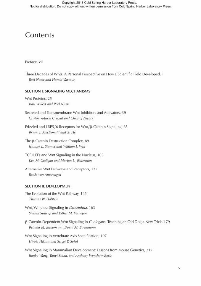
Contents
Preface, vii
Three Decades of Wnts: A Personal Perspective on How a Scientific Field Developed, 1
Roel Nusse and Harold Varmus
SECTION I: SIGNALING MECHANISMS
Wnt Proteins, 25
Karl Willert and Roel Nusse
Secreted and Transmembrane Wnt Inhibitors and Activators, 39
Cristina-Maria Cruciat and Christof Niehrs
Frizzled and LRP5/6 Receptors for Wnt/b-Catenin Signaling, 65
Bryan T. MacDonald and Xi He
The b-Catenin Destruction Complex, 89
Jennifer L. Stamos and William I. Weis
TCF/LEFs and Wnt Signaling in the Nucleus, 105
Ken M. Cadigan and Marian L. Waterman
Alternative Wnt Pathways and Receptors, 127
Renee van Amerongen
SECTION II: DEVELOPMENT
The Evolution of the Wnt Pathway, 145
Thomas W. Holstein
Wnt/Wingless Signaling in Drosophila, 163
Sharan Swarup and Esther M. Verheyen
b-Catenin-Dependent Wnt Signaling in C. elegans: Teaching an Old Dog a New Trick, 179
Belinda M. Jackson and David M. Eisenmann
Wnt Signaling in Vertebrate Axis Specification, 197
Hiroki Hikasa and Sergei Y. Sokol
Wnt Signaling in Mammalian Development: Lessons from Mouse Genetics, 217
Jianbo Wang, Tanvi Sinha, and Anthony Wynshaw-Boris
v
Copyright 2013 Cold Spring Harbor Laboratory Press. Not for distribution. Do not copy without written permission from Cold Spring Harbor Laboratory Press.
SECTION III: STEM CELL BIOLOGY AND DIFFERENTIATION
Wnt Pathway Regulation of Embryonic Stem Cell Self-Renewal, 233
Bradley J. Merrill
Wnt Signaling, Stem Cells, and Cancer of the Gastrointestinal Tract, 251
Arnout Schepers and Hans Clevers
Wnt Signaling in Bone Development and Disease: Making Stronger Bone with Wnts, 265
Jean B. Regard, Zhendong Zhong, Bart O. Williams, and Yingzi Yang
Wnt Signaling in the Vertebrate Central Nervous System: From Axon Guidance
to Synaptic Function, 283
Patricia C. Salinas
Wnt Signaling and Forebrain Development, 297
Susan J. Harrison-Uy and Samuel J. Pleasure
Wnt Signaling in Normal and Malignant Hematopoiesis, 309
William Lento, Kendra Congdon, Carlijn Voermans, Marcie Kritzik, and Tannishtha Reya
Wnt Signaling in Skin Development, Homeostasis, and Disease, 319
Xinhong Lim and Roel Nusse
Wnt Signaling in Mammary Glands: Plastic Cell Fates and Combinatorial Signaling, 345
Caroline M. Alexander, Shruti Goel, Saja A. Fakhraldeen, and Soyoung Kim
Wnt Signaling in Neuromuscular Junction Development, 373
Kate Koles and Vivian Budnik
SECTION IV: DISEASE AND INTERVENTION
Wnt Signaling in Cancer, 395
Paul Polakis
Wnt Signaling and Injury Repair, 409
Jemima L. Whyte, Andrew A. Smith, and Jill A. Helms
Targeting Wnt Pathways in Disease, 423
Zachary F. Zimmerman, Randall T. Moon, and Andy J. Chien
Index, 447
Contents
vi
Copyright 2013 Cold Spring Harbor Laboratory Press. Not for distribution. Do not copy without written permission from Cold Spring Harbor Laboratory Press.
Preface
SIGNALING BY THE WNT FAMILY OF SECRETED PROTEINS has come to be recognized as one of the most fun-damental and conserved regulatory systems in animals. Wnt signaling controls many critical
aspects of development and adult tissue homeostasis and its deregulation is intimately linked to abroad spectrum of human diseases, ranging from congenital defects to osteoporosis and cancer.As such, Wnt-related research has become a major area for targeted drug discovery and therapeuticdevelopment.
Since the identification of the first Wnt gene 30 years ago, research in the Wnt field has takenflight—as reflected by more than 13,000 publications in Medline to date—and has progressedinto numerous exciting areas. Wnt-related investigations continue to reveal fascinating principlesof embryonic patterning, cell growth and differentiation, the wiring of the nervous system, the patho-genic mechanisms underlying cancer as well as degenerative disease, stem cell and regenerativebiology, and potential therapeutic applications. This book on Wnt signaling is therefore bothtimely and long overdue.
When the plan for this book was first conceived, we felt that it should cover the scope of the Wntfield as broadly as we could envision. We have therefore organized the various chapters to include thebiochemistry and molecular mechanisms that control Wnt signal transduction (Section I), in parallelto the evolutionary origin of Wnt signaling and its functions in the development of model organisms(Section II). A major focus lies on the role of Wnt signaling in stem cell biology and differentiation(Section III), as our knowledge in this area has increased tremendously over the last decade or so.Finally, the implications of Wnt signal transduction for pathogenesis and treatment of human dis-eases are presented in Section IV. As this book is published in the same year as the 30th anniversaryof the discovery of the int1/wnt1 gene, we have included a separate chapter describing the history ofthe Wnt field, in the form of a personal perspective by Roel Nusse and Harold Varmus. This chapterwas previously published by EMBO J (doi:10.1038/emboj.2012.146) and is reprinted with kindpermission from the editor.
The pace of progress in the Wnt field has been astonishing, and several chapters had to be revisedeven at the production stage to include the latest exciting discoveries that just appeared in the litera-ture. However, in spite of its broad range, this book does not pretend to cover all of the fascinatinginsights we have gained in Wnt biology over the years. It is our hope that this volume serves as a step-ping-stone for the reader to guide and encourage further exploration and, perhaps, to open up novelavenues of investigation, particularly applications in the fields of bioengineering, regenerative medi-cine, and cancer treatment.
A big thank you goes to all of the authors who contributed timely chapters so enthusiastically tothis project. We are very grateful to Richard Sever and in particular to Barbara Acosta at CSHL Press,who guided and coordinated the entire process and who showed tremendous patience in managingour deadlines.
ROEL NUSSE
XI HE
RENEE VAN AMERONGEN
vii
Copyright 2013 Cold Spring Harbor Laboratory Press. Not for distribution. Do not copy without written permission from Cold Spring Harbor Laboratory Press.

Copyright 2013 Cold Spring Harbor Laboratory Press. Not for distribution. Do not copy without written permission from Cold Spring Harbor Laboratory Press.
Three Decades of Wnts: A PersonalPerspective on How a ScientificField Developed1
Roel Nusse2 and Harold Varmus3
2Department of Developmental Biology, Howard Hughes Medical Institute, Stanford UniversitySchool of Medicine, Stanford, California 94305
3National Cancer Institute, National Institutes of Health, Bethesda, Maryland 20892
Correspondence: [email protected]
Wnt genes and components of Wnt signalling pathways have been implicated in a widespectrum of important biological phenomena, ranging from early organismal developmentto cell behaviours to several diseases, especially cancers. Emergence of the field of Wntsignalling can be largely traced back to the discovery of the first mammalian Wnt gene in1982. In this essay, we mark the thirtieth anniversary of that discovery by describing some ofthe critical scientific developments that led to the flowering of this field of research.
INTRODUCTION: HOW WE LEARNABOUT WHAT WE KNOW
Knowledge differs from the growth of knowl-edge. To learn the facts about a subject, such
as Wnt genes or the Wnt signalling pathway, onecan consult an encyclopaedia, a textbook, or aconventional review article. To understand howthose facts were unearthed and assembled intocoherent concepts, it is necessary to probe thehistory of a field—to learn about the sequenceof events, the logical and illogical connectionsbetween those events, and the people who par-ticipated in them.
We have approached this essay with moreattention to historical development than to a
full repertoire of facts. While interesting exper-imental results about Wnt genes and their effectson cells and organisms continue to appear at anaccelerating pace, we believe that there is a greatdeal to learn about the scientific enterprise morebroadly by looking back on the unusual way inwhich the knowledge about those genes hasgrown over the past three decades. Some under-standing of Wnt signalling is now required ofthose who aspire to succeed in many prominentfields of biology—including organismal devel-opment, cancer research, and stem-cell biology.There is also a sizeable subset of biologists whodefine themselves primarily as students of Wntgenes, while also aligning themselves with can-cer, developmental, or stem-cell biologists orwith communities devoted to the fruit fly,worm, amphibians, mouse, or Homo sapiens.This cannot be said of those working on manyother genes or signalling pathways, raising theinteresting questions of how and why scientists
1First published in EMBO J 31: 2670–2684 (22 May 2012)doi:10.1038/emboj.2012.146, and reprinted with expresspermission from the authors, Roel Nusse and Harold Var-mus.
1
Copyright 2013 Cold Spring Harbor Laboratory Press. Not for distribution. Do not copy without written permission from Cold Spring Harbor Laboratory Press.
organize themselves in unusual ways and viewtheir subjects through certain kinds of lenses.
We have composed this essay on the occa-sion of the thirtieth anniversary of the pub-lished report that announced our discovery ofwhat proved to be the first mammalian Wntgene (Nusse and Varmus 1982). Taking that re-port as an arbitrary starting point in the historyof this field, we have tried to highlight the mostsignificant ways in which the field increased inknowledge, enlarged in scope, and grew in dis-ciples. As we emphasize, some of these advanceswere logical and straightforward, others weretechnically difficult and protracted, and yet oth-ers were serendipitous and surprising. We alsonote more briefly how the shape of the field wasdetermined by certain beneficial attitudes andbehaviours that may be worthy of emulation.
THE “PRE-HISTORY”: MOUSE MODELS FORBREAST CANCER AND CANCER-CAUSINGRETROVIRUSES PRECEDED KNOWLEDGEOF WNT GENES
All modern science is built on earlier science.Accordingly, the discovery that launched theintense study of Wnt genes 30 years ago depend-ed on at least two earlier and closely related linesof enquiry: mouse models of cancer and onco-genic retroviruses.
It had been known since the 1930s that cer-tain strains of laboratory mice are highly suscep-tible to breast cancer, and that the disease isusually transmitted from mothers to offspringmice through the milk (Bittner 1936; Korteweg1936). Later, the tumour-inducing activity waspurified from the milk (Lyons and Moore 1962),and the milk-transmitted factor was shown to bea morphologically atypical retrovirus, called theMouse Mammary Tumour Virus or MMTV.
Although the study of oncogenic retrovirus-es can be tracked to the first decade of the 20thcentury, the basis of their cancer-causing prop-erties came into focus only in the century’s sec-ond half. The first great advances came fromtissue culture assays for viral growth and celltransforming capacities and from the biochem-ical and genetic analysis of the RNA genomes ofretroviruses isolated from chickens, mice, rats,
and other experimental animals. These methodsled to the discovery of distinct viral oncogenes,such as Src, Myc, and Ras, and their cellularprecursors, called proto-oncogenes, a term de-noting any cellular genes that could be convertedto active cancer-causing genes (Bishop and Var-mus 1985). Conversion to oncogenicity couldoccur by the mechanisms that produced highlyoncogenic retroviruses or, as was shown in timeand described later in this essay, by a variety ofother mechanisms, most commonly somaticmutations of several types.
The proto-oncogenes initially discovered bytracing viral oncogenes to their cellular originswere generally not related to each other, but theyshared several properties. They had been con-served during evolution and were converted tocancer-causing genes by gain-of-function mu-tations—as first shown for retroviral oncogenesand later for activated cellular oncogenes foundin human cancers. The rapid onset of cancercaused by many retroviruses reflected the abilityof active viral oncogenes to transform many in-fected cells (Bishop and Varmus 1985).
Before RN came to UCSF in 1980 to workwith HV, we had both been interested in breastcancer in the mouse and in the general prop-erties of retroviruses isolated from animals.Such viruses often cause haematological cancersand sarcomas, relatively infrequent types of can-cers in human beings; only rarely do they in-duce epithelial carcinomas, the most commonhuman cancers. So, we were curious about themechanism by which MMTV might cause car-cinoma of the breast. Unlike the most intensive-ly studied cancer-causing retroviruses, howev-er, there was no readily identifiable oncogenein the viral genome. But, like other retroviruses,MMTV was known to insert a DNA copy of itsRNA genome into the host cell genome dur-ing infection. Moreover, MMTV caused cancerslowly, over the course of several months, unlikeoncogene-containing retroviruses, suggestingthat viral infection alone, while required for tu-mour induction, was not sufficient to transforma host cell—a property that had also been rec-ognized in the study of certain leukaemia-in-ducing retroviruses found in birds and mice(Teich et al. 1982).
R. Nusse and H. Varmus
2 Cite this article as Cold Spring Harb Perspect Biol doi: 10.1101/cshperspect.a007856
Copyright 2013 Cold Spring Harbor Laboratory Press. Not for distribution. Do not copy without written permission from Cold Spring Harbor Laboratory Press.
It was known, however, that tumours in-duced by such viruses were composed of cellclones defined by shared proviral integrationsites (Cohen et al. 1979; Payne et al. 1981) im-plying that tumours consisted of the descen-dants of a single infected cell and were thus theoutcome of rare events. The pattern of clonalityraised the possibility that one of many infectedcells had randomly acquired a provirus thatcould initiate tumourigenesis. For instance, theinsertion of viral DNA might cause a mutationof a gene in the vicinity of the integration site,and the change might confer a growth advantageto that cell. While it was possible that such mu-tations caused loss-of-function mutations, bydisrupting a host cell gene, an alternative andmore attractive model was that a host cell genewas transcriptionally activated by the incom-ing provirus. The latter model was inspired byfindings in yeast and prokaryotes, where trans-posable elements, which are often functionallyand structurally similar to retroviral proviruses(Brown and Varmus 1989), were known to acti-vate residing host cell genes (Errede et al. 1980).
The model of gene activation by proviral in-sertion gained momentum when Bill Hayward’sgroup showed that B-cell lymphomas causedby the slowly oncogenic Avian Leukosis Virus(ALV) commonly contained proviruses insertednear the c-myc gene and that c-myc was overex-pressed in those tumours (Hayward et al. 1981).Initially, activation of c-myc appeared to be theconsequence of a transcriptional promoter pres-ent in an ALV provirus inserted upstream of theprotein- coding exons of c-myc in the same tran-scriptional orientation (Hayward et al. 1981),but research in HV’s group showed that provi-ruses could also enhance c-myc expression fromthe endogenous c-myc promoter when inserteddownstream of the gene or upstream in eitherorientation (Payne et al. 1982).
The discoveries concerning c-myc and ALVwere of critical importance in cancer research, asit was the first time that a cellular homologueof a viral oncogene was shown to be activated incancer by mutation. Soon thereafter, manykinds of mutations other than viral insertionmutations—gene amplifications, chromosom-al translocations, and point mutations—were
shown to activate c-myc and many other pro-genitors of retroviral oncogenes, in human aswell as in animal tumours, without the interven-tion of retroviruses (Weinberg 1983). The re-sults with ALV and c-myc also set the stage forlooking for a similar mechanism in tumourscaused by other retroviruses, such as MMTV,with the prospect of finding proto-oncogenesthat are not related to known viral oncogenes.
MOLECULAR CLONING OF INT1, THE FIRSTNOVEL PROTO-ONCOGENE IDENTIFIEDBY PROVIRAL TAGGING
To seek host cell genes that are activated by in-sertions of MMTV proviruses, we decided to doa systematic screen rather than to look for acti-vation of only those proto-oncogenes knownat the time, such as c-myc. We assumed, perhapsnaively, that only one or perhaps a few genes inthe mouse genome would confer tumourigenicgrowth when activated by MMTV in mammarycancer. This assumption implied that by com-paring MMTV proviruses in multiple differenttumours we would find common integrationsites, or at least common regions, near thoseunusual genes. This segment of the genomewould represent a so-called “common integra-tion site.” But the commonality would not re-flect a predisposition to integrate MMTVat pre-ferred locations in the mouse genome—all data,then and now, suggest that retroviral integrationoccurs quasi-randomly in host genomes—butinstead would result from selection of cells thatacquired a growth advantage when a provirusactivated a nearby proto-oncogene.
The goal then became to isolate an integrat-ed MMTV provirus plus its adjacent host cellDNA from a tumour by molecular cloning. Wereasoned that we should start from a tumourwith just one MMTV insertion. Most tumoursin the strain we were using (C3H) carried mul-tiple newly acquired proviruses, in addition to afew copies of inherited “endogenous” MMTVDNA that were invariant among the arising tu-mours because they were products of ancientinfections of the mouse germ line (Bentvelzenet al. 1970; Cohen and Varmus 1979). Since itwas difficult to know which of the multiple new
Three Decades of Wnts: A Personal Perspective
Cite this article as Cold Spring Harb Perspect Biol doi: 10.1101/cshperspect.a007856 3
Copyright 2013 Cold Spring Harbor Laboratory Press. Not for distribution. Do not copy without written permission from Cold Spring Harbor Laboratory Press.
proviruses would be functionally important, welooked for tumours carrying only one new pro-virus; this single insert should, according to themodel, be near the relevant gene. Restrictionfragments of host DNA derived from the vicin-ity of the provirus from that tumour would thenbe used as probes for two purposes: to screenother tumours for integrations in the same do-main and to measure transcription of localgenes, both in tumours and in normal mamma-ry tissue. Of course, today, with complete se-quences of normal mouse genomes and power-ful methods for sequencing tumour DNA inhand, precise mapping of insertion sites has be-come a relatively simple exercise. But at thetime, these tools were not available, so a morecumbersome method was required.
We initiated the screen in October 1980,gathering over 30 MMTV-infected C3H micewith mammary tumours, and we found justone tumour (#18) with a single new provirus.Happily, that single tumour proved to sufficefor our purposes. From the genome of this tu-mour, we cloned a “junctional” fragment, con-taining a part of an MMTV provirus joinedto adjacent chromosomal DNA, then derivedsmaller fragments containing only host cellDNA. With those fragments as probes, we“walked” along the chromosome in either direc-tion from the original proviral insertion site toobtain more probes for a broader region of themouse genome (Fig. 1). These were then usedto examine DNA from other tumours for dis-ruptions caused by MMTV proviruses. Indeed,
Figure 1. Aworking map of the mouse int1 locus as drawn by RN and used from 1982 to 1984, with the positionof various cloned genomic restriction fragments. Red lines indicate the presence of int1 exons, mapped in 1984.Note the location of Probe C, a genomic fragment hybridizing with int1 mRNA in mouse mammary tumours.At the bottom, the position of MMTV proviruses mapped in different tumours, with the position of the provirusin tumour #18, the starting point of the cloning of the locus, indicated at the right hand end.
R. Nusse and H. Varmus
4 Cite this article as Cold Spring Harb Perspect Biol doi: 10.1101/cshperspect.a007856
Copyright 2013 Cold Spring Harbor Laboratory Press. Not for distribution. Do not copy without written permission from Cold Spring Harbor Laboratory Press.
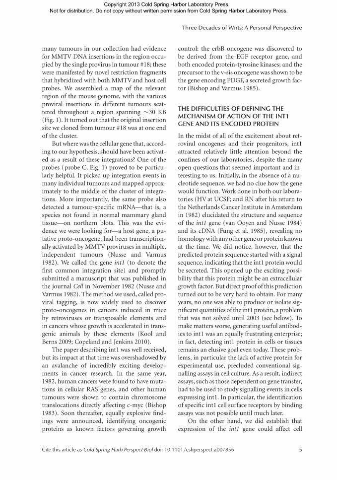
many tumours in our collection had evidencefor MMTV DNA insertions in the region occu-pied by the single provirus in tumour #18; thesewere manifested by novel restriction fragmentsthat hybridized with both MMTV and host cellprobes. We assembled a map of the relevantregion of the mouse genome, with the variousproviral insertions in different tumours scat-tered throughout a region spanning �30 KB(Fig. 1). It turned out that the original insertionsite we cloned from tumour #18 was at one endof the cluster.
But where was the cellular gene that, accord-ing to our hypothesis, should have been activat-ed as a result of these integrations? One of theprobes (probe C, Fig. 1) proved to be particu-larly helpful. It picked up integration events inmany individual tumours and mapped approx-imately to the middle of the cluster of integra-tions. More importantly, the same probe alsodetected a tumour-specific mRNA—that is, aspecies not found in normal mammary glandtissue—on northern blots. This was the evi-dence we were looking for—a host gene, a pu-tative proto-oncogene, had been transcription-ally activated by MMTV proviruses in multiple,independent tumours (Nusse and Varmus1982). We called the gene int1 (to denote thefirst common integration site) and promptlysubmitted a manuscript that was published inthe journal Cell in November 1982 (Nusse andVarmus 1982). The method we used, called pro-viral tagging, is now widely used to discoverproto-oncogenes in cancers induced in miceby retroviruses or transposable elements andin cancers whose growth is accelerated in trans-genic animals by these elements (Kool andBerns 2009; Copeland and Jenkins 2010).
The paper describing int1 was well received,but its impact at that time was overshadowed byan avalanche of incredibly exciting develop-ments in cancer research. In the same year,1982, human cancers were found to have muta-tions in cellular RAS genes, and other humantumours were shown to contain chromosometranslocations directly affecting c-myc (Bishop1983). Soon thereafter, equally explosive find-ings were announced, identifying oncogenicproteins as known factors governing growth
control: the erbB oncogene was discovered tobe derived from the EGF receptor gene, andboth encoded protein-tyrosine kinases; and theprecursor to the v-sis oncogene was shown to bethe gene encoding PDGF, a secreted growth fac-tor (Bishop and Varmus 1985).
THE DIFFICULTIES OF DEFINING THEMECHANISM OF ACTION OF THE INT1GENE AND ITS ENCODED PROTEIN
In the midst of all of the excitement about ret-roviral oncogenes and their progenitors, int1attracted relatively little attention beyond theconfines of our laboratories, despite the manyopen questions that seemed important and in-teresting to us. Initially, in the absence of a nu-cleotide sequence, we had no clue how the genewould function. Work done in both our labora-tories (HV at UCSF; and RN after his return tothe Netherlands Cancer Institute in Amsterdamin 1982) elucidated the structure and sequenceof the int1 gene (van Ooyen and Nusse 1984)and its cDNA (Fung et al. 1985), revealing nohomology with anyother gene or protein knownat the time. We did notice, however, that thepredicted protein sequence started with a signalsequence, indicating that the int1 protein wouldbe secreted. This opened up the exciting possi-bility that this protein might be an extracellulargrowth factor. But direct proof of this predictionturned out to be very hard to obtain. For manyyears, no one was able to produce or isolate sig-nificant quantities of the int1 protein, a problemthat was not solved until 2003 (see below). Tomake matters worse, generating useful antibod-ies to int1 was an equally frustrating enterprise;in fact, detecting int1 protein in cells or tissuesremains an elusive goal even today. These prob-lems, in particular the lack of active protein forexperimental use, precluded conventional sig-nalling assays in cell culture. As a result, indirectassays, such as those dependent on gene transfer,had to be used to study signalling events in cellsexpressing int1. In particular, the identificationof specific int1 cell surface receptors by bindingassays was not possible until much later.
On the other hand, we did establish thatexpression of the int1 gene could affect cell
Three Decades of Wnts: A Personal Perspective
Cite this article as Cold Spring Harb Perspect Biol doi: 10.1101/cshperspect.a007856 5
Copyright 2013 Cold Spring Harbor Laboratory Press. Not for distribution. Do not copy without written permission from Cold Spring Harbor Laboratory Press.
behaviour in a fashion that resembled conven-tional transformation and provided a biologicalassay: various mammary epithelial cell linescould be morphologically altered by overexpres-sion of int1, albeit in a subtle way and rarelyleading to formation of cells capable of growinginto a tumour (Brown et al. 1986; Rijsewijk et al.1987b). More dramatically, Ann Tsukamoto inHV’s laboratory was able to recapitulate the on-cogenic effect of int1 in mice without resortingto virus infection: mice expressing an int1 trans-gene under the influence of an MMTV tran-scriptional regulator developed cancer in themammary gland within about 6 months of age(Tsukamoto et al. 1988). This established thatint1 is a bona fide proto-oncogene. Thesemice—and several others expressing int1 underthe control of inducible promoters—have sincebecome widely used mouse models for studyingbreast carcinogenesis and for finding genes thatcan cooperate with the int1 transgene duringoncogenesis (MacArthur et al. 1995).
DEVELOPMENTAL GENETICS HELPED TOREVEAL THE FUNCTION OF INT1 WHEN THEGENE WAS DISCOVERED TO BE THEHOMOLOGUE OF THE DROSOPHILASEGMENT POLARITY GENE, WINGLESS
How could the function of int1 protein and thecomponents of its signalling pathway be deci-phered without a direct, convenient biochemi-cal or cell-based assay? Fortunately, int1 didhave one advantage: a high degree of conserva-tion across species. The human int1 protein se-quence turned out to be almost completely(99%) identical to that of the mouse homo-logue (Van Ooyen et al. 1985). Moreover, int1-related sequences appeared to be present in theDNA of Drosophila melanogaster as judged frommolecular hybridization (Nusse et al. 1984).
At the time, the new molecular methods thatwere responsible for unveiling the genes centralto cancer research had also re-energized effortsto understand embryogenesis, using the richtreasury of Drosophila developmental mutants.During the 1970s, genetic screens in Drosophilahad unveiled a set of genes that were essential forthe development of the embryo. Nusslein-Vol-
hard and Wieschaus (1980) showed specific pat-terning defects, ranging from abnormal seg-ment numbers to polarity changes, in mutantsfor many of these genes. They coined the term“segment polarity genes” for one class of mu-tants that shared a similar patterning pheno-type during embryogenesis. One of the genesin this group was called Wingless; others, Arma-dillo and Arrow. The Wingless gene had actuallybeen identified earlier as a weak mutant alleleleading to loss of wing tissue, hence the nameWingless (Sharma and Chopra 1976). Subse-quent to the genetic screens, many of the seg-mentation genes were molecularly cloned, gen-erating a treasure trove of reagents to studydevelopmental mechanisms. For example, theexpression patterns of these genes often pro-duced stripes corresponding to body segments.By examining the expression pattern of one genein the background of mutations in other genes,hierarchies of genetic interactions were uncov-ered, providing unparalleled insights into howembryos develop (Ingham 1988).
RN and his colleagues cloned the Drosophilaint1 homologue and used polytene chromo-some mapping (the genomics technology at thetime) to locate the gene. It turned out to mapclose to Wingless, one of the segment polaritygenes; a striped expression pattern observedwith a Drosophila int1 probe also suggested arole in segmentation (Rijsewijk et al. 1987a).Around the same time, Baker (1987) had clonedthe Wingless gene bya P-element transposon tag-ging,amethod akinto theproviral taggingmeth-ods we had used for int1. The gene he cloned hadrestriction maps matching our Drosophila int1clone. The genes were identical; the int1 homo-logue in Drosophila was Wingless, one of the firstexamples of a gene involved in development andalso activated in cancer (Rijsewijk et al. 1987a).This was an exciting discovery in its own right. Inaddition, the membership ofWingless in theseg-ment polarity group promised to open doors todiscovering the mechanisms of action of int1/Wingless, since it seemed likely that other genesin the group would interact with int1/Winglessgenetically and biochemically. As we now under-stand, the core of its signalling pathway is indeedbased on genetic relationships between segment
R. Nusse and H. Varmus
6 Cite this article as Cold Spring Harb Perspect Biol doi: 10.1101/cshperspect.a007856
Copyright 2013 Cold Spring Harbor Laboratory Press. Not for distribution. Do not copy without written permission from Cold Spring Harbor Laboratory Press.
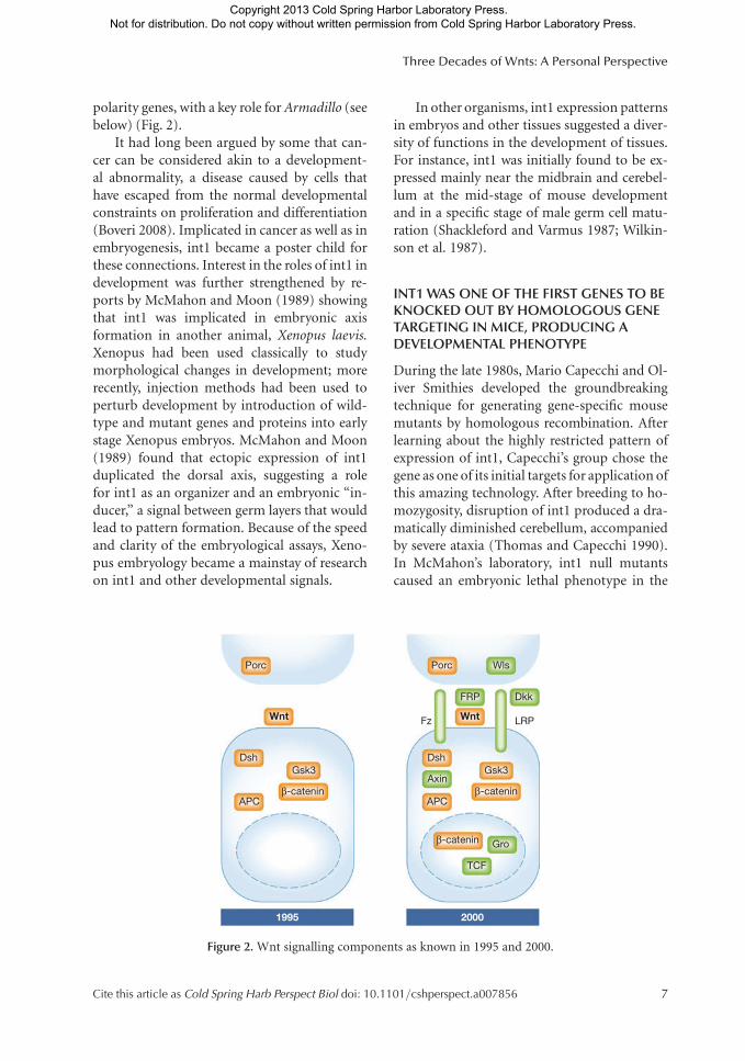
polarity genes, with a key role for Armadillo (seebelow) (Fig. 2).
It had long been argued by some that can-cer can be considered akin to a development-al abnormality, a disease caused by cells thathave escaped from the normal developmentalconstraints on proliferation and differentiation(Boveri 2008). Implicated in cancer as well as inembryogenesis, int1 became a poster child forthese connections. Interest in the roles of int1 indevelopment was further strengthened by re-ports by McMahon and Moon (1989) showingthat int1 was implicated in embryonic axisformation in another animal, Xenopus laevis.Xenopus had been used classically to studymorphological changes in development; morerecently, injection methods had been used toperturb development by introduction of wild-type and mutant genes and proteins into earlystage Xenopus embryos. McMahon and Moon(1989) found that ectopic expression of int1duplicated the dorsal axis, suggesting a rolefor int1 as an organizer and an embryonic “in-ducer,” a signal between germ layers that wouldlead to pattern formation. Because of the speedand clarity of the embryological assays, Xeno-pus embryology became a mainstay of researchon int1 and other developmental signals.
In other organisms, int1 expression patternsin embryos and other tissues suggested a diver-sity of functions in the development of tissues.For instance, int1 was initially found to be ex-pressed mainly near the midbrain and cerebel-lum at the mid-stage of mouse developmentand in a specific stage of male germ cell matu-ration (Shackleford and Varmus 1987; Wilkin-son et al. 1987).
INT1 WAS ONE OF THE FIRST GENES TO BEKNOCKED OUT BY HOMOLOGOUS GENETARGETING IN MICE, PRODUCING ADEVELOPMENTAL PHENOTYPE
During the late 1980s, Mario Capecchi and Ol-iver Smithies developed the groundbreakingtechnique for generating gene-specific mousemutants by homologous recombination. Afterlearning about the highly restricted pattern ofexpression of int1, Capecchi’s group chose thegene as one of its initial targets for application ofthis amazing technology. After breeding to ho-mozygosity, disruption of int1 produced a dra-matically diminished cerebellum, accompaniedby severe ataxia (Thomas and Capecchi 1990).In McMahon’s laboratory, int1 null mutantscaused an embryonic lethal phenotype in the
Porc
DshGsk3
β-catenin
Gsk3
β-catenin
β-catenin
APC
Axin
Dsh
APC
Gro
TCF
Wnt
20001995
Wnt
FRP
Fz
Dkk
LRP
Porc Wls
Figure 2. Wnt signalling components as known in 1995 and 2000.
Three Decades of Wnts: A Personal Perspective
Cite this article as Cold Spring Harb Perspect Biol doi: 10.1101/cshperspect.a007856 7
Copyright 2013 Cold Spring Harbor Laboratory Press. Not for distribution. Do not copy without written permission from Cold Spring Harbor Laboratory Press.
mid-brain also affecting the development ofthe cerebellum (McMahon and Bradley 1990).Soon thereafter, it was also shown that a classicalmouse mutation, Swaying (Lane 1967), was anallele of the int1 gene; when int1 was clonedfrom Swaying mice, it proved to have a frame-shift mutation (Thomas et al. 1991).
These several discoveries about int1 mutantorganisms in flies and mice were very striking.In a review article published in 1992 (Nusse andVarmus 1992), we wrote: “With the benefit ofhindsight, we now recognize that phenomenastudied for several decades are the consequencesof Wnt gene mutations. Viral insertion muta-tions regularly promote mammary tumours inlaboratory mice (Bittner 1936; Korteweg 1936),a spontaneous frameshift mutation of mice(swaying) impairs cerebellar structure and func-tion (Lane 1967; Thomas et al. 1991) and wing-less mutations in Drosophila can transform awing to a notum or disrupt segment polarity(Sharma and Chopra 1976; Nusslein-Volhardand Wieschaus 1980).”
RENAMING INT1 AS WNT1 ANDRECOGNITION OF A WNT GENE FAMILY
Around 1990, it became clear that the int no-menclature had become inadequate and confus-ing. For example, additional screens for MMTVproviral insertion sites in tumours had yieldedother activated genes, called int2, int3, and int4(Dickson et al. 1984; Gallahan and Callahan1987; Roelink et al. 1990). But by sequence com-parisons, these genes were not usually related toint1. One MMTV target gene, initially calledint4, did prove to be related to int1 (Roelinket al. 1990). But the frequently activated int2gene, first identified by Clive Dickson and Gor-don Peters (Dickson et al. 1984), turned out tobe a member of the FGF family (Dickson andPeters 1987). Interestingly, FGF genes were alsoimplicated in normal development at about thistime, sometimes in coordination with int1-related genes, as in mesoderm formation (Ki-melman et al. 1992). Moreover, int1 and int2are sometimes co-activated in MMTV-inducedbreast tumours (Peters et al. 1986). int3 wasshown to be related to Notch, another impor-
tant developmental regulator (Gallahan andCallahan 1997).
At the same time that the int gene no-menclature was becoming unworkable, variousexperiments, including PCR-based homologyscreens, had revealed a large family of genes re-lated to int1 (Gavin et al. 1990). It would havebeen confusing to christen all these genes withthe term “int,” whether or not they had beenactivated by proviral integration. To avoid fur-ther confusion, all those working on int1 and itsrelatives, including Wingless, consented to anew hybrid name “Wnt” (for Wingless-relatedintegration site) to denote genes belonging tothe int1/Wingless family, with int1, now calledWnt1, as the founding member (Nusse et al.1991). (In accord with other recognized rela-tionships, int2 is now called FGF3, the int3gene is Notch4, and int4 is Wnt3A.)
THE WNT FAMILY AS A VANTAGE POINTTO STUDY GENE EVOLUTION ANDDEVELOPMENT IN METAZOANS
With the complete sequences of the genomes ofmany multicellular animals in hand, we nowrealize that vertebrates contain a family of 19Wnt-related genes; pairs of these genes can of-ten be placed in subfamilies that are highly sim-ilar to each other, perhaps reflecting gene dupli-cations relatively recently in evolution (Gavinet al. 1990). Each of these genes seems likely tohave a specific role in development or otherprocesses; they are generally expressed in differ-ent cells and at different times in maturation(Gavin et al. 1990). Many of the Wnt geneshave now been deliberately mutated in themouse, almost always leading to striking pheno-types, including limb polarity defects and sexualdimorphic abnormalities (Parr and McMahon1995, 1998).
Even representatives of the earliest branchesof the animal kingdom, such as Hydra and Ne-matostella, have the same number of Wnt sub-families as vertebrates (Kusserow et al. 2005).Readily recognized orthologues of specific Wntgenes—for example, Wnt1—have been foundthroughout the entire animal kingdom, often ex-pressed in tantalizing patterns (Kusserow et al.
R. Nusse and H. Varmus
8 Cite this article as Cold Spring Harb Perspect Biol doi: 10.1101/cshperspect.a007856
Copyright 2013 Cold Spring Harbor Laboratory Press. Not for distribution. Do not copy without written permission from Cold Spring Harbor Laboratory Press.
2005). As a result, evolutionary biologists spec-ulate that the early amplification and diversifica-tion of the Wnt family were at the roots of theincreased complexity of animal body plans(Sidow 1992; Holstein 2012). It appears fromsuch findingsthat Wnt genes were probably pres-ent in genomes prior to the split of the animalkingdom into protostomes and deuterostomes,are therefore at least 600 M years old, and mayhave a universal role in setting up the primaryaxis of animals (Petersen and Reddien 2009;Niehrs 2010; Holstein 2012). However, it is alsoclear that single-cell organisms do not containWnt genes, nor do plants.
In the midst of all of these genes and fami-lies, it remains striking that Wnt1 is one of thekey Wnt family members and may have been theprimordial one. Wnt1 is the true orthologue ofWingless, a gene in Drosophila with numerousfunctions in later development as well as earlyembryogenesis. While there are six other Wntsin Drosophila, the others each play a minor rolecompared with Wingless. Moreover, there arevery useful temperature-sensitive alleles ofWingless to study its numerous functions. As aresult of these attributes, Wingless has been arich source for understanding developmentalprocesses. In other organisms that have multipleWnts, Planaria in particular, the true Wnt1 or-thologue also has a special place because of therequirement for it in regeneration (Gurley et al.2010). While we do not understand why Wnt1 isthe most frequently activated gene in MMTV-induced breast cancer, it should be noted that wenow know that Wnt1 is closely linked to anotherWnt gene that is often insertionally activated,Wnt10B (Lee et al. 1995). Thus, some MMTVinserts may have activated both genes, providinga greater growth advantage.
UNEXPECTED FINDINGS REVEAL THEIMPORTANCE OF THE WNT PATHWAYIN HUMAN CANCERS
From the time that Wnt1 was discovered as aninitiating gene in mouse mammary carcinogen-esis, it remained of great interest and impor-tance to establish whether Wnt genes were in-volved in any human cancer. Early tests for
aberrant expression of Wnt genes in humanbreast cancer gave ambiguous results, at best,and no Wnt genes appeared to be mutated inany kinds of human tumours by DNA rear-rangements or (as more recently documentedby next generation sequencing of whole exomesor whole genomes) by point mutations (http://www.sanger.ac.uk/genetics/CGP/cosmic).
But work from a different angle changedour perceptions about the role of Wnt genesin human cancer, demonstrating that down-stream components of the Wnt signalling path-way, rather than the Wnt genes themselves at theupstream end of the pathway, were commonlyaltered in several types of human cancer. Thefirst news came from the study of colon cancer,and it was dramatic.
Around 1990, significant advances had beenmade in positional cloning of inherited humandisease genes, including genes predisposing toseveral types of cancer. Among the hereditaryforms of human cancer, adenomatous polyposiscoli (APC), a trait associated with multiple pol-yps in the colon, often leads to colon cancer ata relatively early age. The corresponding mu-tations—often nonsense or frameshift muta-tions that produce truncated proteins—werefound in an enormous gene called APC, whichwas cloned from human chromosome 5 in 1991(Groden et al. 1991; Kinzler et al. 1991). Identi-fying the human APC gene led to the cloning ofa mouse homologue, subsequently shown byBill Dove’s group to be mutated in a mousestrain called Min (Multiple intestinal neoplasia)(Su et al. 1993). Just as in human families, thecancer trait in the Min mouse is produced by anAPC truncating mutation, inherited in an auto-somal dominant manner. But despite the newgenetic insights into intestinal cancer, the func-tion of the large APC protein posed a biochem-ical mystery.
Soon thereafter, the groups of Paul Polakis,Bert Vogelstein, and Ken Kinzler establishedthat APC interacted in cells with a proteincalled b-catenin (Rubinfeld et al. 1993; Suet al. 1993). At the time, b-catenin had justbeen characterized by Masatoshi Takeichi andRolf Kemler as a protein binding to the cyto-plasmic domain of the adhesion molecule
Three Decades of Wnts: A Personal Perspective
Cite this article as Cold Spring Harb Perspect Biol doi: 10.1101/cshperspect.a007856 9
Copyright 2013 Cold Spring Harbor Laboratory Press. Not for distribution. Do not copy without written permission from Cold Spring Harbor Laboratory Press.
E-cadherin (Ozawa et al. 1989; Takeichi 1990).Intriguingly, Pierre McCrea and Barry Gumbi-ner had found that b-catenin gene was a verte-brate homologue of the segment polarity gene,Armadillo (McCrea et al. 1991), while Peiferand Wieschaus (1990) had established similar-ity between Plakoglobin (a b-catenin-relatedadhesion complex member) and Armadillo.Together, these findings suggested that APCand b-catenin/Armadillo were involved inregulating adhesion between vertebrate cells.Given the role of Armadillo in segment polari-ty, a function shared with Wingless, a modelemerged in which Wnt/Wingless signallingcontrolled cell adhesion in development (Peiferand Wieschaus 1990; Peifer et al. 1993), and thesame adhesion-based mechanism could controlgrowth of cells in tissues and cause cancer whenmisregulated.
While this was tantalizing, there were alsoreports that b-catenin/Armadillo was presentin the nucleus, as well as at the cell membrane(Funayama et al. 1995). Other publicationsmentioned that injection of antibodies tob-cat-enin/Armadillo could induce dorsal axis du-plication in Xenopus (McCrea et al. 1993), pos-sibly by stabilizing the b-catenin/Armadilloprotein; yet others claimed that depletion ofmaternal b-catenin/Armadillo could eliminatethe dorsal axis (Heasman et al. 1994). Theseobservations would ultimately all make sense:b-catenin is a key participant of Wnt signalling,but the molecular mechanisms remained unex-plained until a few years later.
In deciphering the cascade of events betweenthe Wnt signal and the role of b-catenin/Arma-dillo, the genetic interactions between a pro-tein kinase called Glycogen Synthase Kinase 3(GSK3) and other Wnt components proved tobe of critical importance. Norbert Perrimon andcolleagues showed that GSK3 (the Drosophilahomologue was called zeste-white 3, also knownas shaggy) was a negative regulator of the path-way; at the genetic level, Wnt/Wingless acted asa GSK3 inhibitor (Siegfried et al. 1992). Untilthen, GSK3 was known for its role in glucosemetabolism (Dent et al. 1989), so its newly dis-covered role in developmental signalling wascertainly surprising.
A WNT SIGNALLING CASCADE FROM THECELL SURFACE TO THE NUCLEUS, ANUNUSUAL PATHWAY EXPERIMENTALLYASSEMBLED FROM SEVERAL DIFFERENTMODELS
By 1995, the combined results from fly andmouse genetics, Xenopus embryology, and flyand mammalian cell culture experiments hadgenerated an outline of a Wnt signalling path-way (Fig. 2). It became clear that Wnt signal-ling was unusual compared with the other path-ways known at the time: those consisted mostlyof successions of protein phosphorylations,with protein associations based on recognitionof phosphorylated domains. In Wnt signalling,the most upstream known component was acytoplasmic protein of uncertain biochemicalfunction, Dishevelled, which then was proposedto inhibit the abundant GSK3 protein kinase(Peifer et al. 1991; Siegfried et al. 1992, 1994;Noordermeer et al. 1994). GSK3 was known tobe a negative regulator of b-catenin/Armadilloand was found in a complex with b-catenin/Armadillo, together with the APC protein (Ru-binfeld et al. 1996). The role of the GSK3 kinaseactivity as a suppressor of Wnt action was con-firmed by injecting dominant-negative (kinase-dead) mutants of GSK3 into early Xenopus em-bryos; this maneuver produced a phenocopy ofthe effect of Wnt1, duplication of the dorsal axis,that had been reported previously by Moon andMacMahon (Dominguez et al. 1995; He et al.1995; Pierce and Kimelman 1996).
A critical next step was to determine howphosphorylation by GSK3 governed b-catenin.This seemed likely to occur by control of the levelof b-catenin protein. In cells activated by Wnt,levels of b-catenin are commonly increased(Riggleman et al. 1990; Peifer et al. 1994; VanLeeuwen et al. 1994) by stabilizing theb-cateninprotein, not by an increase in its synthesis. Sev-eral highly conserved Ser/Thr phosphorylationsites near the amino terminus of b-catenin wereproposed as possible targets for phosphoryla-tion by GSK3 (Peifer et al. 1994). As shown byRolf Kemler’s and Randall Moon’s groups, phos-phorylated b-catenin is targeted for degrada-tion by the ubiquitination/proteasome pathway
R. Nusse and H. Varmus
10 Cite this article as Cold Spring Harb Perspect Biol doi: 10.1101/cshperspect.a007856
Copyright 2013 Cold Spring Harbor Laboratory Press. Not for distribution. Do not copy without written permission from Cold Spring Harbor Laboratory Press.
(Yost et al. 1996; Aberle et al. 1997; Orford et al.1997), with a critical role for F-box proteins (Ji-ang and Struhl 1998). Eliminating one or moreof the N-terminal phosphorylation sites stabiliz-esb-catenin, producing abundant protein high-ly active in Xenopus axis formation assays (Yostet al. 1996). As a result, there are striking parallelsbetween the Wnt, Hedgehog, and NF-kB signal-ling pathways; in all three cases, regulated signal-ling depends on degradation of a key pathwaycomponent by the ubiquitination/proteasomepathwayafter phosphorylation (Jiang and Struhl1998; Maniatis 1999).
The study of the molecular pathology of co-lon cancers then offered a remarkable exampleof the predictive power of knowledge about sig-nalling. As mentioned earlier, inherited muta-tions in the APC gene were known to cause thefamilial disease APC, and somatic mutations inAPC were found in most (ca. 85%) but not in allsporadic colorectal cancers. Why not all? Couldmutations affecting other components of theWnt signalling pathway substitute for APC mu-tations? Or did other mutant signalling path-ways drive those tumours? Paul Polakis, BertVogelstein, Hans Clevers, and their colleagueslooked specifically for altered b-catenin genesin APC wild-type tumours, on the suppositionthat b-catenin protein could be stabilized bymutations affecting the N-terminus as well asby loss of APC. Indeed they found that about5–10% of sporadic colon cancers had muta-tions, often short deletions, that removed orchanged the phosphorylation sites that targetb-catenin for degradation (Korinek et al. 1997;Morin et al. 1997; Rubinfeld et al. 1997). Sub-sequently, mutations have also been found inanother component of the degradation com-plex, Axin, in colorectal and other types of can-cers (Satoh et al. 2000).
This other component of the b-catenin/Ar-madillo/GSK3/APC complex has an inter-esting history. Cloned by Frank Costantini asa mouse developmental mutant called fused(Zeng et al. 1997), the Axin gene encodes a pro-tein that shares homology with Dishevelled,suggesting possible participation in the Wntpathway. This turned out to be the case. Axinis now known to participate in the b-catenin
destruction complex, together with APC andGSK3 (Behrens et al. 1998; Ikeda et al. 1998).Using a novel cell-free system for studying b-catenin degradation, Marc Kirschner and col-leagues showed that Axin is the rate-determin-ing component of the complex, even though itwas the most recently identified (Lee et al.2003). Axin has a similar role in intact mamma-lian cells (Li et al. 2012).
After these several elements were implicatedin Wnt signalling, two apparent and importantgaps in the pathway remained, one at each endof the pathway (Fig. 2). On the upstream end,there were no proteins known to recognizeextracellular Wnt proteins and transmit a sig-nal to the cell’s interior (Wnt receptors). Onthe downstream end, the anticipated effects ongene expression through transcriptional controlcould not be explained because b-catenin doesnot have the expected physical attributes of atranscription factor, and no established tran-scription factor was known to partner with it.Then, in a single year, 1996, these gaps wereclosed, generating excitement in the growingWnt field (Fig. 2).
WNT RECEPTOR PROTEINS WERE KNOWNIN OTHER CONTEXTS BEFORE THEIR ROLESIN WNT SIGNALLING WERE UNCOVERED
After many trial-and-error searches, one classof the elusive Wnt receptors was identified:the Frizzled transmembrane proteins. Original-ly found in mutant screens by Calvin Bridges(Bridges and Brehme 1944), Frizzled had beenidentified in Drosophila as a gene requiredfor planar polarity (Gubb and Garcia-Bellido1982; Vinson and Adler 1987), the orientationof cells in tissues. Paul Adler and colleaguesshowed Frizzled to encode a seven-pass trans-membrane protein (Vinson et al. 1989). Genet-ically, at least, Frizzled interacted with Dishev-elled, which was shown by Perrimon andMahowald (1987) to be involved in Winglesssignalling as well. While this suggested that Friz-zled could mediate Wingless signalling, the ab-sence of an embryonic segment polarity pheno-type in Frizzled mutants indicated otherwise.
Three Decades of Wnts: A Personal Perspective
Cite this article as Cold Spring Harb Perspect Biol doi: 10.1101/cshperspect.a007856 11
Copyright 2013 Cold Spring Harbor Laboratory Press. Not for distribution. Do not copy without written permission from Cold Spring Harbor Laboratory Press.
Here serendipity stepped in. Jeremy Na-thans and his colleagues found a Frizzled ho-mologue among components of a human reti-nal cDNA library that had been made to pursuetheir interests in the molecular biology of vi-sion. When the cDNA was used to seek homo-logues in a library of Drosophila DNA, a secondDrosophila Frizzled gene (Dfz2) was cloned, andthe Dfz2 gene displayed a striped pattern of geneexpression in the embryo, implying that itmight be directly involved in segment polarity.A collaboration between the Nathans and RNlaboratories revealed that the Wingless protein,which RN’s laboratory had solubilized at thetime, could bind to Dfz2 and, more weakly, toFrizzled itself (Bhanot et al. 1996). Moreover, incultured Drosophila cells that did not expressFrizzled genes, transfection of an expressionvector containing Frizzled genes conferred ac-tive signalling, as demonstrated by an increasein Armadillo (b-catenin) levels (Bhanot et al.1996). Genetic and other interactions betweenFrizzleds and Wnts were also reported by thegroups of Randall Moon and Robert Horvitz(Sawa et al. 1996; Yang-Snyder et al. 1996).
Just like Wnts, Frizzleds form a large genefamily in all branches of metazoan animals. Ge-netically, Frizzled genes are often redundant anddisplay phenotypes only when mutated in com-bination with other family members (Ye et al.2011). In Drosophila, this was shown for Friz-zled and Dfz2 using dsRNA interference tech-nology (Kennerdell and Carthew 1998). (Inter-estingly, this occurred in the same year that thisrevolutionizing method to inhibit gene expres-sion was first reported by Andy Fire and CraigMello; Fire et al. 1998). By using loss-of-functionmutations in Frizzled and Dfz2, Eric Wieschaus,Gary Struhl, Ken Cadigan, and Krishna Bhat un-covered a segment polarity phenotype indistin-guishable from phenotypes characteristic of theother genes in that class—but only as doublemutants, explaining why these receptor geneswere not in the original Nusslein-Volhard/Wie-schaus collection (Bhat 1998; Bhanot et al. 1999;Chen and Struhl 1999; Muller et al. 1999). De-spite the evidence for redundancy, Frizzled pro-teins have different affinities for different Wnts(Rulifson et al. 2000), indicating a high degree of
specificity in their interactions. However, persis-tent experimental problems with the biochemis-try of Wnt proteins have hampered systematicsurveys of the interactions.
To complement the Wnt receptor story, theDrosophila gene Arrow, one of the last segmentpolarity genes to be identified, was cloned by Ste-phen DiNardo and colleagues a few years later.Arrow proved to be a member of the Low densitylipoprotein receptor-Related Protein (LRP) fam-ily of receptors (Wehrli et al. 2000). Based on ad-ditional genetic data from Bill Skarnes, who madeLRP mouse mutants, and biochemical experi-ments from Xi He’s laboratory, a model emergedin which Arrow/LRP is a co-receptor for Wnts,physically adjacent to Frizzleds in the cell mem-brane(Pinsonetal.2000;Tamaietal.2000).Whensignalling to downstream components, however,Arrow/LRP may be the key player. Its cytoplasmictail is phosphorylated as a consequence of Wntbinding and interacts directly with GSK3 andAxin (Mao et al. 2001b; Tamai et al. 2004; David-son et al. 2005; Zeng et al. 2005) and Frizzled’sintracellular role in signalling may be limited tobinding Dishevelled (Macdonald et al. 2009).Arrow/LRP is also the target of several Wnt an-tagonists including the protein Dickkopf, isolat-ed by Christof Niehrs (Glinka et al. 1998; Baficoet al. 2001; Mao et al. 2001a; Semenov et al.2001). The Dickkopf-Wnt antagonism is con-served across many animal phyla (Guder et al.2006), illustrating the ancient nature of Wnt sig-nalling in animal development and evolution.
Eddy De Robertis, Jeremy Nathans, Jeff Ru-bin, and their co-workers have uncovered sev-eral other Wnt antagonists, in addition to Dick-kopf. These are secreted molecules usuallyconsisting of Wnt receptor domains that bindto Wnt itself. Some of these molecules havenames such as FRP or FRZB, reflecting theirsimilarity to the Frizzled receptor (Finch et al.1997; Leyns et al. 1997; Rattner et al. 1997).Others, such as the WIF protein (Hsieh et al.1999), are unrelated to Frizzled. These proteinsare likely involved in fine-tuning the concentra-tion of active Wnt outside cells.
The tandem arrays of Frizzled and LRP arenot the only Wnt receptors, as there are variousmembers of the trans-membrane tyrosine kinase
R. Nusse and H. Varmus
12 Cite this article as Cold Spring Harb Perspect Biol doi: 10.1101/cshperspect.a007856
Copyright 2013 Cold Spring Harbor Laboratory Press. Not for distribution. Do not copy without written permission from Cold Spring Harbor Laboratory Press.
family that serve to receive Wnts; these includethe ROR (Oishi et al. 2003; Mikels and Nusse2006) and Derailed/RYK (Yoshikawa et al.2003) proteins. Interestingly, these two classesof molecules have different Wnt binding mod-ules: the RORs contain a CRD domain similar tothe Frizzled CRD, while Derailed/RYK is relatedto the WIF protein mentioned above (Patthy,2000). Wnt interactions with these receptorsoften lead to effects in cells that are unrelatedto b-catenin, possibly mediating “non-canoni-cal Wnt signalling” (van Amerongen et al. 2008).
ANOTHER UNEXPECTED BUT PREVIOUSLYWELL-KNOWN PROTEIN, TCF/LEF1,EXPLAINS THE ROLE OF b-CATENIN IN THEWNT SIGNALLING PATHWAY
The TCF/Lef1 protein proved to be the long-sought Wnt transcription factor in the nucleusDiscovery of the critical interaction between thisprotein and b-catenin highlights one of thethemes of this essay: historically, the map ofWnt signalling was assembled by merging evi-dence from several different cell types and organ-isms. TCF/Lef1, an HMG box-containing tran-scription factor, was first implicated in immuneT-cell gene expression (Travis et al. 1991; van deWetering et al. 1991; Waterman et al. 1991) with-out any evident link to Wnt signalling. Work-ing separately on C. elegans, Jim Priess and col-leagues identified an HMG-box family member,POP1, involved in mesoderm specification inthe worm embryo (Lin et al. 1995), initiallyalso without connections to the Wnt pathway.
Soon thereafter, a surprising discovery wasreported: TCF/Lef1 could interact with b-cate-nin, considered at that time to be an adhesionmolecule. Hans Clevers extended his earlierwork on TCF/Lef1 to make this finding (Mole-naar et al. 1996), while Walter Birchmeier (Beh-rens et al. 1996) and Rolf Kemler (Huber et al.1996) started from b-catenin to establish bind-ing to TCF/Lef1. Using Drosophila, MariannBienz and Rudi Grosschedl (Riese et al. 1997)found that Wingless signalling was mediatedby TCF/Lef1 while Konrad Basler used muta-genesis screens to find a gene called Pangolin(Brunner et al. 1997), the single TCF/Lef1 ho-
mologue in Drosophila. Around the same time,continued investigations into C. elegans em-bryogenesis by Jim Priess, Craig Mello, andBruce Bowerman unveiled that the set ofMOM genes implicated in lineage choices weremembers of the Wnt pathway, including Wntitself (MOM2), Porcupine (MOM1), and Friz-zled (MOM5). All of these MOMs converged onPOP1 as a transcription factor and WRM1 as ab-catenin-related gene (Rocheleau et al. 1997,1999; Thorpe et al. 1997).
In many contexts, TCF/Lef1 can switch be-tween two states. When bound to Groucho, itacts as a repressor of target genes; but whenGroucho is displaced byb-catenin, the same tar-get genes are transcriptionally activated (Cavalloet al. 1998; Daniels and Weis, 2005). Crystallo-graphic studies by Bill Weis and Wenqing Xurevealed the molecular details of the bindingbetween TCF, b-catenin, and other proteins.The structure of b-catenin contains a groovemade by the “Armadillo” repeats in the protein,explaining how b-catenin can interact with sev-eral different partners, including TCF/ Lef1, E-cadherin, and APC (Huber et al. 1997; Grahamet al. 2000).
These discoveries about the interaction be-tween b-catenin and TCF/Lef1 were in morethan one respect very significant, as they notonly closed the gaps in the Wnt pathway butalso provided unparalleled tools for experi-ments. TCF/Lef1 recognizes a well-definedDNA binding site. By multimerizing this se-quence, Hans Clevers and colleagues generatedvery convenient luciferase-based Wnt reporters,called Top-FLASH and now widely used in theWnt field to measure signalling (Korinek et al.1997). There are now numerous genes known tohave TCF/Lef1 binding sites in their promotersand hence likely to be transcriptional targets forthe Wnt signalling pathway in at least some celltypes; among these genes are several implicatedin cancer, such as c-myc (He et al. 1998).
AT LAST, THE PURIFICATION OFACTIVE WNT PROTEIN
As we have recounted, by the end of the 20thcentury we had a blueprint of the Wnt signalling
Three Decades of Wnts: A Personal Perspective
Cite this article as Cold Spring Harb Perspect Biol doi: 10.1101/cshperspect.a007856 13
Copyright 2013 Cold Spring Harbor Laboratory Press. Not for distribution. Do not copy without written permission from Cold Spring Harbor Laboratory Press.
pathway and a readout for the pathway, both ofwhich were missing in the two previous decadesin which Wnt genes were intensively studied(Fig. 2). Still lacking, however, was the purifica-tion of any active Wnt protein, a problem thatwe and many other researchers had been work-ing on since the initial cloning of Wnt1. Whywere Wnt proteins so much more refractory tobiochemical purification than many other se-cretory proteins? Were they modified in a fash-ion that rendered them insoluble or highly ad-herent?
It was known from Norbert Perrimon’s workand Drosophila genetics that export likelyrequired a specialized protein encoded by Por-cupine. Evidence that Porcupine encoded aputative acyltransferase (Kadowaki et al. 1996;Hofmann 2000) suggested that detergentsmight be needed to keep lipid-modified Wntproteins soluble during extraction from cells.With the help of assays that judged Wnt activityin extracts based on increasedb-catenin levels incells, Karl Willert in RN’s laboratory finallymanaged to break through the purification bar-riers (Willert et al. 2003). (Coincidentally, Wil-lert had obtained his PhD working with HV atUCSF.) Wnts were found to be indeed covalentlyattached to lipids, explaining to some extenttheir resistance to biochemical manipulation(Willert et al. 2003). It is now recognized thatsecretion of Wnt proteins is a complex process,involving a dedicated enzyme (Porcupine; Ka-dowaki et al. 1996) and secretory proteins thatare specific for Wnt signals. Among these is themultiple-pass transmembrane protein Wntless/Evi, identified by the groups of Konrad Baslerand Michael Boutros, once again using the Dro-sophila genetic resources that have over the yearsbeen so instrumental in understanding the de-tails of Wnt signalling (Banziger et al. 2006;Bartscherer et al. 2006; Port and Basler 2010).
A GROWING AND VERY ACTIVE FIELDOF WNT SIGNALLING
The availability of Wnt proteins and morequantitative reporters (e.g., TopFlash) as reli-able end points for signalling have simplifiedthe study of Wnt signalling in cell culture, at-
tracting many new investigators. The generationof Wnt reporter mice, initially Top-gal animalsfrom Elaine Fuchs (DasGupta and Fuchs 1999)and later animals with transgenic markers driv-en by Axin 2 promoters (Lustig et al. 2002; vanAmerongen et al. 2012), provided yet more ex-perimental opportunities. One can now traceWnt-responding cells in any tissue of themouse, examine the origin of these cells, andfollow their fate in normal settings or after in-jury (Barker et al. 2007, 2010; van Amerongenet al. 2012). These new experimental tools haveled to a rapidly growing list of Wnt signallingcomponents, built on the core pathway.
Wnt signalling is clearly complicated andunusual when compared with other growth fac-tor cascades. At various nodes in the Wnt path-way, there are links to cyto-architectural pro-teins, such as those involved in adhesion andcell polarity (Nelson and Nusse 2004). TheWnt pathway is clearly important for cell fatechanges and the control of gene expression, butWnt signalling can also influence how cells areshaped and polarized and how they divide (Vee-man et al. 2003). Hitoshi Sawa, Bruce Bower-man, and Craig Mello have provided conclusiveevidence for a major role for Wnt signalling inasymmetric cell division in C. elegans and theannelid worm Platynereis dumerilii (Rocheleauet al. 1999; Schneider and Bowerman 2007; Su-gioka et al. 2011). Given the multiple roles of theWnt pathway in development, these cell biolog-ical phenotypes are perhaps not surprising andhave opened fertile ground for further research.
SOCIOLOGY OF THE FIELD OF WNTSIGNALLING: ANNUAL MEETINGS,SHARING INFORMATION, AND ADEDICATED WEBSITE
A historyof Wnt signalling would be incompletewithout a few comments on the sociology of thefield, which, we believe, has several unusual as-pects. Those features have contributed to oneof the overarching characteristics of this field:the propensity of investigators working on Wntgenes and Wnt signalling to identify themselves,at least in part and often primarily, as studentsof Wnts, regardless of whether they are cancer
R. Nusse and H. Varmus
14 Cite this article as Cold Spring Harb Perspect Biol doi: 10.1101/cshperspect.a007856
Copyright 2013 Cold Spring Harbor Laboratory Press. Not for distribution. Do not copy without written permission from Cold Spring Harbor Laboratory Press.
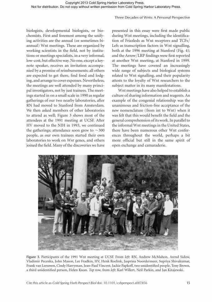
biologists, developmental biologists, or bio-chemists. First and foremost among the unify-ing activities are the annual (or sometimes bi-annual) Wnt meetings. These are organized byworking scientists in the field, not by institu-tions or meetings specialists, in a very informal,low-cost, but effective way. No one, except a key-note speaker, receives an invitation accompa-nied by a promise of reimbursements; all othersare expected to get there, find food and lodg-ing, and arrange to cover expenses. Nevertheless,the meetings are well attended by many princi-pal investigators, not by just trainees. The meet-ings started in on a small scale in 1990 as regulargatherings of our two nearby laboratories, afterRN had moved to Stanford from Amsterdam.We then asked members of other laboratoriesto attend as well; Figure 3 shows most of theattendees at the 1991 meeting at UCSF. AfterHV moved to the NIH in 1993, we continuedthe gatherings; attendance soon grew to �300people, as our own trainees started their ownlaboratories to work on Wnt genes, and othersjoined the field. Many of the discoveries we have
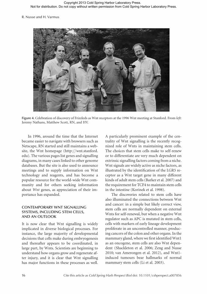
presented in this essay were first made publicduring Wnt meetings, including the identifica-tion of Frizzleds as Wnt receptors and TCFs/Lefs as transcription factors in Wnt signalling,both at the 1996 meeting at Stanford (Fig. 4);and the Arrow/LRP findings were first reportedat another Wnt meeting, at Stanford in 1999.The meetings have covered an increasinglywide range of subjects and biological systemsrelated to Wnt signalling, and their popularityattests to the loyalty of Wnt researchers to thesubject matter in its many manifestations.
Wnt meetings have also helped to establish aculture of sharing information and reagents. Anexample of the congenial relationship was theunanimous and friction-free acceptance of thenew nomenclature (from int to Wnt) when itwas felt that this would benefit the field and thegeneral comprehension of its work. In parallel tothe informal Wnt meetings in the United States,there have been numerous other Wnt confer-ences throughout the world, perhaps a bitmore official but still in the same spirit ofopen exchange and camaraderie.
Figure 3. Participants of the 1991 Wnt meeting at UCSF. From left: RN, Andrew McMahon, Arend Sidow,Vladimir Pecenka, John Mason, Lee Fradkin, HV, Henk Roelink, Jasprina Noordermeer, Supriya Shivakumar,Frank van Leeuwen, Cindy Harryman, Jean-Paul Vincent, Jackie Papkoff, two unidentified people, Tony Brown,a third unidentified person, Helen Kwan. Top row, from left: Karl Willert, Neil Parkin, and Jan Kitajewski.
Three Decades of Wnts: A Personal Perspective
Cite this article as Cold Spring Harb Perspect Biol doi: 10.1101/cshperspect.a007856 15
Copyright 2013 Cold Spring Harbor Laboratory Press. Not for distribution. Do not copy without written permission from Cold Spring Harbor Laboratory Press.
In 1996, around the time that the Internetbecame easier to navigate with browsers such asNetscape, RN started and still maintains a web-site, the Wnt homepage (http://wnt.stanford.edu). The various pages list genes and signallingdiagrams, in many cases linked to other genomedatabases. But the site is also used to announcemeetings and to supply information on Wnttechnology and reagents, and has become apopular resource for the world-wide Wnt com-munity and for others seeking informationabout Wnt genes, as appreciation of their im-portance has expanded.
CONTEMPORARY WNT SIGNALLINGSYSTEMS, INCLUDING STEM CELLS,AND AN OUTLOOK
It is now clear that Wnt signalling is widelyimplicated in diverse biological processes. Forinstance, the large majority of developmentaldecisions that cells make during embryogenesisand thereafter appears to be coordinated, inlarge part, by Wnts. Scientists are beginning tounderstand how organs grow and regenerate af-ter injury, and it is clear that Wnt signallinghas major functions in these processes as well.
A particularly prominent example of the cen-trality of Wnt signalling is the recently recog-nized role of Wnts in maintaining stem cells.The choices that stem cells make to self-renewor to differentiate are very much dependent onextrinsic signalling factors coming from a niche.Wnt signals are widely active as niche factors, asillustrated by the identification of the LGR5 re-ceptor as a Wnt target gene in many differentkinds of adult stem cells (Barker et al. 2007) andthe requirement for TCF4 to maintain stem cellsin the intestine (Korinek et al. 1998).
The discoveries related to stem cells havealso illuminated the connections between Wntand cancer: in a simple but likely correct view,stem cells are normally dependent on externalWnts for self-renewal, but when a negative Wntregulator such as APC is mutated in stem cells,cells with markers of early lineage developmentproliferate in an uncontrolled manner, produc-ing cancers of the colon and other organs. In themammary gland, where we first identified Wnt1as an oncogene, stem cells are also Wnt depen-dent (Shackleton et al. 2006; Zeng and Nusse2010; van Amerongen et al. 2012), and Wnt1-induced tumours bear hallmarks of normalmammary stem cells (Li et al. 2003).
Figure 4. Celebration of discovery of Frizzleds as Wnt receptors at the 1996 Wnt meeting at Stanford. From left:Jeremy Nathans, Matthew Scott, RN, and HV.
R. Nusse and H. Varmus
16 Cite this article as Cold Spring Harb Perspect Biol doi: 10.1101/cshperspect.a007856
Copyright 2013 Cold Spring Harbor Laboratory Press. Not for distribution. Do not copy without written permission from Cold Spring Harbor Laboratory Press.
Wnt signalling mutations have also beenimplicated in a growing list of degenerative dis-eases. Important among these are bone densityabnormalities with dysfunctional LRP recep-tors (Gong et al. 2001) and retinal degenerationwith Frizzled mutations (Robitaille et al. 2002).Some metabolic disorders, including diabetesmellitus, have been associated with alterationsin Wnt pathway genes (Grant et al. 2006).
It has been gratifying to witness growth ofthe Wnt field, from the finding of a single cancergene in a mouse model to a rich system branch-ing out to influence so many aspects of metazo-an biology and human disease. While outsidersmay be intimidated by the current size of thefield and the biochemical complexities of Wntsignalling, we suggest that there are still manyfundamental aspects of Wnt-related biology tobe discovered, understood, and exploited. In-creasingly, structures of Wnt signalling com-ponents are being elucidated, often in com-plexes with their partners (Dann et al. 2001;Schwarz-Romond et al. 2007; Ahn et al. 2011;Chen et al. 2011; Cheng et al. 2011); but im-portant aspects of the pathway’s molecular ma-chinery and biochemical regulators remain in-completely defined.
Progress in the Wnt field is much more rap-id today than it was in the early history of thisfield, thanks to more sophisticated tools: Wnt-specific assays and materials and more generalmethods in structural biology, genetics, and cellbiology. For instance, recognition of the role ofWnt signalling in stem-cell regulation has al-ready led to the use of Wnt proteins or Wntagonists to expand stem cells in culture (Satoet al. 2010; Zeng and Nusse, 2010; ten Bergeet al. 2011). On the other hand, despite theevidence for widespread involvement of Wntsignaling in human carcinogenesis, the kindsof targeted cancer therapies that are now beingdeveloped against components of several othersignaling pathways have not yet been producedto interfere with the Wnt pathway. Among themost significant challenges in future research inthe Wnt field is the identification of effectiveand specific Wnt pathway inhibitors for use incancer and other diseases. We expect that fur-ther understanding of the intricacies and varie-
ties of Wnt signaling will help to achieve theseimportant goals.
ACKNOWLEDGMENTS
We thank Thomas Schwarz-Romond and Reneevan Amerongen for inviting us to write this es-say. We thank Elizabeth Matthews for com-ments on various drafts and for suggesting thename Wnt during the nomenclature discus-sions over 20 years ago.
CONFLICT OF INTEREST
The authors declare that they have no conflict ofinterest.
REFERENCES�Reference is also in this collection.
Aberle H, Bauer A, Stappert J, Kispert A, Kemler R. 1997.b-Catenin is a target for the ubiquitin-proteasome path-way. EMBO J 16: 3797–3804.
Ahn VE, Chu ML, Choi HJ, Tran D, Abo A, Weis WI. 2011.Structural basis of Wnt signaling inhibition by Dickkopfbinding to LRP5/6. Dev Cell 21: 862–873.
Bafico A, Liu G, Yaniv A, Gazit A, Aaronson SA. 2001. Novelmechanism of Wnt signalling inhibition mediated byDickkopf-1 interaction with LRP6/Arrow. Nat Cell Biol3: 683–686.
Baker NE. 1987. Molecular cloning of sequences from wing-less, a segment polarity gene in Drosophila: The spatialdistribution of a transcript in embryos. EMBO J 6:1765–1773.
Banziger C, Soldini D, Schutt C, Zipperlen P, Hausmann G,Basler K. 2006. Wntless, a conserved membrane proteindedicated to the secretion of Wnt proteins from signalingcells. Cell 125: 509–522.
Barker N, Huch M, Kujala P, van de Wetering M, SnippertHJ, van Es JH, Sato T, Stange DE, Begthel H, van de BornM, et al. 2010. Lgr5þve stem cells drive self-renewal inthe stomach and build long-lived gastric units in vitro.Stem Cell 6: 25–36.
Barker N, van Es JH, Kuipers J, Kujala P, van de Born M,Cozijnsen M, Haegebarth A, Korving J, Begthel H, PetersPJ, et al. 2007. Identification of stem cells in small intes-tine and colon by marker gene Lgr5. Nature 449:1003–1007.
Bartscherer K, Pelte N, Ingelfinger D, Boutros M. 2006.Secretion of Wnt ligands requires Evi, a conserved trans-membrane protein. Cell 125: 523–533.
Behrens J, Jerchow BA, Wurtele M, Grimm J, Asbrand C,Wirtz R, Kuhl M, Wedlich D, Birchmeier W. 1998. Func-tional interaction of an axin homolog, conductin, withb-catenin, APC, and GSK3b. Science 280: 596–599.
Three Decades of Wnts: A Personal Perspective
Cite this article as Cold Spring Harb Perspect Biol doi: 10.1101/cshperspect.a007856 17
Copyright 2013 Cold Spring Harbor Laboratory Press. Not for distribution. Do not copy without written permission from Cold Spring Harbor Laboratory Press.
Behrens J, von Kries JP, Kuhl M, Bruhn L, Wedlich D, Gros-schedl R, Birchmeier W. 1996. Functional interaction ofb-catenin with the transcription factor LEF-1. Nature382: 638–642.
Bentvelzen P, Daams JH, Hageman P, Calafat J. 1970. Genetictransmission of viruses that incite mammary tumor inmice. Proc Natl Acad Sci USA 67: 377–384.
Bhanot P, Brink M, Samos CH, Hsieh JC, Wang Y, Macke JP,Andrew D, Nathans J, Nusse R. 1996. A new member ofthe frizzled family from Drosophila functions as a Wing-less receptor. Nature 382: 225–230.
Bhanot P, Fish M, Jemison JA, Nusse R, Nathans J, CadiganKM. 1999. Frizzled and Dfrizzled-2 function as redun-dant receptors for Wingless during Drosophila embry-onic development. Development 126: 4175–4186.
Bhat KM. 1998. Frizzled and frizzled 2 play a partiallyredundant role in wingless signaling and have similar re-quirements to wingless in neurogenesis. Cell 95: 1027–1036.
Bishop JM. 1983. Cellular oncogenes and retroviruses. AnnuRev Biochem 52: 301–354.
Bishop JM, Varmus H. 1985. Functions and origins of ret-roviral transforming genes. In RNA tumor viruses (ed.Weis R, Teich N, Varmus H, Coffin JM), pp. 249–356.Cold Spring Harbor Laboratory Press, Cold SpringHarbor, NY.
Bittner J. 1936. Some possiule effects of nursing on themammary. gland tumor incidence in mice. Science 84:162.
Boveri T. 2008. Concerning the origin of malignant tumours.The Company of Biologists Limited and Cold SpringHarbor Laboratory Press, Cold Spring Harbor, NY.
Bridges C, Brehme K. 1944. The mutants of Drosophilamelanogaster. Carnegie Institute, Washington, DC.
Brown AM, Wildin RS, Prendergast TJ, Varmus HE. 1986. Aretrovirus vector expressing the putative mammary on-cogene int-1 causes partial transformation of a mamma-ry epithelial cell line. Cell 46: 1001–1009.
Brown P, Varmus H. 1989. Retroviruses. In Mobile DNA (ed.Berg D, Howe M). American Society for MicrobiologyPress, Washington, DC.
Brunner E, Peter O, Schweizer L, Basler K. 1997. Pangolinencodes a Lef-1 homologue that acts downstream of Ar-madillo to transduce the Wingless signal in Drosophila.Nature 385: 829–833.
Cavallo RA, Cox RT, Moline MM, Roose J, Polevoy GA,Clevers H, Peifer M, Bejsovec A. 1998. Drosophila Tcfand Groucho interact to repress Wingless signalling ac-tivity. Nature 395: 604–608.
Chen CM, Struhl G. 1999. Wingless transduction by theFrizzled and Frizzled2 proteins of Drosophila. Develop-ment 126: 5441–5452.
Chen S, Bubeck D, Macdonald BT, Liang WX, Mao JH,Malinauskas T, Llorca O, Aricescu AR, Siebold C, He X,et al. 2011. Structural and functional studies of LRP6ectodomain reveal a platform for Wnt signaling. DevCell 21: 848–861.
Cheng Z, Biechele T, Wei Z, Morrone S, Moon RT, Wang L,Xu W. 2011. Crystal structures of the extracellular domainof LRP6 and its complex with DKK1. Nat Struct Mol Biol18: 1204–1210.
Cohen JC, Shank PR, Morris VL, Cardiff R, Varmus HE.1979. Integration of the DNA of mouse mammary tumorvirus in virus-infected normal and neoplastic tissue of themouse. Cell 16: 333–345.
Cohen JC, Varmus HE. 1979. Endogenous mammary tu-mour virus DNA varies among wild mice and segregatesduring inbreeding. Nature 278: 418–423.
Copeland NG, Jenkins NA. 2010. Harnessing transposonsfor cancer. gene discovery. Nat Rev Cancer 10: 696–706.
Daniels DL, Weis WI. 2005. b-Catenin directly displacesGroucho/TLE repressors from Tcf/Lef in Wnt-mediatedtranscription activation. Nat Struct Mol Biol 12: 364–371.
Dann CE, Hsieh JC, Rattner A, Sharma D, Nathans J, LeahyDJ. 2001. Insights into Wnt binding and signalling fromthe structures of two Frizzled cysteine-rich domains. Na-ture 412: 86–90.
DasGupta R, Fuchs E. 1999. Multiple roles for activatedLEF/TCF transcription complexes during hair follicle de-velopment and differentiation. Development 126: 4557–4568.
Davidson G, Wu W, Shen J, Bilic J, Fenger U, Stannek P,Glinka A, Niehrs C. 2005. Casein kinase 1g couplesWnt receptor activation to cytoplasmic signal transduc-tion. Nature 438: 867–872.
Dent P, Campbell DG, Hubbard MJ, Cohen P. 1989. Multi-site phosphorylation of the glycogen-binding subunit ofprotein phosphatase-1G by cyclic AMP-dependent pro-tein kinase and glycogen synthase kinase-3. FEBS Lett248: 67–72.
Dickson C, Peters G. 1987. Potential oncogene product re-lated to growth factors [letter]. Nature 326: 833.
Dickson C, Smith R, Brookes S, Peters G. 1984. Tumorigen-esis by mouse mammary tumor virus: Proviral activationof a cellular gene in the common integration region int-2.Cell 37: 529–536.
Dominguez I, Itoh K, Sokol SY. 1995. Role of glycogen syn-thase kinase 3b as a negative regulator of dorsoventralaxis formation in Xenopus embryos. Proc Natl Acad SciUSA 92: 8498–8502.
Errede B, Cardillo TS, Sherman F, Dubois E, Deschamps J,Wiame JM. 1980. Mating signals control expression ofmutations resulting from insertion of a transposable re-petitive element adjacent to diverse yeast genes. Cell 22:427–436.
Finch PW, He X, Kelley MJ, Uren A, Schaudies RP, PopescuNC, Rudikoff S, Aaronson SA, Varmus HE, Rubin JS.1997. Purification and molecular cloning of a secreted,Frizzled-related antagonist of Wnt action. Proc Natl AcadSci USA 94: 6770–6775.
Fire A, Xu S, Montgomery MK, Kostas SA, Driver SE, MelloCC. 1998. Potent and specific genetic interference bydouble-stranded RNA in Caenorhabditis elegans. Nature391: 806–811.
Funayama N, Fagotto F, McCrea P, Gumbiner BM. 1995.Embryonic axis induction by the armadillo repeat do-main of b-Catenin: Evidence for intracellular signaling.J Cell Biol 128: 959–968.
Fung YK, Shackleford GM, Brown AM, Sanders GS, VarmusHE. 1985. Nucleotide sequence and expression in vitro of
R. Nusse and H. Varmus
18 Cite this article as Cold Spring Harb Perspect Biol doi: 10.1101/cshperspect.a007856
Copyright 2013 Cold Spring Harbor Laboratory Press. Not for distribution. Do not copy without written permission from Cold Spring Harbor Laboratory Press.
cDNA derived from mRNA of int-1, a provirally activatedmouse mammary oncogene. Mol Cell Biol 5: 3337–3344.
Gallahan D, Callahan R. 1987. Mammary tumorigenesis inferal mice-identification of a new int locus in mousemammary tumor virus (Czech II)-induced mammarytumors. J Virol 61: 66–74.
Gallahan D, Callahan R. 1997. The mouse mammary tumorassociated gene INT3 is a unique member of the NOTCHgene family (NOTCH4). Oncogene 14: 1883–1890.
Gavin BJ, McMahon JA, McMahon AP. 1990. Expression ofmultiple novel Wnt-1/int-1-related genes during fetaland adult mouse development. Genes Dev 4: 2319–2332.
Glinka A, Wu W, Delius H, Monaghan AP, Blumenstock C,Niehrs C. 1998. Dickkopf-1 is a member of a new familyof secreted proteins and functions in head induction.Nature 391: 357–362.
Gong Y, Slee RB, Fukai N, Rawadi G, Roman-Roman S,Reginato AM, Wang H, Cundy T, Glorieux FH, Lev D,et al. 2001. LDL receptor- related protein 5 (LRP5) affectsbone accrual and eye development. Cell 107: 513–523.
Graham TA, Weaver C, Mao F, Kimelman D, Xu W. 2000.Crystal structure of a b-catenin/Tcf complex. Cell 103:885–896.
Grant SF, Thorleifsson G, Reynisdottir I, Benediktsson R,Manolescu A, Sainz J, Helgason A, Stefansson H, Emils-son V, Helgadottir A, et al. 2006. Variant of transcriptionfactor 7-like 2 (TCF7L2) gene confers risk of type 2 dia-betes. Nat Genet 38: 320–323.
Groden J, Thliveris A, Samowitz W, Carlson M, Gelbert L,Albertsen H, Joslyn G, Stevens J, Spirio L, Robertson M,et al. 1991. Identification and characterization of thefamilial adenomatous polyposis coli gene. Cell 66:589–600.
Gubb D, Garcia-Bellido A. 1982. A genetic analysis of thedetermination of cuticular polarity during developmentin Drosophila melanogaster. J Embryol Exp Morphol 68:37–57.
Guder C, Pinho S, Nacak TG, Schmidt HA, Hobmayer B,Niehrs C, Holstein TW. 2006. An ancient Wnt-Dickkopfantagonism in Hydra. Development 133: 901–911.
Gurley KA, Elliott SA, Simakov O, Schmidt HA, HolsteinTW, Alvarado AS. 2010. Expression of secreted Wnt path-way components reveals unexpected complexity of theplanarian amputation response. Dev Biol 347: 24–39.
Hayward WS, Neel BG, Astrin SM. 1981. Activation of acellular onc gene by promoter insertion in ALV-inducedlymphoid leukosis. Nature 290: 475–480.
He X, Saint-Jeannet JP, Woodgett JR, Varmus HE, Dawid IB.1995. Glycogen synthase kinase-3 and dorsoventral pat-terning in Xenopus embryos. Nature 374: 617–622.
He TC, Sparks AB, Rago C, Hermeking H, Zawel L, da CostaLT, Morin PJ, Vogelstein B, Kinzler KW. 1998. Identifica-tion of c-MYC as a target of the APC pathway. Science281: 1509–1512.
Heasman J, Crawford A, Goldstone K, Garner-Hamrick P,Gumbiner B, McCrea P, Kintner C, Noro CY, Wylie C.1994. Overexpression of cadherins and underexpressionof b-catenin inhibit dorsal mesoderm induction in earlyXenopus embryos. Cell 79: 791–803.
Hofmann K. 2000. A superfamily of membrane-boundO-acyltransferases with implications for wnt signaling.Trends Biochem Sci 25: 111–112.
� Holstein TW. 2012. The evolution of the Wnt pathway. ColdSpring Harb Perspect Biol doi: 10.1101/cshperspect.a007922.
Hsieh JC, Kodjabachian L, Rebbert ML, Rattner A, Small-wood PM, Samos CH, Nusse R, Dawid IB, Nathans J.1999. A new secreted protein that binds to Wnt proteinsand inhibits their activities. Nature 398: 431–436.
Huber O, Korn R, McLaughlin J, Ohsugi M, Herrmann BG,Kemler R. 1996. Nuclear localization of b-catenin byinteraction with transcription factor LEF-1. Mech Dev59: 3–10.
Huber AH, Nelson WJ, Weis WI. 1997. Three-dimensionalstructure of the armadillo repeat region of b-catenin. Cell90: 871–882.
Ikeda S, Kishida S, Yamamoto H, Murai H, Koyama S, Ki-kuchi A. 1998. Axin, a negative regulator of the Wntsignaling pathway, forms a complex with GSK-3b andb-catenin and promotes GSK-3b-dependent phosphor-ylation of b-catenin. EMBO J 17: 1371–1384.
Ingham PW. 1988. The molecular genetics of embryonicpattern formation in Drosophila. Nature 335: 25–34.
Jiang J, Struhl G. 1998. Regulation of the Hedgehog andWingless signalling pathways by the F-box/WD40-repeatprotein Slimb. Nature 391: 493–496.
Kadowaki T, Wilder E, Klingensmith J, Zachary K, PerrimonN. 1996. The segment polarity gene porcupine encodes aputative multitransmembrane protein involved in Wing-less processing. Genes Dev 10: 3116–3128.
Kennerdell JR, Carthew RW. 1998. Use of dsRNA-mediatedgenetic interference to demonstrate that frizzled and friz-zled 2 act in the wingless pathway. Cell 95: 1017–1026.
Kimelman D, Christian JL, Moon RT. 1992. Synergistic prin-ciples of development: Overlapping patterning systems inXenopus mesoderm induction. Development 116: 1–9.
Kinzler KW, Nilbert MC, Su LK, Vogelstein B, Bryan TM,Levy DB, Smith KJ, Preisinger AC, Hedge P, McKechnieD, et al. 1991. Identification of FAP locus genes fromchromosome 5q21. Science 253: 661–665.
Kool J, Berns A. 2009. High-throughput insertional muta-genesis screens in mice to identify oncogenic networks.Nat Rev Cancer 9: 389–399.
Korinek V, Barker N, Morin PJ, van Wichen D, de Weger R,Kinzler KW, Vogelstein B, Clevers H. 1997. Constitutivetranscriptional activation by a b-catenin-Tcf complex inAPC2/2 colon carcinoma. Science 275: 1784–1787.
Korinek V, Barker N, Moerer P, van Donselaar E, Huls G,Peters PJ, Clevers H. 1998. Depletion of epithelial stem-cell compartments in the small intestine of mice lackingTcf-4. Nat Genet 19: 379–383.
Korteweg R. 1936. On the manner in which the dispositionto carcinoma of the mammary gland is inherited in mice.Genetics 18: 350–371.
Kusserow A, Pang K, Sturm C, Hrouda M, Lentfer J, SchmidtHA, Technau U, von Haeseler A, Hobmayer B, Martin-dale MQ, et al. 2005. Unexpected complexity of the Wntgene family in a sea anemone. Nature 433: 156–160.
Lane P. 1967. Mouse Newsletter 36: 40.
Three Decades of Wnts: A Personal Perspective
Cite this article as Cold Spring Harb Perspect Biol doi: 10.1101/cshperspect.a007856 19
Copyright 2013 Cold Spring Harbor Laboratory Press. Not for distribution. Do not copy without written permission from Cold Spring Harbor Laboratory Press.
Lee FS, Lane TF, Kuo A, Shackleford GM, Leder P. 1995.Insertional mutagenesis identifies a member of the Wntgene family as a candidate oncogene in the mammaryepithelium of int-2/Fgf-3 transgenic mice. Proc NatlAcad Sci USA 92: 2268–2272.
Lee E, Salic A, Kruger R, Heinrich R, Kirschner MW. 2003.The roles of APC and Axin derived from experimentaland theoretical analysis of the Wnt pathway. PLoS Biol1: E10.
Leyns L, Bouwmeester T, Kim SH, Piccolo S, De RobertisEM. 1997. Frzb-1 is a secreted antagonist of Wnt signal-ing expressed in the Spemann organizer. Cell 88: 747–756.
Li Y, Welm B, Podsypanina K, Huang S, Chamorro M, ZhangX, Rowlands T, Egeblad M, Cowin P, Werb Z, et al. 2003.Evidence that transgenes encoding components of theWnt signaling pathway preferentially induce mammarycancers from progenitor cells. Proc Natl Acad Sci USA100: 15853–15858.
Li V, Ng SS, Boersema P, Low T, Karthaus W, Gerlach J,Mohammed S, Hweck A, Maurice M, Mahmoudi T, etal. 2012. Wnt signaling inhibits proteasomal b-catenindegradation within a compositionally intact Axin1 com-plex. Cell 149: 1245–1256.
Lin R, Thompson S, Priess JR. 1995. pop-1 encodes anHMG box protein required for the specification of a me-soderm precursor in early C. elegans embryos. Cell 83:599–609.
Lustig B, Jerchow B, Sachs M, Weiler S, Pietsch T, Karsten U,van de Wetering M, Clevers H, Schlag PM, Birchmeier W,et al. 2002. Negative feedback loop of Wnt signalingthrough upregulation of conductin/axin2 in colorectaland liver tumors. Mol Cell Biol 22: 1184–1193.
Lyons MJ, Moore DH. 1962. Purification of the mousemammary tumour virus. Nature 194: 1141–1142.
MacArthur CA, Shankar DB, Shackleford GM. 1995. Fgf-8,activated by proviral insertion, cooperates with the Wnt-1 transgene in murine mammary tumorigenesis. J Virol69: 2501–2507.
Macdonald BT, Tamai K, He X. 2009. Wnt/b-catenin sig-naling: Components, mechanisms, and diseases. Dev Cell17: 9–26.
Maniatis T. 1999. A ubiquitin ligase complex essential for theNF-kB, Wnt/Wingless, and Hedgehog signaling path-ways. Genes Dev 13: 505–510.
Mao B, Wu W, Li Y, Hoppe D, Stannek P, Glinka A, Niehrs C.2001a. LDL-receptor-related protein 6 is a receptor forDickkopf proteins. Nature 411: 321–325.
Mao J, Wang J, Liu B, Pan W, Farr GH III, Flynn C, Yuan H,Takada S, Kimelman D, Li L, et al. 2001b. Low-densitylipoprotein receptor-related protein-5 binds to Axin andregulates the canonical Wnt signaling pathway. Mol Cell7: 801–809.
McCrea PD, Turck CW, Gumbiner B. 1991. A homolog ofthe armadillo protein in Drosophila (plakoglobin) asso-ciated with E-cadherin. Science 254: 1359–1361.
McCrea PD, Brieher WM, Gumbiner BM. 1993. Inductionof a secondary body axis in Xenopus by antibodies tob-catenin. J Cell Biol 123: 477–484.
McMahon AP, Bradley A. 1990. The Wnt-1 (int-1) proto-oncogene is required for development of a large region ofthe mouse brain. Cell 62: 1073–1085.
McMahon AP, Moon RT. 1989. Ectopic expression of theprotooncogene int-1 in Xenopus embryos leads to dupli-cation of the embryonic axis. Cell 58: 1075–1084.
Mikels AJ, Nusse R. 2006. Purified Wnt5a protein activatesor inhibits b-catenin-TCF signaling depending on recep-tor context. PLoS Biol 4: e115.
Molenaar M, van de Wetering M, Oosterwegel M, Peterson-Maduro J, Godsave S, Korinek V, Roose J, Destree O,Clevers H. 1996. XTcf-3 transcription factor mediatesb-catenin-induced axis formation in Xenopus embryos.Cell 86: 391–399.
Morin PJ, Sparks AB, Korinek V, Barker N, Clevers H, Vo-gelstein B, Kinzler KW. 1997. Activation of b-catenin-Tcfsignaling in colon cancer by mutations in b-catenin orAPC. Science 275: 1787–1790.
Muller HA, Samanta R, Wieschaus E. 1999. Wingless signal-ing in the Drosophila embryo: Zygotic requirements andthe role of the frizzled genes. Development 126: 577–586.
Nelson WJ, Nusse R. 2004. Convergence of Wnt, b-catenin,and cadherin pathways. Science 303: 1483–1487.
Niehrs C. 2010. On growth and form: A Cartesian coordi-nate system of Wnt and BMP signaling specifies bilater-ian body axes. Development (Cambridge, England) 137:845–857.
Noordermeer J, Klingensmith J, Perrimon N, Nusse R. 1994.dishevelled and armadillo act in the wingless signallingpathway in Drosophila. Nature 367: 80–83.
Nusse R, Varmus HE. 1992. Wnt genes. Cell 69: 1073–1087.
Nusse R, Varmus HE. 1982. Many tumors induced by themouse mammary tumor virus contain a provirus inte-grated in the same region of the host genome. Cell 31:99–109.
Nusse R, van Ooyen A, Cox D, Fung YK, Varmus HE. 1984.Mode of proviral activation of a putative mammary on-cogene (int-1) on mouse chromosome 15. Nature 307:131–136.
Nusse R, Brown A, Papkoff J, Scambler P, Shackleford G,McMahon A, Moon R, Varmus H. 1991. A new nomen-clature for int-1 and related genes: The Wnt gene family.Cell 64: 231.
Nusslein-Volhard C, Wieschaus E. 1980. Mutations affectingsegment number and polarity in Drosophila. Nature 287:795–801.
Oishi I, Suzuki H, Onishi N, Takada R, Kani S, Ohkawara B,Koshida I, Suzuki K, Yamada G, Schwabe GC, et al. 2003.The receptor tyrosine kinase Ror2 is involved in non-canonical Wnt5a/JNK signalling pathway. Genes Cells8: 645–654.
Orford K, Crockett C, Jensen JP, Weissman AM, Byers SW.1997. Serine phosphorylation-regulated ubiquitinationand degradation of b-catenin. J Biol Chem 272:24735–24738.
Ozawa M, Baribault H, Kemler R. 1989. The cytoplasmicdomain of the cell adhesion molecule uvomorulin asso-ciates with three independent proteins structurally relat-ed in different species. EMBO J 8: 1711–1717.
R. Nusse and H. Varmus
20 Cite this article as Cold Spring Harb Perspect Biol doi: 10.1101/cshperspect.a007856
Copyright 2013 Cold Spring Harbor Laboratory Press. Not for distribution. Do not copy without written permission from Cold Spring Harbor Laboratory Press.
Parr BA, McMahon AP. 1995. Dorsalizing signal Wnt-7arequired for normal polarity of D-V and A-P axes ofmouse limb. Nature 374: 350–353.
Parr BA, McMahon AP. 1998. Sexually dimorphic develop-ment of the mammalian reproductive tract requires Wnt-7a. Nature 395: 707–710.
Patthy L. 2000. The WIF module. Trends Biochem Sci 25:12–13.
Payne GS, Courtneidge SA, Crittenden LB, Fadly AM, Bish-op JM, Varmus HE. 1981. Analysis of avian leukosis virusDNA and RNA in bursal tumours: Viral gene expressionis not required for maintenance of the tumor state. Cell23: 311–322.
Payne GS, Bishop JM, Varmus HE. 1982. Multiple arrange-ments of viral DNA and an activated host oncogene inbursal lymphomas. Nature 295: 209–214.
Peifer M, Orsulic S, Pai LM, Loureiro J. 1993. A modelsystem for cell adhesion and signal transduction in Dro-sophila. Development (Suppl 1): 163–176.
Peifer M, Wieschaus E. 1990. The segment polarity genearmadillo encodes a functionally modular protein thatis the Drosophila homolog of human plakoglobin. Cell 63:1167–1176.
Peifer M, Rauskolb C, Williams M, Riggleman B, WieschausE. 1991. The segment polarity gene armadillo interactswith the wingless signaling pathway in both embryonicand adult pattern formation. Development 111: 1029–1043.
Peifer M, Pai LM, Casey M. 1994. Phosphorylation of theDrosophila adherens junction protein Armadillo: Rolesfor wingless signal and zeste-white 3 kinase. Dev Biol166: 543–556.
Perrimon N, Mahowald AP. 1987. Multiple functions of seg-ment polarity genes in Drosophila. Dev Biol 119: 587–600.
Peters G, Lee AE, Dickson C. 1986. Concerted activation oftwo potential proto-oncogenes in carcinomas induced bymouse mammary tumour virus. Nature 320: 628–631.
Petersen CP, Reddien PW. 2009. Wnt signaling and the po-larity of the primary body axis. Cell 139: 1056–1068.
Pierce SB, Kimelman D. 1996. Overexpression of Xgsk-3disrupts anterior ectodermal patterning in Xenopus.Dev Biol 175: 256–264.
Pinson KI, Brennan J, Monkley S, Avery BJ, Skarnes WC.2000. An LDL-receptor-related protein mediates Wntsignalling in mice. Nature 407: 535–538.
Port F, Basler K. 2010. Wnt trafficking: New insights into Wntmaturation, secretion and spreading. Traffic, Copenhagen,Denmark.
Rattner A, Hsieh JC, Smallwood PM, Gilbert DJ, CopelandNG, Jenkins NA, Nathans J. 1997. A family of secretedproteins contains homology to the cysteine-rich ligand-binding domain of frizzled receptors. Proc Natl Acad SciUSA 94: 2859–2863.
Riese J, Yu X, Munnerlyn A, Eresh S, Hsu SC, Grosschedl R,Bienz M. 1997. LEF-1, a nuclear factor coordinating sig-naling inputs from wingless and decapentaplegic. Cell 88:777–787.
Riggleman B, Schedl P, Wieschaus E. 1990. Spatial expres-sion of the Drosophila segment polarity gene armadillo is
posttranscriptionally regulated by wingless. Cell 63:549–560.
Rijsewijk F, Schuermann M, Wagenaar E, Parren P, Weigel D,Nusse R. 1987a. The Drosophila homolog of the mousemammary oncogene int-1 is identical to the segmentpolarity gene wingless. Cell 50: 649–657.
Rijsewijk F, van Deemter L, Wagenaar E, Sonnenberg A,Nusse R. 1987b. Transfection of the int-1 mammary on-cogene in cuboidal RAC mammary cell line resultsin morphological transformation and tumorigenicity.EMBO J 6: 127–131.
Robitaille J, MacDonald ML, Kaykas A, Sheldahl LC, ZeislerJ, Dube MP, Zhang LH, Singaraja RR, Guernsey DL,Zheng B, et al. 2002. Mutant frizzled-4 disrupts retinalangiogenesis in familial exudative vitreoretinopathy. NatGenet 32: 326–330.
Rocheleau CE, Downs WD, Lin R, Wittmann C, Bei Y, ChaYH, Ali M, Priess JR, Mello CC. 1997. Wnt signaling andan APC-related gene specify endoderm in early C. elegansembryos. Cell 90: 707–716.
Rocheleau CE, Yasuda J, Shin TH, Lin R, Sawa H, Okano H,Priess JR, Davis RJ, Mello CC. 1999. WRM-1 activates theLIT-1 protein kinase to transduce anterior/posterior po-larity signals in C. elegans. Cell 97: 717–726.
Roelink H, Wagenaar E, Lopes da Silva S, Nusse R. 1990.Wnt-3, a gene activated by proviral insertion in mousemammary tumors, is homologous to int-1/Wnt-1 and isnormally expressed in mouse embryos and adult brain.Proc Natl Acad Sci USA 87: 4519–4523.
Rubinfeld B, Souza B, Albert I, Muller O, Chamberlain SH,Masiarz FR, Munemitsu S, Polakis P. 1993. Associationof the APC gene product with b-catenin. Science(New York, NY) 262: 1731–1734.
Rubinfeld B, Albert I, Porfiri E, Fiol C, Munemitsu S, PolakisP. 1996. Binding of GSK3b to the APC-b-catenin com-plex and regulation of complex assembly. Science(New York, NY) 272: 1023–1026.
Rubinfeld B, Robbins P, El-Gamil M, Albert I, Porfiri E,Polakis P. 1997. Stabilization of b-catenin by genetic de-fects in melanoma cell lines. Science 275: 1790–1792.
Rulifson EJ, Wu CH, Nusse R. 2000. Pathway specificity bythe bifunctional receptor frizzled is determined by affin-ity for wingless. Mol Cell 6: 117–126.
Sato T, van Es JH, Snippert HJ, Stange DE, Vries RG, van denBorn M, Barker N, Shroyer NF, van de Wetering M, Clev-ers H. 2010. Paneth cells constitute the niche for Lgr5stem cells in intestinal crypts. Nature 469: 415–418.
Satoh S, Daigo Y, Furukawa Y, Kato T, Miwa N, Nishiwaki T,Kawasoe T, Ishiguro H, Fujita M, Tokino T, et al. 2000.AXIN1 mutations in hepatocellular carcinomas, andgrowth suppression in cancer cells by virus-mediatedtransfer of AXIN1. Nat Genet 24: 245–250.
Sawa H, Lobel L, Horvitz HR. 1996. The Caenorhabditiselegans gene lin-17, which is required for certain asym-metric cell divisions, encodes a putative seven-trans-membrane protein similar to the Drosophila frizzled pro-tein. Genes Dev 10: 2189–2197.
Schneider SQ, Bowerman B. 2007. b-Catenin asymmetriesafter all animal/vegetal-oriented cell divisions in Platy-nereis dumerilii embryos mediate binary cell-fate speci-fication. Dev Cell 13: 73–86.
Three Decades of Wnts: A Personal Perspective
Cite this article as Cold Spring Harb Perspect Biol doi: 10.1101/cshperspect.a007856 21
Copyright 2013 Cold Spring Harbor Laboratory Press. Not for distribution. Do not copy without written permission from Cold Spring Harbor Laboratory Press.
Schwarz-Romond T, Fiedler M, Shibata N, Butler PJ, Kiku-chi A, Higuchi Y, Bienz M. 2007. The DIX domain ofDishevelled confers Wnt signaling by dynamic polymer-ization. Nat Struct Mol Biol 14: 484–492.
Semenov MV, Tamai K, Brott BK, Kuhl M, Sokol S, He X.2001. Head inducer Dickkopf-1 is a ligand for Wnt co-receptor LRP6. Curr Biol 11: 951–961.
Shackleford GM, Varmus HE. 1987. Expression of theproto-oncogene int-1 is restricted to postmeiotic malegerm cells and the neural tube of mid-gestational embry-os. Cell 50: 89–95.
Shackleton M, Vaillant F, Simpson KJ, Stingl J, Smyth GK,Asselin- Labat ML, Wu L, Lindeman GJ, Visvader JE.2006. Generation of a functional mammary gland froma single stem cell. Nature 439: 84–88.
Sharma RP, Chopra VL. 1976. Effect of the Wingless (wg1)mutation on wing and haltere development in Drosophilamelanogaster. Dev Biol 48: 461–465.
Sidow A. 1992. Diversification of the Wnt gene family on theancestral lineage of vertebrates. Proc Natl Acad Sci USA89: 5098–5102.
Siegfried E, Chou TB, Perrimon N. 1992. Wingless signalingacts through zeste-white 3, the Drosophila homolog ofglycogen synthase kinase-3, to regulate engrailed and es-tablish cell fate. Cell 71: 1167–1179.
Siegfried E, Wilder EL, Perrimon N. 1994. Components ofwingless signalling in Drosophila. Nature 367: 76–80.
Su LK, Kinzler KW, Vogelstein B, Preisinger AC, Moser AR,Luongo C, Gould KA, Dove WF. 1992. Multiple intestinalneoplasia caused by a mutation in the murine homologof the APC gene. Science 256: 668–670.
Su LK, Vogelstein B, Kinzler KW. 1993. Association of theAPC tumor suppressor protein with catenins. Science262: 1734–1737.
Sugioka K, Mizumoto K, Sawa H. 2011. Wnt regulates spin-dle asymmetry to generate asymmetric nuclear b-cateninin C. elegans. Cell 146: 942–954.
Takeichi M. 1990. Cadherins: A molecular family importantin selective cell-cell adhesion. Annu Rev Biochem 59:237–252.
Tamai K, Semenov M, Kato Y, Spokony R, Liu C, KatsuyamaY, Hess F, Saint-Jeannet JP, He X. 2000. LDL-receptor-related proteins in Wnt signal transduction. Nature407: 530–535.
Tamai K, Zeng X, Liu C, Zhang X, Harada Y, Chang Z, He X.2004. A mechanism for Wnt coreceptor activation. MolCell 13: 149–156.
Teich N, Wyke J, Mak T, Bernstein A, Hardy W. 1982. Path-ogenesis of Retrovirus-indiced disease. In RNA tumorviruses (ed. Weis R, Teich N, Varmus H, Coffin JM), pp.785–998. Cold Spring Harbor Laboratory Press, ColdSpring Harbor, NY.
ten Berge D, Kurek D, Blauwkamp T, Koole W, Maas A,Eroglu E, Siu RK, Nusse R. 2011. Embryonic stem cellsrequire Wnt proteins to prevent differentiation to epi-blast stem cells. Nat Cell Biol 13: 1070–1075.
Thomas KR, Capecchi MR. 1990. Targeted disruption of themurine int-1 proto-oncogene resulting in severe abnor-malities in mid-brain and cerebellar development. Nature346: 847–850.
Thomas KR, Musci TS, Neumann PE, Capecchi MR. 1991.Swaying is a mutant allele of the proto-oncogene Wnt-1.Cell 67: 969–976.
Thorpe CJ, Schlesinger A, Carter JC, Bowerman B. 1997.Wnt signaling polarizes an early C. elegans blastomereto distinguish endoderm from mesoderm. Cell 90:695–705.
Travis A, Amsterdam A, Belanger C, Grosschedl R. 1991.LEF-1, a gene encoding a lymphoid-specific proteinwith an HMG domain, regulates T-cell receptor a en-hancer function [corrected]. Genes Dev 5: 880–894.
Tsukamoto AS, Grosschedl R, Guzman RC, Parslow T, Var-mus HE. 1988. Expression of the int-1 gene in transgenicmice is associated with mammary gland hyperplasia andadenocarcinomas in male and female mice. Cell 55:619–625.
van Amerongen R, Mikels A, Nusse R. 2008. Alternativewnt signaling is initiated by distinct receptors. Sci Signal1: re9.
van Amerongen R, Bowman A, Nusse R. 2012. Develop-mental stage and time dictate the fate of Wnt/b-cateninresponsive stem cells in the mammary gland. Cell StemCell (in press).
van de Wetering M, Oosterwegel M, Dooijes D, Clevers H.1991. Identification and cloning of TCF-1, a T lympho-cyte-specific transcription factor containing a sequence-specific HMG box. EMBO J 10: 123–132.
Van Leeuwen F, Harryman Samos C, Nusse R. 1994. Biolog-ical activity of soluble wingless protein in cultured Dro-sophila imaginal disc cells. Nature 368: 342–344.
van Ooyen A, Nusse R. 1984. Structure and nucleotide se-quence of the putative mammary oncogene int-1; provi-ral insertions leave I the protein-encoding domain intact.Cell 39: 233–240.
van Ooyen A, Kwee V, Nusse R. 1985. The nucleotide se-quence of the human int-1 mammary oncogene; evolu-tionary conservation of coding and non-coding se-quences. EMBO J 4: 2905–2909.
Veeman MT, Axelrod JD, Moon RT. 2003. A second canon.Functions and mechanisms of b-catenin-independentWnt signaling. Dev Cell 5: 367–377.
Vinson CR, Adler PN. 1987. Directional non-cell autonomyand the transmission of polarity information by the friz-zled gene of Drosophila. Nature 329: 549–551.
Vinson CR, Conover S, Adler PN. 1989. A Drosophila tissuepolarity locus encodes a protein containing seven poten-tial transmembrane domains. Nature 338: 263–264.
Waterman ML, Fischer WH, Jones KA. 1991. A thymus-specific member of the HMG protein family regulatesthe human T cell receptor C a enhancer. Genes Dev 5:656–669.
Wehrli M, Dougan ST, Caldwell K, O’Keefe L, Schwartz S,Vaizel-Ohayon D, Schejter E, Tomlinson A, DiNardo S.2000. arrow encodes an LDL-receptor-related protein es-sential for Wingless signalling. Nature 407: 527–530.
Weinberg RA. 1983. A molecular basis of cancer. Sci Am 249:126–142.
Wilkinson DG, Bailes JA, McMahon AP. 1987. Expression ofthe proto-oncogene int-1 is restricted to specific neuralcells in the developing mouse embryo. Cell 50: 79–88.
R. Nusse and H. Varmus
22 Cite this article as Cold Spring Harb Perspect Biol doi: 10.1101/cshperspect.a007856
Copyright 2013 Cold Spring Harbor Laboratory Press. Not for distribution. Do not copy without written permission from Cold Spring Harbor Laboratory Press.
Willert K, Brown JD, Danenberg E, Duncan AW, WeissmanIL, Reya T, Yates JR, Nusse R. 2003. Wnt proteins arelipid-modified and can act as stem cell growth factors.Nature 423: 448–452.
Yang-Snyder J, Miller JR, Brown JD, Lai CJ, Moon RT. 1996.A frizzled homolog functions in a vertebrate Wnt signal-ing pathway. CurrBiol 6: 1302–1306.
Ye X, Wang Y, Rattner A, Nathans J. 2011. Genetic mosaicanalysis reveals a major role for frizzled 4 and frizzled 8 incontrolling ureteric growth in the developing kidney. De-velopment 138: 1161–1172.
Yoshikawa S, McKinnon RD, Kokel M, Thomas JB. 2003.Wnt-mediated axon guidance via the Drosophila De-railed receptor. Nature 422: 583–588.
Yost C, Torres M, Miller JR, Huang E, Kimelman D, MoonRT. 1996. The axis-inducing activity, stability, and sub-
cellular distribution of b-catenin is regulated in Xenopusembryos by glycogen synthase kinase 3. Genes Dev 10:1443–1454.
Zeng YA, Nusse R. 2010. Wnt proteins are self-renewal fac-tors for mammary stem cells and promote their long-term expansion in culture. Stem Cell 6: 568–577.
Zeng L, Fagotto F, Zhang T, Hsu W, Vasicek TJ, Perry WL III,Lee JJ, Tilghman SM, Gumbiner BM, Costantini F. 1997.The mouse Fused locus encodes Axin, an inhibitor of theWnt signaling pathway that regulates embryonic axis for-mation. Cell 90: 181–192.
Zeng X, Tamai K, Doble B, Li S, Huang H, Habas R, Oka-mura H, Woodgett J, He X. 2005. A dual-kinase mecha-nism for Wnt co-receptor phosphorylation and activa-tion. Nature 438: 873–877.
Three Decades of Wnts: A Personal Perspective
Cite this article as Cold Spring Harb Perspect Biol doi: 10.1101/cshperspect.a007856 23
Copyright 2013 Cold Spring Harbor Laboratory Press. Not for distribution. Do not copy without written permission from Cold Spring Harbor Laboratory Press.

Copyright 2013 Cold Spring Harbor Laboratory Press. Not for distribution. Do not copy without written permission from Cold Spring Harbor Laboratory Press.
Index
AAardvark, 147
AD. See Alzheimer’s diseaseAdenomatous polyposis coli (APC), 9–11, 16
axon terminal remodeling, 287b-catenin
binding sites, 94
destruction role, 93–98mutation in cancer, 254, 395–396
Adenomatous polyposis coli down-regulated-1(APCDD1), Wnt signaling inhibition, 51
ALV. See Avian leukosis virus
Alzheimer’s disease (AD)LRP6 Alzheimer’s disease variants, 293Wnt signaling and therapeutic targeting, 426, 430
Androgen receptor (AR)b-catenin binding, 118
Wnt cross-talk in cancer, 400AP-1, b-catenin-independent Wnt signaling, 136APC. See Adenomatous polyposis coliAPCDD1. See Adenomatous polyposis coli
down-regulated-1AR. See Androgen receptorArmadillo, 10–11Arrow
history of study, 12
homologs. See Lipoprotein receptor-related proteinmutants, 69structure, 70
Avian leukosis virus (ALV), history of study, 3Axin, 11, 14, 34, 70
b-catenindestruction complex, 92–93phosphorylation inhibition after receptor
activation, 79–82domains, 92–93
recruitment, 75therapeutic targeting, 435tumor suppressor genes, 396Wnt signaling overview, 74
Axis specificationanteroposterior axis specification
Dkk1 and patterning, 42–43, 45, 201–202, 221mouse, 220–221zygotic Wnt signaling, 201–202
cross-talk between Wnt and other pathways, 205–207dorsoventral axis specification, 199–201, 203
pathway switch mechanisms, 204–205prospects for study, 207TCF function regulation, 203–204
transcriptional targets of Wnt pathway, 202–203Wnt signaling overview, 197–199
Axon. See Central nervous system
BBAR-1, 184–186Basal cell carcinoma (BCC), Wnt signaling, 338BCC. See Basal cell carcinoma
B cell, Wnt signaling in development, 314–315b-Catenin
APC interactions, 9–10Caenorhabditis elegans
BAR-1 pathway, 186
b-catenin-independent signaling, 186destruction complex, 184types, 184–186Wnt/b-catenin asymmetry pathway, 188–189
canonical Wnt signalingoutside of TCF/LEF, 118–119overview, 65–66
destruction complexAPC role, 93–94
axin as phosphorylation scaffold, 92b-catenin phosphorylation, 94–95cycling, 95–96kinases, 91localization, 98–99
overview, 89–90proteasomal degradation, 90–91, 96–97sequestration of b-catenin, 97–98tumor suppressor genes, 396–397
embryonic stem cell
Oct4–b-catenin interaction, 240–241stability role in glycogen synthase kinase-3
inhibition, 239–240Tcf-dependent mechanisms and Wnt-stabilized
b-catenin, 242–246
histone modification regulation, 112–114phosphorylation inhibition after receptor
activation, 79–82signaling cascade from cell surface to nucleus, 10–11
TCF/LEF binding domain, 106–107transcriptional targets in axis specification, 202–203
447
Copyright 2013 Cold Spring Harbor Laboratory Press. Not for distribution. Do not copy without written permission from Cold Spring Harbor Laboratory Press.
Biglycan, 32BMP. See Bone morphogenetic proteinsBone
anatomy, 267
developmentDickkopf role, 45–46overview, 265–267Wnt/b-catenin signaling in cartilage and
joint formation, 271–272
lithium effects, 436Wnt/b-catenin signaling
cancer and metastasis, 276–277fracture healing, 275–276human bone disease, 268–270
osteoblastdifferentiation and maturation, 270–271function, 272–274
osteocyte, 274–275
regenerative medicine, 432Bone morphogenetic proteins (BMPs), axis
specification and Wnt cross-talk,205–207
Brachyury, 148
Brain. See Central nervous systemBreast cancer. See also Mammary gland
growth and Wnt signalingE-cadherin loss, 354–355nuclear b-catenin, 354
overview, 352–354Wnt-dependent subpopulations, 354
human cell studies of Wnt signaling, 402–403
CCaenorhabditis elegans, Wnt signaling
b-catenin-independent signaling, 186
components of canonical signalingb-catenins, 184–186BAR-1 pathway, 186destruction complex, 184Dishevelled, 184
Frizzled, 183–184table, 180TCF/LEF, 185–186Wnt ligands and inhibitors, 181–183
conserved metazoan components, 180–181
embryonic endoderm induction, P2/EMSsignaling, 189–191
neuromuscular junction development. SeeNeuromuscular junction
overview, 179–180prospects for study, 191–192QL neuroblast progeny migration regulation,
186–188Wnt/b-catenin asymmetry pathway, 188–189
E-Cadherin, loss in breast cancer, 354–355
Calcium/calmodulin-dependent kinase-II (CaMKII),synaptic function, 289–290
Calcium flux, b-catenin-independent Wntsignaling, 137
CaMKII. See Calcium/calmodulin-dependent kinase-IICancer. See also specific cancers
history of Wnt studies, 9–10oncogenes, 395–396tumor suppressor genes, 395–397
Wnt signalingchronic injury and prolonged Wnt signaling,
415–416cross-talk with other pathways, 399–401defects, 398, 424
epigenetic modulation, 397–399human cell studies, 401–404therapeutic targeting
non-canonical signaling, 425–426
overview, 424patient survival correlation with b-catenin,
424–425Cancer stem cell (CSC)
intestine, 258–259
stomach, 259Casein kinase-1 (CK1)
b-catenin destruction complex, 91phosphorylation of LRP6, 72, 75–78therapeutic targeting, 436
CBP. See CREB-binding proteinCdc42, b-catenin-independent Wnt signaling,
135–136CE. See Convergent extensionCentral nervous system (CNS), Wnt function
axon guidance, 284–285axon terminal remodeling, 287disease and deregulation, 293forebrain development
cell adhesion, 303–304expression patterns, 298–299lamination, 304overview, 297–298patterning, 299–302
prospects for study, 304–305radial glia regulation and intermediate
progenitor fate, 302–303ventral forebrain neurogenesis, 304
neuronal activity regulation of Wnt release and
receptor localization, 290–291prospects for study, 293–294synapse formation
excitatory versus inhibitory synapses, 289–290overview, 287
postsynaptic assembly, 289presynaptic assembly, 287–289
synapse plasticity, 291–293topographical map formation in brain, 285–287
Index
448
Copyright 2013 Cold Spring Harbor Laboratory Press. Not for distribution. Do not copy without written permission from Cold Spring Harbor Laboratory Press.
Cerberusknockout mice, 44overview, 49–50
CK1. see Casein kinase-1
Clozapine, Wnt signaling effects, 436CNS. See Central nervous systemConvergent extension (CE)
b-catenin-independent Wnt signaling, 129, 135–136core proteins, 135
effectors, 135–136CREB-binding protein (CBP), cancer mutations, 397Cripto, 221CSC. See Cancer stem cell
DDACT3, repression in colorectal cancer, 399Derailed, history of study, 13Destruction complex. See b-CateninDevelopment. See Axis specification; Bone; Central
nervous system; Mouse development;
Neuromuscular junction; SkinDickkopf (Dkk)
Alzheimer’s disease expression, 293cancer expression of DKK1, 276–277developmental function
anteroposterior patterning and Dkk1, 42–43, 45,201–202, 221
limb, bone, and eye formation, 45–46Dkk2 inhibition and activation of Wnt signaling
pathway, 42
injury repair therapeutic targeting of DKK1, 417knockout mice, 44mechanism of action, 40–42pathology, 46
structure, 40therapeutic targeting, 434, 437
DISC1, 293, 426Dishevelled (DVL)
b-catenin-independent Wnt signaling, 132–133
Caenorhabditis elegans, 184dorsoventral axis specification, 200Frizzled interactions, 74–75structure, 67therapeutic targeting, 434–435
Wnt signaling overview, 73–74DKK. See DickkopfDrosophila. See Neuromuscular junction; WinglessDVL. See Dishevelled
EEGFR. See Epidermal growth factor receptor
Embryonic stem cell (ESC)comparison of mouse and human, 234–235culture, 233–234
transcription factors in self-renewal, 235Wnt signaling
cell differentiation inhibition, 235–237glycogen synthase kinase-3 inhibition
b-catenin stability role, 239–240pluripotency state dependence, 238–239
Oct4–b-catenin interaction, 240–241prospects for study, 246self-renewal stimulation, 237–238
Tcf-dependent mechanismsTcf3 in self-renewal, 241–242Wnt-stabilized b-catenin, 242–246
Emx2, forebrain patterning, 301–302Ephrin, topographical map formation in brain, 286–287
Epidermal growth factor receptor (EGFR), 166, 400Epigenetics, modulation of Wnt signaling, 397–399ESC. See Embryonic stem cellEvi. See Wntless
Evolution, Wnt pathwaybilaterians
Deuterostomia, 153–154Ecdysozoa, 153gene distribution, 152
Lophotrochozoa, 151, 153gene synteny and evolution, 154Hydra organizer
local restriction, 157short- and long-range signaling, 155–157
metazoan conserved elements, 180–181overview, 145–146pre-bilaterians
Cnidaria, 150–151Ctenophora, 150
overview, 148Placozoa, 149–150Porifera, 148–149
pre-metazoans
ligands of Wnt, 146receptors and mediators, 146–148transcription factors and target genes, 148
FFamilial adenomatous polyposis (FAP)
gene mutations. See Adenomatous polyposis coli
Wnt pathwayactivating mutations, 254therapeutic targeting, 432–433
FAP. See Familial adenomatous polyposisFDH. See Focal dermal hypoplasia
FGF. See Fibroblast growth factorFibroblast growth factor (FGF)
axis specification and Wnt cross-talk, 206–207forebrain patterning, 301
skin development, 319Focal dermal hypoplasia (FDH), 28
Index
449
Copyright 2013 Cold Spring Harbor Laboratory Press. Not for distribution. Do not copy without written permission from Cold Spring Harbor Laboratory Press.
Fracture, Wnt/b-catenin signaling in healing, 275–276Frizzled
axon guidance, 284b-catenin-independent Wnt signaling, 131–132
Caenorhabditis elegans, 183–184complex with Wnt and LRP5/6, 72–73Dishevelled interactions, 74–75history of study, 11–12human genes, 66
neuromuscular junction development, 383–384planar cell polarity role, 66presynaptic assembly role, 288therapeutic targeting, 434trafficking, 31
Wnt–cysteine-rich domain interactions, 66–69Fumagillin, Wnt signaling effects, 438
GGastrulation, mouse Wnt signaling, 219–221Gli1, forebrain patterning, 300Glycogen synthase kinase-3 (GSK-3), 10, 34
b-catenindestruction complex, 91
phosphorylation inhibition after receptor acti-vation, 79–82
embryonic stem cell inhibitionb-catenin stability role, 239–240pluripotency state dependence, 238–239
evolution, 146–147inhibitors, 436–437phosphorylation of LRP6, 75–76
Glypicans, 32Gpr49. See Lgr5
G-proteins, b-catenin-independent Wnt signaling,136–137
GSK-3. See Glycogen synthase kinase-3
HHair follicle (HF), Wnt signaling
anatomy, 320cycling changes, 328–333development, 319–322, 328
diseases, 337–338mouse mutation models, 323–327orientation in mouse, 225prospects for study, 338–339
Hematopoietic stem cell (HSC)
hematopoiesisoverview, 309–310Wnt signaling activation, 310–312
lymphocyte development and Wnt signaling,314–315
niche, 311–312Wnt signaling manipulation
gain-of-function studies, 312–313loss-of-function studies, 313–314
HF. See Hair follicleHIPK2, phosphorylation of TCF/LEF, 116–117
Histone modification, regulation by TCF/LEF-b-catenin, 112–114
HIV. See Human immunodeficiency virusHMP-2, 184–185HSC. See Hematopoietic stem cell
Human immunodeficiency virus (HIV), Wntsignaling, 423
IIGFBP-4. See Insulin-like growth factor-binding
protein-4Implantation, mouse Wnt signaling, 218Inflammation, Wnt signaling, 413–414Injury repair, Wnt signaling
chronic injury, prolonged signaling, and cancer,415–416
fracture, 275–276inflammatory response, 413–414overview, 409–411
prospects for study, 417–418regeneration of tissues
capacity across species, 410Wnt signaling, 411–412
therapeutic applications, 416–417
wound repair phase responseinitial phase, 412–413proliferative phase, 414–415
Insulin-like growth factor-binding protein-4 (IGFBP-4)knockout mice, 44
Wnt signaling mediation, 50Int1
developmental genetics, 6–7history of study, 3–8
knockout mouse, 7–8mechanism elucidation, 5–6molecular cloning, 3–5Wnt1 renaming, 8, 25
Intestinal stem cell
anatomy overview, 252–253cancer stem cells, 258–259characteristics, 254–257derivation, 431maintenance by TCF4, 16
markers, 255–257R-spondin signaling mediation by Lgr receptors,
259–260Wnt signaling
activating mutations in cancer, 254
crypt self-renewal, 253–254IWP, 34IWR, 34
Index
450
Copyright 2013 Cold Spring Harbor Laboratory Press. Not for distribution. Do not copy without written permission from Cold Spring Harbor Laboratory Press.
JJNK. See Jun N-terminal kinaseJun N-terminal kinase (JNK), b-catenin-independent
Wnt signaling, 135–136
KKremen, Dickkopf binding, 40–42
LLEF. See TCF/LEFLeukemia, Wnt signaling
myeloid leukemia, 315–316T-cell leukemia, 316
Lgr5gastric stem cell expression, 257–258intestinal stem cell expression, 255–257R-spondin signaling mediation by Lgr receptors,
259–260
Lhx2, forebrain patterning, 301Lin-17, 389Lin-44, 388Lipoprotein receptor-related protein (LRP)
Dickkopf binding, 40–42
history of study, 12LRP5/6
Alzheimer’s disease LRP6 variants, 293complex with Frizzled and Wnt, 72–73extracellular domain, 69, 71
mouse mutant studies of development,220–221
osteoporosis pseudoglioma syndrome LRP5mutations, 268–269
phosphorylation of LRP6axin recruitment, 75kinases and regulation, 75–78models, 77sites, 72
signalosome, 78–79structure, 70Wnt-binding surfaces of LRP6, 71–72
mutations, 17R-spondin binding, 52–53
therapeutic targeting, 434, 437LIT1/NLK, phosphorylation of TCF/LEF, 116–117Lithium, Wnt signaling effects, 436LRP. See Lipoprotein receptor-related proteinLung cancer, human cell studies of Wnt signaling, 403
Lymphoid enhancer factor. See TCF/LEFLymphoma, Wnt signaling, 316
MMammary gland. See also Breast cancer
anatomy in mouse, 346
canonical versus non-canonical Wnt signaling,350–352
cell fate plasticity and Wnt signaling, 347–349expression and function of cell surface Wnt signaling
components, 356–365mouse models for Wnt studies, 345–347prospects for study, 355, 366stem cell niche and Wnt signaling, 349–350Wnt4 function, 352
MBOAT. See Membrane-bound O-acyltransferasesMeetings, Wnt research, 14–16Melanocyte, Wnt signaling, 335–337Membrane-bound O-acyltransferases (MBOAT),
Wnt processing, 28
Microphthalmia-associated transcription factor(MITF), 335–337
MIG-14. See WntlessMITF. See Microphthalmia-associated
transcription factorMMTV. See Mouse mammary tumor virusMOM1. See PorcupineMOM2. See WntMOM5. See Frizzled
Morphogenesis, mouse non-canonical Wntsignaling, 225
Mouse developmentcanonical Wnt signaling in early development
anteroposterior axis specification, 220–221
gastrulation, 219–221implantation, 218overview, 217–218suppression in anterior patterning, 221tail bud formation, somitogenesis, and left-right
determination, 222–223neuromuscular junction. See Neuromuscular
junctionplanar cell polarity and non-canonical
Wnt signalingepiblast cell polarity, 223–224hair follicle orientation, 225limb development, 226–228morphogenesis, 225
neural tube closure, 225nodal cilia, 224overview, 223stereociliary bundle orientation, 224–225
prospects for study, 228
Mouse mammary tumor virus (MMTV), historyof study, 2–5, 9
c-Myc, history of study, 3
NNanog, embryonic stem cell self-renewal, 235Neural tube, closure and mouse non-canonical
Wnt signaling, 225
Index
451
Copyright 2013 Cold Spring Harbor Laboratory Press. Not for distribution. Do not copy without written permission from Cold Spring Harbor Laboratory Press.
Neuromuscular junction (NMJ)invertebrate development and Wnt signaling
Caenorhabditis elegans, 388–389Drosophila larva, 381–388
vertebrate developmentoverview, 376Wnt signaling
components, 376–377negative regulation, 380–381
positive regulation, 377–380Wnt signaling
overview, 373–375prospects for study, 389–390
Nexins, sorting nexins, 31
NFAT, b-catenin-independent Wnt signaling, 137NMJ. See Neuromuscular junctionNodal, 220, 223Norrin
developmental function, 55knockout mice, 45Wnt signaling activation, 55
Notch, Wnt cross-talk in cancer, 400–401
OOct4
b-catenin interaction in stem cells, 240–241
embryonic stem cell self-renewal, 235, 238Odonto-onycho-dermal dysplasia, Wnt
signaling, 338OPPG. See Osteoporosis pseudoglioma syndromeOsteoblast, Wnt/b-catenin signaling
differentiation and maturation, 270–271function, 272–274
Osteocyte, Wnt/b-catenin signaling, 274–275Osteoporosis, Wnt/b-catenin signaling mutations,
268–269Osteoporosis pseudoglioma syndrome (OPPG), Wnt/
b-catenin signaling, 268–269, 432Oto, Wnt processing, 28
PPatterning. See Axis specification
PCP. See Planar cell polarityPhenylmethimazole, Wnt signaling effects, 438Planar cell polarity (PCP)
b-catenin-independent Wnt signaling, 128–129,135–136
core proteins, 135effectors, 135–136Frizzled role, 66mouse non-canonical Wnt signaling
epiblast cell polarity, 223–224hair follicle orientation, 225limb development, 226–228
morphogenesis, 225neural tube closure, 225nodal cilia, 224overview, 223
stereociliary bundle orientation, 224–225POP1, 116, 185, 190–191PORCN, 28, 34Porcupine
Caenorhabditis elegans, 182
history of study, 13Prickle, 135Prostaglandins, Wnt cross-talk in cancer, 399–400PSD95, 289Pyrivinium, Wnt signaling inhibition, 433
Pyruvate kinase, Wnt cross-talk in cancer, 400
RRA. See Rheumatoid arthritisRac, b-catenin-independent Wnt signaling, 135–136
Radial glia. See Central nervous systemRegeneration. See Injury repairRheumatoid arthritis (RA), Wnt signaling and
therapeutic targeting, 431Rho, b-catenin-independent Wnt
signaling, 135–136Rhomboid, 166Riluzole, melanoma trials, 438ROR
b-catenin-independent Wnt signaling, 133–134history of study, 13
Rspo. See R-spondinR-spondin (Rspo)
cancer role, 55
developmental functionfemale sex determination and Rspo1, 54limb, craniofacial, and lung development, 54nail development and Rspo4, 55vasculature and Rspo3, 54–55
injury repair therapeutic application, 416–417knockout mice, 44–45Lgr receptor mediation of signaling in stem cells,
259–260mechanism of action, 52–54
structure, 52Ryk
b-catenin-independent Wnt signaling, 134topographical map formation in brain, 285–287
SSchizophrenia, Wnt signaling, 293, 426Sclerostin (SOST)
bone disease mutations, 268–266knockout mouse, 44, 437
overview, 49
Index
452
Copyright 2013 Cold Spring Harbor Laboratory Press. Not for distribution. Do not copy without written permission from Cold Spring Harbor Laboratory Press.
Secreted Frizzled-related proteins (SFRPs)Caenorhabditis elegans, 183developmental function, 47–48implantation role in mouse, 218
knockout mice, 44mechanism of action, 46–47structure, 46therapeutic targeting, 437tumor suppression, 47
SFRPs. See Secreted Frizzled-related proteinsShisa
knockout mice, 44Wnt signaling inhibition, 50–51
Skin, Wnt signaling
anatomy, 320development, 319–322, 328diseases, 337–338hair follicle cycling changes, 328–333
interfollicular epidermis, 333–335melanocyte, 335–337mouse mutation models, 323–327prospects for study, 338–339
SLE. See Systemic lupus eyrthematosus
Smoothened, 66Sociology, Wnt research, 14–16Somitogenesis, mouse Wnt signaling, 222–223SOSDC. See WiseSOST. See Sclerostin
Sox2, embryonic stem cell self-renewal, 235Stem cells. See Cancer stem cell; Embryonic stem
cell; Hematopoietic stem cell;Intestinal stem cell; Lgr5;Mammary gland
Stomachcancer stem cells, 259gastric stem cells, 257–258human versus mouse, 251–252
Synapse. See Central nervous systemSYS-1, 184–185Systemic lupus eyrthematosus (SLE),
Wnt signaling and therapeutictargeting, 431
TTail bud, mouse Wnt signaling, 222–223Tankyrase, therapeutic targeting, 435
T cell, Wnt signaling in development, 314–315T-cell factor. See TCF/LEFTCF/LEF
axis specification and TCF function regulation,
203–204b-catenin-binding domain, 106–107Caenorhabditis elegans, 185–186canonical Wnt signaling outside of TCF/LEF,
118–119
C clamp, 107–108DNA-binding domain, 106embryonic stem cell
Tcf3 in self-renewal, 241–242
Wnt-stabilized b-catenin in Tcf-dependentmechanisms, 242–246
evolution, 109expression regulation, 114–118Groucho/transducin-like enhancer of split-binding
domain, 108–109histone modification regulation, 112–114history of study, 13intestinal stem cell maintenance by TCF4, 16isoforms, 109–110, 115
LEF-1forebrain patterning, 300–301melanocyte function, 335
overview, 105–106
TCF4, cancer mutations, 397therapeutic targeting, 434–436transcriptional switch, 110–112
Tiki1, Wnt signaling inhibition, 51–52TNIK, phosphorylation of TCF/LEF, 116–117
VVangl, 226–227VDR. See Vitamin D receptor
Vestigial (Vg), 169Vg. See VestigialVitamin D receptor (VDR), b-catenin
binding, 119
Vps26, 31Vps35, 31
WWaif1/5T4. See Wnt-activated inhibitory
factor-1WIF1. See Wnt inhibitory factor-1Wingless
developmental genetics, 6–7embryo signaling, 164–168gradient formation, 171–172morphogen activity, 169–171mutants, 167
neuromuscular junction development, 382,385–387
prospects for study, 173target genes, 172–173
vertebrate Wnt comparison, 164wing imaginal disc signaling, 168–169
Wiseknockout mice, 44overview, 48–49
Wls. See Wntless
Index
453
Copyright 2013 Cold Spring Harbor Laboratory Press. Not for distribution. Do not copy without written permission from Cold Spring Harbor Laboratory Press.
Wntagonists and antagonists. See also specific modulators
protein studies, 33small molecule studies, 34
b-catenin-independent signalingdevelopment, 128–129, 135–136mechanisms, 129–135
canonical signaling overview, 65–66Drosophila. See Wingless
evolution of pathway. See Evolution, Wnt pathwayextracellular interactions, 32–33genes and protein product prediction, 26–27Int1 history of study, 3–8intracellular processing, 27–28
purification, 13–14, 33secretion, 30–32signaling cascade from cell surface to nucleus,
10–11
structure, 28–29therapeutic targeting
activation strategies, 436–438autoimmune disease and inflammation,
430–431
b-catenin-independent signaling, 438cancer, 424–426
clinical trials, 429–430inhibitors
downstream signaling inhibition,435–436
identification, 433–434overview, 429, 432–433proximal signaling inhibition, 434–435
neurologic disease, 426, 430regenerative medicine, 431–432
tissue functions. See specific tissuesWeb page, 16Wnt1 gene evolution and development in
metazoans, 8–9Wnt-activated inhibitory factor-1 (Waif1/5T4)
knockout mice, 44Wnt signaling inhibition, 51
Wnt inhibitory factor-1 (WIF1)knockout mice, 44
overview, 31, 48Wntless (Wls), 30–32, 182, 386Wound healing. See Fracture; Injury repair
ZZNRF3, 53–54
Index
454
Copyright 2013 Cold Spring Harbor Laboratory Press. Not for distribution. Do not copy without written permission from Cold Spring Harbor Laboratory Press.