Embed Size (px)
Citation preview
IDENTIFICATION OF TEETH, WRIST AND FEMUR BONE FEATURES FOR AGE AND GENDER IDENTIFICTION
Vidyashree H S
Department of Computer Science and Engineering, Bapuji Institute of Engineering and Technology,
Davanagere, India. Email: [email protected]
Pradeep N
Department of Computer Science and Engineering, Bapuji Institute of Engineering and Technology,
Davanagere, India. Email: [email protected]
Abstract: Personal identification is defined as establishing the identity of an individual. The need for personal identification arises in natural mass disasters like earth quakes, tsunamis, landslides, floods etc., and in man-made disasters such as terrorist attacks, bomb blasts, mass murders, and in cases when the body is highly decomposed or dismembered to deliberately conceal the identity of the individual. Computers have been widely used in the field of medical research over the past few decades. Machine Learning and Biomedical Image Processing Techniques have enormously contributed in the field of Medical Image Analysis, classification and recognition. But fewer contributions have been made in the area of forensics by researchers. There is a scope for researches, where they can make their contributions especially in medical image analysis where they can estimate age and identify gender using digital X-ray images. In this paper, we are identifying features of Femur, Teeth and Wrist features that are helpful for age and gender identification.
Keywords- Forensic Science, Femur Bone Age, Gender Estimation, teeth, wrist bone age and gender comparison.
I.INTRODUCTION The age of an individual is often a fundamental piece of data in connection with forensic identification of
unidentified bodies. Age determination of unknown human bodies is important in the setting of a crime investigation or a mass disaster because the age at death as well as gender can guide investigators to the correct identity of human. Age determination by Teeth and bones is one of the main means of determining gender and age estimation. The histological approach is useful among the various methodologies for this purpose, but it is still unclear as to what is the best technique, as almost all the researchers suggest the use of the approach they have tested. Traditional morphological methods used by anthropologists for estimation of age and identification of gender are often imprecise and take time to give result. Age determination is a highly researched area, and new methods using the dentition and other skeletal parameters continue to be developed. Estimating age is an important step in establishing identity of post-mortem remains.
Teeth are one of the strongest structures in the human body. Teeth are frequently better preserved than other materials of human body, they are used more efficiently for identification of an individual age and gender, and also Teeth offer an excellent material for age determination by stages of development below the age of 25 years and secondary changes after the age of 25 years.
Wrist bone has more unique features than any other bone features of human. Ossification of all Carpal bones, lower ends of radius and Ulna bones are the key feature in Wrist which helps us for estimation of age and gender identification.
Vidyashree H S et al. / International Journal of Computer Science Engineering (IJCSE)
ISSN : 2319-7323 Vol. 6 No.01 Jan 2017 29
Long bones are important for the assessment of gender, the male bones tends to be longer and more massive than the female. However the only long bone in human body available is Femur bone. The length of the Femur bone, the diameter of the head, and the width of condyles are the best criteria in age estimation and gender identification.
II. TEETH FEATURES FOR IDENTICATION OF AGE AND GENDER
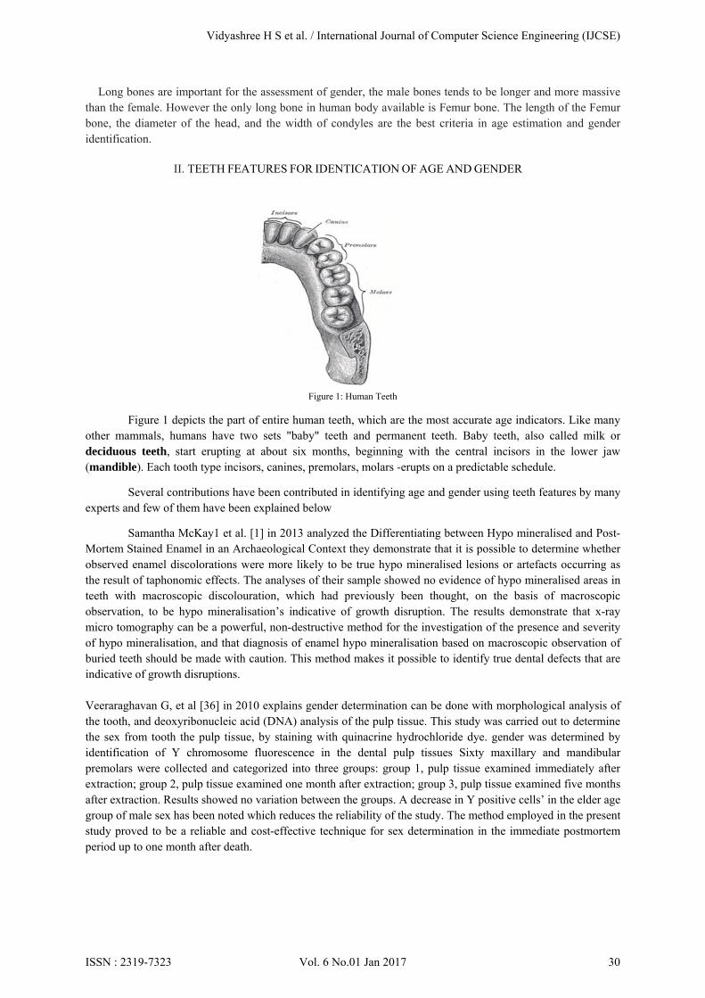
Figure 1: Human Teeth
Figure 1 depicts the part of entire human teeth, which are the most accurate age indicators. Like many other mammals, humans have two sets "baby" teeth and permanent teeth. Baby teeth, also called milk or deciduous teeth, start erupting at about six months, beginning with the central incisors in the lower jaw (mandible). Each tooth type incisors, canines, premolars, molars -erupts on a predictable schedule.
Several contributions have been contributed in identifying age and gender using teeth features by many experts and few of them have been explained below
Samantha McKay1 et al. [1] in 2013 analyzed the Differentiating between Hypo mineralised and Post-Mortem Stained Enamel in an Archaeological Context they demonstrate that it is possible to determine whether observed enamel discolorations were more likely to be true hypo mineralised lesions or artefacts occurring as the result of taphonomic effects. The analyses of their sample showed no evidence of hypo mineralised areas in teeth with macroscopic discolouration, which had previously been thought, on the basis of macroscopic observation, to be hypo mineralisation’s indicative of growth disruption. The results demonstrate that x-ray micro tomography can be a powerful, non-destructive method for the investigation of the presence and severity of hypo mineralisation, and that diagnosis of enamel hypo mineralisation based on macroscopic observation of buried teeth should be made with caution. This method makes it possible to identify true dental defects that are indicative of growth disruptions.
Veeraraghavan G, et al [36] in 2010 explains gender determination can be done with morphological analysis of the tooth, and deoxyribonucleic acid (DNA) analysis of the pulp tissue. This study was carried out to determine the sex from tooth the pulp tissue, by staining with quinacrine hydrochloride dye. gender was determined by identification of Y chromosome fluorescence in the dental pulp tissues Sixty maxillary and mandibular premolars were collected and categorized into three groups: group 1, pulp tissue examined immediately after extraction; group 2, pulp tissue examined one month after extraction; group 3, pulp tissue examined five months after extraction. Results showed no variation between the groups. A decrease in Y positive cells’ in the elder age group of male sex has been noted which reduces the reliability of the study. The method employed in the present study proved to be a reliable and cost-effective technique for sex determination in the immediate postmortem period up to one month after death.
Vidyashree H S et al. / International Journal of Computer Science Engineering (IJCSE)
ISSN : 2319-7323 Vol. 6 No.01 Jan 2017 30
Nolla’s method
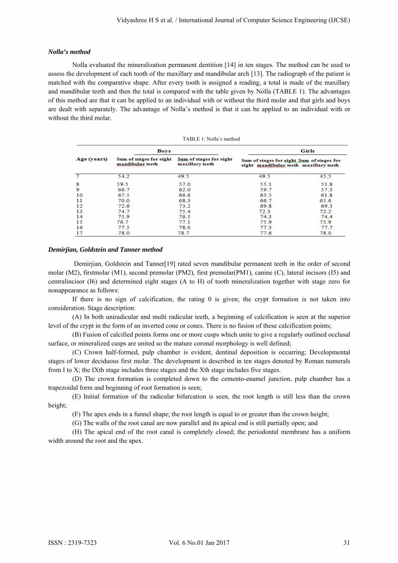
Nolla evaluated the mineralization permanent dentition [14] in ten stages. The method can be used to assess the development of each tooth of the maxillary and mandibular arch [13]. The radiograph of the patient is matched with the comparative shape. After every tooth is assigned a reading, a total is made of the maxillary and mandibular teeth and then the total is compared with the table given by Nolla (TABLE 1). The advantages of this method are that it can be applied to an individual with or without the third molar and that girls and boys are dealt with separately. The advantage of Nolla’s method is that it can be applied to an individual with or without the third molar.
TABLE 1: Nolla’s method
Demirjian, Goldstein and Tanner method
Demirjian, Goldstein and Tanner[19] rated seven mandibular permanent teeth in the order of second molar (M2), firstmolar (M1), second premolar (PM2), first premolar(PM1), canine (C), lateral incisors (I5) and centralincisor (I6) and determined eight stages (A to H) of tooth mineralization together with stage zero for nonappearance as follows:
If there is no sign of calcification, the rating 0 is given; the crypt formation is not taken into consideration. Stage description:
(A) In both uniradicular and multi radicular teeth, a beginning of calcification is seen at the superior level of the crypt in the form of an inverted cone or cones. There is no fusion of these calcification points;
(B) Fusion of calcified points forms one or more cusps which unite to give a regularly outlined occlusal surface, or mineralized cusps are united so the mature coronal morphology is well defined;
(C) Crown half-formed, pulp chamber is evident, dentinal deposition is occurring; Developmental stages of lower deciduous first molar. The development is described in ten stages denoted by Roman numerals from I to X; the IXth stage includes three stages and the Xth stage includes five stages.
(D) The crown formation is completed down to the cemento-enamel junction, pulp chamber has a trapezoidal form and beginning of root formation is seen;
(E) Initial formation of the radicular bifurcation is seen, the root length is still less than the crown height;
(F) The apex ends in a funnel shape; the root length is equal to or greater than the crown height; (G) The walls of the root canal are now parallel and its apical end is still partially open; and (H) The apical end of the root canal is completely closed; the periodontal membrane has a uniform
width around the root and the apex.
Vidyashree H S et al. / International Journal of Computer Science Engineering (IJCSE)
ISSN : 2319-7323 Vol. 6 No.01 Jan 2017 31
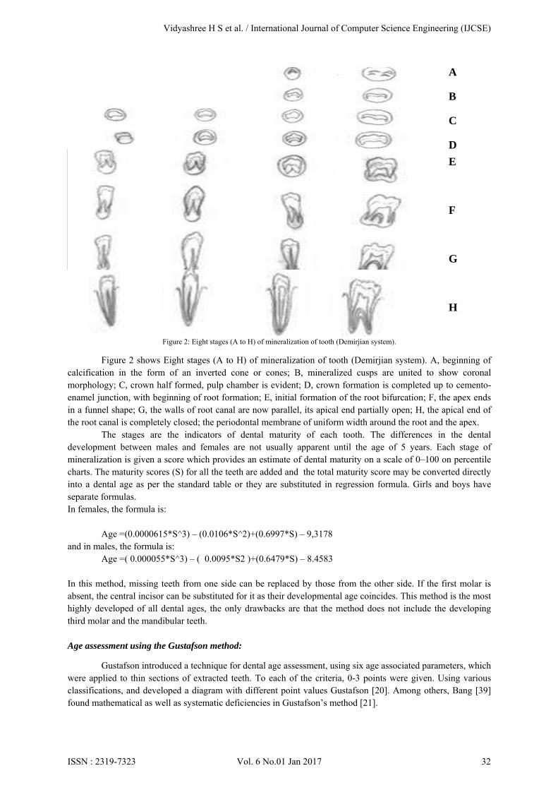
Figure 2: Eight stages (A to H) of mineralization of tooth (Demirjian system).
Figure 2 shows Eight stages (A to H) of mineralization of tooth (Demirjian system). A, beginning of
calcification in the form of an inverted cone or cones; B, mineralized cusps are united to show coronal morphology; C, crown half formed, pulp chamber is evident; D, crown formation is completed up to cemento-enamel junction, with beginning of root formation; E, initial formation of the root bifurcation; F, the apex ends in a funnel shape; G, the walls of root canal are now parallel, its apical end partially open; H, the apical end of the root canal is completely closed; the periodontal membrane of uniform width around the root and the apex.
The stages are the indicators of dental maturity of each tooth. The differences in the dental development between males and females are not usually apparent until the age of 5 years. Each stage of mineralization is given a score which provides an estimate of dental maturity on a scale of 0–100 on percentile charts. The maturity scores (S) for all the teeth are added and the total maturity score may be converted directly into a dental age as per the standard table or they are substituted in regression formula. Girls and boys have separate formulas. In females, the formula is:
Age =(0.0000615*S^3) – (0.0106*S^2)+(0.6997*S) – 9,3178 and in males, the formula is:
Age =( 0.000055*S^3) – ( 0.0095*S2 )+(0.6479*S) – 8.4583
In this method, missing teeth from one side can be replaced by those from the other side. If the first molar is absent, the central incisor can be substituted for it as their developmental age coincides. This method is the most highly developed of all dental ages, the only drawbacks are that the method does not include the developing third molar and the mandibular teeth. Age assessment using the Gustafson method:
Gustafson introduced a technique for dental age assessment, using six age associated parameters, which were applied to thin sections of extracted teeth. To each of the criteria, 0-3 points were given. Using various classifications, and developed a diagram with different point values Gustafson [20]. Among others, Bang [39] found mathematical as well as systematic deficiencies in Gustafson’s method [21].
A
B
C
D
E
F
G
H
Vidyashree H S et al. / International Journal of Computer Science Engineering (IJCSE)
ISSN : 2319-7323 Vol. 6 No.01 Jan 2017 32
Age assessment using quantification of tooth cementum annulation:
The root cementum of human teeth is subject to a physiological lamination process. Different layers of various types of root cementum are formed during continuous physiological change. These patterns can be examined employing thin section light microscopy. Different studies have addressed the application of this technique to human teeth, which was originally commonly used only in biology and veterinarian medicine. Lipsinic [22] as well as Pilloud [23] found in accuracies of this method, especially in older individuals, whereas for example Charles and Maat [24,25] found a good correlation between cementum layering and chronological age in humans.
Age assessment using the aspartic acid racemization:
Stereo chemical changes in amino acids from human tissues can be used for age assessment. These methods employ the time-dependent conversion processes between different enantiomers. Different methods found a high correlation between the extent of racemization of specific amino acids and age, especially in dental hard tissue (e.g. dentin) [25-27].
III. WRIST FEATURES FOR IDENTICATION OF AGE AND GENDER
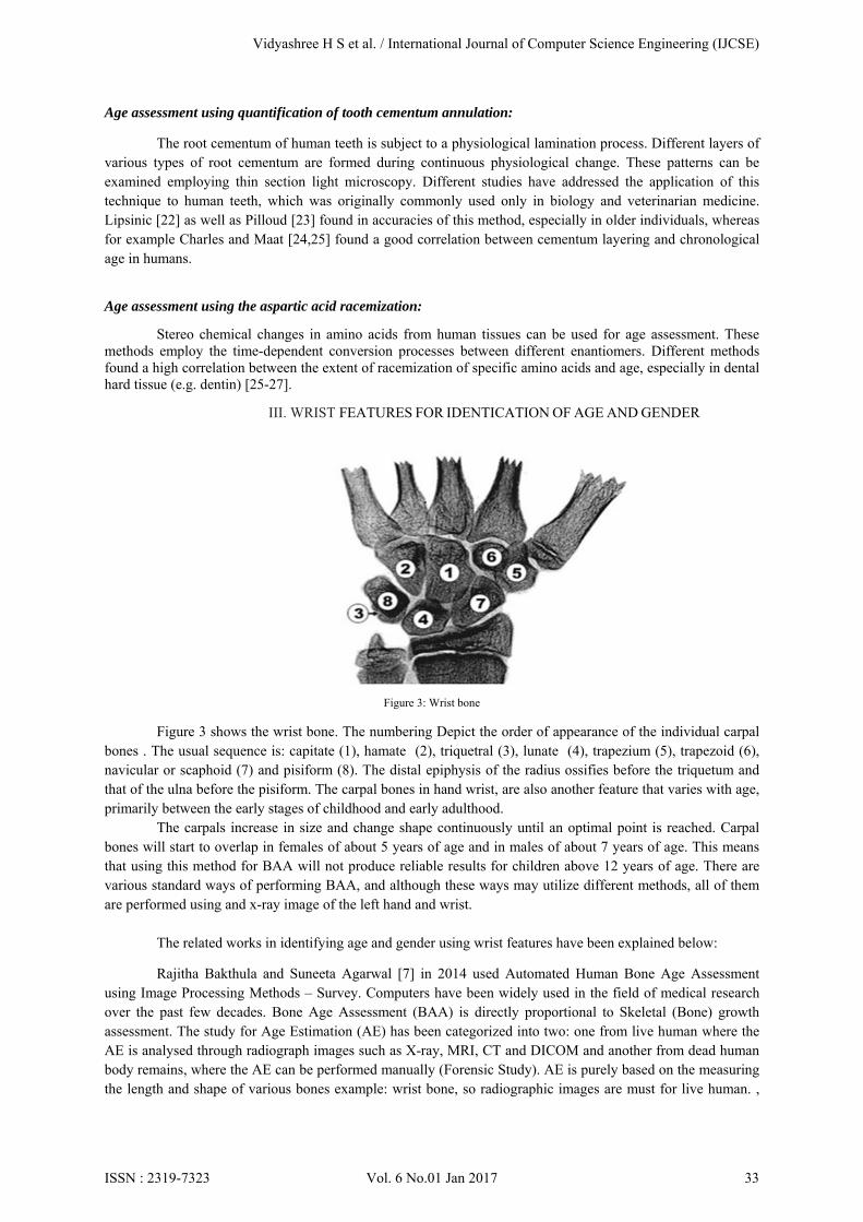
Figure 3: Wrist bone
Figure 3 shows the wrist bone. The numbering Depict the order of appearance of the individual carpal bones . The usual sequence is: capitate (1), hamate (2), triquetral (3), lunate (4), trapezium (5), trapezoid (6), navicular or scaphoid (7) and pisiform (8). The distal epiphysis of the radius ossifies before the triquetum and that of the ulna before the pisiform. The carpal bones in hand wrist, are also another feature that varies with age, primarily between the early stages of childhood and early adulthood.
The carpals increase in size and change shape continuously until an optimal point is reached. Carpal bones will start to overlap in females of about 5 years of age and in males of about 7 years of age. This means that using this method for BAA will not produce reliable results for children above 12 years of age. There are various standard ways of performing BAA, and although these ways may utilize different methods, all of them are performed using and x-ray image of the left hand and wrist.
The related works in identifying age and gender using wrist features have been explained below:
Rajitha Bakthula and Suneeta Agarwal [7] in 2014 used Automated Human Bone Age Assessment using Image Processing Methods – Survey. Computers have been widely used in the field of medical research over the past few decades. Bone Age Assessment (BAA) is directly proportional to Skeletal (Bone) growth assessment. The study for Age Estimation (AE) has been categorized into two: one from live human where the AE is analysed through radiograph images such as X-ray, MRI, CT and DICOM and another from dead human body remains, where the AE can be performed manually (Forensic Study). AE is purely based on the measuring the length and shape of various bones example: wrist bone, so radiographic images are must for live human. ,
Vidyashree H S et al. / International Journal of Computer Science Engineering (IJCSE)
ISSN : 2319-7323 Vol. 6 No.01 Jan 2017 33
this study present’s an overview of various image processing methods applied for automated bone age estimation of live human and discuss some major challenges in storing the medical image datasets.
Mohd faaizie bin darmawan et al. [4] in 2000 proposed on Determination of Age and Gender Based on Length of Hand wrist Bone: Limitation of determination techniques for certain population is among problems in the determination process. They presents age and gender determination using regression and discriminant function respectively. The body part used is left hand-wrist where the length is measured manually. The result shows that the best part of bone in the left hand-wrist that can be used to determine age is proximal phalanx on third finger for female and distal phalanx on third finger for male. For gender, result shows that the length for distal phalanx on first finger and third finger can be used to determine gender. To conclude, for age determination, regression is suitable for age determination based on the correlation between age and length of bone. For gender determination, discriminant function is suitable to determine gender based on the percentage of accuracy of the value of cut of point compared to real data.
Fuzzy Logic Based System is one of the Age assessment based on carpal bones alone was given by Aifeng Zhang and et. al. in the year 2007 [38]. Background is removed by thresholding. Carpal ROI is segmented using the horizontal line crossing the middle two metacarpal bones, vertical lines crossing the corner carpal bones and the line crossing the bottom forearm bones. Then the image is smoothed by using the filter proposed by Malik and Perona [38]. This method works on diffusion i.e. removing the noise by preserving the border information. Thereafter the edge is found using canny edge function. Then the image is refined to get only the carpal bones by excluding the unwanted bony pixels (morphological operators are used for this). Centroid is estimated for capitate bone. ROI is divided into five sectors with respect to the centroid for checking the presence of bones and their size and shape. Finally a fuzzy logic classifier is used for age assessment by giving these results as inputs. According to the authors the success rate was near to 100% for age>=2 and 80% for age<2 years. Authors had included radiographs of 0-7 years of males and 0-5 years for females. After the age above 7 years for males and 5 years for females the some of the carpal bones may overlap so, these age groups are excluded from research.
Neural Network Based System of Another automated approach was given by Jian Liu et. al [39] in the year 2008. The authors used particle swam optimisation method for segmenting the image by matching different templates from the dataset. Then features were analysed using the edge detection followed by neural network training using one hidden layer with back propagation. Authors claim that the manual reading and automated readings were differing in coefficient variation of RUS and Carpal bones. Final results proved that the carpal bones are best to differentiate for age<=9 years and RUS for age>9 years. Overall the implemented concept was complicated with PSO and NN. Krit Somkantha et. al [40] used support vector machines and neural networks for extracting the borders of carpal bones for age assessment by comparing the atlas of [41].
Greulich and Pyle Method (GP):
Left hand image is widely used for age assessment or skeleton assessment. Since the right hand is used too much to work it effects on the growth of bones. So the Left hand is preferred for analysis. The hand bone development stages were given in as an Atlas by W. W. Greulich and S. I. Pyle [37] in 1959. The atlas has been published by the name "Atlas of Skeletal Development of the Hand and Wrist. Large number of children of different age groups was enrolled for this study. Different Radiographs of left shoulder, elbow, hand, hip and knee were taken. These Radiographs were taken at the interval of three months up to five years and thereafter taken on yearly basis. The data collection process was carried out from 1931 to 1942 years comprising around 1000 children radiographs as a source. Finally in the year 1950, research Atlas has been published. For Male and Female different Atlas has been proposed since they mature at different rates. These atlases are the base model for analysing age related changes in the human bone architectural structure. This method is popularly referred as GP method by most of the authors.
Vidyashree H S et al. / International Journal of Computer Science Engineering (IJCSE)
ISSN : 2319-7323 Vol. 6 No.01 Jan 2017 34
Advantages of Greulich and Pyle Method (GP) are:
1. Simple and reliable method to use, just by comparing the x-ray with the atlas.
2. Once found the age group the observer have to check for older and younger stages for exact age.
Disadvantages of Greulich and Pyle Method (GP) are:
1. Time consuming process.
2. Around 22 joints have to be processed.
3. The image must be very clear for estimating the age.
4. Might lead to miss classification
5. The observer must be an expert
Tanner and Whitehouse Method (TW):
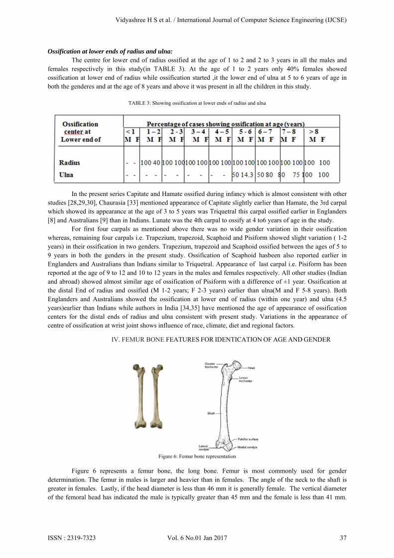
Another atlas has been proposed by Tanner and Whitehouse (TW: TW1, TW2, TW3)[34][35] in the year 1962. Here the study was focused on the age estimation but relies on the bone standard maturity. The TW method used bone joints location as ROIs for bone maturity (shown in Figure.3, Figure 4 and Figure.5). Each ROI is further divided into three parts: epiphysis, diaphysis and metaphysis. Out of these three, the epiphysis ossifies from the age zero to teen age and later gets combined with diaphysis. So the age assessment of the TW and GP methods is only up to 19 years. Epiphysis has 9 stages in total starting with A (as no epiphysis bone) to B, C, D E F G H and I as shown in Figure.5. Further alternative methods for skeleton growth assessment system are stated as below, 1) TW2 (20 bones): This is a modified method in the year 1975 of initial TW1 method. It uses 20 ROIs for bone analysis including first, middle, fifth fingers and the carpal bones. 2) RUS (Radius, Ulna and Short bones): This is same as TW2 method but excludes the carpal bones. 3) CARPAL: This study is using the carpal bones alone. These bones ossify till the age of 9 years. So the age assessment using this method is limited to 9 years GP and TW2 methods are the base models with initial Atlas and look-up-tables for human age assessment. In the radiograph image epiphysis-ROI or carpal-ROI index stage must be identified and manually checked with the hand atlas. So, many researchers tried to develop fully automated systems for verifying and validating (digitally) their results using these two methods. A new atlas has been developed by combining children of both sexes of different countries [36] [37]. Advantages of Tanner and Whitehouse Method (TW):
1. Unlike GP it is based on maturity of skeleton bones.
2. Based on numerical score assigned for each bone (rather than just shape).
3. The age can be estimated using RUS or Carpal bones or Phalangeal bones (20) separately.
4. Accurate than GP method. Disadvantages of Tanner and Whitehouse Method (TW):
1. Time consuming process.
2. Complex procedure (maturity scores are 8 for each bone).
3. Here also the image has to be clear enough for classification.
4. The observer must be an expert.
Vidyashree H S et al. / International Journal of Computer Science Engineering (IJCSE)
ISSN : 2319-7323 Vol. 6 No.01 Jan 2017 35
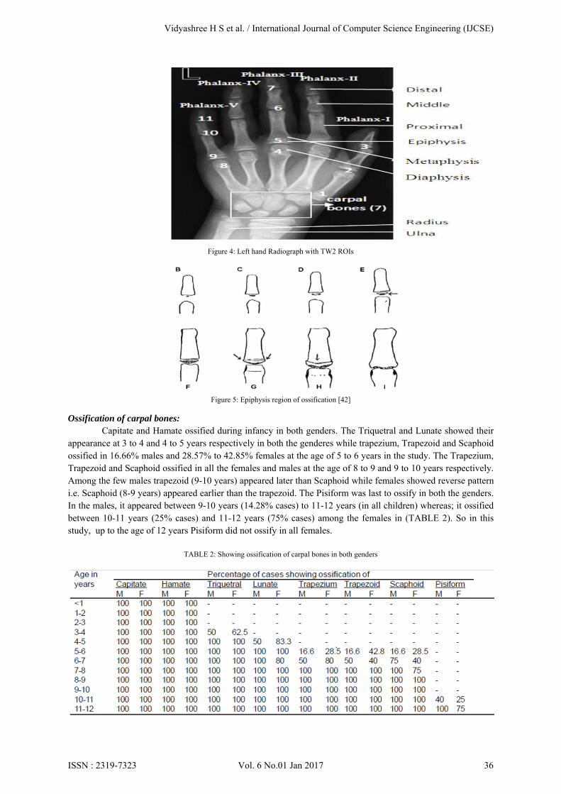
Figure 4: Left hand Radiograph with TW2 ROIs
Figure 5: Epiphysis region of ossification [42]
Ossification of carpal bones:
Capitate and Hamate ossified during infancy in both genders. The Triquetral and Lunate showed their appearance at 3 to 4 and 4 to 5 years respectively in both the genderes while trapezium, Trapezoid and Scaphoid ossified in 16.66% males and 28.57% to 42.85% females at the age of 5 to 6 years in the study. The Trapezium, Trapezoid and Scaphoid ossified in all the females and males at the age of 8 to 9 and 9 to 10 years respectively. Among the few males trapezoid (9-10 years) appeared later than Scaphoid while females showed reverse pattern i.e. Scaphoid (8-9 years) appeared earlier than the trapezoid. The Pisiform was last to ossify in both the genders. In the males, it appeared between 9-10 years (14.28% cases) to 11-12 years (in all children) whereas; it ossified between 10-11 years (25% cases) and 11-12 years (75% cases) among the females in (TABLE 2). So in this study, up to the age of 12 years Pisiform did not ossify in all females.
TABLE 2: Showing ossification of carpal bones in both genders
Vidyashree H S et al. / International Journal of Computer Science Engineering (IJCSE)
ISSN : 2319-7323 Vol. 6 No.01 Jan 2017 36
Ossification at lower ends of radius and ulna: The centre for lower end of radius ossified at the age of 1 to 2 and 2 to 3 years in all the males and
females respectively in this study(in TABLE 3). At the age of 1 to 2 years only 40% females showed ossification at lower end of radius while ossification started ,it the lower end of ulna at 5 to 6 years of age in both the genderes and at the age of 8 years and above it was present in all the children in this study.
TABLE 3: Showing ossification at lower ends of radius and ulna
In the present series Capitate and Hamate ossified during infancy which is almost consistent with other
studies [28,29,30], Chaurasia [33] mentioned appearance of Capitate slightly earlier than Hamate, the 3rd carpal which showed its appearance at the age of 3 to 5 years was Triquetral this carpal ossified earlier in Englanders [8] and Australians [9] than in Indians. Lunate was the 4th carpal to ossify at 4 to6 years of age in the study.
For first four carpals as mentioned above there was no wide gender variation in their ossification whereas, remaining four carpals i.e. Trapezium, trapezoid, Scaphoid and Pisiform showed slight variation ( 1-2 years) in their ossification in two genders. Trapezium, trapezoid and Scaphoid ossified between the ages of 5 to 9 years in both the genders in the present study. Ossification of Scaphoid hasbeen also reported earlier in Englanders and Australians than Indians similar to Triquetral. Appearance of last carpal i.e. Pisiform has been reported at the age of 9 to 12 and 10 to 12 years in the males and females respectively. All other studies (Indian and abroad) showed almost similar age of ossification of Pisiform with a difference of ±1 year. Ossification at the distal End of radius and ossified (M 1-2 years; F 2-3 years) earlier than ulna(M and F 5-8 years). Both Englanders and Australians showed the ossification at lower end of radius (within one year) and ulna (4.5 years)earlier than Indians while authors in India [34,35] have mentioned the age of appearance of ossification centers for the distal ends of radius and ulna consistent with present study. Variations in the appearance of centre of ossification at wrist joint shows influence of race, climate, diet and regional factors.
IV. FEMUR BONE FEATURES FOR IDENTICATION OF AGE AND GENDER
Figure 6: Femur bone representation
Figure 6 represents a femur bone, the long bone. Femur is most commonly used for gender determination. The femur in males is larger and heavier than in females. The angle of the neck to the shaft is greater in females. Lastly, if the head diameter is less than 46 mm it is generally female. The vertical diameter of the femoral head has indicated the male is typically greater than 45 mm and the female is less than 41 mm.
Vidyashree H S et al. / International Journal of Computer Science Engineering (IJCSE)
ISSN : 2319-7323 Vol. 6 No.01 Jan 2017 37
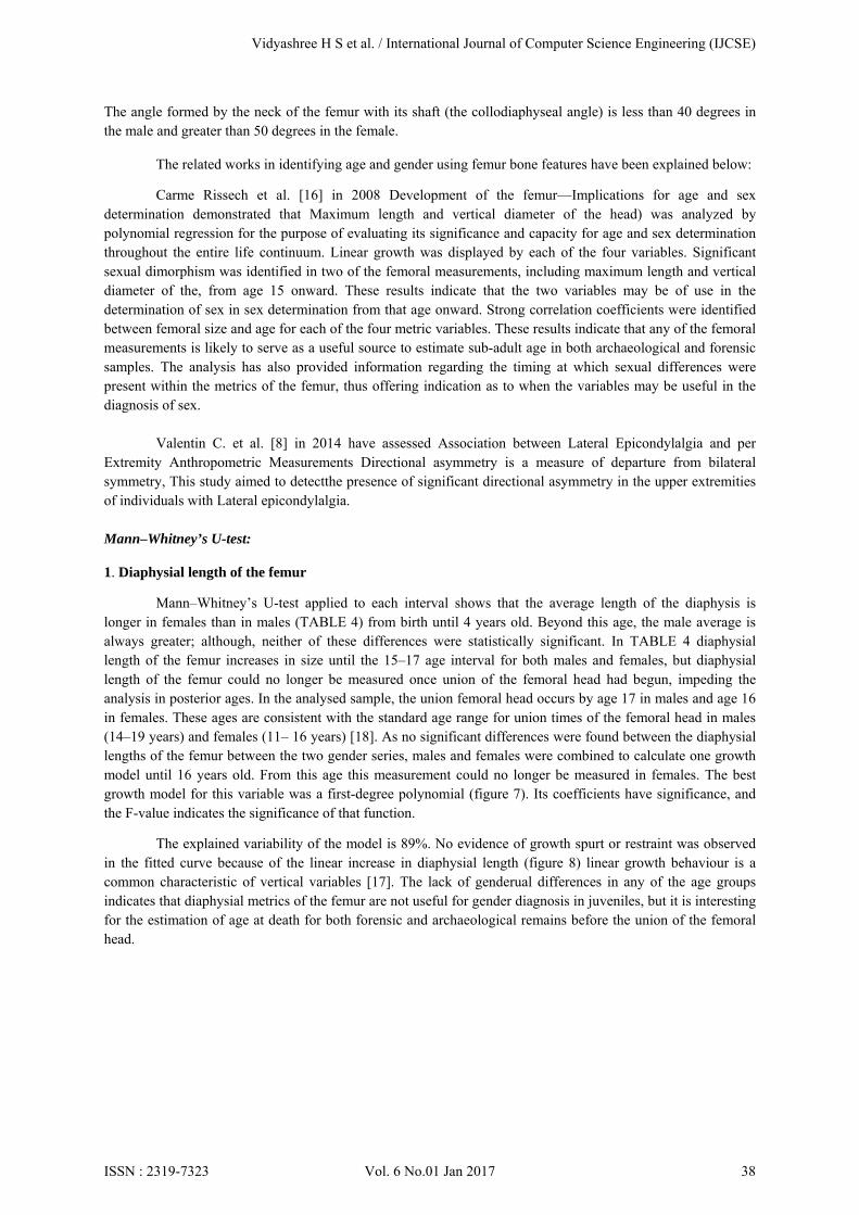
The angle formed by the neck of the femur with its shaft (the collodiaphyseal angle) is less than 40 degrees in the male and greater than 50 degrees in the female.
The related works in identifying age and gender using femur bone features have been explained below:
Carme Rissech et al. [16] in 2008 Development of the femur—Implications for age and sex determination demonstrated that Maximum length and vertical diameter of the head) was analyzed by polynomial regression for the purpose of evaluating its significance and capacity for age and sex determination throughout the entire life continuum. Linear growth was displayed by each of the four variables. Significant sexual dimorphism was identified in two of the femoral measurements, including maximum length and vertical diameter of the, from age 15 onward. These results indicate that the two variables may be of use in the determination of sex in sex determination from that age onward. Strong correlation coefficients were identified between femoral size and age for each of the four metric variables. These results indicate that any of the femoral measurements is likely to serve as a useful source to estimate sub-adult age in both archaeological and forensic samples. The analysis has also provided information regarding the timing at which sexual differences were present within the metrics of the femur, thus offering indication as to when the variables may be useful in the diagnosis of sex.
Valentin C. et al. [8] in 2014 have assessed Association between Lateral Epicondylalgia and per Extremity Anthropometric Measurements Directional asymmetry is a measure of departure from bilateral symmetry, This study aimed to detectthe presence of significant directional asymmetry in the upper extremities of individuals with Lateral epicondylalgia.
Mann–Whitney’s U-test:
1. Diaphysial length of the femur
Mann–Whitney’s U-test applied to each interval shows that the average length of the diaphysis is longer in females than in males (TABLE 4) from birth until 4 years old. Beyond this age, the male average is always greater; although, neither of these differences were statistically significant. In TABLE 4 diaphysial length of the femur increases in size until the 15–17 age interval for both males and females, but diaphysial length of the femur could no longer be measured once union of the femoral head had begun, impeding the analysis in posterior ages. In the analysed sample, the union femoral head occurs by age 17 in males and age 16 in females. These ages are consistent with the standard age range for union times of the femoral head in males (14–19 years) and females (11– 16 years) [18]. As no significant differences were found between the diaphysial lengths of the femur between the two gender series, males and females were combined to calculate one growth model until 16 years old. From this age this measurement could no longer be measured in females. The best growth model for this variable was a first-degree polynomial (figure 7). Its coefficients have significance, and the F-value indicates the significance of that function.
The explained variability of the model is 89%. No evidence of growth spurt or restraint was observed in the fitted curve because of the linear increase in diaphysial length (figure 8) linear growth behaviour is a common characteristic of vertical variables [17]. The lack of genderual differences in any of the age groups indicates that diaphysial metrics of the femur are not useful for gender diagnosis in juveniles, but it is interesting for the estimation of age at death for both forensic and archaeological remains before the union of the femoral head.
Vidyashree H S et al. / International Journal of Computer Science Engineering (IJCSE)
ISSN : 2319-7323 Vol. 6 No.01 Jan 2017 38
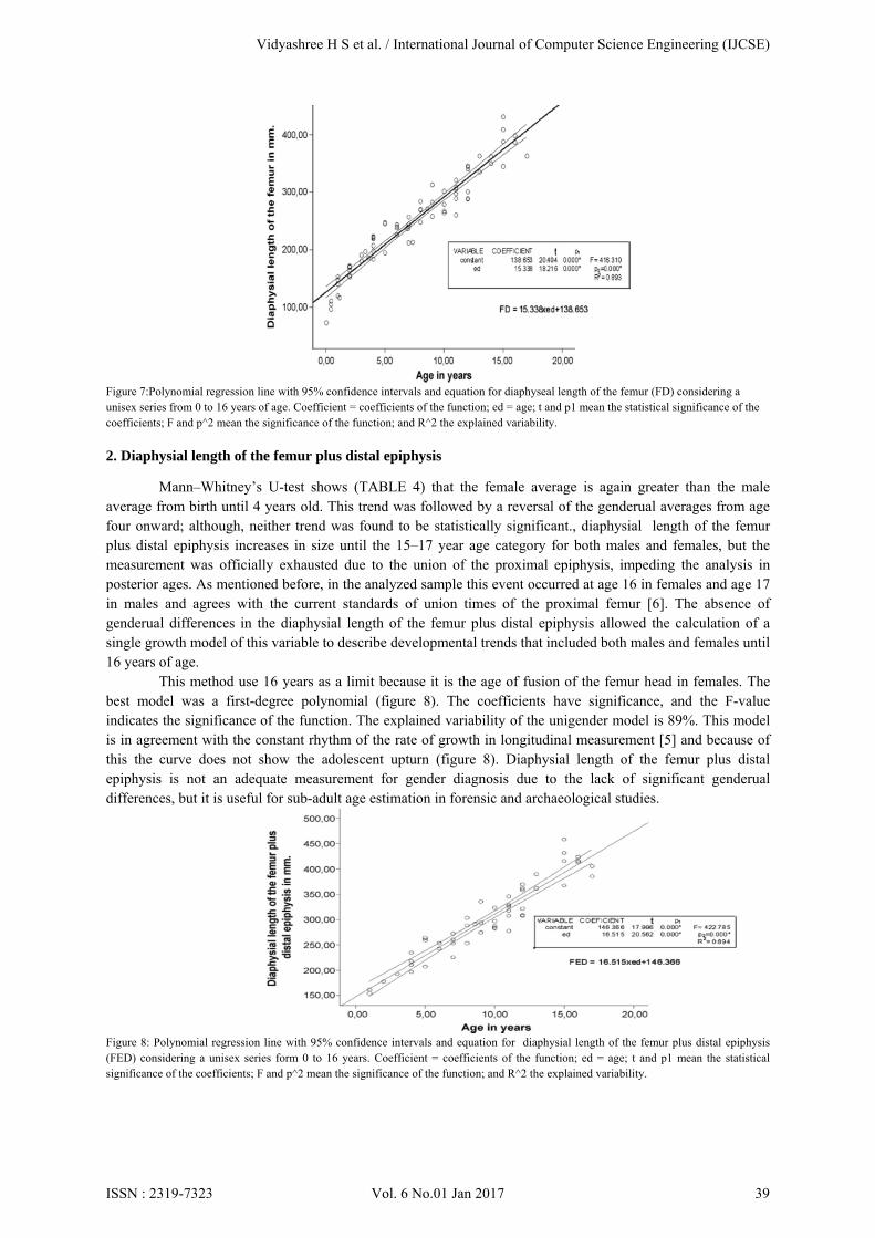
Figure 7:Polynomial regression line with 95% confidence intervals and equation for diaphyseal length of the femur (FD) considering a unisex series from 0 to 16 years of age. Coefficient = coefficients of the function; ed = age; t and p1 mean the statistical significance of the coefficients; F and p^2 mean the significance of the function; and R^2 the explained variability. 2. Diaphysial length of the femur plus distal epiphysis
Mann–Whitney’s U-test shows (TABLE 4) that the female average is again greater than the male average from birth until 4 years old. This trend was followed by a reversal of the genderual averages from age four onward; although, neither trend was found to be statistically significant., diaphysial length of the femur plus distal epiphysis increases in size until the 15–17 year age category for both males and females, but the measurement was officially exhausted due to the union of the proximal epiphysis, impeding the analysis in posterior ages. As mentioned before, in the analyzed sample this event occurred at age 16 in females and age 17 in males and agrees with the current standards of union times of the proximal femur [6]. The absence of genderual differences in the diaphysial length of the femur plus distal epiphysis allowed the calculation of a single growth model of this variable to describe developmental trends that included both males and females until 16 years of age.
This method use 16 years as a limit because it is the age of fusion of the femur head in females. The best model was a first-degree polynomial (figure 8). The coefficients have significance, and the F-value indicates the significance of the function. The explained variability of the unigender model is 89%. This model is in agreement with the constant rhythm of the rate of growth in longitudinal measurement [5] and because of this the curve does not show the adolescent upturn (figure 8). Diaphysial length of the femur plus distal epiphysis is not an adequate measurement for gender diagnosis due to the lack of significant genderual differences, but it is useful for sub-adult age estimation in forensic and archaeological studies.
Figure 8: Polynomial regression line with 95% confidence intervals and equation for diaphysial length of the femur plus distal epiphysis (FED) considering a unisex series form 0 to 16 years. Coefficient = coefficients of the function; ed = age; t and p1 mean the statistical significance of the coefficients; F and p^2 mean the significance of the function; and R^2 the explained variability.
Vidyashree H S et al. / International Journal of Computer Science Engineering (IJCSE)
ISSN : 2319-7323 Vol. 6 No.01 Jan 2017 39
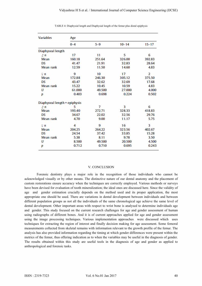
TABLE 4: Diaphysial length and Diaphysial length of the femur plus distal epiphysis
V. CONCLUSION
Forensic dentistry plays a major role in the recognition of those individuals who cannot be acknowledged visually or by other means. The distinctive nature of our dental anatomy and the placement of custom restorations ensure accuracy when the techniques are correctly employed. Various methods or surveys have been devised for evaluation of tooth mineralization; the ideal ones are discussed here. Since the validity of age and gender estimation crucially depends on the method used and its proper application, the most appropriate one should be used. There are variations in dental development between individuals and between different population groups as not all the individuals of the same chronological age achieve the same level of dental development. Other important areas with respect to wrist bone is analysed to determine individuals age and gender. This study focused on the current research challenges for age and gender assessment of human using radiographs of different bones. And it is of current approaches applied for age and gender assessment using the image processing techniques. Various implementation approaches were discussed which uses techniques for extracting the region of interest and finally decision making for age assessment. Some femoral measurements collected from skeletal remains with information relevant to the growth profile of the femur. The analysis has also provided information regarding the timing at which gender differences were present within the metrics of the femur, thus offering indication as to when the variables may be useful in the diagnosis of gender. The results obtained within this study are useful tools in the diagnosis of age and gender as applied to anthropological and forensic tasks.
Vidyashree H S et al. / International Journal of Computer Science Engineering (IJCSE)
ISSN : 2319-7323 Vol. 6 No.01 Jan 2017 40
VI. REFERENCES
[1] Samantha McKay1, Rami Farah2, Jonathan M. Broadbent3, Nancy Tayles4, Sian E. Halcrow4(2013) Differentiating between Hypomineralised and Post-Mortem Stained Enamel in an Archaeological Context.
[2] R.G Aykroyd, d. lucy, a.m. pollard, and t. solheim3 (1997)Technical Note: “Regression Analysis in Adult Age Estimation”. [3] Baccino E, Ubelaker DH, Hayek L-AC, Zerilli A. Evaluation of seven methods of estimating age at death from mature human skeletal
remains. J Forensic Sci 1999;44(5):931–936. “Evaluation of Seven Methods of Estimating Age at Death from Mature Human Skeletal Remains”.
[4] Mohd Faaizie Bin Darmawan, Assoc. Prof. Dr. Habibollah bin Haron, Dr. Suhaila Mohammad Yusof Faculty of Computer Science and Information Systems, “Determination of age and gender based on length of hand- wrist bone: malaysian case study” .
[5] F. Twiesselmann, De´veloppement biome` trique de l’enfant a´ l’adulte, Presses Q1 Universitaires de Bruxelles-Librairie Maloine, Paris, 1969.
[6] L. Scheuer, S. Black, Developmental Juvenile Osteology, Academic Press, London, 2000. [7] Rajitha Bakthula Suneeta Agarwal CSED CSED MNNIT Allahabad, Uttar Pradesh, India. MNNIT Allahabad, Uttar Pradesh, Ind in
2014 “Automated Human Bone Age Assessment using Image Processing Methods – Survey”. [8] Valentin C. Dones, Karen Grimmer-Somers, Steven Milanese and Alvin P. Atlas in 2014, “Association between Lateral
Epicondylalgia and Upper Extremity Anthropometric Measurements”: A Case Control Study [9] Tanuj Kanchan and Kewal Krishan Department of Forensic Medicine, Kasturba Medical College, Mangalore (A Constituent College
of Manipal University), India in 2013, “Personal Identification in Forensic Examinations”. [10] Charles DK, Condon K, Cheverud JM, Buikstra J (1986) “Cementum annulation and age determination in homo sapiens”. I. Tooth
variability and observer error. Am J Phys Anthropol71: 311–320 [11] Carme Rissech a,, Maureen Schaefer b, Assumpcio´ Malgosa aba Unitat d’Antropologia Biologica, Department of Biologia Animal,
Vegetal i Ecololgia, Universitat Auto`noma de Barcelona, 08193-Bellaterra, Spain b Department Anatomy and Forensic Anthropology, University of Dundee, Scotland, Complex Dow Street, Dundee DD1 5EH, UK(2008).
[12] Iman F. Gaballah, Alaa M. Shehab , Khaled A. Bayoumi Department of Forensic Medicine and Clinical Toxicology, Faculty of Medicine, Cairo University, Egypt Received 28 February 2014; “Gender determination in femurs of modernEgyptians”: A comparative study betweenmetric measurements and SRY gene detection revised 7 July 2014; accepted 7 August 2014 Available online 5 September 2014.
[13] Panchbhai “ Dental radiographic indicators, a key toage estimation”Department of Oral Medicine and Radiology,SPD College & Hospital, DMIMSU, Sawangi-M, Wardha, India(2011).
[14] Nolla CM. “The development of permanent teeth”. J Dent Child1960; 27: 254–266(2001) [15] Singaraju S, Sharda P. Age estimation using pulp-tooth arearatio: A digital image analysis. J Forensic Dent Sci 2009; 1: 37–41. [16] Cameriere R, Ferrante L, Belcastro M, Bonfiaglioli B, Rastelli E,Cingolani M. Age estimation by pulp/tooth ratio in canines
byperiapical X-rays. J Forensic Sci 2007; 52: 166–170. [17] F. Twiesselmann, De´veloppement biome` trique de l’enfant a´ l’adulte, Presses Q1Universitaires de Bruxelles-Librairie Maloine,
Paris, 1969 [18] L. Scheuer, S. Black, Developmental Juvenile Osteology, Academic Press, London,2000. [19] Demirjian A, Goldstein H, Tanner JM. A new system of dentalage assessment. Hum Biol 1973; 45: 211–227. [20] Gustafson G (1950) “Age determination on teeth”. J Am Dent Assoc 41: 45-54. [21] Bang G, Ramm E (1970) Determination of age in humans from root dentintransparency. Acta Odontol Scand 28: 3-35. [22] Mandojana JM, Martin-de las Heras S, Valenzuela A, Valenzuela M, Luna JD(2001) Differences in morphological age-related dental
changes depending onpostmortem interval. J Forensic Sci 46: 889-892. [23] Pilloud S (2004) Can there be age determination on the basis of the dentalcementum also in older individuals as a significant context
between histologicaland real age determination. Anthropologischer Anzeiger 62: 231-239. [24] Charles DK, Condon K, Cheverud JM, Buikstra JE (1986) Cementum annulations and age determination in Homo sapiens. I. Tooth
variability and observer error. Am J Phys Anthropol 71: 311-320. [25] Helfman PM, Bada JL (1975) “Aspartic acid racemization in tooth enamel fromliving humans”. Proc Natl Acad Sci U S A 72: 2891-
2894. [26] Bada JL, Helfman PM (1975) “Amino acid racemization dating of fossil bones”. World Archaeol 7: 160-173. [27] Ohtani S, Yamamoto K (1987) “Age estimation using the racemization ofaspartic acid on human dentin”. Nihon Hoigaku Zasshi 41:
181-190. [28] Krishnan MKR.Krishnan’s “Handbook ofForensic Medicine and Toxicology”. 8th ed.:Hyderabad, Paras Medical Books: 1984;32. [29] Davies DA and parson 1. Of anatomy 1927;62.58. [30] Fleoker H. Anatomical Society of India. Vol.1932;67, Oct. [31] Survey committee Survey Committee Reporton Medico-Legal Practice in India, DirectorGeneral of Health Services, Ministry Of
Health,New Delhi.1964. [32] Chaurasia BD. “Human Anatomy”- Regional andApplied vol. I, 1st ed.: CBS Publisher, New Delhi 1979. [33] Rao NG: Textbook of “Forensic Medicine andToxicology”. 1st ed.: Jaypee Brothers (P) Ltd.,New Delhi: 2000;89. [34] Chaurasia BD. “Human Anatomy”- Regional andApplied vol. I, 1st ed.: CBS Publisher, New Delhi 1979. [35] Nandy A. Principles Of Forensic Medicine, 1sted.: Calcutta, New Central Book Agency (P)Ltd.1995. [36] Determination of sex from tooth pulp tissue Veeraraghavan G, Lingappa A, Shankara SP, Mamatha GP, Sebastian BT, Mujib A
Libyan J Med 2010;5:10 [37] W. W. Greulich and S. I. Pyle, Radiographic Atlas of Skeletal Development of Hand Wrist, 2 ed. Palo Alto, CA: Stanford Univ. Press,
19 [38] Perona P, Malik J, Scale-space and edge detection using anisotropic diffusion, PAMI 1990:12(7). [39] Jian Liu, Jing Qi, Zhao Liu, Qin Ning, Xiaoping Luo, Automatic bone age assessment based on intelligent algorithms and comparison
with TW3 method, Comput Med Imaging and Graphics 32 (2008) 678–684 [40] Krit Somakanta, Nipon Theera Umpon, Sansanee Auephanwiriyakul, Bone age assessment in young children using automatic carpal
bone feature extraction and support vector regression, J. Digit Imaging, 2011, 24:1044-1058. [41] Gilsanz V, Ratib O, Hand Bone age: A Digital Atlas of Skeletal maturity, 2005. [42] Concetto Spampinato, Skeletal Bone Age Assessment, University of Catania, Viale Andrea Doria, 6 95125, 1995.
Vidyashree H S et al. / International Journal of Computer Science Engineering (IJCSE)
ISSN : 2319-7323 Vol. 6 No.01 Jan 2017 41


![english.zereg.netenglish.zereg.net/English VI word quizzes.docx · Web viewstomach – strong – tail – thigh – toe - tongue – tooth [teeth] – weak - wing – wrist – English](https://img.pdfslide.net/doc/110x75/5cd105fa88c9930c558bcffc/vi-word-quizzesdocx-web-viewstomach-strong-tail-thigh-toe-.jpg)