Embed Size (px)
Citation preview

IEEE TRANSACTIONS ON BIOMEDICAL ENGINEERING, VOL. XX, NO. X, MONTH 2013 1
A Neuro-Fuzzy Approach for Medical Image FusionSudeb Das∗, and Malay Kumar Kundu, Senior Member, IEEE,
Abstract—This paper addresses a novel approach to the multi-sensor, multimodal medical image fusion (MIF) problem, employ-ing multiscale geometric analysis of non-subsampled contourlettransform and fuzzy-adaptive reduced pulse-coupled neural net-work (RPCNN). The linking strengths of the RPCNNs’ neuronsare adaptively set by modelling them as the fuzzy membershipvalues, representing their significance in the corresponding sourceimage. Use of RPCNN with less complex structure and havingless number of parameters, leads to computational efficiency,an important requirement of point-of-care (POC) health caretechnologies. The proposed scheme is free from the commonshortcomings of the state-of-the-art MIF techniques: contrastreduction, loss of image fine details and unwanted image degra-dations etc. Subjective and objective evaluations show better per-formance of this new approach compared to existing techniques.
Index Terms—Image fusion, artificial neural network, fuzzylogic, medical imaging, image analysis.
I. INTRODUCTION
THE rapid and significant advancements in medical imag-ing technologies and sensors, lead to new uses of medical
images in various healthcare and bio-medical applicationsincluding diagnosis, research, treatment and education etc.Different modalities of medical images reflect different in-formation of human organs and tissues, and have their re-spective application ranges. For instance, structural imageslike magnetic resonance imaging (MRI), computed tomog-raphy (CT), ultrasonography (USG) and magnetic resonanceangiography (MRA) etc., provide high-resolution images withexcellent anatomical detail and precise localization capability.Whereas, functional images such as position emission tomog-raphy (PET), single-photon emission computed tomography(SPECT) and functional MRI (fMRI) etc., provide low-spatialresolution images with functional information, useful for de-tecting cancer and related metabolic abnormalities. A singlemodality of medical image cannot provide comprehensive andaccurate information. Therefore, it is necessary to correlateone modality of medical image to another to obtain therelevant information. Moreover, the manual process of inte-grating several modalities of medical images is rigorous, time-consuming, costly, subject to human error, and requires yearsof experience. Therefore, automatically combining multimodalmedical images through image fusion (IF) has become thefocus of imaging research and processing [1], [2].
In recent years, many IF and medical image fusion (MIF)techniques have been proposed by various researchers. It hasbeen found that the pixel-level spatial domain methods usually
Manuscript received Month XX, 2013; revised Month XX, 2013. Asteriskindicate corresponding author.
∗S. Das and M. K. Kundu are with the Machine Intelligence Unit,Indian Statistical Institute, 203 B.T.Road, Kolkata-108, India e-mail: ([email protected], [email protected]).
lead to contrast reduction. Approaches based on intensity-hue-saturation (IHS), principal component analysis (PCA),and the Brovey transform offer better results, but suffer fromspectral degradation. Pyramidal IF schemes fail to introduceany spatial orientation selectivity in the decomposition pro-cess, and hence often cause blocking effects [3], [4]. Most ofthe above mentioned schemes are modality specific with itsown limitations [5]. The problem with widely used Wavelettransform (WT) is that it can preserve spectral informationefficiently but cannot express spatial characteristics well [6]–[8]. As a result, WT based fusion schemes fail to preservethe salient features of the source images efficiently, but in-troduce artifacts and inconsistencies in the fused results [9].Recently, to overcome these problems, many improved IF/MIFmethods based on multiscale geometric analysis (MGA) tools(like curvelet, contourlet, ripplet etc.) are proposed [9], [10].However, measuring the importance/contribution of individualsource image in the fused image, and finding effective way ofcombining them is still an open problem.
Pulse-coupled neural network (PCNN) is a novel neural net-work model that simulates the processing mechanism of cat’svisual cortex and characterized by global coupling and pulsesynchronization of neurons [11]–[13]. PCNN and its modifiedversions have been used in the IF/MIF domains by variousresearchers [3], [5], [14], [15]. Even though, PCNN basedIF/MIF methods outperform other conventional schemes, butthese approaches also suffer from several shortcomings: con-trast reduction, loss of image fine details and unwanted degra-dations etc. Moreover, PCNN has several parameters withcomplex structures, and optimal estimation of these parametersis a major limitation to automatization and generalization ofPCNN. In most of the PCNN based IF/MIF techniques, theseparameters are kept same and set as a constant. But, accordingto human visual system (HVS), the responses to a regionwith notable features are stronger than a region with non-notable features. Thus, the parameters of PCNN’s neuronsshould be related to the importance (significance) of thefeatures of either the corresponding pixel (in spatial domain)or coefficient (in transform domain) of the image [16]–[18].But, the problem remains: how to measure the importance(significance) of the pixel (coefficient) in the correspondingimage. Therefore, we not only need a way to adaptively andautomatically set the values of the parameters of the PCNN,but also to make the fusion scheme free from the commonproblems faced by the existing techniques [19]. This paperexplores the use of fuzzy logic for building a simultaneousfusion and enhancement technique based on the HVS modelto address these above mentioned problems. In this regard,the main contributions of this article are as follows: (1)A novel MIF scheme employing non-subsampled contourlettransform (NSCT) and reduced PCNN (RPCNN) with adaptive

IEEE TRANSACTIONS ON BIOMEDICAL ENGINEERING, VOL. XX, NO. X, MONTH 2013 2
linking strengths based on the corresponding image’s localfeatures. (2) Following the subjectivity of HVS, fuzzy logic isused to enable the proposed scheme to produce high qualityfused images with higher contrast, more clarity and moreuseful subtle detail information. (3) Without involving anyprior training and trials the less complex RPCNN having lessnumber of parameters leads to computational efficiency, whichis suitable for real time image processing applications (likepoint-of-care (POC) health care technologies). Both subjectiveand objective performance evaluations are made and verifiedin the paper. Performance comparisons with state-of-the-artschemes show that the proposed method performs better.
The rest of the paper is organized as follows. NSCT isdescribed in Section II. In Section III, the RPCNN modelis briefly reviewed. Section IV, presents the proposed MIFscheme. Experimental results and comparisons are given inSection V, and we draw conclusion in Section VI.
II. NON-SUBSAMPLED CONTOURLET TRANSFORM
NSCT is a fully shift-invariant, multiscale, and multi-direction expansion with fast implementability [20]. NSCTachieves the shift-invariance property (not present in contourlettransform (CNT)) by using non-subsampled pyramid filterbank (NSP) and non-subsampled DFB (NSDFB).
A. Non-subsampled Pyramid Filter Bank (NSP)
NSP ensures the multiscale property of NSCT, and has nodown-sampling or up-sampling, and hence is shift-invariant.It is constructed by iterated two channel non-subsampledfilter bank, and one low-frequency and one high-frequencyimage can be produced at each NSP decomposition level.The subsequent NSP decomposition stages are carried out todecompose the low-frequency component available iterativelyto capture the singularities in the image. As a result, NSPcan result in k + 1 sub-images, which consist of one low-frequency image and k high-frequency images whose sizesare all the same as the source image; k denotes the numberof decomposition levels.
B. Non-subsampled Directional Filter Bank (NSDFB)
The NSDFB is constructed by eliminating the downsam-plers and upsamplers of the DFB [20]. This results in atree composed of two-channel non-subsampled filter banks(NSFBs). The NSDFB allows direction decomposition withl stages in high-frequency images from NSP at each scale,and produces 2l directional sub-images with the same sizeas the source image. Thus, NSDFB enables NSCT with themulti-direction property and provides more precise directionaldetail information. The outputs of the first level and secondlevel filters are combined to get the directional frequencydecompositions. The synthesis filter bank is obtained similarly.All filter banks in the NSDFB tree structure are obtained froma single NSFB with fan filters. To obtain multi directionaldecomposition, the NDFBs are iterated, and to get the nextlevel decomposition, all filters are up sampled by a quincunxmatrix given by QM = [1 1; 1 − 1].
NSCT is obtained by combining the NSP and the NSDFBas described by the Fig. 1(a). The resulting filtering structure
(a)
(b)Fig. 1: NSCT (a) NSFB structure that implements the NSCT.(b) Frequency partitioning obtained with the proposed struc-ture.
approximates the ideal partition of the frequency plane dis-played in Fig. 1(b). It must be noted that different from thecontourlet expansion, the NSCT has a redundancy given byR =
∑jj=0 2
lj , where, 2lj is the number of directions at scalej. For further information on NSCT see [20].
In the proposed method the decomposition parameter ofNSCT is set at levels = [1, 2, 2] and we use ‘pyrexc’ and‘vk’ as the pyramid filter and orientation filter, respectively.With this decomposition configuration, the number of subbandimages obtained is 11. Each obtained subband is of the samesize as the source medical image. This helps in finding therelationship between different subbands, which is good fordesigning the fusion rule and helpful for avoiding the pseudo-Gibbs phenomenon. Moreover, NSCT has better frequencyselectivity and regularity than the other MGA tools and iscapable of capturing the fine details present in the image.Furthermore, NSCT provides a sparse representation of signalsand structurally conforms to the frequency sensitivity distri-bution of the HVS. These facts motivate us to utilize NSCTto develop our proposed MIF scheme.
III. REDUCED PULSE COUPLED NEURAL NETWORK
PCNN is a single layered, 2D, laterally connected neuralnetwork of pulse coupled neurons [12], [21]. The neuronconsists of an input part (dendritic tree), linking part and apulse generator as shown in Fig. 2. The neuron receives theinput signals from feeding and linking inputs. Considering theapplications of multimodal MIF, and in order to reduce thecomputational complexity, we use a RPCNN adapted slightlyfrom [22]:
Fi,j [n] = Si,j (1)
Li,j [n] =∑k,l
Wi,j,k,lYi,j [n− 1] (2)
Ui,j [n] = Fi,j [n](1 + βLi,j [n]) (3)
Yi,j [n] =
{1, Ui,j [n] > Ti,j [n− 1]0, otherwise (4)
Ti,j [n] = e−αT Ti,j [n− 1] + VTYi,j [n] (5)
where, the indexes i and j refer to the pixel (coefficient)location in the image. k and l refer to the dislocation in a

IEEE TRANSACTIONS ON BIOMEDICAL ENGINEERING, VOL. XX, NO. X, MONTH 2013 3
Fig. 2: Structure of PCNN.
symmetric neighborhood around one pixel (coefficient), andn denotes the current iteration. F and L are called feedingand linking input, respectively. Wi,j,k,l represents the synapticweight coefficient and Si,j denotes the external stimulus. Thelinking modulation is given in Eq.(3), where Ui,j [n] is theinternal state of the neuron and β is the linking strengthparameter. The pulse generator determines the firing eventsin the model in Eq.(4). The value of Yi,j [n] depends on theinternal state and threshold. The dynamic threshold of theneuron is given in Eq.(5), where VT and αT are normalizedconstant and time constant, respectively.
Compared to the 9 parameters of the standard PCNN model,the RPCNN contains only 4 key parameters: Wi,j,k,l, βi,j , VT
and αT (for details see [12], [21], [22]). Moreover, Wi,j,k,l isusually kept unchanged and we set these to the reciprocal ofsquare distance between two pixels (coefficients). Among theremaining three parameters, the linking coefficient β can varythe weighting of the linking channel in the internal activity,and hence is application dependant. Keeping this in mind, wepropose to adaptively set the values of the linking strengthsβ, based on fuzzy logic approach, and set the values of theother two parameters, heuristically.
IV. PROPOSED METHOD
In the proposed method, coefficients of both low frequencysubbands (LFSs) and high frequency subbands (HFSs) arefused in a similar way using RPCNNs with fuzzy-adaptivelinking strengths. The notations used are as follows: I =(X,Y,R) where X , Y , R represents the two source im-ages and the resultant fused image, respectively. The valueBI
s,d(i, j) indicates a coefficient of the subband B of the imageI at the scale s (= 1, ...., S) and direction d, where S is thecoarsest scale, and (i, j) denotes the spatial location of thecoefficient in the subband. The method can be easily extendedto more than two images.
A. Fuzzy Adaptive Linking Strength
From PCNN related literature we know that the linkingstrength (β) reflects the pixel’s (coefficient) characteristics,and should be adaptive to the importance (significance) ofthe corresponding pixel (coefficient). Moreover, from the HVSmodel related literature, it has been found that the contrastenhancement mechanism and incremental visual threshold canbe effectively model as a non-linear system, which following
the HVS decide visually significant or insignificant pixels withrespect to its neighbors [17], [23], [24]. The uncertainty existsin deciding the visual quality (significance) of the image’spixel (coefficient) and the subjectivity of the HVS response issuccessfully handled by fuzzy logic approaches [18], [25].
Keeping these in mind, we propose to use a novel fuzzybased technique to adaptively set the value of β, by es-timating each coefficient’s significance (importance) in thecorresponding image. If a coefficient’s “local average energy”is large or its “local information entropy” is large, then thecoefficient has more importance in the image. We considerLAEI
s,d(i, j) and LIEIs,d(i, j) as the representations of a
coefficient’s “local average energy” and its “local informa-tion entropy”, respectively. LAE gives information about theexistence of edges, contours and textures in an image. Sim-ilarly, LIE indicates the complexity or unpredictability of alocal region. Regions corresponding to high signal complexitytend to have flatter distributions hence higher entropy andthese regions are considered to be the important regions(edges, contours and texture information) of the image [26].We compute two fuzzy membership values correspondingto each coefficient BI
s,d(i, j) using the ‘S-type’ membershipfunction. Considering, µ1(B
Is,d(i, j)) and µ2(B
Is,d(i, j)) as
the fuzzy membership values associated with LAEIs,d(i, j)
and LIEIs,d(i, j), respectively, we compute, µ(BI
s,d(i, j)) asthe membership value associated with the coefficient’s larger“local average energy” or larger “local information entropy”.This µ(BI
s,d(i, j)), reflecting the importance of the coefficientBI
s,d(i, j) in the corresponding image I , is used as the linkingstrength βs,d,I
i,j .For a coefficient BI
s,d(i, j), LAEIs,d(i, j) and LIEI
s,d(i, j)are computed according to the Eq. (6) and Eq. (7), respectively,considering a window of size M × N centered around thecoefficient:
LAEIs,d(i, j) =
1
M ×N
M∑m=1
N∑n=1
BIs,d(m,n)2 (6)
LIE(BIs,d(i, j)) = −
∑p(BI
s,d(i, j)) log2 p(BIs,d(i, j)) (7)
where, p(BIs,d(i, j)) is the probability of occurrence of the
coefficient BIs,d(i, j).
The fuzzy membership values µ1(BIs,d(i, j)) and
µ2(BIs,d(i, j)) are computed as follows:
µ1(BIs,d(i, j)) =
0, LAEIs,d(i, j) ≤ a1
2(LAEI
s,d(i,j)−a1c1−a1
)2, a1 ≤ LAEIs,d(i, j) ≤ b1
1 − 2(LAEI
s,d(i,j)−a1c1−a1
)2, b1 ≤ LAEIs,d(i, j) ≤ c1
1, LAEIs,d(i, j) ≥ c1
(8)
and
µ2(BIs,d(i, j)) =
0, LIEIs,d(i, j) ≤ a2
2(LIEI
s,d(i,j)−a2c2−a2
)2, a2 ≤ LIEIs,d(i, j) ≤ b2
1 − 2(LIEI
s,d(i,j)−a2c2−a2
)2, b2 ≤ LIEIs,d(i, j) ≤ c2
1, LIEIs,d(i, j) ≥ c2
(9)
where, b1 = average(LAEIs,d), c1 = b1 + max(|b1 −
max(LAEIs,d)|, |b1 − min(LAEI
s,d)|), a1 = 2b1 − c1, andsimilarly, b2 = average(LIEI
s,d), c2 = b2 + max(|b2 −max(LIEI
s,d)|, |b2 −min(LIEIs,d)|), a2 = 2b2 − c2. Here, bi
is the cross-over point, ci is the shoulder point and ai is the

IEEE TRANSACTIONS ON BIOMEDICAL ENGINEERING, VOL. XX, NO. X, MONTH 2013 4
feet point of S type membership curve, i = 1, 2 (consideringtwo source images).
The linking strength βs,d,Ii,j corresponding to the coefficient
BIs,d(i, j) is then computed as follows:
βs,d,Ii,j = max(µ1(B
Is,d(i, j)), µ2(B
Is,d(i, j))) (10)
B. Algorithm
Assuming that the medical images to be fused are co-registered to ensure that the corresponding pixels are aligned,here we outlines the salient steps of the proposed MIF method:
1) Decompose the registered source medical images X andY by NSCT to get the LFSs and HFSs.
2) Compute the linking strengths βs,d,Ii,j , I = (X,Y ) as
described in Section IV-A.3) Input the coefficients of the subbands to motivate the
RPCNNs and generate pulse of neurons using Eqs.(1)–(4), and compute the firing times T s,d,I
i,j [n], I = (X,Y )by Eq.(5).
4) At n = N (total number of iterations), determine thefused coefficient BR
s,d(i, j) following the fusion rule:
BRs,d(i, j) =
{BX
s,d(i, j), T s,d,Xi,j [N ] ≥ T s,d,Y
i,j [N ]
BYs,d(i, j), otherwise
(11)
5) Apply inverse NSCT on the fused coefficients to get thefinal fused medical image R.
The block diagram of the proposed MIF scheme is shown inFig. 3.
V. EXPERIMENTAL RESULTS AND COMPARISONS
To evaluate the performance of the proposed technique,extensive experiments were carried out on 5 differentgroups of medical images combining different modalities(CT/MRI, T1 W MR/MRA, FLAIR/DW, FDG PET/MR andFDG PET/CT). Each group contains at least 7 image pairs:one pair from one patient and one slice per image. Forsimplicity, we term the proposed neuro-fuzzy hybrid MIFmethod based on NSCT as NFHF-NSCT.
A. Experimental Setup
We implemented the proposed technique in MATLAB, andexperiments were done on a PC with 2.66 GHz CPU and 4 GBRAM. Parameters of PCNN were set as k×l = 3×3, αθ = 0.2,Vθ = 20, W = [0.7071 1 0.7071; 1 0 1; 0.7071 1 0.7071] andN = 200. The size of the window for computing the localaverage energy and the local information entropy, was set as3× 3.
The selected quantitative criterions used in the objectiveanalysis are described next briefly. Standard deviation (STD)is used to measure the image contrast (high STD means bettercontrast). To measure the image information content entropy(EN) is used. If entropy of fused image is higher than sourceimages then it indicates that the fused image contains moreinformation. Spatial frequency (SF) can be used to measurethe overall activity and clarity level of an image. Larger SFvalue denotes better fusion result. Mutual information (MI)measures the degree of dependence of the two images. Alarger MI implies better quality [27]. The other objective IFperformance measures used in the experiments are QAB/F and
Fig. 3: Block Diagram of the proposed MIF method.
Q0 [28]. Values of both of these two measures should be nearto 1, which indicate better fused image.
The visual and quantitative results for 5 pairs of sourceimages from 5 different combinations described previously,are given in this section. For simplicity, we have termed thefive pairs of source medical images as ‘Group 1’ to ‘Group5’, and these are shown in the first two columns of Fig. 4.In ‘Group 1’ the CT image in Fig. 4(a1) shows the bonestructure, and the MRI image in Fig. 4(b1) displays the softtissue information. The T1-weighted MR image in Fig. 4(a2)contains the soft tissues, and a lesion in the brain, but thevascular nature of the lesion is not clear. The vascular nature ofthe lesion is evident in MRA image of Fig. 4(b2), but the tissueinformation is low. The fluid attenuated inversion recovery(FLAIR) MR image of Fig. 4(a3) shows symmetrical signalhyper-intensity of the occipito-parietal cortical ribbon, and thediffusion-weighted (DW) image of Fig. 4(b3) shows increasedsignal in the areas of the FLAIR abnormality. In Fig. 4(a4) thecoronal F-18 fluorodeoxyglucose (FDG) PET image providesthe metabolic information, whereas, the coronal MR-T1 imageof Fig. 4(b4) shows the structural information. The FDG-PET image of Fig. 4(a5) shows a lesion in the right lungthat shows increased FDG uptake, and the CT image ofFig. 4(b5) shows the structural information with exact locationof the lesion within the right lung. We have compared ourproposed method with five state-of-the-art MIF schemes bothsubjectively and objectively. The performance of the proposedNFHF-NSCT method, is also compared with the effectivenessof other MGA-tools such as contourlet (NFHF-CNT) andcurvelet (NFHF-CVT). Even though we provide the visual andquantitative results for 5 pairs of medical images, for the othersource images we got similar results.
B. Subjective Analysis and Discussion
An expert radiologist was asked to subjectively evaluatethe effectiveness of the proposed method. Both the fusedimages obtained by our proposed method, and the fusedimages obtained by the compared schemes were shown to theradiologist. According to the clinician opinion, it can be seenfrom the given results of Fig. 4, that apart from our proposedmethod (Fig. 4: j1-j5) and the schemes of [6] (Fig. 4: g1-g5)and [14] (Fig. 4: e1-e5), all the other compared techniquessuffer from the problem of contrast reduction. Moreover, hefound that the fused images obtained by the schemes of [7]

IEEE TRANSACTIONS ON BIOMEDICAL ENGINEERING, VOL. XX, NO. X, MONTH 2013 5
(a1) (b1) (c1) (d1) (e1) (f1) (g1) (h1) (i1) (j1)
(a2) (b2) (c2) (d2) (e2) (f2) (g2) (h2) (i2) (j2)
(a3) (b3) (c3) (d3) (e3) (f3) (g3) (h3) (i3) (j3)
(a4) (b4) (c4) (d4) (e4) (f4) (g4) (h4) (i4) (j4)
(a5) (b5) (c5) (d5) (e5) (f5) (g5) (h5) (i5) (j5)
Fig. 4: Visual results for the five pairs (ak, bk) of source images, (k = 1, 2, 3, 4, 5). Fused images obtained: c1-c5 by scheme [7],d1-d5 by scheme [5], e1-e5 by scheme [14], f1-f5 by scheme [9], g1-g5 by scheme [6], h1-h5 by NFHF-CNT, i1-i5 byNFHF-CVT, j1-j5 by proposed scheme.
(Fig. 4: c1-c5), [14] (Fig. 4: e1-e5), [9] (Fig. 4: f1-f5) and [6](Fig. 4: g1-g5) have lost large amount of image details.Furthermore, he observed that the methods of [9] (Fig. 4:f1-f5) and [6] (Fig. 4: g1-g5) suffer from the problem ofblocking effect (as evident from the lower portions of theimages) and contain unwanted image degradations. Whereas,in his opinion, the fused image Fig. 4(j1) obtained by ourproposed scheme for ‘Group 1’ source images, contains boththe bone structure (from the CT image of Fig. 4(a1)) and thesoft tissue information (from the MRI image of Fig. 4(b1)).The lesion and its vascular nature along with the soft tissueinformation are evident in the fused image Fig. 4(j2) of‘Group 2’. Both the complementary information from thesource images of ‘Group 3’ can be clearly seen in the fusedimage Fig. 4(j3). For ‘Group 4’, the fused image shown inFig. 4(j4) contains the metabolic information from the FDG-PET image of Fig. 4(a4) and the structural information fromthe T1-weighted MR image of Fig. 4(b4). The fused imageFig. 4(j5) of ‘Group 5’ shows both the structural information(exact location) and the metabolic activity of the lesion in thesame image. Finally, after careful inspection of all the resultantimages, the clinician conformed to the effectiveness of theproposed scheme. He found that the fused images obtainedby the proposed scheme, are clearer, informative and have
higher contrast than the source medical images. For evaluatingthe efficiency of NSCT over CNT and CVT, we also showedthe zoomed in versions of the fused images to the clinician.The zoomed in version resultant images of ‘Group 1’ images,obtained by NFHF-CNT, NFHF-CVT and NFHF-NSCT areshown in Fig. 5. The clinician conformed that even though,the fused images obtained by NFHF-CNT and NFHF-CVTlook similar to the fused images produced by our proposedtechnique. But, both of these transforms cause blurring ofedges and unwanted image degradations as shown in Fig. 5.We can clearly see from the resultant images given in Figs. 4and 5 that the proposed MIF method results in low contrastreduction, high clarity and high information content. Theproposed MIF scheme also causes less unwanted degradationsin the fused images, as well as is free from the problem ofblocking effect.
C. Objective Analysis and Discussion
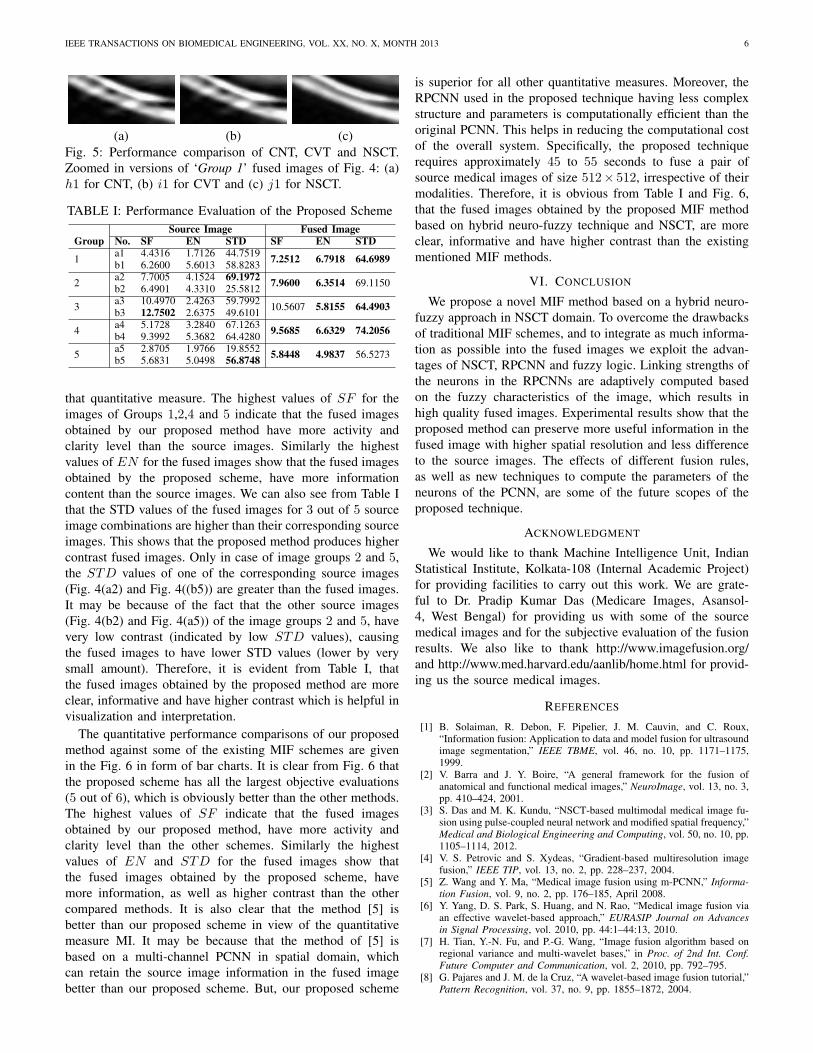
For the five pairs of source medical images the detailedquantitative evaluations are given in Table I and Fig.6.Columns 3 to 5 in the Table I, show the SF, EN and STD ofthe source medical images, respectively. The values of thesequantitative measures of the fused images obtained by theproposed technique are given in columns 6 to 8 of Table I.The ‘bold’ values indicate the highest values in Table I, for
IEEE TRANSACTIONS ON BIOMEDICAL ENGINEERING, VOL. XX, NO. X, MONTH 2013 6
(a) (b) (c)Fig. 5: Performance comparison of CNT, CVT and NSCT.Zoomed in versions of ‘Group 1’ fused images of Fig. 4: (a)h1 for CNT, (b) i1 for CVT and (c) j1 for NSCT.
TABLE I: Performance Evaluation of the Proposed SchemeSource Image Fused Image
Group No. SF EN STD SF EN STD
1 a1 4.4316 1.7126 44.7519 7.2512 6.7918 64.6989b1 6.2600 5.6013 58.8283
2 a2 7.7005 4.1524 69.1972 7.9600 6.3514 69.1150b2 6.4901 4.3310 25.5812
3 a3 10.4970 2.4263 59.7992 10.5607 5.8155 64.4903b3 12.7502 2.6375 49.6101
4 a4 5.1728 3.2840 67.1263 9.5685 6.6329 74.2056b4 9.3992 5.3682 64.4280
5 a5 2.8705 1.9766 19.8552 5.8448 4.9837 56.5273b5 5.6831 5.0498 56.8748
that quantitative measure. The highest values of SF for theimages of Groups 1,2,4 and 5 indicate that the fused imagesobtained by our proposed method have more activity andclarity level than the source images. Similarly the highestvalues of EN for the fused images show that the fused imagesobtained by the proposed scheme, have more informationcontent than the source images. We can also see from Table Ithat the STD values of the fused images for 3 out of 5 sourceimage combinations are higher than their corresponding sourceimages. This shows that the proposed method produces highercontrast fused images. Only in case of image groups 2 and 5,the STD values of one of the corresponding source images(Fig. 4(a2) and Fig. 4((b5)) are greater than the fused images.It may be because of the fact that the other source images(Fig. 4(b2) and Fig. 4(a5)) of the image groups 2 and 5, havevery low contrast (indicated by low STD values), causingthe fused images to have lower STD values (lower by verysmall amount). Therefore, it is evident from Table I, thatthe fused images obtained by the proposed method are moreclear, informative and have higher contrast which is helpful invisualization and interpretation.
The quantitative performance comparisons of our proposedmethod against some of the existing MIF schemes are givenin the Fig. 6 in form of bar charts. It is clear from Fig. 6 thatthe proposed scheme has all the largest objective evaluations(5 out of 6), which is obviously better than the other methods.The highest values of SF indicate that the fused imagesobtained by our proposed method, have more activity andclarity level than the other schemes. Similarly the highestvalues of EN and STD for the fused images show thatthe fused images obtained by the proposed scheme, havemore information, as well as higher contrast than the othercompared methods. It is also clear that the method [5] isbetter than our proposed scheme in view of the quantitativemeasure MI. It may be because that the method of [5] isbased on a multi-channel PCNN in spatial domain, whichcan retain the source image information in the fused imagebetter than our proposed scheme. But, our proposed scheme
is superior for all other quantitative measures. Moreover, theRPCNN used in the proposed technique having less complexstructure and parameters is computationally efficient than theoriginal PCNN. This helps in reducing the computational costof the overall system. Specifically, the proposed techniquerequires approximately 45 to 55 seconds to fuse a pair ofsource medical images of size 512× 512, irrespective of theirmodalities. Therefore, it is obvious from Table I and Fig. 6,that the fused images obtained by the proposed MIF methodbased on hybrid neuro-fuzzy technique and NSCT, are moreclear, informative and have higher contrast than the existingmentioned MIF methods.
VI. CONCLUSION
We propose a novel MIF method based on a hybrid neuro-fuzzy approach in NSCT domain. To overcome the drawbacksof traditional MIF schemes, and to integrate as much informa-tion as possible into the fused images we exploit the advan-tages of NSCT, RPCNN and fuzzy logic. Linking strengths ofthe neurons in the RPCNNs are adaptively computed basedon the fuzzy characteristics of the image, which results inhigh quality fused images. Experimental results show that theproposed method can preserve more useful information in thefused image with higher spatial resolution and less differenceto the source images. The effects of different fusion rules,as well as new techniques to compute the parameters of theneurons of the PCNN, are some of the future scopes of theproposed technique.
ACKNOWLEDGMENT
We would like to thank Machine Intelligence Unit, IndianStatistical Institute, Kolkata-108 (Internal Academic Project)for providing facilities to carry out this work. We are grate-ful to Dr. Pradip Kumar Das (Medicare Images, Asansol-4, West Bengal) for providing us with some of the sourcemedical images and for the subjective evaluation of the fusionresults. We also like to thank http://www.imagefusion.org/and http://www.med.harvard.edu/aanlib/home.html for provid-ing us the source medical images.
REFERENCES
[1] B. Solaiman, R. Debon, F. Pipelier, J. M. Cauvin, and C. Roux,“Information fusion: Application to data and model fusion for ultrasoundimage segmentation,” IEEE TBME, vol. 46, no. 10, pp. 1171–1175,1999.
[2] V. Barra and J. Y. Boire, “A general framework for the fusion ofanatomical and functional medical images,” NeuroImage, vol. 13, no. 3,pp. 410–424, 2001.
[3] S. Das and M. K. Kundu, “NSCT-based multimodal medical image fu-sion using pulse-coupled neural network and modified spatial frequency,”Medical and Biological Engineering and Computing, vol. 50, no. 10, pp.1105–1114, 2012.
[4] V. S. Petrovic and S. Xydeas, “Gradient-based multiresolution imagefusion,” IEEE TIP, vol. 13, no. 2, pp. 228–237, 2004.
[5] Z. Wang and Y. Ma, “Medical image fusion using m-PCNN,” Informa-tion Fusion, vol. 9, no. 2, pp. 176–185, April 2008.
[6] Y. Yang, D. S. Park, S. Huang, and N. Rao, “Medical image fusion viaan effective wavelet-based approach,” EURASIP Journal on Advancesin Signal Processing, vol. 2010, pp. 44:1–44:13, 2010.
[7] H. Tian, Y.-N. Fu, and P.-G. Wang, “Image fusion algorithm based onregional variance and multi-wavelet bases,” in Proc. of 2nd Int. Conf.Future Computer and Communication, vol. 2, 2010, pp. 792–795.
[8] G. Pajares and J. M. de la Cruz, “A wavelet-based image fusion tutorial,”Pattern Recognition, vol. 37, no. 9, pp. 1855–1872, 2004.

IEEE TRANSACTIONS ON BIOMEDICAL ENGINEERING, VOL. XX, NO. X, MONTH 2013 7
Fig. 6: Objective Performance Comparisons
[9] L. Yang, B. L. Guo, and W. Ni, “Multimodality medical image fusionbased on multiscale geometric analysis of contourlet transform,” Neu-rocomputing, vol. 72, no. 1-3, pp. 203–211, 2008.
[10] S. Das, M. Chowdhury, and M. K. Kundu, “Medical image fusion basedon ripplet transform type-I,” Progress In Electromagnetics Research B,vol. 30, pp. 355–370, 2011.
[11] R. Eckhorn, H. J. Reitboeck, M. Arndt, and P. Dicke, “Feature linkingvia synchronization among distributed assemblies: Simulations of resultsfrom cat visual cortex,” Neural Computation, vol. 2, pp. 293–307, 1990.
[12] F. C. Z. Wang, Y. Ma and L. Yang, “Review of pulse-coupled neuralnetworks,” Image Vision Comput., vol. 28, no. 1, pp. 5–13, 2010.
[13] J. L. Johnson, “Time signatures of images,” in Proc. of IEEE NeuralNetworks, vol. 2, 1994, pp. 1279–1284.
[14] Q. Xiao-Bo, Y. Jing-Wen, X. Hong-Zhi, and Z. Zi-Qian, “Image fusionalgorithm based on spatial frequency-motivated pulse coupled neural
networks in nonsubsampled contourlet transform domain,” Acta Auto-matica Sinica, vol. 34, no. 12, pp. 1508–1514, 2008.
[15] Z. Wang, Y. Ma, and J. Gu, “Multi-focus image fusion using PCNN,”Pattern Recognition, vol. 43, no. 6, pp. 2003–2016, June 2010.
[16] J. M. Kinser, “Foveation by a pulse-coupled neural network,” IEEE NN,vol. 10, no. 3, pp. 621–626, 1999.
[17] M. K. Kundu and S. K. Pal, “A note on the transformation betweenintensity and gray level,” Pattern Recognition Letters, vol. 8, pp. 257–269, 1988.
[18] M. K. Kundu and S. K. Pal, “Automatic selection of object enhancementoperator with quantitative justification based on fuzzy set theoreticmeasure,” Pattern Recognition Letters, vol. 11, pp. 811–829, 1990.
[19] J. M. Kinser, “Pulse coupled image fusion,” Optical Engineering,vol. 36, no. 3, pp. 737–742, 1997.
[20] A. da Cunha, J. Zhou, and M. Do, “The nonsubsampled contourlettransform: Theory, design, and applications,” IEEE TIP, vol. 15, no. 10,pp. 3089 –3101, 2006.
[21] J. Johnson and M. Padgett, “PCNN models and applications,” IEEETNN, vol. 10, no. 3, pp. 480–498, may 1999.
[22] J. A. Karvonen, “Baltic sea ice SAR segmentation and classificationusing modified pulse-coupled neural networks,” IEEE TGRS, vol. 42,no. 7, pp. 1566–1574, 2004.
[23] G. Buchsbaum, “An analytical derivation of visual nonlinearity,” IEEETBME, vol. 27, no. 5, pp. 237–242, 1980.
[24] M. K. Kundu and S. K. Pal, “Thresholding for edge detection usinghuman psycho visual phenomena,” Pattern Recognition Letters, vol. 4,pp. 433–441, 1986.
[25] H. Cheng and H. Xu, “A novel fuzzy logic approach to contrastenhancement,” Pattern Recognition, vol. 33, no. 5, pp. 809–819, 2000.
[26] T. Kadir and M. Brady, “Saliency, scale and image description,” Inter-national Journal of Computer Vision, vol. 45, no. 2, pp. 83–105, 2001.
[27] G. H. Qu, D. L. Zhang, and P. F. Yan, “Information measure forperformance of image fusion,” Electronic Letters, vol. 38, no. 7, pp.313–315, 2002.
[28] C. S. Xydeas and V. Petrovic, “Objective image fusion performancemeasure,” Electronic Letters, vol. 36, no. 4, pp. 308–309, 2000.
Sudeb Das received the M.C.A. degree from WBUT,India, in 2008. He is currently working towardhis Ph.D. from Calcutta University in ComputerSci. and doing his research in Indian StatisticalInstitute, Kolkata, India. He has contributed about15 research papers to well-known and prestigiousarchival journals, international refereed conferences.His research areas include Pattern Recognition, Dig-ital Watermarking, CBIR, biomedical imaging andSoft computing etc.
Malay Kumar Kundu (M’90-SM’99) received hisB. Tech., M. Tech. and Ph.D (Tech.) degrees inRadio physics and Electronics all are from the Uni-versity of Calcutta. Currently he is a full professor inthe Machine Intelligence Unit of the Indian Statis-tical Institute, Kolkata, India. He had been the headof the Machine Intelligence Unit from September1993 to November 1995 and Professor In-charge(Chairman) of the Computer and CommunicationSciences Division of the Institute during 2004 to2006.He is a Fellow of a The International Asso-
ciation for Pattern Recognition, USA (FIAPR), Indian National Academyof Engineering (FNAE), National Academy of Sciences (FNASc.), Indiaand the Institute of Electronics and Telecommunication Engineers (FIETE),India. A senior member of the IEEE, USA and the founding life memberand Vice President of the Indian Unit for Pattern Recognition and ArtificialIntelligence (IUPRAI).He was selected as INAE Distinguished Professor in2013. He received the prestigious VASVIK award for industrial researchin the field of Electronic Sciences and Technology for the year 1999.He also received the Sir. J. C. Bose memorial award of the Institute ofElectronics and Telecommunication Engineers (IETE), India in the year 1986.His current research interest includes Image/video processing and analysis,soft computing, computer vision, machine intelligence and data security. Hehas contributed 3 book volumes, about 140 research papers in well knownand prestigious archival journals, international refereed conferences and in theedited monograph volumes. He is the holder of nine U.S patents.