Embed Size (px)
Citation preview

SUBMITTED TO IEEE TRANSACTIONS ON BIOMEDICAL ENGINEERING 1
Healthy versus Pathological Learning Transferabilityin Shoulder Muscle MRI Segmentation using Deep
Convolutional Encoder-DecodersPierre-Henri Conze?, Sylvain Brochard, Valerie Burdin, Frances T. Sheehan and Christelle Pons
Abstract—Automatic segmentation of pathological shouldermuscles in patients with musculo-skeletal diseases is a challengingtask due to the huge variability in muscle shape, size, location,texture and injury. A reliable fully-automated segmentationmethod from magnetic resonance images could greatly helpclinicians to plan therapeutic interventions and predict inter-ventional outcomes while eliminating time consuming manualsegmentation efforts. The purpose of this work is three-fold. First,we investigate the feasibility of pathological shoulder musclesegmentation using deep learning techniques, given a very limitedamount of available annotated pediatric data. Second, we addressthe learning transferability from healthy to pathological databy comparing different learning schemes in terms of modelgeneralizability. Third, extended versions of deep convolutionalencoder-decoder architectures using encoders pre-trained onnon-medical data are proposed to improve the segmentationaccuracy. Methodological aspects are evaluated in a leave-one-outfashion on a dataset of 24 shoulder examinations from patientswith obstetrical brachial plexus palsy and focus on 4 differentmuscles including deltoid as well as infraspinatus, supraspinatusand subscapularis from the rotator cuff. The most relevantsegmentation model is partially pre-trained on ImageNet andjointly exploits inter-patient healthy and pathological annotateddata. Its performance reaches Dice scores of 82.4%, 82.0%,71.0% and 82.8% for deltoid, infraspinatus, supraspinatus andsubscapularis muscles. Absolute surface estimation errors are allbelow 83mm2 except for supraspinatus with 134.6mm2. Thesecontributions offer new perspectives for force inference in thecontext of musculo-skeletal disorder management.
Index Terms—Shoulder muscle segmentation, musculo-skeletaldisorders, deep convolutional encoder-decoders, healthy versuspathological transferability, obstetrical brachial plexus palsy
I. INTRODUCTION
P ixel-wise segmentation is a crucial task in medical im-age analysis with many applications such as computer-
assisted diagnosis, surgery planning, visual augmentation,image-guided interventions or extraction of quantitative in-dices from images. The rapid development of non-invasiveimaging technologies over the last decades has opened newhorizons in studying both healthy and pathological anatomy.However, the analysis of complex Magnetic Resonance (MR)
P.-H. Conze is with IMT Atlantique, Inserm UMR 1101, Brest, France. E-mail: [email protected]. S. Brochard is with RehabilitationMedicine Department, University Hospital of Brest, Inserm UMR 1101,Fondation ILDYS, Brest, France. E-mail : [email protected]. Burdin is with IMT Atlantique, Inserm UMR 1101, Brest, France. E-mail: [email protected]. F. T Sheehan is with RehabilitationMedicine, National Institutes of Health, Bethesda, USA. E-mail : [email protected]. C. Pons is with SSR pediatric, Fondation ILDYS, Brest,France. E-mail : [email protected]. Asterisk indicates corre-sponding author. Manuscript received —- XX, 2017; revised —- XX, 2017.
imaging datasets could be tedious and time-consuming forradiologist, clinicians or researchers. Thus, computerized as-sistance methods including robust automatic image segmen-tation techniques are needed to guide and improve imageinterpretation and clinical decision making.
In particular, the need for accurate muscle delineationsemerges for the management of musculo-skeletal disorders.The task becomes difficult when the pathology alters thesize, shape, texture and global MR appearance of muscles,especially when a large variability across patients arises dueto age-related development and injury. Obstetrical brachialplexus palsy (OBPP), one of the most common birth injury[1], is such a pathology. As for many other musculo-skeletaldiseases affecting children or adults, the limited amount ofavailable data in OBPP represents an additional challenge.
Characterized by the disorder of the peripheral nervoussystem conducting signals from the spinal cord to shoulders,arms and hands, OBPP occurs most often during the deliveryphase when lateral tractions are applied to the head to permitshoulder clearance [2]. Its incidence is estimated around 1.4every 1000 live births [3]. While full recovery is possible,prognosis is variable and up to 35% of children may havefrom weak to severe life-long functional impairments [2].Shoulder involvment is the main cause of morbidity. At thislevel, nerve injuries lead to variable muscle denervations andinduce muscle degeneration with fatty and fibrosis infiltration,disruption of growth, muscle atrophy and force imbalancesbetween agonist and antagonist muscle pairs [4].
Treatment and prevention of shoulder muscle strength im-balances are main therapeutic goals for children with OBPPwho do not fully recover [5]. Therapeutic interventions mainlyaim at restoring a physiological balance between muscleslength and strength to prevent or treat joint deformity andfacilitate movement of the upper limb [6]. Patient-specificinformation related to the degree of muscle atrophy acrossshoulder muscles is therefore needed to plan interventions andpredict interventional outcomes [7]. However, clinical exam-inations do not allow the distinction and precise evaluationof each shoulder muscle. Conversely, recent works report aclear relationship between muscle atrophy and strength lossfor children with OBPP [1], showing the need for musclemorphological evaluation to better understand and treat shoul-der problems. In this direction, shoulder muscle segmentationon MR images is useful to both quantify individual muscleinvolvement and analyze shoulder strength balance profiles ofnon-dominant and dominant shoulders for children with OBPP.
arX
iv:1
901.
0162
0v1
[cs
.CV
] 6
Jan
201
9

SUBMITTED TO IEEE TRANSACTIONS ON BIOMEDICAL ENGINEERING 2
Considering the large variability in shoulder muscle shape,size, location, texture and injury, muscle segmentation fromMR images has been commonly noticed as challenging [8].Both healthy and pathological muscles are prone to subtleor more severe changes regarding their visual properties andcould be difficult to delineate near their insertion areas dueto the vicinity with other structures (Fig.1). In the case ofpathological muscles, changes in muscle shape and signaloccur because of muscle degeneration which can render iden-tification of muscle boundaries even more difficult. To cir-cumvent these difficulties, muscle segmentation is traditionallyperformed manually in a slice-by-slice fashion [9]. However,manual segmentation is a time-consuming task, prone to errorsdue to intra and inter-expert variability. There has been arecent and growing interest in developing fully-automatedtechniques for muscle segmentation. This question is all themore significant as many efforts have been recently devotedto pixel-wise segmentation based on deep learning techniquesusing convolutional encoder-decoders [10].
In this direction, the purpose of our study is three-fold.First, we aim at studying the feasibility of automatic patho-logical shoulder muscle segmentation using deep convolutionalencoder-decoder networks, given the small amount of availableannotated data in children with OBPP. To our knowledge,shoulder muscle delineations have never been achieved auto-matically through deep learning techniques. Second, our workaddresses the learning transferability from healthy to patho-logic data and focuses in particular on how data available onboth healthy and pathological shoulder muscles can be jointlyexploited for pathological shoulder muscle delineation. Third,extended versions of deep convolutional encoder-decoder ar-chitectures using encoders pre-trained on non-medical dataare investigated to improve the segmentation accuracy. Ex-periments extend preliminary results [11] to four shouldermuscles including deltoid, infraspinatus, supraspinatus andsubscapularis. Our contributions reach a robust and fully-automated muscle segmentation pipeline which provides newinsights for the management of musculo-skeletal diseases.
II. RELATED WORKS
To get quantitative muscle volume measures, from whichforces can be derived [1], muscle segmentation is traditionallyperformed manually in a slice-by-slice fashion [9] from MRimages. This task is extremely time-consuming and requirestens of minutes to get accurate delineations for one single mus-cle. It is not usable for a great amount of data in research andclinical practice. In addition, manual segmentation is prone tointra and inter-expert variability given the variability of muscleshapes and the lack of clearly visible boundaries betweenmuscles and surrounding anatomy [12]. To ease the process, asemi-automatic processing based on transversal propagationsof manually-drawn masks can be considered [13]. It consistsof several ascending and descending non-linear registrationsapplied to manual masks to finally achieve volumetric resultsin less time than entire manual segmentation.
A model-based muscle segmentation incorporating a priorstatistical shape model can be employed to delineate muscles
axial coronal sagittal
—-
delto
id–
infr
aspi
natu
s–
supr
aspi
natu
s–
subs
capu
lari
s
Fig. 1: Groundtruth segmentations of pathological shouldermuscles including deltoid as well as infraspinatus, supraspina-tus and subscapularis from the rotator cuff. Axial, coronaland sagittal slices are extracted from a 3D MR examinationacquired for a child with obstetrical brachial plexus palsy.
boundaries in MR images. A patient-spectific 3D geometry isthus reached based on the deformation of a parametric ellipsefitted to muscle contours, starting from a reduced set of initialslices [14], [15]. Segmentation models can be further improvedby exploiting a-priori knowledge of shape information relyingon internal shape fitting and auto-correction to guide muscledelineation [16]. Baudin et al. associated in [17] a statisticalshape atlas with a random walks graph-based algorithm toautomatically segment individual muscles through iterativelinear optimization. Andrews et al. [18] used a probabilisticshape representation called generalized log-ratio representationthat included adjacency information along with a rotationallyinvariant boundary detector to segment thigh muscles.
Aligning and merging manually segmented images intospecific atlas coordinate spaces can be a reliable alternative.In this context, various single and multi-atlas methods havebeen proposed for quadriceps muscle segmentation [19], [20]relying on non-linear registration. Engstrom et al. [21] useda statistical shape model constrained using probabilistic MRatlases to automatically segment quadratus lumborum. Seg-mentation of muscle versus fatty tissues has been also per-formed through possibilistic clustering [22], histogram-basedthresholding followed by region growing [23] and active con-tours [24] techniques. However, all the previously describedmethods (except [18]) are not perfectly suited for high inter-subject shape variability and significant differences of tissueappearance due to injury. Moreover, many of the previouslydescribed methods are semi-automatic and hence require priorknowledge, usually associated with high computational costs.
Therefore, developing an accurate and robust muscle seg-mentation method remains an open and challenging issue,especially when dealing with pathological pediatric data. Hugeprogress has been recently made for automatic image seg-

SUBMITTED TO IEEE TRANSACTIONS ON BIOMEDICAL ENGINEERING 3
mentation using deep Convolutional Neural Networks (CNN).Introduced in [25], deep CNNs are entirely data-driven super-vised learning models formed by multi-layer neural networks.In contrast to conventional machine learning which requireshand-crafted features and hence specialized knowledge, deepCNNs automatically learn complex hierarchical features di-rectly from data. CNNs obtained outstanding performance formany medical image segmentation tasks [10] which suggeststhat robust fully-automated delineation of shoulder musclesfrom MR images may be achieved using CNN-based segmen-tation. To our knowledge, no other study has been conductedon shoulder muscle segmentation using deep learning methods.
III. METHOD
A. Deep convolutional encoder-decoders
The simplest way to perform segmentation using deepCNNs consists in classifying each pixel individually by work-ing on patches extracted around them [26]. Since input patchesfrom neighboring pixels have large overlaps, the same convo-lutions are computed many time. By replacing fully connectedlayers with convolutional layers, a Fully Convolutional Net-work (FCN) can take entire images as inputs and producelikelihood maps instead of single pixel outputs. It removes theneed to select representative patches and eliminates redundantcalculations due to patch overlaps. To avoid outputs withfar lower resolution than input shapes, FCNs can be appliedto shifted versions of input images [27]. Multiple resultingoutputs are stitched together to get results at full resolution.
Further improvements can be reached with architecturescomprising a regular FCN to extract features and capture con-text, followed by an up-sampling part that enables to recoverthe input resolution using up-convolutions [10]. Compared topatch-based or shift-and-stitch methods, it allows a preciselocalization in one single pass while taking into account thefull image context. Such architecture consisting of pairednetworks is called Convolutional Encoder-Decoder (CED).
U-Net [28] is the most well-known CED in the medicalimage analysis community. U-Net has a symmetrical architec-ture with equal amount of down-sampling and up-samplinglayers between contracting and expanding paths (Fig.3a). Theencoder gradually reduces the spatial dimension with poolinglayers whereas the decoder gradually recovers object detailsand spatial dimension. One key aspect of U-Net is the use ofskip connections which concatenate features from the encoderto the decoder to help in recovering object details better whileimproving localization accuracy. By allowing information toflow directly from low-level to high-level feature maps, fasterconvergence is achieved. This architecture can be exploited for3D volume segmentation [29] by replacing all 2D operationswith their 3D counterparts but at the cost of computationalspeed and GPU memory consumption. Processing 2D slicesindependently before reconstructing 3D medical volumes re-mains a simpler alternative. Instead of cross-entropy measuresused as loss function, the extension of U-Net proposed in [30]directly minimizes a segmentation error measure to to handleclass imbalance between foreground and background.
healthy
pathological all
HP
A
P
train test
Fig. 2: Three different learning schemes (P, HP, A) involvedin a leave-one-out setting for deep learning-based pathologicalshoulder muscle segmentation.
B. Healthy versus pathological learning transferability
In this work, we investigate the feasibility of automaticpathological shoulder muscle segmentation using deep CEDs(Sect.III-A), given a small amount of available annotateddata. In the OBPP context, the availability of both healthyand pathological data for image segmentation brings newqueries related to the learning transferability from healthy topathological structures. This aspect is particulary suitable tomusculo-skeletal pathologies for two reasons. First, despitedifferent shapes and sizes due to growth and atrophy, healthyand pathological muscles may share common characteristicssuch as anatomic locations and overall aspects. Second, suchmedical applications suffer from a limited amount of availableannotated data, usable for segmentation purposes. Combininghealthy and pathological data for deep learning-based segmen-tation can thus act as a smart data augmentation strategy.
In exploring the joint use of healthy and pathological datafor pathological muscle segmentation, determining the optimallearning scheme is crucial. Thus, three different learningschemes (Fig.2) employed with deep CEDs are considered:
- pathological only (P): the most common configurationconsists in exploiting groundtruth annotations made onimpaired shoulder muscles only, assuming that CED fea-tures extracted from healthy examinations are not suitedenough for pathological anatomies.
- healthy transfer to pathological (HP): another strategydeals with transfer learning and fine tuning from healthyto pathological muscles. In this context, a first CED istrained using groundtruth segmentations from unaffectedshoulders only. The weights of the resulting model arethen used as initialization for a second CED networkwhich is trained using pathological inputs only.
- simultaneous healthy and pathological (A): the last con-figuration consists in training a CED with a groundtruthdataset comprising annotations made on both healthy andpathological shoulder muscles, which allows to benefitfrom a more consequent dataset.
By comparing these different training strategies, we evaluatethe benefits brought by combining healthy and pathologicaldata together in terms of model generalizability. The balancebetween data augmentation and healthy versus pathological
SUBMITTED TO IEEE TRANSACTIONS ON BIOMEDICAL ENGINEERING 4
—-
a)U-Net
[28]
256
256
256
256
256
256
256
128
128
128
64 256
256
128
128
128
128
64
64
32
32
321
64
64
64
64
32
32
32
32 1
–b)v16pU-Net
512
512
512
512
512
512
512
512
256
256
256
256
128
512
512
512
256
256
256
256
256
128
128
64
64
643
128
128
128
128
64
64
64
64 1
3x3 conv + ReLU 3x3 conv (pre-trained) + ReLU 2x2 max pooling 1x1 conv + sigmoid 2x2 up conv copy and concatenate
Fig. 3: Extension of U-Net [28] by exploiting as encoder a slightly modified VGG-16 [31] with weights pre-trained on ImageNet[32], following [33], [34]. The decoder is modified to get an exactly symmetrical construction while keeping skip connections.
muscle variability is a crucial question which has never beeninvestigated for muscle segmentation. These three differentschemes, referred as P (pathological only), HP (healthy trans-fer to pathological) and A (simultaneous healthy and pathologi-cal), are illustrated Fig.2 in a leave-one-out setting. The overallshoulder dataset is divided into healthy and pathologicalMR examinations. Iteratively, one pathological examination isextracted from the pathological dataset and considered as testexamination for muscle segmentation. It will therefore not beused for training contrary to remaining pathological images.
C. Extended architectures with pre-trained encoders
Contrary to deep classification networks which are usuallypre-trained on a very large image dataset, CED architecturesused for segmentation are typically trained from scratch, rely-ing on randomly initialized weights. Reaching a generic modelwithout over-fitting is therefore tedious, especially when only asmall amount of images is available. As suggested in [33], theencoder part of a deep CED network can be replaced by a well-known classification network whose weights are pre-trainedon an initial classification task. It allows to exploit transferlearning from large datasets such as ImageNet [32] for deeplearning-based segmentation. In the literature, the encoder partof a deep CED has been already replaced by pre-trained VGG-11 [33] and ABN WideResnet-38 [34] with improvementscompared to their randomly weighted counterparts.
Following this idea, we propose to extend the standardU-Net architecture (Sect.III-A) by exploiting another simplenetwork from the VGG family [31] as encoder, namely theVGG-16 architecture. To improve performance, this encoderbranch is pre-trained on ImageNet [32]. This database has beendesigned for object recognition purposes and contains morethan 1 million natural images from 1000 classes. Pre-trainingour deep CED dedicated to muscle image segmentation usingnon-medical data is an efficient way to reduce the data scarcityissue while improving model generalizability [35]. Pre-trainedmodels can not only improve predictive performance but alsorequire less training time to reach convergence for the targettask. In particular, low-level features captured by first convolu-
tional layers are usually shared between different image typeswhich explains the success of transfer learning between tasks.
The VGG-16 encoder (Fig.3b) consists of sequential layersincluding 3× 3 convolutional layers followed by RectifiedLinear Unit (ReLU) activation functions. Reducing the spatialsize of the representation is handled by 2 × 2 max pool-ing layers. Compared to standard U-Net (Fig.3a), the firstconvolutional layer generates 64 channels instead of 32. Asthe network deepens, the number of channels doubles aftereach max pooling until it reaches 512 (256 for classical U-Net). After the second max pooling operation, the numberof convolutional layers differ from U-Net with patterns of 3consecutive convolutional layers instead of 2, following theoriginal VGG-16 architecture. In addition, input images areextended from one single greyscale channel to 3 channels byrepeating the same content in order to respect the dimensionsof the RGB ImageNet images used for encoder pre-training.The only differences with VGG-16 rely in the fact that thelast convolutional layer as well as top layers including fully-connected layers and softmax have been omitted. The two lastconvolutional layers taken from VGG-16 serve as central partof the CED and separate both contracting and expanding paths.
The extension of the U-Net encoder is transferred to thedecoder branch by adding 2 convolutional layers as well asmore features channels to get an exactly symmetrical con-struction while keeping skip connections. Contrary to encoderweights which are initialized using pre-training performed onImageNet, decoder weights are set randomly before fine tun-ing. As for U-Net, a final 1×1 convolutional layer followed bya sigmoid activation function achieves pixel-wise segmentationmasks whose resolution are the same as input slices.
IV. DATA AND EXPERIMENTAL RESULTS
A. Imaging dataset
Experimental data have been collected from a previousstudy [1] investigating the muscle volume-strength relationshipin 12 children with unilateral OPBB (averaged age 12.1± 3.3years). In this IRB approved study, informed consents and

SUBMITTED TO IEEE TRANSACTIONS ON BIOMEDICAL ENGINEERING 5
——
——
–P
——
——
–HP
——
——
–A
Fig. 4: Pathological deltoid segmentation using U-Net [28] embedded with learning schemes P, HP and A. Groundtruth andestimated delineations are in green and red. Displayed results cover the whole muscle spatial extent for L-P-0103 examination.
assents have been obtained for all subjects. For each patient,two 3D axial-plane T1-weighted gradient-echo MR imageshave been acquired: one for the affected shoulder and anotherfor the unaffected one. For each healthy or pathological MRimage, spatially equally distributed 2D axial slices have beenselected for four different shoulder muscles including deltoidas well as infraspinatus, supraspinatus and subscapularis fromthe rotator cuff. These slices were annotated by an expert inpediatric physical medicine and rehabilitation to reach pixel-wise groundtruth delineations. Image size for axial slices areconstant for each subject (416× 312 pixels). Image resolutionvaries from 0.55× 0.55 to 0.63× 0.63mm, allowing a finerresolution for smaller subjects. The number of axial slicesfluctuates from 192 to 224 whereas slice thickness remainsunchanged (1.2mm). Overall, we have 374 (resp. 395) anno-tated axial slices for deltoid, 306 (367) for infraspinatus, 238(208) for supraspinatus and 388 (401) for subscapularis across2400 (2448) axial slices arising from 12 pathological (healthy)examinations. Among these 24 MR images, pairings betweenaffected and unaffected shoulders are known. Due to sparseannotations (Fig.1), deep CEDs exploit as inputs 2D axialslices and produce 2D segmentation masks which can be thenstacked to recover a 3D volume for clinical purposes. Amongpathological examinations, 8 are right shoulders (R-P-0134,0684, 0382, 0447, 0660, 0737, 0667, 0277) whereas4 correspond to left shoulders (L-P-0103, 0351, 0922,0773). Training images displaying a right (left) shoulder areflipped when a left (right) shoulder is considered for test.
B. Segmentation assessmentTo assess both healthy versus pathological learning trans-
ferability (Sect.IV-C) and extended pre-trained deep convo-
lutional architectures (Sect.IV-D), the accuracy of automaticpathological shoulder muscle segmentation is quantified basedon Dice ( 2TP
2TP+FP+FN ), sensitivity ( TPTP+FN ), specificity
( TNTN+FP ) and Jaccard ( TP
TP+FP+FN ) similarity scores whereTP, FP, TN and FN are the number of true or false positive andnegative pixels. We also exploit an absolute surface estimationerror (ASE) which compares groundtruth and estimated musclesurfaces defined in mm2 from segmentation masks. Thesescores tend to provide a complete assessment of the ability ofCED models to provide contours identical to those manuallyperformed. Results provided are averaged among all annotatedslices arising from the 12 pathological shoulder examinations.
C. Healthy versus pathological learning transferability
The learning transferability from healthy to pathologicaldata (Sect.III-B) is addressed by comparing learning schemesP (pathological only), HP (healthy transfer to pathological) andA (simultaneous healthy and pathological) in a leave-one-outfashion (Fig.2). To avoid any biais for HP/A, annotated datafrom the healthy shoulder of the patient whose pathologicalshoulder is considered for test is not used during training.
For all schemes, deep CED networks are trained usingdata augmentation since the amount of available training datais limited. To teach the network the desired invariance androbustness properties [28], training 2D axial slices undergorandom scaling, rotation, shearing and shifting on both di-rections. 100 augmented images are thus produced for onesingle training axial slice. Comparisons between P, HP andA schemes are performed using standard U-Net [28] with 10epochs, a batch size of 10 images, an Adam optimizer with10−4 as learning rate for stochastic optimization, a fuzzy Dicescore as loss function and randomly initialized weights for

SUBMITTED TO IEEE TRANSACTIONS ON BIOMEDICAL ENGINEERING 6
metric scheme P HP Anetwork U-Net [28] v16U-Net v16pU-Net
dice
deltoid 68.94±29.9 71.05±29.5 78.32±24.4 80.05±23.1 82.42±20.4infraspinatus 71.38±24.7 77.00±22.5 81.58±18.3 81.91±19.0 81.98±18.6supraspinatus 64.94±28.0 65.69±29.6 65.68±30.7 67.30±29.4 70.98±28.7subscapularis 78.10±18.1 74.55±25.2 81.41±15.0 81.58±15.2 82.80±14.4
sens
deltoid 70.85±30.5 70.74±29.5 78.92±25.4 81.45±23.7 83.80±21.3infraspinatus 72.12±26.4 79.45±23.1 84.61±18.2 83.74±18.6 83.48±19.0supraspinatus 64.02±31.8 63.16±33.2 65.55±34.5 67.21±33.0 68.60±32.3subscapularis 78.89±19.7 74.75±27.3 82.53±18.1 81.75±18.8 84.36±16.5
spec
deltoid 99.61±0.80 99.56±1.07 99.85±0.19 99.82±0.22 99.84±0.22infraspinatus 99.82±0.23 99.82±0.22 99.84±0.18 99.86±0.17 99.86±0.18supraspinatus 99.86±0.18 99.90±0.13 99.88±0.15 99.86±0.17 99.91±0.12subscapularis 99.86±0.13 99.83±0.28 99.87±0.13 99.88±0.12 99.86±0.15
jacc
deltoid 59.27±29.7 61.68±29.3 69.48±26.0 71.46±24.9 74.00±22.8infraspinatus 60.32±25.6 66.91±24.0 72.00±20.4 72.63±20.6 72.71±21.0supraspinatus 53.61±27.1 55.27±29.3 55.70±30.1 56.98±28.7 61.31±28.7subscapularis 66.93±19.6 64.31±24.7 70.83±17.6 71.13±17.7 72.72±17.16
ASE
deltoid 252.0±421.6 268.0±507.8 105.5±178.9 94.23±139.2 80.38±127.5infraspinatus 156.8±228.7 92.37±105.9 74.47±92.8 80.11±96.2 79.17±96.9supraspinatus 174.8±164.0 159.9±153.5 153.9±146.0 147.5±129.4 134.6±135.5subscapularis 94.56±95.5 102.0±110.7 95.19±109.0 94.06±111.3 82.95±86.88
TABLE I: Quantitative assessment of deep CEDs (U-Net [28], v16U-Net, v16pU-Net) embedded with learning schemesP, HP and A over the pathological dataset in Dice, sensivity, specificity, Jaccard scores (%) and absolute surface error (ASE)in mm2. Best results are in bold. Italic underlined scores highlight best results among learning schemes employed with U-Net.
convolutional filters. Models were implemented using Kerasand trained on a recent desktop PC with a single NvidiaGeForce GTX 1080 Ti GPU with 11Gb/s. Once training isperformed, predictions for one single axial slice take around28ms only which is suitable for routine clinical practice.
We present Tab.I a comparative assessment of P, HP and Aaveraged over the whole pathological dataset. Reported resultscorrespond to the best results among those obtained after eachof the 10 training epochs. Italic underlined scores highlightbest results achieved among the three configurations.
Dice results show higher performance when both healthyand pathological data are simultaneously used for training (A)with scores of 78.32% for deltoid, 81.58% for infraspinatusand 81.41% for subscapularis. Scheme A outperforms transferlearning and fine tuning (HP) from 4 to 7% in terms ofDice since HP only obtains 71.05% for deltoid, 77% forinfraspinatus and 74.55% for subscapularis. However, thisconclusion does not apply to supraspinatus for which A and HPschemes get the same performance in Dice (≈65.7%). In par-ticular, A increases the sensitivity (65.55% instead of 63.16%)but provides a slightly smaller specificity compared to HP.Comparing ASE from HP to A reveals improvements for allshoulder muscles including deltoid whose surface estimationerror decreases from 268 to 105.5mm2. The same finding arisewhen studying Jaccard scores whose gains are 7.8% and 6.5%for deltoid and subscapularis. Directly combining healthy andpathological data therefore appears as a better strategy thandividing training into two parts focusing on first healthy andthen pathological data via transfer learning. Another findingbrought by Tab.I is that exploiting annotations made on patho-logical shoulder muscles only (P) is the worst training strategy,especially for deltoid (Dice loss of 10% from A to P). It provesthat CED features extracted from healthy examinations aresuited enough for pathological anatomies while acting as an
efficient data augmentation strategy. Results for subscapularisdeviate from this rule with higher similarity scores comparedto HP combined with the best ASE (94.56mm2).
Accuracy scores for supraspinatus are globally worse thanfor other shoulder muscles since its thin and elongated shapecan strongly vary across patients [16]. Moreover, we notice thepresence of a single severely atrophied supraspinatus muscle(L-P-0922) among the set of pathological examinations.Dice results for this single muscle is 42.99% for P against38.59% and 32.33% for HP and A. It suggests that musclesundergoing very strong degrees of injury must be processedseparately, relying on pathological data only or manual de-lineations. Nevetheless, A appears globally better suited fromweak to moderately severe muscle impairments.
Dedicated to the deltoid muscle, a comparison betweenP, HP and A is provided Fig.5 for each annotated slices ofthe whole pathological dataset. The top raw displays Dicescores with respect to normalized axial slice number obtainedby linearly scaling slice number from [zmin, zmax] to [0, 1]where {zmin, zmax} are the minimal and maximal axialslice number displaying the deltoid. Overall, the bell-shapecurves indicate that segmentation results are more accuratefor mid-muscle regions than for both base and apex wheremuscles appear smaller with strong appearance similaritieswith surrounding tissues. Above conclusions (A>HP>P) areconfirmed with much more individual Dice results groupedon the interval [75, 95%] for A. By studying the concordancebetween predicted and groundtruth deltoid surfaces (bottomrow), we observe a stronger correlation for A than for P andHP with individual estimations closer to the line of perfectconcordance (L-P-0773 is the most telling example).
Evaluation is supplemented by qualitative results given Fig.4for deltoid (P, HP and A) and Fig.6 for rotator cuff muscles(A only). Displayed results cover the whole muscle spatial
SUBMITTED TO IEEE TRANSACTIONS ON BIOMEDICAL ENGINEERING 7
Fig. 5: Deltoid segmentation accuracy using U-Net [28] with learning schemes P, HP and A for each annotated slices of thewhole pathological dataset. Top raw show Dice scores with respect to normalized axial slice number. Bottom row displaysconcordance between groundtruth and predicted muscle surfaces in mm2. Black line indicates perfect concordance.
——
-in
fras
pina
tus
——
-su
pras
pina
tus
——
-su
bsca
pula
ris
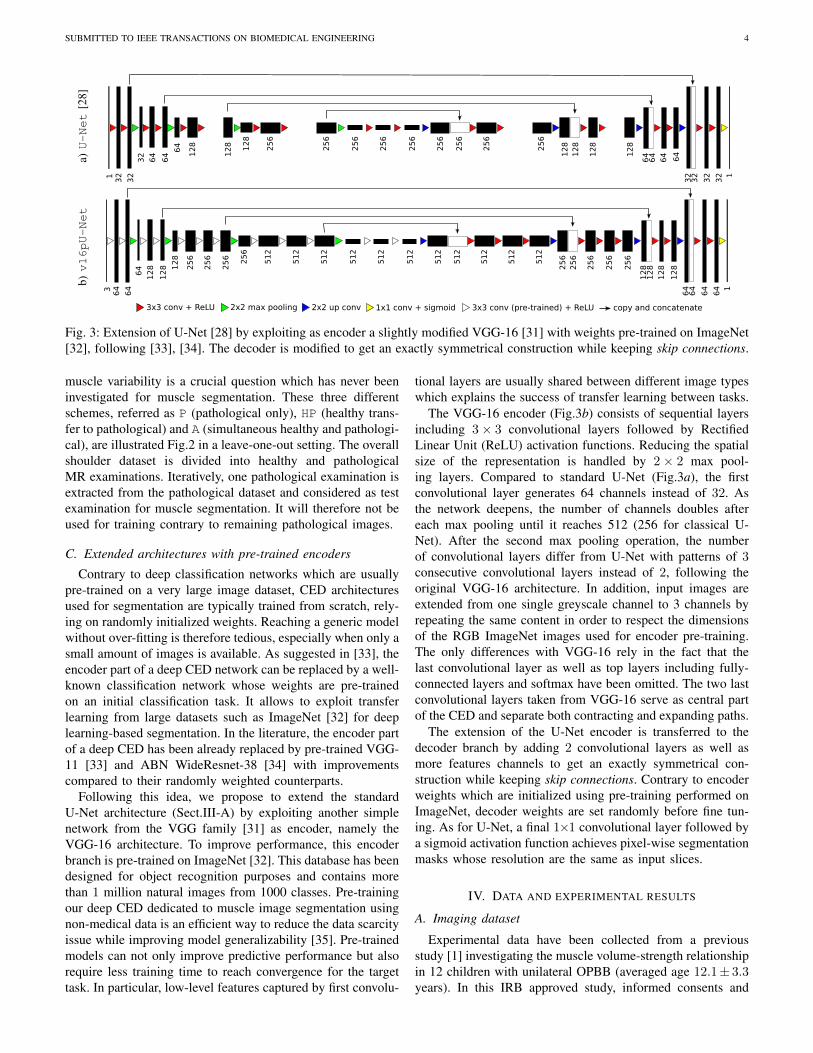
Fig. 6: Automatic pathological segmentation of infraspinatus, supraspinatus and subscapularis using U-Net [28] with trainingon both healthy and pathological data simultaneously (A). Groundtruth and estimated delineations are resp. in green and red.Displayed results cover whole muscle spatial extents resp. for R-P-0447, R-P-0660 and R-P-0134 examinations.

SUBMITTED TO IEEE TRANSACTIONS ON BIOMEDICAL ENGINEERING 8
extents. We notice Fig.4 a very accurate deltoid delineationfor A whereas P and HP tend to under-segment the musclearea. Complex muscle shapes and subtle contours illustratedFig.6 are relatively well captured. In addition, we can noticeoutstanding performance near muscle insertion regions whosecontours are usually hard to extract due to the vicinity withother structures. These results confirm that using simulta-neously healthy and pathological data for training helps inproviding good model generalizability despite the data scarcityissue combined with large appearance variabilities.
D. Extended deep convolutional architectures
The standard U-Net architecture [28] is extended Sect.III-Cby exploiting VGG-16 as encoder, without (v16U-Net) orwith (v16pU-Net) pre-trained weights based on ImageNet.Let us compare these three deep CEDs for pathological shoul-der muscle segmentation through leave-one-out experiments.We rely on the best obtained training scheme (Sect.IV-C)combining healthy and pathological data (A). As previously,networks are trained with data augmentation, 10 epochs, abatch size of 10 images, Adam optimizer and a fuzzy Dicescore as loss function. Learning rates change from U-Netand v16pU-Net (10−4) to v16U-Net (5 × 10⁻⁵) to avoiddivergence for deep networks trained with random weights.
Tab.I indicate the performance reached by U-Net,v16U-Net and v16pU-Net over the whole pathologi-cal dataset with best scores in bold. It clearly shows thatv16pU-Net globally outperforms U-Net and v16U-Netwith Dice scores of 82.42% for deltoid, 81.98% for infraspina-tus, 70.98% for supraspinatus and 82.80% for subscapularis.On the contrary, v16U-Net (resp. U-Net) obtains 80.05%(78.32%) for deltoid, 81.91% (81.58%) for infraspinatus,67.30% (65.68%) for supraspinatus and 81.58% (81.41%)for subscapularis. In one hand, despite slightly worse scorescompared with U-Net for infraspinatus in terms of sensitivity(83.74 against 84.61) and ASE (80.11 against 74.47mm2),v16U-Net is most likely to provide good predictive perfor-mance and model generalizability thanks to its deeper archi-tecture. On the other hand, comparisons between v16U-Netand v16pU-Net reveals that pre-training the encoder usingImageNet brings non-negligible improvements. For instance,v16pU-Net provides significant gains for deltoid (resp.supraspinatus) whose Jaccard score goes from 71.46 (56.98) to74% (61.31%). Surface estimation errors are among the lowestobtained with only 80.38mm2 for deltoid and 82.95mm2
for subscapularis. Despite their non-medical nature, the largeamount of ImageNet images used for pre-training makes thenetwork converge towards a better solution. v16pU-Net istherefore the most able to efficiently discriminate individualmuscles from surrounding anatomical structures, compared toU-Net and v16U-Net. In average among the four shouldermuscles, gains for Dice, sensivity and Jaccard reaches 2.8, 2.7and 3.2% from U-Net to v16pU-Net.
Fig.7 focuses on the deltoid muscle to compare U-Net,v16U-Net and v16pU-Net for each annotated slices ofthe whole pathological dataset. As previously, comparisons areperformed according to Dice scores (top row) and predictedversus groundtruth surface concordance (bottom row). From
U-Net to v16pU-Net, we can notice that individual Diceresults are slightly pushed towards the upper limit (100%)with less variability and an increased overall consistency alongaxial slices, as for R-P-0737 and L-P-0773. Extreme axialslices are much better handled in the case v16pU-Net,especially when normalized slice numbers approach zero.In addition, compared to U-Net and v16U-Net, a slightlystronger correlation between predicted and groundtruth deltoidsurface can be seen for v16pU-Net. Great improvements forR-P-0737 and L-P-0773 can be also highlighted.
Automatic pathological segmentation results given Fig.8for deltoid, infraspinatus, supraspinatus and subscapularis us-ing U-Net, v16U-Net and v16pU-Net allows to visuallyconfirm the conclusions (v16pU-Net>v16U-Net>U-Net)given above. 8 pathological examinations among the 12 avail-able are involved to give a complete overview. Globally,better contour adherence and shape consistency are reachedby v16pU-Net whose ability to mimmic expert annotationsis notable. The great diversity in terms of textures (smoothin R-P-0684 versus granular in R-P-0737) is accuratelycaptured despite high similar visual properties with surround-ing structures. Visual results also reveal that v16pU-Nethas a good behavior for complex muscle insertion regions(R-P-0447). Despite a satisfactory overall quality, U-Netand v16U-Net are frequently prone to under- (R-P-0134,R-P-0277) or over-segmentation (R-P-0684). Some ex-amples report inconsistent shapes (R-P-0667, R-P-0737),sometimes combined with false positive areas located faraway from the groundtruth muscle location (R-P-0447,L-P-0773). Using a pre-trained and complex architecturesuch as v16pU-Net to process simultaneously healthy andpathological data automatically provides accurate delineationsof pathological shoulder muscles for patients with OPBB.
V. CONCLUSION
In this work, we successfully addressed automatic patho-logical shoulder muscle MRI segmentation for patients withobstetrical brachial plexus palsy by means of deep convolu-tional encoder-decoders. In particular, we studied healthy topathological learning transferability by comparing differentlearning schemes in terms of model generalizability againstlarge muscle shape, size, location, texture and injury variabil-ity. Moreover, convolutional encoder-decoder networks wereexpanded using VGG-16 encoders pre-trained on ImageNet toimprove the accuracy reached by standard U-Net architectures.Our contributions were evaluated on four different shouldermuscles. First, results clearly show that features extractedfrom unimpaired limbs are suited enough for pathologicalanatomies while acting as an efficient data augmentationstrategy. Compared to transfer learning, combining healthy andpathological data for training provides the best segmentationaccuracy together with outstanding delineation performancefor muscle boundaries including insertion areas. Second, ex-periments reveal that convolutional encoder-decoders involv-ing a pre-trained VGG-16 encoder strongly outperforms U-Net. Despite the non-medical nature of pre-training data, suchdeeper networks are able to efficiently discriminate individual

SUBMITTED TO IEEE TRANSACTIONS ON BIOMEDICAL ENGINEERING 9
Fig. 7: Deltoid segmentation accuracy using U-Net [28], v16U-Net and v16pU-Net with learning scheme A for eachannotated slices of the whole pathological dataset. Top raw show Dice scores with respect to normalized axial slice number.Bottom row displays concordance between groundtruth and predicted muscle surfaces. Black line indicates perfect concordance.
deltoid infraspinatus supraspinatus subscapularis
——
U-N
et[2
8]—
—v16U-Net
—–v16pU-Net
R-P-0134 R-P-0447 R-P-0667 R-P-0737 L-P-0773 R-P-0684 L-P-0103 R-P-0277
Fig. 8: Automatic pathological segmentation of deltoid, infraspinatus, supraspinatus and subscapularis using U-Net [28],v16U-Net and v16pU-Net with training on both healthy and pathological data simultaneously (A). Groundtruth and estimateddelineations are resp. in green and red.

SUBMITTED TO IEEE TRANSACTIONS ON BIOMEDICAL ENGINEERING 10
muscles from surrounding anatomical structures. The proposedapproach can be easily extended to other muscle types andimaging modalities to provide clinical decision support invarious applications including neuro-muscular diseases, sportsrelated injuries or any other muscle disorders. Theses conclu-sions offer new perspectives for the management of musculo-skeletal diseases, even if a small and heterogeneous set ofdata is available. It paves the way for automatic inference ofindividual morphological parameters which are not accessiblewith simple clinical examinations. Our method could be usefulto distinguish between pathologies, evaluate the effect oftreatments and facilitate surveillance of neuromuscular diseaseprogression. It could also be integrated into biomechanicalmodels to improve the understanding of complex pathologiesand help clinicians for intervention planning.
REFERENCES
[1] C. Pons, F. T. Sheehan, H. S. Im, S. Brochard, and K. E. Alter, “Shouldermuscle atrophy and its relation to strength loss in obstetrical brachialplexus palsy,” Clinical Biomechanics, vol. 48, pp. 80–87, 2017.
[2] P. OBerry, M. Brown, L. Phillips, and S. H. Evans, “Obstetrical brachialplexus palsy,” Current Problems in Pediatric and Adolescent HealthCare, vol. 47, no. 7, pp. 151–155, 2017.
[3] S. P. Chauhan, S. B. Blackwell, and C. V. Ananth, “Neonatal brachialplexus palsy: incidence, prevalence, and temporal trends,” in Seminarsin Perinatology, vol. 38, no. 4, 2014, pp. 210–218.
[4] S. Brochard, K. Alter, and D. Damiano, “Shoulder strength profilesin children with and without brachial plexus palsy,” Muscle & Nerve,vol. 50, no. 1, pp. 60–66, 2014.
[5] P. M. Waters, J. T. Monica, B. E. Earp, D. Zurakowski, and D. S.Bae, “Correlation of radiographic muscle cross-sectional area withglenohumeral deformity in children with brachial plexus birth palsy,”The Journal of Bone and Joint Surgery, vol. 91, no. 10, p. 2367, 2009.
[6] S. H. Kozin, “The evaluation and treatment of children with brachialplexus birth palsy,” The Journal of Hand Surgery, vol. 36, no. 8, pp.1360–1369, 2011.
[7] A. Aydin, A. Bicer, T. Ozkan, B. Mersa, S. Ozkan, and Z. H. Yildirim,“Does primary brachial plexus surgery alter palliative tendon transfersurgery outcomes in children with obstetric paralysis?” BMC Muscu-loskeletal Disorders, vol. 12, no. 1, p. 74, 2011.
[8] Y. Barnouin, G. Butler-Browne, T. Voit, D. Reversat, N. Azzabou,G. Leroux, A. Behin, J. S. McPhee, P. G. Carlier, and J.-Y. Hogrel,“Manual segmentation of individual muscles of the quadriceps femorisusing MRI: a reappraisal,” Journal of Magnetic Resonance Imaging,vol. 40, no. 1, pp. 239–247, 2014.
[9] M. J. Tingart, M. Apreleva, J. T. Lehtinen, B. Capell, W. E. Palmer,and J. J. Warner, “Magnetic resonance imaging in quantitative analysisof rotator cuff muscle volume,” Clinical Orthopaedics and RelatedResearch, vol. 415, pp. 104–110, 2003.
[10] G. Litjens, T. Kooi, B. E. Bejnordi, A. A. A. Setio, F. Ciompi,M. Ghafoorian, J. A. van der Laak, B. Van Ginneken, and C. I. Sanchez,“A survey on deep learning in medical image analysis,” Medical ImageAnalysis, vol. 42, pp. 60–88, 2017.
[11] P.-H. Conze, C. Pons, V. Burdin, F. T. Sheehan, and S. Brochard, “Deepconvolutional encoder-decoders for deltoid segmentation using healthyversus pathological learning transferability,” in IEEE International Sym-posium on Biomedical Imaging, 2019.
[12] C. Pons, B. Borotikar, M. Garetier, V. Burdin, D. B. Salem, M. Lem-pereur, and S. Brochard, “Quantifying skeletal muscle volume and shapein humans using mri: A systematic review of validity and reliability,”PloS one, vol. 13, no. 11, 2018.
[13] A. Ogier, M. Sdika, A. Foure, A. Le Troter, and D. Bendahan, “Individ-ual muscle segmentation in MR images: A 3D propagation through 2Dnon-linear registration approaches,” in IEEE International Engineeringin Medicine and Biology Conference, 2017, pp. 317–320.
[14] I. Sudhoff, J. A. de Guise, A. Nordez, E. Jolivet, D. Bonneau, V. Khoury,and W. Skalli, “3D-patient-specific geometry of the muscles involvedin knee motion from selected MRI images,” Medical & BiologicalEngineering & Computing, vol. 47, no. 6, pp. 579–587, 2009.
[15] E. Jolivet, E. Dion, P. Rouch, G. Dubois, R. Charrier, C. Payan,and W. Skalli, “Skeletal muscle segmentation from MRI dataset usinga model-based approach,” Computer Methods in Biomechanics andBiomedical Engineering: Imaging & Visualization, vol. 2, no. 3, pp.138–145, 2014.
[16] S. Kim, D. Lee, S. Park, K.-S. Oh, S. W. Chung, and Y. Kim, “Automaticsegmentation of supraspinatus from MRI by internal shape fitting andautocorrection,” Computer Methods and Programs in Biomedicine, vol.140, pp. 165–174, 2017.
[17] P.-Y. Baudin, N. Azzabou, P. G. Carlier, and N. Paragios, “Priorknowledge, random walks and human skeletal muscle segmentation,” inInternational Conference on Medical Image Computing and Computer-Assisted Intervention, 2012, pp. 569–576.
[18] S. Andrews and G. Hamarneh, “The generalized log-ratio transforma-tion: learning shape and adjacency priors for simultaneous thigh musclesegmentation,” IEEE Transactions on Medical Imaging, vol. 34, no. 9,pp. 1773–1787, 2015.
[19] E. Ahmad, M. H. Yap, H. Degens, and J. S. McPhee, “Atlas-registrationbased image segmentation of MRI human thigh muscles in 3D space,”in Medical Imaging: Image Perception, Observer Performance, andTechnology Assessment, vol. 9037, 2014.
[20] A. Le Troter, A. Foure, M. Guye, S. Confort-Gouny, J.-P. Mattei,J. Gondin, E. Salort-Campana, and D. Bendahan, “Volume measure-ments of individual muscles in human quadriceps femoris using atlas-based segmentation approaches,” Magnetic Resonance Materials inPhysics, Biology and Medicine, vol. 29, no. 2, pp. 245–257, 2016.
[21] C. M. Engstrom, J. Fripp, V. Jurcak, D. G. Walker, O. Salvado, andS. Crozier, “Segmentation of the quadratus lumborum muscle usingstatistical shape modeling,” Journal of Magnetic Resonance Imaging,vol. 33, no. 6, pp. 1422–1429, 2011.
[22] V. Barra and J.-Y. Boire, “Segmentation of fat and muscle from MRimages of the thigh by a possibilistic clustering algorithm,” ComputerMethods and Programs in Biomedicine, vol. 68, no. 3, 2002.
[23] S. Purushwalkam, B. Li, Q. Meng, and J. McPhee, “Automatic seg-mentation of adipose tissue from thigh magnetic resonance images,” inInternational Conference Image Analysis and Recognition, 2013, pp.451–458.
[24] S. Orgiu, C. L. Lafortuna, F. Rastelli, M. Cadioli, A. Falini, andG. Rizzo, “Automatic muscle and fat segmentation in the thigh fromT1-Weighted MRI,” Journal of Magnetic Resonance Imaging, vol. 43,no. 3, pp. 601–610, 2016.
[25] Y. LeCun, L. Bottou, Y. Bengio, and P. Haffner, “Gradient-based learningapplied to document recognition,” Proceedings of the IEEE, vol. 86,no. 11, pp. 2278–2324, 1998.
[26] D. Ciresan, A. Giusti, L. M. Gambardella, and J. Schmidhuber, “Deepneural networks segment neuronal membranes in electron microscopyimages,” in Advances in Neural Information Processing Systems, 2012,pp. 2843–2851.
[27] J. Long, E. Shelhamer, and T. Darrell, “Fully convolutional networks forsemantic segmentation,” in IEEE Conference on Computer Vision andPattern Recognition, 2015, pp. 3431–3440.
[28] O. Ronneberger, P. Fischer, and T. Brox, “U-Net: Convolutional net-works for biomedical image segmentation,” in International Conferenceon Medical Image Computing and Computer-Assisted Intervention,2015, pp. 234–241.
[29] O. Cicek, A. Abdulkadir, S. S. Lienkamp, T. Brox, and O. Ronneberger,“3D U-Net: learning dense volumetric segmentation from sparse anno-tation,” in International Conference on Medical Image Computing andComputer-Assisted Intervention, 2016, pp. 424–432.
[30] F. Milletari, N. Navab, and S.-A. Ahmadi, “V-Net: Fully convolutionalneural networks for volumetric medical image segmentation,” in Inter-national Conference on 3D Vision, 2016, pp. 565–571.
[31] K. Simonyan and A. Zisserman, “Very deep convolutional networks forlarge-scale image recognition,” arXiv preprint arXiv:1409.1556, 2014.
[32] O. Russakovsky, J. Deng, H. Su, J. Krause, S. Satheesh, S. Ma,Z. Huang, A. Karpathy, A. Khosla, M. Bernstein et al., “ImageNet largescale visual recognition challenge,” International Journal of ComputerVision, vol. 115, no. 3, pp. 211–252, 2015.
[33] V. Iglovikov and A. Shvets, “TernausNet: U-Net with VGG11 en-coder pre-trained on imagenet for image segmentation,” arXiv preprintarXiv:1801.05746, 2018.
[34] V. Iglovikov, S. Seferbekov, A. Buslaev, and A. Shvets, “TernausNetV2:Fully convolutional network for instance segmentation,” arXiv preprintarXiv:1806.00844, 2018.
[35] J. Yosinski, J. Clune, Y. Bengio, and H. Lipson, “How transferable arefeatures in deep neural networks?” in Advances in Neural InformationProcessing Systems, 2014, pp. 3320–3328.