Embed Size (px)
Citation preview
RESEARCH Open Access
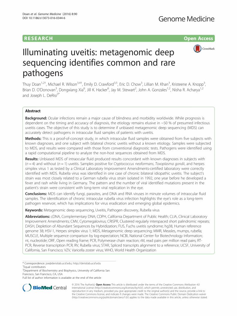
Illuminating uveitis: metagenomic deepsequencing identifies common and rarepathogensThuy Doan1,2†, Michael R. Wilson3,4†, Emily D. Crawford3,5, Eric D. Chow3, Lillian M. Khan3, Kristeene A. Knopp3,Brian D. O’Donovan3, Dongxiang Xia6, Jill K. Hacker6, Jay M. Stewart2, John A. Gonzales1,2, Nisha R. Acharya1,2
and Joseph L. DeRisi3*
Abstract
Background: Ocular infections remain a major cause of blindness and morbidity worldwide. While prognosis isdependent on the timing and accuracy of diagnosis, the etiology remains elusive in ~50 % of presumed infectiousuveitis cases. The objective of this study is to determine if unbiased metagenomic deep sequencing (MDS) canaccurately detect pathogens in intraocular fluid samples of patients with uveitis.
Methods: This is a proof-of-concept study, in which intraocular fluid samples were obtained from five subjects withknown diagnoses, and one subject with bilateral chronic uveitis without a known etiology. Samples were subjectedto MDS, and results were compared with those from conventional diagnostic tests. Pathogens were identified usinga rapid computational pipeline to analyze the non-host sequences obtained from MDS.
Results: Unbiased MDS of intraocular fluid produced results concordant with known diagnoses in subjects with(n = 4) and without (n = 1) uveitis. Samples positive for Cryptococcus neoformans, Toxoplasma gondii, and herpessimplex virus 1 as tested by a Clinical Laboratory Improvement Amendments-certified laboratory were correctlyidentified with MDS. Rubella virus was identified in one case of chronic bilateral idiopathic uveitis. The subject’sstrain was most closely related to a German rubella virus strain isolated in 1992, one year before he developed afever and rash while living in Germany. The pattern and the number of viral identified mutations present in thepatient’s strain were consistent with long-term viral replication in the eye.
Conclusions: MDS can identify fungi, parasites, and DNA and RNA viruses in minute volumes of intraocular fluidsamples. The identification of chronic intraocular rubella virus infection highlights the eye’s role as a long-termpathogen reservoir, which has implications for virus eradication and emerging global epidemics.
Keywords: Metagenomic deep sequencing, Uveitis, Pathogen discovery, Rubella virus
Abbreviations: cDNA, Complementary DNA; CDPH, California Department of Public Health; CLIA, Clinical LaboratoryImprovement Amendments; CMV, Cytomegalovirus; CRISPR, Clustered regularly interspaced short palindromic repeats;DASH, Depletion of Abundant Sequences by Hybridization; FUS, Fuchs uveitis syndrome; hg38, Human referencegenome 38; HSV-1, Herpes simplex virus 1; MDS, Metagenomic deep sequencing; MMR, Measles, mumps, rubella;MUSCLE, Multiple sequence comparison by log-expectation; NCBI, National Center for Biotechnology Information;nt, nucleotide; ORF, Open reading frame; PCR, Polymerase chain reaction; rM, read pairs per million read pairs; RT-PCR, Reverse transcription PCR; RV, Rubella virus; STAR, Spliced transcripts alignment to a reference; UCSF, University ofCalifornia, San Francisco; VZV, Varicella zoster virus; WHO, World Health Organization
* Correspondence: [email protected]; http://derisilab.ucsf.edu†Equal contributors3Department of Biochemistry and Biophysics, University of California SanFrancisco, San Francisco, CA, USAFull list of author information is available at the end of the article
© 2016 The Author(s). Open Access This article is distributed under the terms of the Creative Commons Attribution 4.0International License (http://creativecommons.org/licenses/by/4.0/), which permits unrestricted use, distribution, andreproduction in any medium, provided you give appropriate credit to the original author(s) and the source, provide a link tothe Creative Commons license, and indicate if changes were made. The Creative Commons Public Domain Dedication waiver(http://creativecommons.org/publicdomain/zero/1.0/) applies to the data made available in this article, unless otherwise stated.
Doan et al. Genome Medicine (2016) 8:90 DOI 10.1186/s13073-016-0344-6
BackgroundOcular infection is an important cause of ocular morbidityand blindness worldwide. However, diagnosis is challengingdue to the multitude of possible pathogens. The sensitivityof culture-based assays ranges from 40 to 70 %, and availablemolecular diagnostic tests target only a fraction of pathogensknown to cause ocular disease [1–3]. These limitations areexacerbated by (1) the inability to collect large intraocularfluid volumes given the eye’s small and delicate anatomy,and (2) the difficulty in distinguishing clinically between in-fectious and non-infectious causes of ocular inflammation.The urgency to develop better diagnostics for uveitis has
been compounded by the recent cases of persistent infec-tion with Ebola virus [4], and possibly Zika virus [5]. Thesecases highlight the eye’s role as a potential reservoir for in-fectious agents, with important public health consequences.It is essential that more sensitive, unbiased, and compre-hensive approaches are developed to efficiently diagnoseocular infections.Rapid advances in sequencing technology and bioinfor-
matics have made metagenomics a fertile area for develop-ing clinical diagnostics [6–8]. This prompted us to evaluatea hypothesis-free approach to identify ocular infections byperforming unbiased metagenomic deep sequencing (MDS)on clinical intraocular samples from patients with uveitis.
MethodsStudy designSix subjects were recruited for a research study using un-biased MDS to identify potential pathogens in intraocular
fluid (aqueous or vitreous) (Table 1). This study wasconducted according to the guidelines laid down in theDeclaration of Helsinki and approved by the Institu-tional Review Board of the University of California, SanFrancisco (UCSF). Five of the six subjects served as con-trols to benchmark the ability of MDS to identify a var-iety of pathogens; subjects 1–3 had ocular infectionswith herpes simplex virus 1 (HSV-1), Cryptococcus neo-formans, and Toxoplasma gondii, respectively. HSV-1and T. gondii-directed qualitative PCRs and cultureswere performed at the Proctor Foundation, a ClinicalLaboratory Improvement Amendments (CLIA)-certifiedlaboratory for ocular testing. Subject 4 had non-infec-tious uveitis clinically demonstrated by the resolution ofintraocular inflammation followed by intraocular injec-tion of a dexamethasone intravitreal implant and theinitiation of systemic immunosuppression with antime-tabolites. Subject 5 had no ocular inflammation but hadintraocular fluid obtained at the time of a retinal mem-brane peel. MDS was also used to investigate subject 6,who had bilateral uveitis that had defied a 16-year diag-nostic work-up at multiple academic centers across twocontinents (Table 1).
Sequencing library preparationSamples were prepared for MDS as previously described[6]. RNA was extracted from 20–50 μL of intraocularfluid using TRIzol LS reagent (ThermoFisher Scientific,PA, USA) and the RNA Clean & Concentrator Kit(Zymo Research, CA, USA) per the manufacturers'
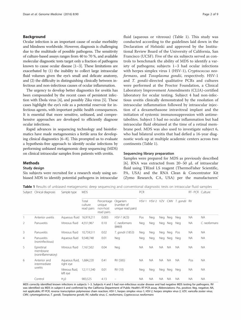
Table 1 Results of unbiased metagenomic deep sequencing and conventional diagnostic tests on intraocular fluid samples
Subject Clinical diagnosis Sample type MDS PCR RT- PCR Culture
Totalcultureread pairs
Percentageuniquenon-hostread pairs
Organism(number ofunique read pairs)
HSV-1 HSV-2 VZV CMV T. gondii RV
1 Anterior uveitis Aqueous fluid 16,919,211 0.003 HSV-1 (423) Pos Neg Neg Neg Neg NA NA
2 Panuveitis Vitreous fluid 4,551,967 0.10 C. neoformans(8469)
Neg Neg Neg Neg Neg NA C. neoformans
3 Panuveitis Vitreous fluid 10,759,511 0.02 T. gondii (1853) Neg Neg Neg Neg Pos NA NA
4 Panuveitis(noninfectious)
Aqueous fluid 9,548,748 0.01 Neg Neg Neg Neg Neg Neg NA NA
5 Epiretinalmembrane(noninflammatory)
Vitreous fluid 7,167,502 0.04 Neg NA NA NA NA NA NA NA
6 Anterior andintermediateuveitis
Aqueous fluid,right eye
1,684,220 0.41 RV (585) NA NA NA NA NA Pos NA
Vitreous fluid,left eye
12,111,540 0.01 RV (10) Neg Neg Neg Neg Neg NA NA
Control H20 983,525 4.13 – NA NA NA NA NA NA NA
MDS correctly identified known infections in subjects 1–3. Subjects 4 and 5 had non-infectious ocular disease and had negative MDS testing for pathogens. RVwas identified via MDS in subject 6 and confirmed by the California Department of Public Health’s RT-PCR assay. Abbreviations: Pos, positive; Neg, negative; NA,not applicable; RT-PCR, reverse transcription polymerase chain reaction; HSV-1, herpes simplex virus-1; HSV-2, herpes simplex virus-2; VZV, varicella zoster virus;CMV, cytomegalovirus; T. gondii, Toxoplasma gondii; RV, rubella virus; C. neoformans, Cryptococcus neoformans
Doan et al. Genome Medicine (2016) 8:90 Page 2 of 9
protocols. Samples were eluted in 20 μL nuclease-freewater. Samples were not subjected to DNase treatment.The NuGEN Ovation v.2 Kit (NuGEN, CA, USA) wasused to randomly amplify 5 μL of the total extractedRNA to double-stranded complementary DNA (cDNA).cDNA was tagmented with the Nextera DNA LibraryPrep Kit (Illumina, CA, USA). Depletion of AbundantSequences by Hybridization (DASH), a novel moleculartechnique using the clustered regularly interspaced shortpalindromic repeats (CRISPR)-associated nuclease Cas9in vitro, selectively depleted human mitochondrialcDNAs from the tagmented library, thus enriching theMDS library for non-human (i.e., microbial) sequences[9]. All samples were subjected to DASH using the sameset of single guide RNAs (sgRNAs) as referenced in Guet al. (2015) [9]. One library was prepared with NewEngland Biolabs’ (NEB) NEBNext RNA First Strand Syn-thesis Module (E7525) and NEBNext Ultra DirectionalRNA Second Strand Synthesis Module (E7550) to gener-ate double-stranded cDNA. The cDNA was converted toIllumina libraries using the NEBNext Ultra II DNA Li-brary Prep Kit (E7645) according to the manufacturer’srecommendation and then amplified with 11 PCR cycles.Library size and concentration were determined usingthe Blue Pippin (Sage Science, MA, USA) and KAPAUniversal Quantitative PCR Kit (Kapa Biosystems, Wo-burn, MA, USA), respectively. Samples were sequencedon an Illumina HiSeq 2500 instrument using 135 nu-cleotide paired-end sequencing [6, 7]. A water (“no-tem-plate”) control was included in each librarypreparation. Microbial sequences from each sampleare located in the National Center for BiotechnologyInformation (NCBI) Sequence Read Archive [acces-sion ID SRP078679].
BioinformaticsSequencing data were analyzed using a rapid computa-tional pipeline developed by the DeRisi Laboratory toclassify MDS reads and identify potential pathogens bycomparison to the entire NCBI nucleotide referencedatabase [6]. The pipeline consists of the following steps.First, an initial human-sequence removal step is accom-plished by alignment of all paired-end reads to the hu-man reference genome 38 (hg38) and the Pantroglodytes genome (panTro4, 2011, UCSC), using theSpliced Transcripts Alignment to a Reference (STAR)aligner (v2.5.1b) [10]. Unaligned reads were quality fil-tered using PriceSeqFilter [11] with the “-rnf 90” and“-rqf 85 0.98” settings. Reads passing quality controlwere then subjected to duplicate removal. The remainingreads that were at least 95 % identical were compressedby cd-hit-dup (v4.6.1) [12]. Paired reads were thenassessed for complexity by compression with theLempel-Ziv-Welch algorithm [13]. Read pairs with a
compression score <0.45 were subsequently removed.Next, a second phase of human removal was conductedusing the very-sensitive-local mode of Bowtie2 (v2.2.4)with the same hg38 and panTro4 references as describedabove [14]. Read pairs in which both members remainedunmapped were then passed on to GSNAPL (v2015-12-31) [15]. At this step, read pairs were aligned to theNCBI nucleotide database (downloaded July 2015,indexed with k = 16mers), and preprocessed to removeknown repetitive sequences with RepeatMasker (vOpen-4.0) (www.repeatmasker.org). Finally, reads were alignedto the NCBI non-redundant database (July 2015) usingthe Rapsearch2 algorithm [16]. On a single 24-core ser-ver, processing time varied between 6 and 20 min, de-pending on the number of non-host reads.Given the small sample size, we implemented a conser-
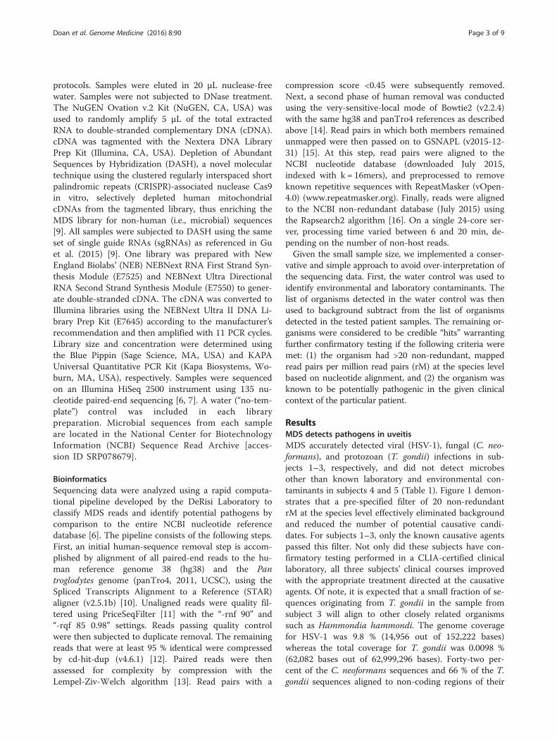
vative and simple approach to avoid over-interpretation ofthe sequencing data. First, the water control was used toidentify environmental and laboratory contaminants. Thelist of organisms detected in the water control was thenused to background subtract from the list of organismsdetected in the tested patient samples. The remaining or-ganisms were considered to be credible “hits” warrantingfurther confirmatory testing if the following criteria weremet: (1) the organism had >20 non-redundant, mappedread pairs per million read pairs (rM) at the species levelbased on nucleotide alignment, and (2) the organism wasknown to be potentially pathogenic in the given clinicalcontext of the particular patient.
ResultsMDS detects pathogens in uveitisMDS accurately detected viral (HSV-1), fungal (C. neo-formans), and protozoan (T. gondii) infections in sub-jects 1–3, respectively, and did not detect microbesother than known laboratory and environmental con-taminants in subjects 4 and 5 (Table 1). Figure 1 demon-strates that a pre-specified filter of 20 non-redundantrM at the species level effectively eliminated backgroundand reduced the number of potential causative candi-dates. For subjects 1–3, only the known causative agentspassed this filter. Not only did these subjects have con-firmatory testing performed in a CLIA-certified clinicallaboratory, all three subjects’ clinical courses improvedwith the appropriate treatment directed at the causativeagents. Of note, it is expected that a small fraction of se-quences originating from T. gondii in the sample fromsubject 3 will align to other closely related organismssuch as Hammondia hammondi. The genome coveragefor HSV-1 was 9.8 % (14,956 out of 152,222 bases)whereas the total coverage for T. gondii was 0.0098 %(62,082 bases out of 62,999,296 bases). Forty-two per-cent of the C. neoformans sequences and 66 % of the T.gondii sequences aligned to non-coding regions of their
Doan et al. Genome Medicine (2016) 8:90 Page 3 of 9
respective genomes, indicating that some genomic DNAwas likely sequenced in addition to RNA. Subject 4 wasa patient with autoimmune-related panuveitis. His in-flammation was controlled with a dexamethasone in-travitreal implant, systemic prednisone, and systemicanti-metabolites. The MDS dataset generated fromsubject 4 contained no pathogen passing our filter(Fig. 1). Subject 5 was a healthy patient who underwentan epiretinal membrane peel and volunteered to do-nate discarded intraocular fluid for testing. While Pre-votella melaninogenica had >20 rM in his sample, aninfection with this organism was not consistent withthis patient’s benign clinical syndrome. Hence, it wasconsidered to be background.In subject 6, MDS detected a single candidate patho-
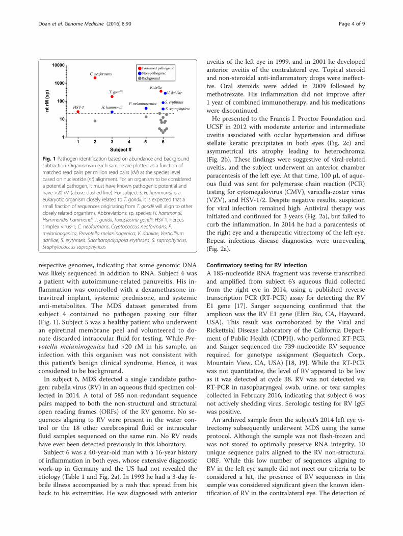
gen: rubella virus (RV) in an aqueous fluid specimen col-lected in 2014. A total of 585 non-redundant sequencepairs mapped to both the non-structural and structuralopen reading frames (ORFs) of the RV genome. No se-quences aligning to RV were present in the water con-trol or the 18 other cerebrospinal fluid or intraocularfluid samples sequenced on the same run. No RV readshave ever been detected previously in this laboratory.Subject 6 was a 40-year-old man with a 16-year history
of inflammation in both eyes, whose extensive diagnosticwork-up in Germany and the US had not revealed theetiology (Table 1 and Fig. 2a). In 1993 he had a 3-day fe-brile illness accompanied by a rash that spread from hisback to his extremities. He was diagnosed with anterior
uveitis of the left eye in 1999, and in 2001 he developedanterior uveitis of the contralateral eye. Topical steroidand non-steroidal anti-inflammatory drops were ineffect-ive. Oral steroids were added in 2009 followed bymethotrexate. His inflammation did not improve after1 year of combined immunotherapy, and his medicationswere discontinued.He presented to the Francis I. Proctor Foundation and
UCSF in 2012 with moderate anterior and intermediateuveitis associated with ocular hypertension and diffusestellate keratic precipitates in both eyes (Fig. 2c) andasymmetrical iris atrophy leading to heterochromia(Fig. 2b). These findings were suggestive of viral-relateduveitis, and the subject underwent an anterior chamberparacentesis of the left eye. At that time, 100 μL of aque-ous fluid was sent for polymerase chain reaction (PCR)testing for cytomegalovirus (CMV), varicella-zoster virus(VZV), and HSV-1/2. Despite negative results, suspicionfor viral infection remained high. Antiviral therapy wasinitiated and continued for 3 years (Fig. 2a), but failed tocurb the inflammation. In 2014 he had a paracentesis ofthe right eye and a therapeutic vitrectomy of the left eye.Repeat infectious disease diagnostics were unrevealing(Fig. 2a).
Confirmatory testing for RV infectionA 185-nucleotide RNA fragment was reverse transcribedand amplified from subject 6’s aqueous fluid collectedfrom the right eye in 2014, using a published reversetranscription PCR (RT-PCR) assay for detecting the RVE1 gene [17]. Sanger sequencing confirmed that theamplicon was the RV E1 gene (Elim Bio, CA, Hayward,USA). This result was corroborated by the Viral andRickettsial Disease Laboratory of the California Depart-ment of Public Health (CDPH), who performed RT-PCRand Sanger sequenced the 739-nucleotide RV sequencerequired for genotype assignment (Sequetech Corp.,Mountain View, CA, USA) [18, 19]. While the RT-PCRwas not quantitative, the level of RV appeared to be lowas it was detected at cycle 38. RV was not detected viaRT-PCR in nasopharyngeal swab, urine, or tear samplescollected in February 2016, indicating that subject 6 wasnot actively shedding virus. Serologic testing for RV IgGwas positive.An archived sample from the subject’s 2014 left eye vi-
trectomy subsequently underwent MDS using the sameprotocol. Although the sample was not flash-frozen andwas not stored to optimally preserve RNA integrity, 10unique sequence pairs aligned to the RV non-structuralORF. While this low number of sequences aligning toRV in the left eye sample did not meet our criteria to beconsidered a hit, the presence of RV sequences in thissample was considered significant given the known iden-tification of RV in the contralateral eye. The detection of
Fig. 1 Pathogen identification based on abundance and backgroundsubtraction. Organisms in each sample are plotted as a function ofmatched read pairs per million read pairs (rM) at the species levelbased on nucleotide (nt) alignment. For an organism to be considereda potential pathogen, it must have known pathogenic potential andhave >20 rM (above dashed line). For subject 3, H. hammondi is aeukaryotic organism closely related to T. gondii. It is expected that asmall fraction of sequences originating from T. gondii will align to otherclosely related organisms. Abbreviations: sp, species; H. hammondi,Hammondia hammondi; T. gondii, Toxoplasma gondii; HSV-1, herpessimplex virus-1; C. neoformans, Cryptococcus neoformans; P.melaninogenica, Prevotella melaninogenica; V. dahliae, Verticilliumdahliae; S. erythraea, Saccharopolyspora erythraea; S. saprophyticus,Staphylococcus saprophyticus
Doan et al. Genome Medicine (2016) 8:90 Page 4 of 9
RV in both eyes corroborated the clinical suspicion ofbilateral viral infection and demonstrated the robustnessof MDS to detect pathogens.
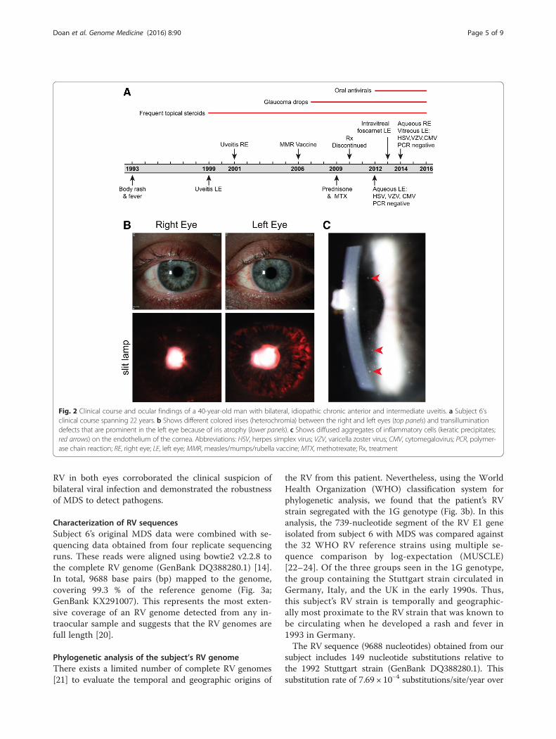
Characterization of RV sequencesSubject 6’s original MDS data were combined with se-quencing data obtained from four replicate sequencingruns. These reads were aligned using bowtie2 v2.2.8 tothe complete RV genome (GenBank DQ388280.1) [14].In total, 9688 base pairs (bp) mapped to the genome,covering 99.3 % of the reference genome (Fig. 3a;GenBank KX291007). This represents the most exten-sive coverage of an RV genome detected from any in-traocular sample and suggests that the RV genomes arefull length [20].
Phylogenetic analysis of the subject’s RV genomeThere exists a limited number of complete RV genomes[21] to evaluate the temporal and geographic origins of
the RV from this patient. Nevertheless, using the WorldHealth Organization (WHO) classification system forphylogenetic analysis, we found that the patient’s RVstrain segregated with the 1G genotype (Fig. 3b). In thisanalysis, the 739-nucleotide segment of the RV E1 geneisolated from subject 6 with MDS was compared againstthe 32 WHO RV reference strains using multiple se-quence comparison by log-expectation (MUSCLE)[22–24]. Of the three groups seen in the 1G genotype,the group containing the Stuttgart strain circulated inGermany, Italy, and the UK in the early 1990s. Thus,this subject’s RV strain is temporally and geographic-ally most proximate to the RV strain that was known tobe circulating when he developed a rash and fever in1993 in Germany.The RV sequence (9688 nucleotides) obtained from our
subject includes 149 nucleotide substitutions relative tothe 1992 Stuttgart strain (GenBank DQ388280.1). Thissubstitution rate of 7.69 × 10⁻⁴ substitutions/site/year over
Fig. 2 Clinical course and ocular findings of a 40-year-old man with bilateral, idiopathic chronic anterior and intermediate uveitis. a Subject 6’sclinical course spanning 22 years. b Shows different colored irises (heterochromia) between the right and left eyes (top panels) and transilluminationdefects that are prominent in the left eye because of iris atrophy (lower panels). c Shows diffused aggregates of inflammatory cells (keratic precipitates;red arrows) on the endothelium of the cornea. Abbreviations: HSV, herpes simplex virus; VZV, varicella zoster virus; CMV, cytomegalovirus; PCR, polymer-ase chain reaction; RE, right eye; LE, left eye; MMR, measles/mumps/rubella vaccine; MTX, methotrexate; Rx, treatment
Doan et al. Genome Medicine (2016) 8:90 Page 5 of 9

the 20-year period is within two-fold of the RV evolu-tionary rate calculated as part of epidemiologic studiesevaluating person-to-person transmission (1.19 × 10⁻³
to 1.94 × 10⁻³ substitutions/site/year) [25]. Of the 149substitutions, 107 were synonymous (Fig. 3a, Additionalfile 1: Table S1). Of the 42 non-synonymous mutations,25 occurred within the coding region for the E1 and E2glycoproteins. Per unit length, the number of non-
synonymous mutations in the E1 and E2 structural pro-teins was 6.3-fold higher than in the non-structuralproteins. Considering all mutations in this region, thesubstitution rate in E1 and E2 was 1.16 × 10⁻³ substitu-tions/site/year. We note that this mutational imbalanceassociated with E1 and E2 compared to the non-structural proteins is consistent with persistent viralreplication under immunological pressure [21].
RVi/Beijing.CHN/79_2ARVi/Beijing.CHN/80_2ARVi/TelAviv.ISR/68_2BRVi/Anqing.Anhui.CHN/00_2BRVi/Seattle.USA/16.00_2BRVi/Moscow.RUS/67/_2CRVi/Moscow.RUS/97_2CRVi/NJ.USA/61_1aRVi/BEL/63_1aRVi/Toyama.JPN/67_1aRVi/PA.USA/64_1aRA27/3_VaccineRVi/Dangshan.Anhui.CHN/00_1FRVi/Linqing.Shandong.CHN/00_1FRVi/Jerusalem.ISR/75_1BRVi/Bene-Berak.ISR/79_1BRVi/Tiberius.ISR/88_1BRVi/Tomsk.RUS/05_1hRVi/Minsk.BLR/28.05/2_1hRVi/Minsk.BLR/29.04/1_1GRVi/Ontario.CAN/05_1GUCSF Uveitis Patient SampleGUZ_GER92 (Stuttgart Germany)RVi/UGA/20.01_1GRVi/Milan.ITA/46.92_1iRVi/Pavia.ITA/21.91_1iRVi/PAN/99_1CRVi/SLV/02_1CRVi/LA.CA.USA/91_1CRVi/Tochigi.JPN/04-1_1jRVs/Miami.FL.USA/32.02_1jRVi/Saitama.JPN/94_1DRVi/Tokio.JPN/90_1DRVi/Dezhou.Shandong.CHN/02_1ERVi/MYS/01_1E
A
B
1 1000
synonymous
non-synonymous
500Coverage
10010
1
2000 3000 4000 5000 6000 7000 8000 9000 9762
RdRpprotease p150
nucleotide position
E2capsid E1WHO
Fig. 3 Identification of rubella virus (RV) by metagenomic deep sequencing (MDS). a Illustrates how the 9688 nucleotide paired-end sequencereads obtained from sequencing the RNA extracted from subject 6’s aqueous fluid aligned to the most closely matched RV genome (GenBankDQ388280.1): 99.3 % of the total RV genome is represented. Positions of synonymous (black vertical lines) and non-synonymous (red vertical lines)variants are shown. Of the 149 substitutions, 107 were synonymous and 42 were non-synonymous. Of the 42 non-synonymous mutations, 25occurred within the coding region for the E1 and E2 glycoproteins. Per unit length, the number of non-synonymous mutations in the E1 and E2proteins was 6.3-fold higher than in the non-structural proteins. The cyan marker above the E1 gene represents the 739-nucleotide sequencewindow recommended by the World Health Organization (WHO) for RV genotyping. b Phylogenetic analysis of subject 6’s RV strain obtained fromMDS with 32 WHO reference strains, GUZ_GER92 (Stuttgart strain), and the RV27/3 vaccine strain, demonstrating that the subject’s RV sequencewas most closely related to the genotype 1G viruses and not the vaccine strain
Doan et al. Genome Medicine (2016) 8:90 Page 6 of 9
DiscussionMDS correctly identified the causative agent in three in-fected positive control subjects (1–3). Only environmen-tal contaminants and sequences associated with non-pathogenic organisms were detected in one uninfectedsubject (patient 5) and one patient with idiopathic uve-itis that was likely autoimmune in nature (patient 4).Furthermore, MDS revealed RV in subject 6 who had a16-year history of idiopathic bilateral uveitis that defiedtreatment with multiple modalities, including prolonged,systemic immunosuppression. Our results demonstratethat a single unbiased MDS assay can detect fungi, para-sites, DNA viruses, and RNA viruses in minute volumesof intraocular fluid from patients with uveitis. The un-biased nature of MDS has potential pitfalls as well. Itcan be difficult to discriminate between microbes thatare present as a result of laboratory or reagent contam-ination and those that are actually causing disease [26].For this reason, we have incorporated a simple but use-ful addition to our analytical pipeline described abovethat attempts to limit over-interpretation of low abun-dance microbes identified via MDS that are also presentin control samples. Lastly, orthogonal assays like culture,PCR, and serology are still critical for confirmation, aswe have highlighted in our cases above.RV is a positive-sense single-stranded RNA virus in
the genus Rubivirus of the Togaviridae family thatcauses transient body rash and fever in healthy adultsbut can also cause devastating birth defects [27]. RV hasalso been associated with Fuchs uveitis syndrome (FUS),a rare form of chronic intraocular inflammation mostoften characterized by mild anterior chamber reaction,iris atrophy with or without heterochromia, late-onsetocular hypertension, and minimal associated visual com-plaints [20, 28–30]. In a subset of patients with FUS, ei-ther RV IgG or small fragments of RV RNA have beendetected in ocular fluid by Goldmann-Witmer coeffi-cient analysis or RT-PCR, respectively [20, 28, 31]. Thesetests are only validated for ocular fluid at a few centersin Europe and are not available as clinical diagnostics inthe USA.The protracted diagnostic challenge in our subject was
three-fold: (1) diagnostic tests are lacking for ocular in-flammation, (2) the subject’s clinical findings were notconsistent with FUS until many years after disease onset,and (3) the subject’s relevant infectious exposure oc-curred 6 years prior to the onset of his ocular symptoms.This case highlights the advantage of a hypothesis-freeapproach in which a single MDS assay can detect amultitude of pathogens that may or may not have beenpreviously associated with a particular clinical syndrome.The identification of RV RNA in our subject’s eyes un-
derscores current challenges in infectious disease sur-veillance and for eradication and elimination programs
[32]. The WHO declared RV eliminated in the USA in2005 as a result of effective and long-standing vaccin-ation policies, but RV remains a threat throughout muchof the world [33, 34]. Our subject’s ocular inflammationpre-dated his measles, mumps, and rubella (MMR) vac-cination by 7 years, and his RV strain most closelymatched the strain circulating in his home country ofGermany at the time of his rash and fever in 1993, andnot the vaccine strain (Fig. 3b). This is consistent withthe notion that RV likely seeded his eyes during this pri-mary infection. Although his immune system cleared theinfection peripherally, RV sequestered in the ocular com-partment and persisted presumably due to relative im-mune privilege. Indeed, our analysis of the RV genomeprovides the first molecular evidence for active RV repli-cation in FUS. Ocular RNA virus sequestration is not aphenomenon relating solely to RV, as Ebola virus was re-cently detected in the ocular fluid of a patient 9 weeksafter resolution of his viremia [4]. Using RT-PCR for RVon our subject’s tears, we were not able to detect shed-ding of RV, although longitudinal studies are required todetermine whether intermittent shedding through tearscan occur. As we devise strategies to rapidly identify andcontrol emerging and re-emerging infectious diseases,expanding the scope of pathogen detection to the eyesand other immune privileged sites may be of criticalimportance.
ConclusionsDiagnostic tests for intraocular infection fundamentallydiffer from those for systemic infections because of thesmall sample volume that can be safely obtained fromthe eye. Unbiased MDS may circumvent this limitation,as it detects many infectious organisms with a singleassay requiring as little as 20 μL of intraocular fluid. Notonly does MDS have the potential to alter the paradigmfor infectious disease diagnostics in ophthalmology, butit may also provide another valuable public health toolto surveil for re-emerging and emerging infectious dis-eases in immune privileged body sites.
Additional file
Additional file 1: Table S1. List of nucleotide substitutions identifiedin subject 6’s RV genome. The patient’s RV genome was aligned with theStuttgart strain (GenBank DQ388280.1). A nucleotide change wasconsidered a substitution only if the change was present in ≥4 reads orin 80 % of the total reads at that nucleotide position. (PDF 120 kb)
AcknowledgementsWe thank Derek Bogdanoff in the UCSF Center for Advanced Technology forhis expertise and assistance operating the Illumina sequencer and Dr. StevenMiller, Director of the UCSF Microbiology Laboratory, for his assistancecoordinating confirmatory laboratory studies. We thank Daniela Munafo andErbay Yigit from New England Biolabs for assistance with the sequencing
Doan et al. Genome Medicine (2016) 8:90 Page 7 of 9
library preparation. We thank the Measles, Mumps, Rubella & HerpesvirusesLaboratory Branch at the Centers for Disease Control, particularly EmilyAbernathy and Dr. Joseph Icenogle, for helpful discussions regarding thepossible public health implications of the RV case and their critical reading ofthe manuscript. We thank Carlos Gonzalez for his assistance in performingSanger sequencing of the 739-nucleotide segment of the RV E1 gene. Wethank the Sandler and William K. Bowes, Jr. Foundations for their generousphilanthropic support. Lastly, we thank our patients for their participation inthis research program.
FundingResearch reported in this publication was supported by the UCSF ResourceAllocation Program for Junior Investigators in Basic and Clinical/TranslationScience (TD); Research to Prevent Blindness Career Development Award (TD);the National Eye Institute of the National Institutes of Health (NIH) underAward Number K08EY026986 (TD); Silicon Valley Community Foundation/Huang Pacific Foundation (TD); UCSF Center for Next-Gen Precision Medicinesupported by the Sandler and William K. Bowes, Jr. Foundations (JLD andMRW); Howard Hughes Medical Institute (JLD); the National Center forAdvancing Translational Sciences of the NIH under Award NumberKL2TR000143 (MRW); the Cooperative Agreement Number U60OE000103,funded by the Centers for Disease Control (CDC) and Prevention throughthe Association of Public Health Laboratories (DX and JKH). Its contents aresolely the responsibility of the authors and do not necessarily represent theofficial views of the CDC, the Department of Health and Human Services, theAssociation of Public Health Laboratories, or the NIH.
Availability of data and materialsThe RV dataset supporting the conclusions of this article is available inthe GenBank repository (accession number KX291007). Microbialsequences from each sample are located in the NCBI Sequence ReadArchive (SRA) under accession ID SRP078679: http://www.ncbi.nlm.nih.gov/sra/SRP078679.
Authors’ contributionsTD and MRW contributed equally and therefore are co-first authors. JLD andNRA conceived the study. JLD, MRW, and TD developed study protocol anddesign, and were responsible for the study implementation and projectmanagement. TD, MRW, LMK, ED Crawford, and ED Chow performed librarypreparation and sequencing. JLD wrote the pipeline analysis software, andJLD, MRW, and TD performed statistical analysis. BDO assisted in thecreation of the pipeline. TD, MRW, and KAK performed rubella RT-PCR. DXand JKH supervised the confirmatory rubella RT-PCR at the CDPH. TD, NRA,JG, and JMS obtained clinical samples and participated in patient care.TD, MRW, and JLD wrote the first draft of the article and produced the figures.Allauthors contributed to the interpretation of the data and the writing andediting of the article.
Competing interestsThe authors declare that they have no competing interests.
Consent for publicationWe have obtained a consent to publish images of the eyes from the studysubject infected with rubella virus.
Ethics approval and consent to participateOur research conformed to the Declaration of Helsinki. The InstitutionalReview Board of the University of California, San Francisco (UCSF) approvedthe study (Study # 14-15275), and informed consent was obtained from allparticipants.
Author details1Francis I. Proctor Foundation, University of California San Francisco, SanFrancisco, CA, USA. 2Department of Ophthalmology, University of CaliforniaSan Francisco, San Francisco, CA, USA. 3Department of Biochemistry andBiophysics, University of California San Francisco, San Francisco, CA, USA.4Department of Neurology, University of California San Francisco, SanFrancisco, CA, USA. 5Howard Hughes Medical Institute, Chevy Chase, MD,USA. 6California Department of Public Health, Richmond, CA, USA.
Received: 28 May 2016 Accepted: 5 August 2016
References1. Sugita S, Ogawa M, Shimizu N, et al. Use of a comprehensive polymerase
chain reaction system for diagnosis of ocular infectious diseases.Ophthalmology. 2013;120(9):1761–8.
2. Taravati P, Lam D, Van Gelder RN. Role of molecular diagnostics in ocularmicrobiology. Curr Ophthalmol Rep. 2013;1(4). doi: 10.1007/s40135-013-0025-1.
3. Endophthalmitis Vitrectomy Study Collaboration. Results of theEndophthalmitis Vitrectomy Study. A randomized trial of immediatevitrectomy and of intravenous antibiotics for the treatment of postoperativebacterial endophthalmitis. Endophthalmitis Vitrectomy Study Group. ArchOphthalmol. 1995;113(12):1479–96.
4. Varkey JB, Shantha JG, Crozier I, et al. Persistence of Ebola virus in ocularfluid during convalescence. N Engl J Med. 2015;372(25):2423–7.
5. de Paula Freitas B, de Oliveira Dias JR, Prazeres J, et al. Ocular findings ininfants with microcephaly associated with presumed Zika virus congenitalinfection in Salvador, Brazil. JAMA Ophthalmol. 2016;134(5):529–535.
6. Wilson MR, Shanbhag NM, Reid MJ, et al. Diagnosing Balamuthiamandrillaris encephalitis with metagenomic deep sequencing. Ann Neurol.2015;78(5):722–30.
7. Wilson MR, Naccache SN, Samayoa E, et al. Actionable diagnosis ofneuroleptospirosis by next-generation sequencing. N Engl J Med. 2014;370(25):2408–17.
8. Pak TR, Kasarskis A. How next-generation sequencing and multiscale dataanalysis will transform infectious disease management. Clin Infect Dis.2015;61(11):1695–702.
9. Gu W, Crawford ED, O’Donovan BD, Wilson MR, Chow ED, Retallack H,DeRisi JL. Depletion of Abundant Sequences by Hybridization (DASH): usingCas9 to remove unwanted high-abundance species in sequencing librariesand molecular counting applications. Genome Biol. 2016;17:41.
10. Dobin A, Davis CA, Schlesinger F, et al. STAR: ultrafast universal RNA-seqaligner. Bioinformatics. 2013;29(1):15–21.
11. Ruby JG, Bellare P, Derisi JL. PRICE: software for the targeted assembly ofcomponents of (Meta) genomic sequence data. G3 (Bethesda).2013;3(5):865–80.
12. Fu L, Niu B, Zhu Z, Wu S, Li W. CD-HIT: accelerated for clustering thenext-generation sequencing data. Bioinformatics. 2012;28(23):3150–2.
13. Ziv J, Lempel A. A universal algorithm for sequential data compression. IEEETrans Inf Theory. 1977;23(3):337–43.
14. Langmead B, Salzberg SL. Fast gapped-read alignment with Bowtie 2. NatMethods. 2012;9(4):357–9.
15. Wu TD, Nacu S. Fast and SNP-tolerant detection of complex variants andsplicing in short reads. Bioinformatics. 2010;26(7):873–81.
16. Zhao Y, Tang H, Ye Y. RAPSearch2: a fast and memory-efficient proteinsimilarity search tool for next-generation sequencing data. Bioinformatics.2012;28(1):125–6.
17. Zhu Z, Xu W, Abernathy ES, et al. Comparison of four methods usingthroat swabs to confirm rubella virus infection. J Clin Microbiol.2007;45(9):2847–52.
18. Namuwulya P, Abernathy E, Bukenya H, et al. Phylogenetic analysis ofrubella viruses identified in Uganda, 2003-2012. J Med Virol.2014;86(12):2107–13.
19. Standardization of the nomenclature for genetic characteristics of wild-typerubella viruses. Wkly Epidemiol Rec. 2005;80(14):126–32.
20. Abernathy E, Peairs RR, Chen MH, Icenogle J, Namdari H. Genomiccharacterization of a persistent rubella virus from a case of Fuch’ uveitissyndrome in a 73 year old man. J Clin Virol. 2015;69:104–9.
21. Abernathy E, Chen MH, Bera J, et al. Analysis of whole genome sequences of16 strains of rubella virus from the United States, 1961-2009. Virol J. 2013;10:32.
22. Dereeper A, Audic S, Claverie JM, Blanc G. BLAST-EXPLORER helps youbuilding datasets for phylogenetic analysis. BMC Evol Biol. 2010;10:8.
23. Dereeper A, Guignon V, Blanc G, et al. Phylogeny.fr: robustphylogenetic analysis for the non-specialist. Nucleic Acids Res.2008;36(Web Server issue):W465–9.
24. Edgar RC. MUSCLE: a multiple sequence alignment method with reducedtime and space complexity. BMC Bioinforma. 2004;5:113.
25. Zhu Z, Rivailler P, Abernathy E, et al. Evolutionary analysis of rubella virusesin mainland China during 2010-2012: endemic circulation of genotype 1Eand introductions of genotype 2B. Sci Rep. 2015;5:7999.
Doan et al. Genome Medicine (2016) 8:90 Page 8 of 9
26. Lee D, Das Gupta J, Gaughan C, et al. In-depth investigation of archival andprospectively collected samples reveals no evidence for XMRV infection inprostate cancer. PLoS One. 2012;7(9):e44954.
27. Lambert N, Strebel P, Orenstein W, Icenogle J, Poland GA. Rubella. Lancet.2015;385(9984):2297–307.
28. Quentin CD, Reiber H. Fuchs heterochromic cyclitis: rubella virus antibodiesand genome in aqueous humor. Am J Ophthalmol. 2004;138(1):46–54.
29. Cunningham Jr ET, Baglivo E. Fuchs heterochromic iridocyclitis–syndrome,disease, or both? Am J Ophthalmol. 2009;148(4):479–81.
30. Birnbaum AD, Tessler HH, Schultz KL, et al. Epidemiologic relationshipbetween fuchs heterochromic iridocyclitis and the United States rubellavaccination program. Am J Ophthalmol. 2007;144(3):424–8.
31. de Groot-Mijnes JD, de Visser L, Rothova A, Schuller M, van Loon AM,Weersink AJ. Rubella virus is associated with fuchs heterochromiciridocyclitis. Am J Ophthalmol. 2006;141(1):212–4.
32. Dunn G, Klapsa D, Wilton T, Stone L, Minor PD, Martin J. Twenty-eight yearsof poliovirus replication in an immunodeficient individual: impact on theglobal polio eradication initiative. PLoS Pathog. 2015;11(8):e1005114.
33. Reef SE, Redd SB, Abernathy E, Kutty P, Icenogle JP. Evidence used tosupport the achievement and maintenance of elimination of rubellaand congenital rubella syndrome in the United States. J Infect Dis.2011;204 Suppl 2:S593–7.
34. Pan American Health Organization. Americas region is declared the world’sfirst to eliminate rubella. 2015. Available from: http://www.paho.org/us/index.php?option=com_content&view=article&id=135%3Aamericas-region-free-of-rubella&Itemid=0&lang=en. Accessed 22 Aug 2016.
• We accept pre-submission inquiries
• Our selector tool helps you to find the most relevant journal
• We provide round the clock customer support
• Convenient online submission
• Thorough peer review
• Inclusion in PubMed and all major indexing services
• Maximum visibility for your research
Submit your manuscript atwww.biomedcentral.com/submit
Submit your next manuscript to BioMed Central and we will help you at every step:
Doan et al. Genome Medicine (2016) 8:90 Page 9 of 9
![Pulmonary Metagenomic Sequencing Suggests Missed ...derisilab.ucsf.edu/pdfs/ciy802.pdf · mold in this patient population [16]. Sample Preparation We tested nucleic acid extraction](https://img.pdfslide.net/doc/110x75/6000075f19be4f382d11dc1a/pulmonary-metagenomic-sequencing-suggests-missed-mold-in-this-patient-population.jpg)