Embed Size (px)
Citation preview
BASIC SCIENCE INVESTIGATIONS
Imaging Cell Death with RadiolabeledAnnexin V in an Experimental Model ofRheumatoid ArthritisAnneke M. Post, BS1; Peter D. Katsikis, MD, PhD2; Jonathan F. Tait, MD, PhD3; Sharon M. Geaghan, MD4;H. William Strauss, MD5; and Francis G. Blankenberg, MD6
1Division of Nuclear Medicine, Department of Radiology, Stanford University, Stanford, California; 2Department of Microbiologyand Immunology, MCP Hahnemann University, Philadelphia, Pennsylvania; 3Department of Laboratory Medicine, University ofWashington, Seattle, Washington; 4Department of Pathology, Stanford University, Stanford, California; 5Division of NuclearMedicine, Department of Radiology, Memorial Sloan-Kettering Hospital, New York, New York; and 6Division of PediatricRadiology, Department of Radiology, Lucile Salter Packard Children’s Hospital, Stanford, California
Rheumatoid arthritis is associated with chronic synovial in-flammation due to the abnormal accumulation of macro-phages and autoreactive T lymphocytes in joints. The auto-reactive cells cause an inflammatory proapoptotic responseto self-antigens resulting in eventual bone, cartilage, andsoft-tissue loss and destruction. The goal of our study was todetermine the timing and intensity of apoptosis in joints using99mTc-labeled annexin V, an in vivo marker of apoptosis, in amurine model of immune arthritis. Methods: We used 99mTc-annexin V and autoradiography to study the extent and se-verity of apoptosis in the front and rear paws of DBA/1 micewith type II collagen-induced rheumatoid arthritis. Results:Compared with control values (n � 10), there was a signifi-cant (P � 0.002) nearly 3-fold increase in uptake of 99mTc-annexin V in the front foot pads, rear toes, rear foot pads, andheels at the time of maximal extremity swelling as determinedby serial caliper measurements at 4 wk after inoculation withtype II bovine collagen (n � 9). The front toes had a 5- to6-fold increase in uptake compared with control values (P �0.001). Histologic analysis revealed only scattered rare lym-phocytes in the periarticular soft tissues, without joint de-struction. Dual autoradiography with 125I-bovine serum albu-min as a control showed that 99mTc-annexin V localizationwas specific. Treatment with methylprednisolone for 1 wk(n � 8) at 4 wk after immunization with type II collagendecreased 99mTc-annexin V uptake by 3- to 6-fold comparedwith control values (P � 0.002). Conclusion: 99mTc-annexin Vcan detect collagen-induced immune arthritis and its re-sponse to steroid therapy before joint destruction.
Key Words: annexin V; rheumatoid arthritis; apoptosis;radionuclide
J Nucl Med 2002; 43:1359–1365
Rheumatoid arthritis (RA) is a chronic progressive dis-ease associated with substantial morbidity and an acceler-ated incidence of atherosclerotic disease due to persistentlyelevated serum levels of homocysteine (1,2). The diseaseoften requires therapy with drugs associated with significantside effects (3). To date, steroids, nonsteroidal anti-inflam-matory drugs, and methotrexate are the most commonagents used to temporarily alleviate symptoms, but theseagents rarely improve the quality of life. Investigators areperforming clinical trials of novel therapies such as anti-bodies directed against tumor necrosis factor-� or interleu-kin-1 in the hope of developing a treatment that will haveless morbidity than those in current use (4,5). The design ofstudies to test these new therapies is complex because thedisease commonly waxes and wanes with respect to inflam-mation and because objective measurements of disease ac-tivity such as plain-film radiography, nuclear scintigraphywith bone scanning and nonspecific inflammatory radio-pharmaceuticals, and contrast-enhanced MRI are either in-sensitive or impractical guides for the assessment of therapyor disease monitoring (6,7).
The inflammation associated with RA is caused by ab-normal collections of immune cells responding inappropri-ately to idiotypic antigens (8,9). These inflammatory cellsoften induce apoptosis of target tissues before their ownapoptotic cell death. The role of apoptosis in both thepathogenesis and the treatment of RA is only now beginningto be understood. Several studies have indicated that there isan increase in apoptotic cell death in the synovial membranein RA (10,11). Apoptosis of both the synovial membranelining and the sublining cells has been demonstrated. There-fore, determining the presence and the degree of apoptoticcell death in affected joints and periarticular tissues mayprove crucial for both the study and the clinical evaluationof RA. Our goal in this investigation was to determine the
Received Oct. 17, 2001; revision accepted Mar. 25, 2002.For correspondence or reprints contact: Francis G. Blankenberg, MD, 725
Welch Rd., Palo Alto, CA 94304.E-mail: [email protected]
ANNEXIN V IMAGING OF ARTHRITIS • Post et al. 1359
by on April 9, 2019. For personal use only. jnm.snmjournals.org Downloaded from
incidence of immune cell and target tissue apoptosis inrelation to the severity of local joint swelling.
To detect apoptosis in vivo, we have previously appliedan imaging technique using radiolabeled annexin V (12,13),a human protein commonly labeled with fluorescent mark-ers for in vitro detection and quantification of apoptotic cells(14,15). These investigations have demonstrated a highcorrelation between the intensity of radiolabeled annexin Vlocalization at sites of apoptosis in vivo as confirmed bystaining with terminal deoxynucleotidyl transferase–medi-ated deoxyuridine triphosphate nick end labeling, a markerof DNA ladder formation and degradation in the apoptoticcell nucleus (16,17).
Annexin V has a reversible, strictly calcium-dependentnanomolar affinity for the membrane aminophospholipid,phosphatidylserine (PS). PS comprises 10%–15% of thetotal phospholipid content of the plasma cell membrane andis normally restricted to the inner leaflet of the plasmamembrane lipid bilayer by an adenosine triphosphate–de-pendent translocase (18,19). With the onset of apoptosis,however, PS is rapidly redistributed onto the cell surface(20). The number of annexin V binding sites per cell withthe onset of apoptosis increases 100- to 1,000-fold duringapoptosis, reaching values of 3–4 million in some cell lines(21,22). PS exposure on the cell surface closely followscaspase-3 activation and occurs well before DNA fragmen-tation (23). Radiolabeled annexin V, therefore, is a sensitivemarker of the early to intermediate phases of apoptosis.Apoptosis has been imaged in vivo after intravenouslyadministered radiolabeled annexin V in experimental mod-els of immune-mediated apoptosis induced by anti-Fas an-tibody (Jo2) and alloreactive T lymphocytes in the course ofacute transplant rejection of the heart (24), liver (25), andlung (26).
In this study, we used intravenously administered 99mTc-annexin V to detect the presence and amount of apoptosis inthe extremities of DBA/1 mice with collagen-induced RA.
MATERIALS AND METHODS
Murine Model of Collagen-Induced RAMale 8- to 12-wk-old DBA/1 mice were purchased (Jackson
Laboratories, Bar Harbor, ME) and were housed and treated in ahumane manner in accordance with National Institutes of Healthand institutional guidelines on animal subjects (27). A total of 27mice were anesthetized with an intraperitoneal sodium pentobar-bital injection of 50 mg/kg, after which they received multipleintradermal injections of an emulsion containing bovine type IIcollagen (1 mg/mL; Biogenesis, Brentwood, NH) suspended incomplete Freund’s adjuvant (0.1 mL total volume; Difco Labora-tories, Detroit, MI) according to the methods of Walmsley et al.(28). After collagen inoculation, the front and rear paws of themice were observed every 2 d for signs of redness and swelling.Maximal swelling was observed at 4 wk. Just before the time ofsacrifice and imaging, electronic caliper measurements of eachpaw were recorded for each mouse. Sacrifice followed by autora-diographic imaging was performed at 2 (n � 5), 3 (n � 5), and 4(n � 9) wk on 19 collagen-inoculated mice. The remaining 8
collagen-inoculated mice were treated with a 5-d course of meth-ylprednisolone (5 mg/kg/d intraperitoneally) starting 4 wk (25 or26 d) after collagen injection (time of maximal swelling). Steroid-treated mice were sacrificed and imaged on day 31. A group of 10uninoculated mice (controls) was sacrificed and imaged at week 4.
Preparation of 99mTc-Hydrazinonicotinamide Annexin VHuman annexin V (molecular weight, 35,806) was produced by
expression in Escherichia coli as previously described (29). Thisrecombinant material binds to membrane-bound PS with an affin-ity equivalent to that of native annexin V. Hydrazinonicotinamide-derivatized annexin V was prepared as previously described with-out affecting membrane-bound PS activity and was radiolabeledwith a 99mTc-tricine precursor complex according to the method ofLarsen et al. (30). After chelation with the 99mTc-tricine precursorcomplex, the volume of the reaction mixture was brought up to 1mL with phosphate-buffered saline, pH 7.4, and was collected in1-mL fractions eluted from a Sephadex G-25 column (Pharmacia,Piscataway, NJ). Fractions 3 and 4 contained 70%–80% of thetotal derivatized protein and 95.7%–99.4% of total 99mTc activityas determined from previously described methods (31). The poolof fractions 3 and 4 had a radiopurity of 92%–97%, determined byinstant thin-layer chromatography using 0.9% phosphate-bufferedsaline as a solvent. The prepared radiolabeled material had calcu-lated specific activities ranging from 3.7 to 7.4 MBq (100–200�Ci)/�g protein.
99mTc-Annexin V AutoradiographyOne hour after intravenous injection of 55–74 MBq (1.5–2.0
mCi) of radiolabeled annexin V, mice were sacrificed by cervicaldislocation and the paws were removed, embedded in optimal-cutting-temperature medium (Miles, Inc., Tarrytown, NY), andfrozen at –20°C on dry ice. Pairs of frozen sections were preparedby alternately cutting 50- and 5-�m sections through the coronalplane of each paw. The 50-�m specimens were then placed on astorage phosphor screen for 18–24 h (PhosphorImager SI systemwith storage phosphor screens; Molecular Dynamics, MountainView, CA). After exposure, the storage phosphor screen imageswere “developed” with a laser digitizer at a resolution of 50 �mper pixel. The remaining 5-�m slices underwent histologic study.
125I-Bovine Serum Albumin (BSA) AutoradiographyTo determine whether 99mTc-annexin V localization was due to
nonspecific membrane permeability, dual-tracer autoradiographicstudies were performed with 125I-BSA in addition to 99mTc-annexinV in a subgroup of 5 mice. Four weeks after collagen injection,these mice were coinjected with 0.74 MBq (20 �Ci) of 125I-BSA(80–120 �g/kg of protein) along with 99mTc-annexin V. Initialautoradiographs (recorded for a total of 21 h, commencing 3 h afterinjection) defined the distribution of 99mTc-annexin V in the paw.The 50-�m histologic sections were then allowed to decay for 4 or5 d and were again placed on a storage phosphor screen for anadditional 4-d exposure to define the albumin (i.e., inert protein)distribution.
Region-of-Interest (ROI) AnalysisThe relative localization of radiotracer in the paws was quanti-
fied by ROI analysis using ImageQuant software (version 5.1;Molecular Dynamics). ROIs were drawn over each digit, paw pad,and Achilles tendon sheath using the corresponding histologicsections to ensure proper anatomic orientation (Fig. 1). The totalnumber of counts was recorded for each digit and pad of the front
1360 THE JOURNAL OF NUCLEAR MEDICINE • Vol. 43 • No. 10 • October 2002
by on April 9, 2019. For personal use only. jnm.snmjournals.org Downloaded from
and rear paws as well as each Achilles tendon sheath. The countsobtained for each anatomic region were then normalized to thetotal counts (also obtained by ROI analysis) of a 5-�L bloodsample taken at the time of sacrifice from each mouse and placed
adjacent to the 50-�m histologic sections during their exposure onthe storage phosphor screen for autoradiography. The digit, pad, orAchilles tendon sheath with the highest 99mTc-annexin V activitywas recorded for each mouse. These maximal values were thenaveraged for each anatomic location and expressed as the meanpercentage of the 5-�L blood standard � 1 SD from the mean.
Histologic AnalysisFive-micrometer coronal frozen histologic sections were stained
with hematoxylin–eosin for microscopic analysis. The presence,degree, and location of inflammatory cells and erosive changeswere qualitatively noted for each paw.
Statistical AnalysisAll variables were expressed as average values � 1 SD from the
mean. All statistical comparisons of average values were per-formed with the Student t test (2-tailed) for significance using thenull hypothesis. P � 0.05 was considered statistically significant.
RESULTS
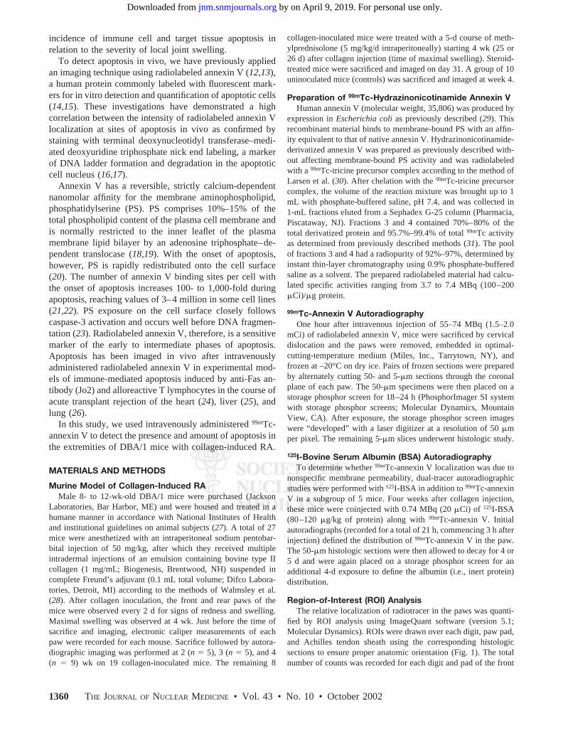
Some degree of swelling and redness of the paw jointsdeveloped in all mice. The degree of swelling and rednessvaried from joint to joint and from mouse to mouse withineach inoculated group of mice. The maximal degree of pawpad swelling occurred between 4 and 5 wk after inoculationas previously described by Walmsley et al. (28). Treatmentwith a short course of methylprednisolone was initiated at 4wk in a group of arthritic mice.
Measurements of Paw Pad ThicknessThe thickness (swelling) of the front and rear pads of
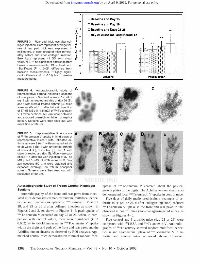
arthritic mice was 11% and 18%, respectively, of baselinevalues by day 13 and increased to maximal values on day 25or 26 (Figs. 2 and 3). Little change in the degree of pawswelling before and after treatment occurred with methyl-prednisolone, although both groups of mice had signifi-cantly thicker front (8%–16%) and rear (35%–36%) padscompared with age-matched control mice.
FIGURE 1. Diagrammatic schema of experiments. First,DBA/1 mice are inoculated with emulsion of killed Mycobacte-rium tuberculosis and bovine type II collagen dissolved in in-complete Freund’s adjuvant. Autoimmune response results inpolyarticular rheumatoid arthritis in 3–4 wk. Second, 99mTc-annexin V is injected, and autoradiographic images are obtainedfrom frozen coronal sections of paw. Third, ROIs are drawn overeach digit, paw pad, and Achilles tendon sheath and are used toanalyze counts.
FIGURE 2. Front pad thickness after col-lagen injection. Bars represent average val-ues of front pad thickness, expressed inmillimeters, of each group of mice immedi-ately before and after collagen injection.Error bars represent �1 SD from meanvalue. N.S. � no significant difference frombaseline measurements; TX � treatment.**Highly significant difference (P � 0.004)from baseline measurements.
ANNEXIN V IMAGING OF ARTHRITIS • Post et al. 1361
by on April 9, 2019. For personal use only. jnm.snmjournals.org Downloaded from
Autoradiographic Study of Frozen Coronal HistologicSections
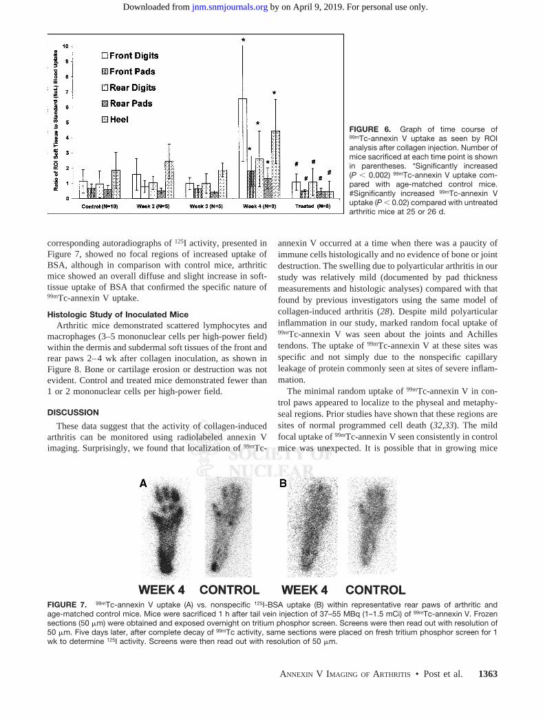
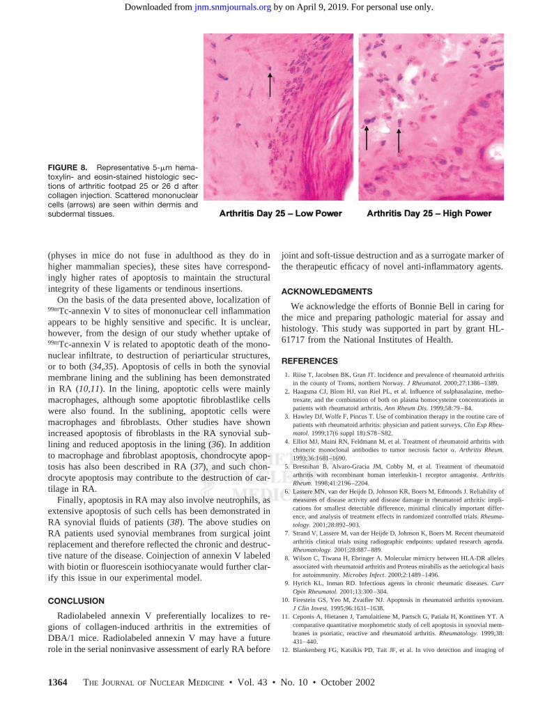
Autoradiography of the front and rear paws from inocu-lated mice demonstrated marked random, multifocal periar-ticular and ligamentous uptake of 99mTc-annexin V at 13,18, and 25 or 26 d after collagen injection as shown inFigures 2 and 3. As shown in Figures 4–6, peak uptake of99mTc-annexin V occurred on day 25 or 26, when, in com-parison with control values, there were significant (P �0.002) 2- to 6-fold increases in 99mTc-annexin V uptakewithin the digits and pads of the front and rear paws and theAchilles tendon sheaths as observed by ROI analysis. Age-matched control mice demonstrated minimal random focal
uptake of 99mTc-annexin V centered about the physealgrowth plates of the digits. The Achilles tendon sheath alsodemonstrated focal 99mTc-annexin V uptake in control mice.
Five days of daily methylprednisolone treatment of ar-thritic mice (25 or 26 d after collagen injection) reduced99mTc-annexin V uptake in the front and rear paws to thatobserved in control mice (non–collagen-injected mice), asshown in Figures 4–6.
Five control and 5 arthritic mice (day 25 or 26) werecoinjected with 125I-BSA and 99mTc-annexin V. Autoradio-graphs of 99mTc activity showed random multifocal periar-ticular and ligamentous uptake of 99mTc-annexin V in ar-thritic and control mice as noted above. However,
FIGURE 3. Rear pad thickness after col-lagen injection. Bars represent average val-ues of rear pad thickness, expressed inmillimeters, of each group of mice immedi-ately before and after collagen injection.Error bars represent �1 SD from meanvalue. N.S. � no significant difference frombaseline measurements; TX � treatment.*Significant (P � 0.05) difference frombaseline measurements. **Highly signifi-cant difference (P � 0.01) from baselinemeasurements.
FIGURE 4. Autoradiographic study ofrepresentative coronal histologic sectionsof front paws of 3 individual mice: 1 control(A), 1 with untreated arthritis at day 26 (B),and 1 with steroid-treated arthritis (C). Micewere sacrificed 1 h after tail vein injectionof 37–55 MBq (1–1.5 mCi) of 99mTc-annexinV. Frozen sections (50 �m) were obtainedand exposed overnight on tritium phosphorscreen. Screens were then read out withresolution of 50 �m.
FIGURE 5. Representative time courseof 99mTc-annexin V uptake in hind paws ofrepresentative mice: 1 with untreated ar-thritis at week 2 (A), 1 with untreated arthri-tis at week 3 (B), 1 with untreated arthritisat week 4 (C), 1 control (D), and 1 withsteroid-treated arthritis (E). Mice were sac-rificed 1 h after tail vein injection of 37–55MBq (1–1.5 mCi) of 99mTc-annexin V. Fro-zen sections (50 �m) were obtained andexposed overnight on tritium phosphorscreen. Screens were then read out withresolution of 50 �m.
1362 THE JOURNAL OF NUCLEAR MEDICINE • Vol. 43 • No. 10 • October 2002
by on April 9, 2019. For personal use only. jnm.snmjournals.org Downloaded from
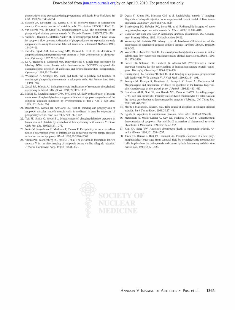
corresponding autoradiographs of 125I activity, presented inFigure 7, showed no focal regions of increased uptake ofBSA, although in comparison with control mice, arthriticmice showed an overall diffuse and slight increase in soft-tissue uptake of BSA that confirmed the specific nature of99mTc-annexin V uptake.
Histologic Study of Inoculated MiceArthritic mice demonstrated scattered lymphocytes and
macrophages (3–5 mononuclear cells per high-power field)within the dermis and subdermal soft tissues of the front andrear paws 2–4 wk after collagen inoculation, as shown inFigure 8. Bone or cartilage erosion or destruction was notevident. Control and treated mice demonstrated fewer than1 or 2 mononuclear cells per high-power field.
DISCUSSION
These data suggest that the activity of collagen-inducedarthritis can be monitored using radiolabeled annexin Vimaging. Surprisingly, we found that localization of 99mTc-
annexin V occurred at a time when there was a paucity ofimmune cells histologically and no evidence of bone or jointdestruction. The swelling due to polyarticular arthritis in ourstudy was relatively mild (documented by pad thicknessmeasurements and histologic analyses) compared with thatfound by previous investigators using the same model ofcollagen-induced arthritis (28). Despite mild polyarticularinflammation in our study, marked random focal uptake of99mTc-annexin V was seen about the joints and Achillestendons. The uptake of 99mTc-annexin V at these sites wasspecific and not simply due to the nonspecific capillaryleakage of protein commonly seen at sites of severe inflam-mation.
The minimal random uptake of 99mTc-annexin V in con-trol paws appeared to localize to the physeal and metaphy-seal regions. Prior studies have shown that these regions aresites of normal programmed cell death (32,33). The mildfocal uptake of 99mTc-annexin V seen consistently in controlmice was unexpected. It is possible that in growing mice
FIGURE 6. Graph of time course of99mTc-annexin V uptake as seen by ROIanalysis after collagen injection. Number ofmice sacrificed at each time point is shownin parentheses. *Significantly increased(P � 0.002) 99mTc-annexin V uptake com-pared with age-matched control mice.#Significantly increased 99mTc-annexin Vuptake (P � 0.02) compared with untreatedarthritic mice at 25 or 26 d.
FIGURE 7. 99mTc-annexin V uptake (A) vs. nonspecific 125I-BSA uptake (B) within representative rear paws of arthritic andage-matched control mice. Mice were sacrificed 1 h after tail vein injection of 37–55 MBq (1–1.5 mCi) of 99mTc-annexin V. Frozensections (50 �m) were obtained and exposed overnight on tritium phosphor screen. Screens were then read out with resolution of50 �m. Five days later, after complete decay of 99mTc activity, same sections were placed on fresh tritium phosphor screen for 1wk to determine 125I activity. Screens were then read out with resolution of 50 �m.
ANNEXIN V IMAGING OF ARTHRITIS • Post et al. 1363
by on April 9, 2019. For personal use only. jnm.snmjournals.org Downloaded from
(physes in mice do not fuse in adulthood as they do inhigher mammalian species), these sites have correspond-ingly higher rates of apoptosis to maintain the structuralintegrity of these ligaments or tendinous insertions.
On the basis of the data presented above, localization of99mTc-annexin V to sites of mononuclear cell inflammationappears to be highly sensitive and specific. It is unclear,however, from the design of our study whether uptake of99mTc-annexin V is related to apoptotic death of the mono-nuclear infiltrate, to destruction of periarticular structures,or to both (34,35). Apoptosis of cells in both the synovialmembrane lining and the sublining has been demonstratedin RA (10,11). In the lining, apoptotic cells were mainlymacrophages, although some apoptotic fibroblastlike cellswere also found. In the sublining, apoptotic cells weremacrophages and fibroblasts. Other studies have shownincreased apoptosis of fibroblasts in the RA synovial sub-lining and reduced apoptosis in the lining (36). In additionto macrophage and fibroblast apoptosis, chondrocyte apop-tosis has also been described in RA (37), and such chon-drocyte apoptosis may contribute to the destruction of car-tilage in RA.
Finally, apoptosis in RA may also involve neutrophils, asextensive apoptosis of such cells has been demonstrated inRA synovial fluids of patients (38). The above studies onRA patients used synovial membranes from surgical jointreplacement and therefore reflected the chronic and destruc-tive nature of the disease. Coinjection of annexin V labeledwith biotin or fluorescein isothiocyanate would further clar-ify this issue in our experimental model.
CONCLUSION
Radiolabeled annexin V preferentially localizes to re-gions of collagen-induced arthritis in the extremities ofDBA/1 mice. Radiolabeled annexin V may have a futurerole in the serial noninvasive assessment of early RA before
joint and soft-tissue destruction and as a surrogate marker ofthe therapeutic efficacy of novel anti-inflammatory agents.
ACKNOWLEDGMENTS
We acknowledge the efforts of Bonnie Bell in caring forthe mice and preparing pathologic material for assay andhistology. This study was supported in part by grant HL-61717 from the National Institutes of Health.
REFERENCES
1. Riise T, Jacobsen BK, Gran JT. Incidence and prevalence of rheumatoid arthritisin the county of Troms, northern Norway. J Rheumatol. 2000;27:1386–1389.
2. Haagsma CJ, Blom HJ, van Riel PL, et al. Influence of sulphasalazine, metho-trexate, and the combination of both on plasma homocysteine concentrations inpatients with rheumatoid arthritis. Ann Rheum Dis. 1999;58:79–84.
3. Hawley DJ, Wolfe F, Pincus T. Use of combination therapy in the routine care ofpatients with rheumatoid arthritis: physician and patient surveys. Clin Exp Rheu-matol. 1999;17(6 suppl 18):S78–S82.
4. Elliot MJ, Maini RN, Feldmann M, et al. Treatment of rheumatoid arthritis withchimeric monoclonal antibodies to tumor necrosis factor �. Arthritis Rheum.1993;36:1681–1690.
5. Bresnihan B, Alvaro-Gracia JM, Cobby M, et al. Treatment of rheumatoidarthritis with recombinant human interleukin-1 receptor antagonist. ArthritisRheum. 1998;41:2196–2204.
6. Lassere MN, van der Heijde D, Johnson KR, Boers M, Edmonds J. Reliability ofmeasures of disease activity and disease damage in rheumatoid arthritis: impli-cations for smallest detectable difference, minimal clinically important differ-ence, and analysis of treatment effects in randomized controlled trials. Rheuma-tology. 2001;28:892–903.
7. Strand V, Lassere M, van der Heijde D, Johnson K, Boers M. Recent rheumatoidarthritis clinical trials using radiographic endpoints: updated research agenda.Rheumatology. 2001;28:887–889.
8. Wilson C, Tiwana H, Ebringer A. Molecular mimicry between HLA-DR allelesassociated with rheumatoid arthritis and Proteus mirabilis as the aetiological basisfor autoimmunity. Microbes Infect. 2000;2:1489–1496.
9. Hyrich KL, Inman RD. Infectious agents in chronic rheumatic diseases. CurrOpin Rheumatol. 2001;13:300–304.
10. Firestein GS, Yeo M, Zvaifler NJ. Apoptosis in rheumatoid arthritis synovium.J Clin Invest. 1995;96:1631–1638.
11. Ceponis A, Hietanen J, Tamulaitiene M, Partsch G, Patiala H, Konttinen YT. Acomparative quantitative morphometric study of cell apoptosis in synovial mem-branes in psoriatic, reactive and rheumatoid arthritis. Rheumatology. 1999;38:431–440.
12. Blankenberg FG, Katsikis PD, Tait JF, et al. In vivo detection and imaging of
FIGURE 8. Representative 5-�m hema-toxylin- and eosin-stained histologic sec-tions of arthritic footpad 25 or 26 d aftercollagen injection. Scattered mononuclearcells (arrows) are seen within dermis andsubdermal tissues.
1364 THE JOURNAL OF NUCLEAR MEDICINE • Vol. 43 • No. 10 • October 2002
by on April 9, 2019. For personal use only. jnm.snmjournals.org Downloaded from
phosphatidylserine expression during programmed cell death. Proc Natl Acad SciUSA. 1998;95:6349–6354.
13. Stratton JR, Dewhurst TA, Kasina S, et al. Selective uptake of radiolabeledannexin V on acute porcine left atrial thrombi. Circulation. 1995;92:3113–3121.
14. van Heerde WL, de Groot PG, Reutelingsperger CPM. The complexity of thephospholipid binding protein annexin V. Thromb Haemost. 1995;73:172–179.
15. Vermes I, Haanen C, Steffens-Nakken H, Reutelingsperger CPM. A novel assayfor apoptosis flow cytometric detection of phosphatidylserine expression on earlyapoptotic cells using fluorescein labelled annexin V. J Immunol Methods. 1995;184:39–51.
16. van den Eijnde SM, Luijsterburg AJM, Boshart L, et al. In situ detection ofapoptosis during embryogenesis with annexin V: from whole mount to ultrastruc-ture. Cytometry. 1997;29:313–320.
17. Li X, Traganos F, Melamed MR, Darzynkiewicz Z. Single-step procedure forlabeling DNA strand breaks with fluorescein- or BODIPY-conjugated de-oxynucleotides: detection of apoptosis and bromodeoxyuridine incorporation.Cytometry. 1995;20:172–180.
18. Williamson P, Schlegel RA. Back and forth: the regulation and function oftransbilayer phospholipid movement in eukaryotic cells. Mol Membr Biol. 1994;11:199–216.
19. Zwaal RF, Schroit AJ. Pathophysiologic implications of membrane phospholipidasymmetry in blood cells. Blood. 1997;89:1121–1132.
20. Martin SJ, Reutelingsperger CPM, McGahon AJ. Early redistribution of plasmamembrane phosphatidylserine is a general feature of apoptosis regardless of theinitiating stimulus: inhibition by overexpression of Bcl-2 Abl. J Exp Med.1995;182:1545–1556.
21. Bennett MR, Gibson DF, Schwartz SM, Tait JF. Binding and phagocytosis ofapoptotic vascular smooth muscle cells is mediated in part by exposure ofphosphatidylserine. Circ Res. 1995;77:1136–1142.
22. Tait JF, Smith C, Wood BL. Measurement of phosphatidylserine exposure inleukocytes and platelets by whole-blood flow cytometry with annexin V. BloodCells Mol Dis. 1999;25:271–278.
23. Naito M, Nagashima K, Mashima T, Tsuruo T. Phosphatidylserine externaliza-tion is a downstream event of interleukin-1�-converting enzyme family proteaseactivation during apoptosis. Blood. 1997;89:2060–2066.
24. Vriens PW, Blankenberg FG, Stoot JH, et al. The use of 99m technetium labeledannexin V for in vivo imaging of apoptosis during cardiac allograft rejection.J Thorac Cardiovasc Surg. 1998;116:844–853.
25. Ogura Y, Krams SM, Martinez OM, et al. Radiolabeled annexin V imaging:diagnosis of allograft rejection in an experimental rodent model of liver trans-plantation. Radiology. 2000;214:795–800.
26. Blankenberg FG, Robbins RC, Stoot JH, et al. Radionuclide imaging of acutelung transplant rejection with annexin V. Chest. 2000;117:834–840.
27. Guide for the Care and Use of Laboratory Animals. Washington, DC: Govern-ment Printing Office; 1985. NIH publication 86-23.
28. Walmsley M, Katsikis PD, Abney E, et al. Interleukin-10 inhibition of theprogression of established collagen induced arthritis. Arthritis Rheum. 1996;39:495–503.
29. Wood BL, Gibson DF, Tait JF. Increased phosphatidylserine exposure in sicklecell disease: flow-cytometric measurement and clinical associations. Blood. 1996;88:1873–1880.
30. Larsen SK, Solomon HF, Caldwell G, Abrams MJ. [99mTc]tricine: a usefulprecursor complex for the radiolabeling of hydrazinonicotinate protein conju-gates. Bioconjug Chemistry. 1995;6:635–638.
31. Blankenberg FG, Katsikis PD, Tait JF, et al. Imaging of apoptosis (programmedcell death) with 99mTc annexin V. J Nucl Med. 1999;40:184–191.
32. Zenmyo M, Komiya S, Kawabata R, Sasaguri Y, Inoue A, Morimatsu M.Morphological and biochemical evidence for apoptosis in the terminal hypertro-phic chondrocytes of the growth plate. J Pathol. 1996;80:430–433.
33. Bronckers ALJJ, Goei W, van Heerde WL, Dumont EAWJ, ReutelingspergerCPM, van den Eijnde SM. Phagocytosis of dying chondrocytes by osteoclasts inthe mouse growth plate as demonstrated by annexin-V labeling. Cell Tissue Res.2000;301:267–272.
34. Morita I, Matsuno H, Sakai K, et al. Time course of apoptosis in collagen-inducedarthritis. Int J Tissue React. 1998;20:37–43.
35. Eguchi K. Apoptosis in autoimmune diseases. Intern Med. 2001;40:275–284.36. Matsumoto S, Muller-Ladner U, Gay RE, Nishioka K, Gay S. Ultrastructural
demonstration of apoptosis, Fas and Bcl-2 expression of rheumatoid synovialfibroblasts. J Rheumatol. 1996;23:1345–1352.
37. Kim HA, Song YW. Apoptotic chondrocyte death in rheumatoid arthritis. Ar-thritis Rheum. 1999;42:1528–1537.
38. Jones ST, Denton J, Holt PJ, Freemont AJ. Possible clearance of effete poly-morphonuclear leucocytes from synovial fluid by cytophagocytic mononuclearcells: implications for pathogenesis and chronicity in inflammatory arthritis. AnnRheum Dis. 1993;52:121–126.
ANNEXIN V IMAGING OF ARTHRITIS • Post et al. 1365
by on April 9, 2019. For personal use only. jnm.snmjournals.org Downloaded from
2002;43:1359-1365.J Nucl Med. BlankenbergAnneke M. Post, Peter D. Katsikis, Jonathan F. Tait, Sharon M. Geaghan, H. William Strauss and Francis G. Rheumatoid ArthritisImaging Cell Death with Radiolabeled Annexin V in an Experimental Model of
http://jnm.snmjournals.org/content/43/10/1359This article and updated information are available at:
http://jnm.snmjournals.org/site/subscriptions/online.xhtml
Information about subscriptions to JNM can be found at:
http://jnm.snmjournals.org/site/misc/permission.xhtmlInformation about reproducing figures, tables, or other portions of this article can be found online at:
(Print ISSN: 0161-5505, Online ISSN: 2159-662X)1850 Samuel Morse Drive, Reston, VA 20190.SNMMI | Society of Nuclear Medicine and Molecular Imaging
is published monthly.The Journal of Nuclear Medicine
© Copyright 2002 SNMMI; all rights reserved.
by on April 9, 2019. For personal use only. jnm.snmjournals.org Downloaded from