Embed Size (px)
Citation preview

8/2/2019 Imaging Parathyroid 2011
http://slidepdf.com/reader/full/imaging-parathyroid-2011 1/12
Imaging techniques in parathyroid surgery for primary
hyperparathyroidism
Arash Mohebati, MD, Ashok R. Shaha, MD⁎
Head and Neck Service, Memorial Sloan Kettering Cancer Center, New York, NY, USA
Received 29 August 2011
Abstract As more patients present with the incidental diagnosis of primary hyperparathyroidism due to
biochemic al screening, treat ment guide lines have been developed for the treat ment of
hyperparathyroidism. Management of primary hyperparathyroidism has evolved in recent years,
with considerable interest in minimally invasive approaches. Successful localization of the diseasedgland(s) by nuclear imaging and anatomical studies, along with rapid intraoperative parathyroid
hormone assay, has allowed for focused and minimally invasive surgical approaches. Patients in
whom the localization studies have identified single-gland adenoma or unilateral disease are
candidates for such focused approaches instead of the traditional approach of bilateral exploration.
These imaging techniques have also been critical in the successful management of patients with
persistent or recurrent disease.\
© 2011 Elsevier Inc. All rights reserved.
1. Introduction
Primary hyperparathyroidism (PHPT) is the most com-
mon cause of hypercalcemia, the treatment of which is primarily surgical resection. It is characterized by hypercal-
cemia due to overproduction of parathyroid hormone (PTH).
In the Western countries, as a result of increasing
biochemical screening, PHPT has evolved from the disease
of “ bones, moans stones and groans” to a disorder that is
most commonly asymptomatic in many patients [1,2].
Primary hyperparathyroidism is a common disease occurring
in about 1% of the adults, and its incidence rises to 2% or
more in population older than 55 years [3]. It is 2 to 3 times
more common in women than in men and peaks around the
fourth and fifth decades of life [1,4].The traditional approach
to parathyroid surgery consists of bilateral neck explorationwith the goal of identifying and visually inspecting all 4
parathyroid glands. The success of this approach exceeds
90% to 95%. However, cervical exploration requires a larger
incision and longer operating time and potentially can have
higher morbidity. Because more than 85% of patients with
PHPT have a single-gland adenoma, 4-gland exploration
may not be necessary in most patients if the enlarged
parathyroid gland can be identified and localized preoper-atively. There has been considerable interest in localization
studies of the abnormal parathyroid gland(s) since the 1980s.
The goal of this approach is to allow the surgeon to perform
minimally invasive parathyroidectomy. The minimally
invasive approach includes small incision parathyroidecto-
my, outpatient parathyroidectomy, endoscopic or video-
assisted parathyroidectomy, and parathyroidectomy under
local anesthesia. Various invasive and noninvasive localiz-
ing tests are available for evaluating PHPT. The traditional
noninvasive imaging modalities include ultrasonography,
computed tomography (CT), magnetic resonance imaging
(MRI); however, more recently, technetium-99m (99m
Tc)sestamibi scan and 99m Tc sestamibi single-emission CT have
been used for localizing the pathologic or enlarged
parathyroid glands.
In this article, we will review the pertinent anatomy,
pathophysiology of the parathyroid glands, and diagnosis of
PHPT. The indications for the management of hyperpara-
thyroidism and treatment options will be reviewed. Further-
more, we will discuss various surgical approaches to
parathyroid surgery. In detail, we will review the role of
Available online at www.sciencedirect.com
American Journal of Otolaryngology–Head and Neck Medicine and Surgery xx (2011) xxx–xxxwww.elsevier.com/locate/amjoto
⁎ Corresponding author. Head and Neck Service, Memorial Sloan
Kettering Cancer Center, 1275 York Ave, New York, NY 10065, USA.
E-mail address: [email protected] (A.R. Shaha).
0196-0709/$ – see front matter © 2011 Elsevier Inc. All rights reserved.
doi:10.1016/j.amjoto.2011.10.010
8/2/2019 Imaging Parathyroid 2011
http://slidepdf.com/reader/full/imaging-parathyroid-2011 2/12
various preoperative localization techniques and the sensi-
tivity and specificity of each technique in localizing the
abnormal parathyroid gland and the concordance of this
identification with the operative and pathologic findings. In
addition, we will evaluate the role of intraoperative
parathormone assay in determining the success of the
image-guided minimally invasive approach.
2. Anatomy and embryology of parathyroid glands
The parathyroid glands are endodermal in origin and
develop from the dorsal wing of the third and fourth
pharyngeal pouches [5,6]. The first detailed anatomical
description of the parathyroid glands was published by
Welsh [7] in 1898 and subsequently by Halsted and Evans
[8] in 1907, making a distinction between the superior and
the inferior glands. They produce PTH, which regulates
the circulating level of calcium through intestinal and renal
absorption and bone remodeling. There are typically 4 parathyroid glands; however, supernumerary glands and
less than 4 glands have been reported. In a reported series
of 428 cases, 0.5% had 6 glands, 25% had 5 glands, 87%
had 4 glands, and 6.1% of the cases had 3 glands [9].
Most supernumerary glands were either rudimentary or
divided, weighing as little as less than 5 mg and near a
normal gland. The normal weight of each parathyroid
gland is about 35 to 40 mg and measuring about 3 to
8 mm [9,10]. The inferior thyroid artery is the predominant
vascular supply to both upper and lower parathyroid
glands in 76% to 86% of the cases [9].
The superior parathyroid glands originate from the fourth pharyngeal pouch. As they lose their attachment with the
pharyngeal wall, they attach to the posterior surface of the
inferiorly migrating thyroid [5,10]. They have a much
shorter migration distance compared with the inferior
parathyroid glands accounting for their more predictable
location. They are generally at the level of the upper two
thirds of the thyroid. In an autopsy study of 503 cases, 80%
of the superior glands were located on the posterior aspect of
the thyroid gland within a circumscribed area of 2 cm in
diameter about 1 cm above the crossing point of the recurrent
laryngeal nerve and inferior thyroid artery [11]. In this study,
the ectopic superior parathyroid glands were found at the
level of the upper pole of the thyroid and above the pole in2% and 0.8% of the subjects, respectively. Other ectopic
positions of superior parathyroid glands such as in the
posterior neck, retropharyngeal, retroesophageal space, and
intrathyroidal position are quite rare and reported in up to 1%
of the cases [11,12].
Although the dorsal wing of the third pharyngeal pouch
gives rise to the inferior parathyroid glands, the ventral wing
gives rise to the thymus during the fifth week of gestation
[5]. Both primitive glands lose their connection with the
pharyngeal wall and join the thymus as it travels caudally
and medially to its final position in the mediastinum [5,13].
This migration of the inferior parathyroid glands with the
thymus results in the inferior parathyroid gland to be in a
plane that is usually ventral to that of the superior
parathyroid glands [13]. For the same reason, ectopic
inferior parathyroid glands can be found anywhere along
this large area of descent up to the superior border of the
pericardium [14]. In a study of 645 parathyroid glands from
160 postmortem subjects, the inferior glands were evenly
distributed between the lower pole of the thyroid and isthmus
[12]. In this study, 42% of the inferior parathyroid glands
were found on the anterior or the posterolateral surface of the
lower pole of the thyroid, whereas 39% were located in the
lower neck in proximity to the thymic tissue, 15% lateral to
the thyroid, and only 2% within the mediastinal thymic
tissue. The persistence of the primitive attachment of the
inferior parathyroid glands to the thymus, during the thymic
migration, may result in a more inferior placement of the
parathyroid glands. In this situation, the inferior parathyroid
glands may be found at the level of the anterior superior
mediastinum near the upper pole of thymic remnants [15].Exploration of the superior mediastinum becomes important
during 4-gland exploration when the inferior parathyroid
glands cannot be identified in the neck.
A rare ectopic location that could be a source of pitfall
during parathyroid surgery for hyperparathyroidism is the
intrathyroid location of the parathyroid glands. The embry-
ologic origin of this ectopic location has been controversial,
but it has been reported to originate from either the superior
or the inferior gland [11,16-18]. The incidence of intrathyr-
oid parathyroid gland is reported between 0.7% and 3.6% in
the literature [17,19,20]. Because of their rarity, the
intrathyroid parathyroid glands can be missed by preoper-ative imaging. This must be kept in mind when meticulous
bilateral neck exploration fails to identify the hyperfunction-
ing gland. However, most of the time, the enlarged
parathyroid glands are in the capsule of the thyroid or in
the crypts of the thyroid tissue and are missed as intrathyroid
parathyroid. In most cases, parathyroid glands are located in
a symmetrical position in the neck. In one study, the
symmetrical position of the superior and inferior glands was
found in 80% and 70% of the cases, respectively, with a
relative symmetry of 60% for all 4 glands [11].
3. Etiology
The most challenging aspect of managing patients with
PHPT is the recognition of the pathologic process that gives
rise to the disorder. The appropriate treatment choice is
directly dependent on the underlying etiology of the
disorder. Primary hyperparathyroidism could be due to
adenoma and 4-gland hyperplasia and rarely due to
carcinoma. Most adults (80%–85%) with PHPT have single
benign parathyroid adenoma, and up to 4% to 5%% are
reported to have double adenomas [21,22]. Four-gland
hyperplasia is reported in up to 15% of patients with PHPT
2 A. Mohebati, A.R. Shaha / American Journal of Otolaryngology – Head and Neck Medicine and Surgery xx (2011) xxx – xxx
8/2/2019 Imaging Parathyroid 2011
http://slidepdf.com/reader/full/imaging-parathyroid-2011 3/12
and parathyroid carcinoma in 0.8% to 2% of the cases
[23,24]. In the Multiple Endocrine Neoplasia Type 1 (MEN-
1) and Endocrine Neoplasia Type 2 (MEN-2) and the
familial syndromes, hyperplasia is the primary etiology of
the hyperparathyroidism. Classically, an adenoma is defined
as a pathologically enlarged gland with a rim of normal
tissue that is most commonly solitary. Many surgeons and
pathologists agree that differentiating adenoma from hyper-
plasia on frozen section is extremely difficult, and it can
primarily be used to distinguish parathyroid tissue from other
tissue; however, routine biopsy of the parathyroid glands is
not recommended [18,25]. In addition, criteria, such as gland
size, shape, and cell density and type, have been shown to be
of little help in distinguishing between adenoma and
hyperplasia [26].
4. Pathophysiology
Parathyroid glands secrete PTH, which is the keyregulator of calcium homeostasis. Parathyroid hormone is
secreted as an 84-amino-acid peptide with a short plasma
half-life (2–4 minutes). It is metabolized to biologically
active N-terminal and inactive C-terminal fragments. The
major regulator of PTH levels is the extracellular calcium
through a feedback mechanism control of the calcium
receptor [27]. Calcitriol, or 1,25-dihydroxyvitamin D (1,25-
(OH)2D), is the other essential mediator of calcium
homeostasis, the synthesis of which is regulated by PTH.
Calcitriol production begins when cholecalciferol (vitamin
D) is generated in the skin exposed to ultraviolet light or
absorbed through the intestine. In the liver, vitamin D ishydroxylated to 25-(OH)D and converted 1,25-(OH)2D3
(calcitriol) in the kidneys. This last step is tightly regulated
by PTH [28]. Parathyroid hormone and calcitriol act through
the gastrointestinal tract, bone, and the kidney to maintain
circulating ionized calcium concentrations. It increases
serum calcium by acting indirectly on the osteoclasts in the
skeleton to promote bone resorption and, thus, release of
calcium into extracellular fluid. In addition to regulating
calcitriol production, PTH also enhances calcium resorption
at the distal nephron of the kidney. Through this series of
checks and balances, extracellular calcium concentration is
carefully maintained in the body and at times at the expense
of skeletal calcium stores [29].
5. Diagnosis
Primary hyperparathyroidism is usually diagnosed by the
physician because of hypercalcemia found on routine
laboratory evaluation with an inappropriately elevated or
normal levels of PTH [30]. Patients with PHPT typically
have low phosphorus because elevated PTH levels decrease
the resorption of phosphorus in the kidneys. Most common-
ly, these patients are asymptomatic; however, a careful
history and physical examination is necessary to rule out
other causes of hypercalcemia. Although PHPT is the most
common cause of hypercalcemia in the ambulatory setting,
the differential diagnosis of hypercalcemia is complex and
includes conditions such as metastatic cancer, multiple
myeloma, sarcoidosis and other granulomatous diseases,
ingestion of calcium or vitamin D, milk-alkali syndrome, and
other less common causes. In addition to PHPT, other
disorders such as familial hypocalciuric hypercalcemia will
result in elevated levels of PTH and calcium. However, 24-
hour urine calcium will show abnormally low levels of
calcium in the urine in patients with familial hypocalciuric
hypercalcemia. Medications such as thiazides and lithium
can result in the elevation of PTH and calcium levels.
Patients with tertiary hyperparathyroidism who had a history
of renal failure and subsequent renal transplantation will also
have elevated levels of PTH and calcium.
Parathyroid hormone levels are measured using immu-
noradiometric assays. The intact PTH assay that measures
the PTH 1–84 and the non–PTH 1–84 and the bioactiveassay that measures the PTH 1–84 and N-terminal PTH
can be used for the diagnosis of PHPT with excellent
correlation [31-33]. Plasma total calcium levels are more
commonly measured compared with ionized calcium
levels; however, the superiority of one test over the other
has been debated. If the total serum calcium level is used,
it needs to be corrected based on the patient's albumin
level. This correction is made by adding 0.8 mg/dL to the
total serum calcium measurement for every 1 g/dL below
the serum albumin concentration of 4 g/dL.
6. Indications for treatment
The clinical presentation of PHPT has changed from
patients with symptomatic disease to those with only
biochemical evidence of disease incidentally found on
routine laboratory examination. Asymptomatic PHT has
spurred investigation into the natural history of this disease
and indications for treatment. It is accepted that surgery is
indicated in patients who present with symptomatic PHPT
such as nephrolithiasis, nephrocalcinosis, renal dysfunction,
osteopenia with fractures, osteitis fibrosa cystica, and altered
neurologic function with obtundation, delirium, or coma [4].
However, the management of patients with asymptomaticPHPT has been controversial. The National Institutes of
Health organized a consensus development conference in
1990 that made recommendations for the management of
patients with asymptomatic PHPT. These recommendations
were subsequently updated in the 2002 National Institutes of
Health Workshop on Asymptomatic PHPT [3,30,34]. The
summary of these recommendations for the surgical man-
agement of patients with PHTP is presented in Table 1. Under
these guidelines, the indications for surgery include a serum
calcium concentration of 1.0 mg/dL above the upper limit of
the normal level, urinary calcium excretion of greater than
3 A. Mohebati, A.R. Shaha / American Journal of Otolaryngology – Head and Neck Medicine and Surgery xx (2011) xxx – xxx
8/2/2019 Imaging Parathyroid 2011
http://slidepdf.com/reader/full/imaging-parathyroid-2011 4/12
400 mg/24 h, a reduction in the creatinine clearance of more
than 30%, a bone mineral density with a t score below 2.5 at any site, age younger than 50 year, or those who cannot
participate in appropriate follow-up [35]. Some authorities,
however, recommend a more liberal guidelines in the
management of these patients owing to the inability to
predict whether the complications or progression of disease
will develop in a specific patient [36]. The 2002 guidelines
for monitoring patients with asymptomatic PHPT who do not
undergo parathyroid surgery are measurement of serum
calcium level every 6 months, annual measurement of serum
creatinine concentration, and annual bone density measure-
ment at all 3 sites (the lumbar spine, the hip, and the distal
third of the radius) [4].
7. Localizing studies
The treatment of hyperparathyroidism involves the
removal of abnormally enlarged parathyroid gland(s) with
the goal of rendering the patient with normal PTH levels and
who is eucalcemic. Four-gland parathyroid exploration has
been the criterion standard for parathyroid surgery until
recently. Doppman [37] made a classic statement in 1986
that “the only localization study indicated in a patient with
untreated primary hyperparathyroidism is the localization of
an experienced parathyroid surgeon.” However, minimallyinvasive and focused parathyroidectomy has become a
widely accepted approach for the treatment of PHPT. The
major limitation of this approach is the imperfection of the
preoperative localization techniques. A summary of various
localization techniques is provided in Table 2. Through the
use of combined preoperative and intraoperative techniques,
this approach has achieved cure rates of greater than 95%
with low morbidity [38]. The success of the localization
studies also depends on the experience of the institution.
Invasive localizing studies, such as angiography and
selective venous sampling, are rarely used now because of
the high success rate of noninvasive approaches. However, if
preoperative noninvasive studies are inconclusive, intrao-
perative or preoperative office-based ultrasound (US)-guided
jugular venous sampling can be helpful in lateralizing the
most hyperfunctioning parathyroid gland(s) [39].
8. Ultrasound
Among anatomical imaging modalities, US is the most
frequently used imaging modality with the lowest cost and
has the advantage of not using ionizing radiation in this
setting. It is, however, highly user-dependent and does not
enable retromanubrial or mediastinal visualization. Normal
parathyroid glands are rarely visualized by ultrasonography
because of their small size and insufficient acoustic
difference compared with adjacent thyroid tissue. However,
parathyroid adenomas, hyperplasia, and carcinomas exhibit a
relatively hypoechogenic pattern because of their compact
cellularity relative to thyroid tissue [40].Parathyroid adeno-
mas are usually well-circumscribed ovoid, are longitudinal inshape, and tend to be solid and homogenously hypoechoic
relative to echogenic thyroid tissue (Fig. 1) [12,41,42].
Overall, the ability to detect a parathyroid adenoma is a
function of its size and the pathology of the adjacent thyroid
tissue [43]. Ultrasound is inexpensive and highly sensitive in
experienced hands. The sensitivity of US for detecting
enlarged parathyroid glands ranges from 70% to 100%
[34,38-40]. False-positive or false-negative sonographic
diagnosis may be due to thyroid nodules, prominent blood
vessels, cervical lymph nodes, esophagus, and longus colli
muscle [41]. The smaller the adenoma, the more difficult it is
to localize radiographically, and in the setting of multi-
nodular goiter, parathyroid adenomas may be over lookeddue to poor sonographic penetration. In the presence of
thyroid gland abnormalities, the sensitivity of US to identify
abnormal parathyroid glands decreases, ranging from 47% to
84% [43-46]. The identified concurrent thyroid disease
should be addressed preoperatively by fine-needle aspiration
or intraoperatively in these patients. Ectopically located
glands, particularly intrathyroidal or retroesophageal glands,
make the sonographic detection of the parathyroid glands
more challenging and difficult. The US sensitivity also
decreases in patients with persistent or recurrent hyperpara-
thyroidism. This sensitivity is reported between 36% and
Table 1
Surgical indications in patients with primary hyperparathyroidism (Adopted
from the 2002 National Institutes of Health Work-shop)
1. Significant bone, renal, gastrointestinal, or neuromuscular symptoms
typical of primary hyperparathyroidism
In asymptomatic patients
2. Serum calcium Elevation by ≥1 mg/dL above the
reference range (ie, ≥11.5 mg/dL
in most laboratories)
3. 24-h urine calcium excretion Marked elevation (eg, N400 mg)
4. Creatinine clearance Decreased levels (eg, reduced by
≥30% compared with age-matched
healthy persons)
5. Bone density Reduction in bone density of N2.5
standard deviations below peak bone
mass at any measured site (hip, lumbar
spine, wrist; ie, “T score” approximately
b2.5 at any of these sites)
6. Age younger than 50 years
Table 2
Parathyroid localization studies
Noninvasive Invasive
High-resolution US Selective venous sampling99m Tc-Sestamibi scan Angiogram
CT scan Digital subtraction
MRI US-guided FNA
SPECT/CT US-guided jugular venous sampling4D-CT
FNA indicates fine-needle aspiration.
4 A. Mohebati, A.R. Shaha / American Journal of Otolaryngology – Head and Neck Medicine and Surgery xx (2011) xxx – xxx

8/2/2019 Imaging Parathyroid 2011
http://slidepdf.com/reader/full/imaging-parathyroid-2011 5/12
63% [47,48]. The sensitivity of surgeon-performed US is
reported to be comparable with radiologist-performed US in
localizing parathyroid adenomas, and this may be advanta-geous for experienced endocrine surgeons [49]. An advan-
tage of this technique is that it provides dynamic imaging in
the hands of the surgeon who has an intimate knowledge of
the cervical anatomy. If the US findings are inconclusive,
additional localizing techniques such as Tc-sestamibi scan
should be used.
9. Sestamibi scan
The thallium technetium scintigraphy was popular in the
late 1980s for the diagnosis of cervical and mediastinal parathyroid adenomas [50]. However, sestamibi (99m Tc-
methoxyisobutyl isonitrile), which was used for evaluation
of myocardial perfusion, was noted to have selective affinity
for abnormal parathyroid glands [51]. Sestamibi has become
the radiopharmaceutical of choice for parathyroid nuclear
localization studies. Two different techniques are used to
differentiate sestamibi uptake by abnormal parathyroid
glands from uptake by the thyroid gland. The first uses
sestamibi as a substitute to Tl-201 in a dual-radionuclide
approach with subtraction imaging (with either radioactive
iodine-123 [123I] or 99m Tc pertechnetate), and the second
approach uses sestamibi alone (single radiotracer) with early
and delayed imaging (dual-phase study). Subtractionimaging is performed with the coadministration of a thyroid
imaging agent, such as 123I or 99m Tc pertechnetate and
sestamibi as a thyroid-parathyroid agent [52]. The thyroid
images generated are then digitally subtracted from the
sestamibi image, and the residual signal after subtraction of
the thyroid image is indicative of parathyroid uptake [53,54].
Various protocols have been described based on the type of
the thyroid imaging tracer used and the sequence of tracer
administration. 123I/ 99m Tc-sestamibi and 99m TcO4
−/ 99m Tc-
sestamibi dual-tracer subtraction techniques, have used in
parathyroid imaging with limitations such as long imaging
time or high thyroid counts [55,56]. The use of a pinhole
collimator in the neck can increase the imaging resolution;
however, it will increase the image acquisition time.Regional perfusion, gland size and functional activity, cell
cycle phase, and prevalence of mitochondria-rich cells are
some of the factors affecting the diagnostic accuracy of 99m Tc-sestamibi imaging of parathyroid glands [57,58].
In the dual-phase technique, the net retention of
sestamibi in thyroid decreases significantly more rapidly
than in parathyroid adenoma over time (1–3 hours
postinjection). This technique relies on the differential
washout of sestamibi from thyroid tissue than from
abnormal parathyroid glands [59,60]. Images are obtained
after the administration of sestamibi and then, again,
approximately 2 hours later (Fig. 2). A greater number of mitochondria in parathyroid tissue sequester sestamibi
intracellularly. The patient's neck immobilization is an
essential step in double-tracer subtraction scintigraphy that
is not required for dual-phase imaging. Technical simplicity
of dual-phase MIBI scintigraphy is a major advantage of
this technique. Conversely, a major limitation of dual-phase
MIBI scan in detecting parathyroid gland pathology is the
association of HPT with thyroid nodular disease. In a study
of 39 patients undergoing dual-phase MIBI scintigraphy,
41% of the patients with positive MIBI scan had thyroid
carcinoma [61]. Thyroid pathology, such as hyperplastic
nodules, chronic thyroiditis, Hürthle cell lesions, and
adenomas can increase sestamibi uptake and retention,resulting in a false-positive study, and high washout from
the parathyroid tissue can result in false-negative findings
[62]. Correlation with anatomical imaging and the use of
subtraction techniques are important in these cases.
Regardless of which technique is used, sestamibi scanning
as a single modality for identifying adenomas has a reported
sensitivity of 54% to 100%, with most series in the 80% or
90% range [63-66]. Many studies looked at the sensitivity
of US compared with sestamibi scanning and found no
significant difference in the sensitivity of the 2 techniques
in detecting abnormal parathyroid glands [65]. In 1 recent
Fig. 1. Hypoechoic left-lower-pole (A) and behind the left-lower-pole (B) parathyroid adenomas identified by US.
5 A. Mohebati, A.R. Shaha / American Journal of Otolaryngology – Head and Neck Medicine and Surgery xx (2011) xxx – xxx

8/2/2019 Imaging Parathyroid 2011
http://slidepdf.com/reader/full/imaging-parathyroid-2011 6/12
study of 516 patients undergoing surgery for PHPT,
surgeon-performed US accurately localized adenomas in
87% of patients, and MIBI correctly identified their
locations in 76% ( P b .001) [67]. In patients who underwent
US first, MIBI provided no additional information in 92%.
The authors concluded that in experienced hands, US is
more accurate than MIBI in predicting the location of
abnormal parathyroid glans in PHPT patients.
The addition of single-photon emission CT (SPECT) is
shown to increase the sensitivity and specificity of
parathyroid scintigraphy, allowing for better localization
of the enlarged glands. The superposition of the 2 radiomarkers results in a more accurate location of the abnormal
gland [66,68,69]. In 1 study, the addition of SPECT
significantly improved the sensitivity for the detection of
parathyroid adenomas to 79% from 54% for MIBI alone.
In principle, SPECT offers the advantage of better
discrimination of focal 99m Tc-sestamibi retention in
thyroid nodules and the parathyroid tissue. This technique
is particularly useful for the localization of ectopic
parathyroid adenomas such as mediastinal glands. In
addition, the sensitivity of this technique is lower for
hyperplastic parathyroid glands compared with parathyroid
adenomas. The sensitivity of identifying hyperplastic
parathyroid glands using these techniques is reported to
be from 25% to 58% compared with 54% to 98% for
parathyroid adenomas [69-71]. When localizing studies are
undertaken, frequently, a sestamibi scan is used in
conjunction with some type of anatomical imaging, US,
or CT, or MRI. Several studies have reported on the use of
SPECT/CT for localization of parathyroid adenomas and
hyperplasia in patients with previously untreated PHPT
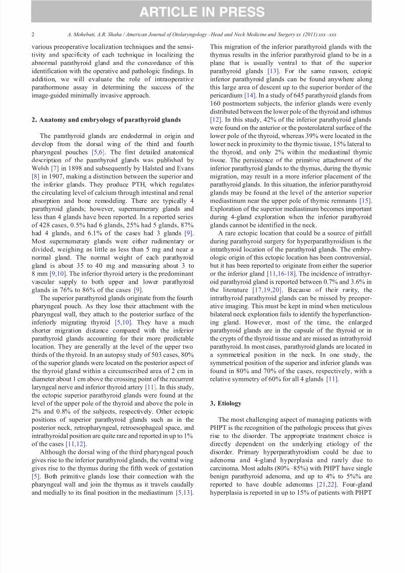
(Fig. 3). The results have been variable, with some series
demonstrating improved sensitivity and positive predictive
value resulting in a change in therapeutic management andothers reporting no additional clinical value when
evaluating patients with previously untreated PHPT [72-
74]. In some reports, SPECT/CT is shown to be valuable
in locating ectopic parathyroid glands [73].
10. Computed tomography
Computed tomography scanning can provide valuable
and complimentary localizing information for the identifi-
cation of abnormal parathyroid glands. This technique
Fig. 2. Image after the administration of sestamibi (2-hour) delay demonstrates left inferior parathyroid adenoma.
6 A. Mohebati, A.R. Shaha / American Journal of Otolaryngology – Head and Neck Medicine and Surgery xx (2011) xxx – xxx
8/2/2019 Imaging Parathyroid 2011
http://slidepdf.com/reader/full/imaging-parathyroid-2011 7/12
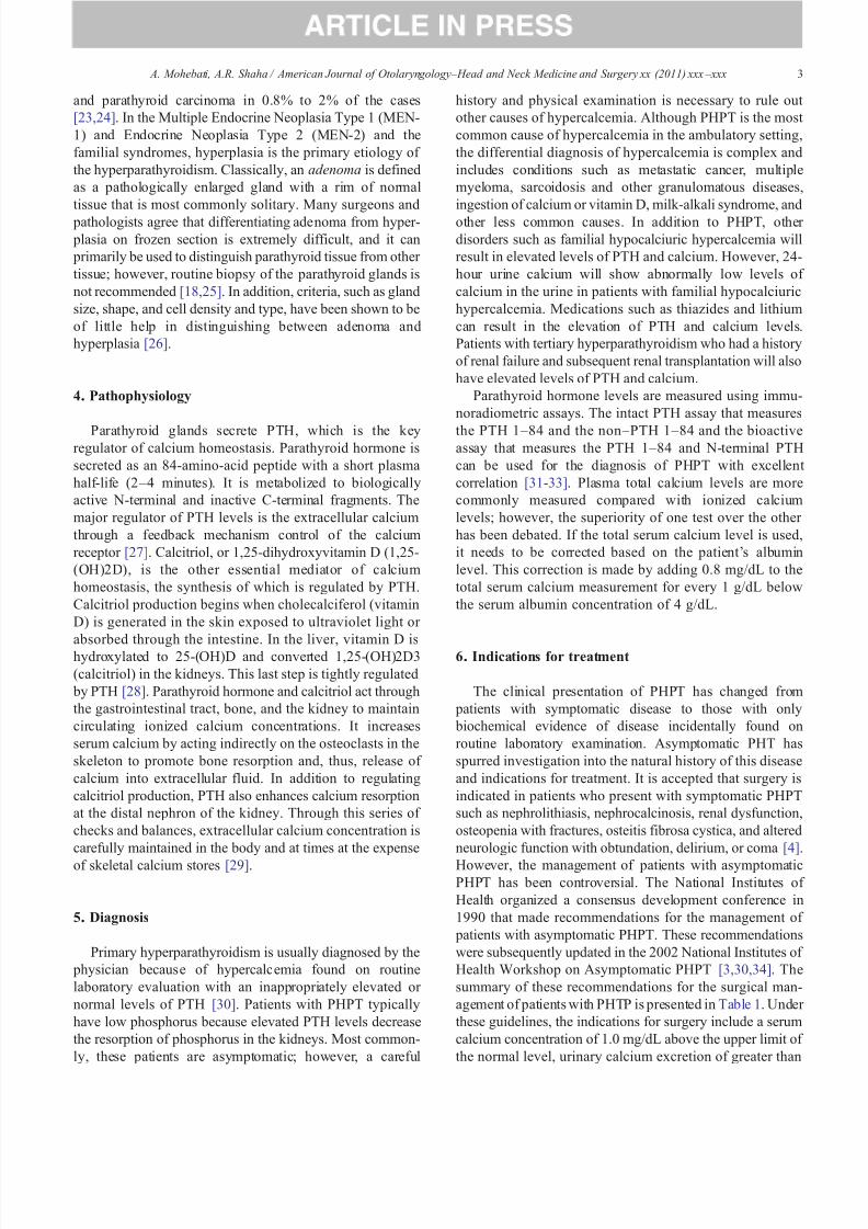
relies on the vascularity of the parathyroid glands and their relative enhancement with contrast compared with the
surrounding structures (Fig. 4). Computed tomography has
reported to have a sensitivity of 40% to 86%, depending
on the size of the lesion, with the accuracy that is
dependent on the technique and the experience of the
radiologist [75-77]. In a study of 63 patients with negative
sestamibi who underwent thin-cut CT scanning, a
sensitivity of 66% and a specificity of 89% for identifying
the exact location of diseased gland were observed. This
allowed for 66% of the patients to undergo focused parathyroid exploration [78]. Parathyroid glands can be
difficult to identify by CT at the area of the sternal notch
and mediastinum because of streak artifact or being
mistaken with lymph nodes. Computed tomography is
also not particularly useful in identifying the parathyroid
glands in patients who are not able to receive intravenous
contrast. Some authors advocate for the use of CT in
patients with inconclusive or discordant US and MIBI
results [75]. In addition, CT scanning may be useful in
identifying ectopic parathyroid glands. In 1 study, CT had
a sensitivity of 40% and a specificity of 88% in identifying
ectopic parathyroid glands [76]. Computed tomography
scanning appears to be effective in localizing parathyroiddisease as an adjunct to conventional first-line imaging
techniques.
In 2006, Rodgers et al [79] published their report of a
new CT protocol for localizing parathyroid glands in
patients with PHPT. They called this 4-dimensional CT
(4D-CT) because of the addition of changes in the
perfusion of contrast over time to the 3-dimensional
anatomical CT images. This allowed for the addition of
functional information to the anatomical images because of
the rapid uptake and washout of the contrast from the
hyperfunctioning gland to the anatomical data. They
Fig. 3. Single-photon emission CT/CT demonstrating a right-sided parathyroid adenoma.
Fig. 4. Computed tomography demonstrating a left-sided parathyroid
adenoma.
7 A. Mohebati, A.R. Shaha / American Journal of Otolaryngology – Head and Neck Medicine and Surgery xx (2011) xxx – xxx

8/2/2019 Imaging Parathyroid 2011
http://slidepdf.com/reader/full/imaging-parathyroid-2011 8/12
demonstrated an improved sensitivity of 88% for 4D-CT
compared with sestamibi (65%) and ultrasonography
(57%) for lateralizing the hyperfunctioning gland and
further allowed for improved localizing to the correct
quadrant compared with the other 2 imaging modalities.
More recently, Kutler et al [80], using a combination of
4D-CT/US technique, reported a sensitivity of 94% and a
specificity of 96% for lateralizing the hyperfunctioning
parathyroid and a sensitivity of 82% for localizing the
gland to the correct quadrant. They reported a positive
predictive value of 92% for a single-gland and 75% for a
multigland disease for the combination of 4D-CT/US. This
combination technique appears to be an excellent and cost-
effective preoperative localizing technique for hyperfunc-
tioning parathyroid glands.
11. Magnetic Resonance Imaging
Another anatomical imaging modality used for detecting
abnormal parathyroid glands is MRI. Parathyroid adeno-
mas are isointense relative to the surrounding muscle and
have low intensity on T1-weighted images. They are
generally hyperintense on T2-weighted images; however,
hyperfunctioning parathyroid glands show intense contrast
enhancement on T1-weighted images (Fig. 5) [81].This
method is preferred by some over CT scanning because of
lack of ionizing radiation. The sensitivity of MRI in
detecting abnormal parathyroid glands is reported to be
from 69% to 88%, with a false-positive rate of1.6% to
10% [82-84]. Factors resulting in false-positive disease are
concomitant thyroid disease and enlarged cervical lymphnodes that may mimic parathyroid adenomas. In a study
comparing the sensitivity of MRI to MIBI and US in
patients with recurrent or persistent hyperparathyroidism,
the overall sensitivity of the 3 modalities were 88%, 80%,
and 58%, respectively, with the accuracy of 84%, 80%,
and 44%, respectively. This accuracy increased to 92%
when MIBI and MRI results were combined [83].
Magnetic resonance imaging is less sensitive and specific
in identifying hyperplastic glands compared with parathy-
roid adenomas [85].
12. Intraoperative PTH assay
The half-life of PTH in the circulation is approximately 2
to 4 min. Generally, more than 50% drop in the PTH level
taken in the operating room after the removal of the
suspected gland indicates that the source of the hyperpara-
thyroidism is removed. The blood drawn after removal of the
suspected adenoma should occur 10 minutes after the
specimen is excised [86,87]. In a study comparing 421
patients who underwent intraoperative PTH (IOPTH) assay-
guided limited parathyroidectomy to 340 patients undergo-
ing bilateral neck exploration, IOPTH-guided limited
parathyroidectomy was successful in 97% of the cases with
3% bilateral exploration for multiglandular disease [88].
Some authors strongly advocate the use of IOPTH in the
management of PHPT and believe that it improves the curerate of patients undergoing minimally invasive parathyroid-
ectomy [89]. However, the benefit of intraoperative
parathormone assay in the presence of concordant preoper-
ative localization studies may be marginal [90]. An analysis
of 210 published series using IOPTH calculated that a
unilateral neck dissection was completed as intended in
94.5% of the cases. Intraoperative PTH levels resulted in
conversion to a bilateral neck exploration in 5.5% of cases.
Persistent hypercalcemia after all cases in which IOPTH was
used was 1.3% [91]. However, some authors have reported
excellent success rates as high as 97% in patients undergoing
minimally invasive parathyroidectomy without IOPTH when preoperative imaging studies are concordant in identifying
the abnormal glands [92,93]. In 1 retrospective study, using
decision tree and cost-analysis model to examine IOPTH
monitoring in localized PHPT, IOPTH marginally increased
the cure rate in minimally invasive parathyroidectomy while
incurring approximately 4% additional cost [94].
In addition, intraoperative or preoperative differential
selective internal jugular venous sampling and measurement
of the PTH level have been reported with good success in
lateralizing the hyperfunctioning gland(s). In a study of 21
patients with unequivocal preoperative surgeon-performed
US, office-based differential jugular venous sampling was
successful in 81% of the cases to lateralize the side of theabnormal gland [39].
13. Treatment options
There are no convincing data to support the long-term
efficacy of medical therapy or observation in the manage-
ment of patients with PHPT. The definitive treatment of
PHPT is the surgical removal of the hyperfunctioning tissue.
Before the widespread use of localization studies and
intraoperative rapid PTH assays, the treatment of PHPTFig. 5. Magnetic resonance imaging demonstrating a left parathyroid
adenoma.
8 A. Mohebati, A.R. Shaha / American Journal of Otolaryngology – Head and Neck Medicine and Surgery xx (2011) xxx – xxx

8/2/2019 Imaging Parathyroid 2011
http://slidepdf.com/reader/full/imaging-parathyroid-2011 9/12
was bilateral neck exploration with the goal of locating and
visually evaluating all 4 parathyroid glands. This technique
yields excellent results and became the criterion standard for
the treatment of PHPT [95-97]. The success of 4-gland
exploration in identifying the hyperfunctioning gland(s) and
treating the patient with PHPT approaches 95% [18,95,98].
Bilateral neck exploration generally involves making an
incision similar to the one used for thyroid surgery. Both
lobes of the thyroid gland are mobilized, both recurrent
laryngeal nerves are identified, and the neck is explored to
identify 4 parathyroid glands. Direct visualization is often
sufficient for the identification of the abnormal gland or
glands based on size and shape. Frozen-section sampling can
also be helpful in comparing an abnormal gland with a
normal parathyroid or nonparathyroid tissue.
It is known that 80% to 90% of patients with PHPT have a
solitary parathyroid adenoma, and in these patients, only 1
gland requires excision for cure [98,99]. In these patients, a
focused surgical approach will result in a reduced risk of
postoperative hypocalcemia and vocal cord injury [100-102].For the minimally invasive and focused surgical approaches
to be successful, preoperative localization techniques are
essential to guide the surgical planning. If a sestamibi scan, in
combination with anatomical imaging such as an US or CT,
indicates a single adenoma, then it is possible to focus the
surgical attention to the area of the diseased gland. In a
retrospective review of 656 consecutive parathyroid explo-
rations, the success of minimally invasive parathyroidectomy
was 98% with a low complication rate of 1.2% compared with
3.0% for the standard bilateral exploration technique [103].
Minimally invasive parathyroidectomy was associated with
decreased hospital length of stay and costs. IntraoperativePTH assay is usually used to confirm the preoperative
imaging findings. This focused approach can be achieved via
unilateral exploration or a single-gland exploration. A
unilateral neck exploration involves making a smaller 3- to
5-cm incision and exploring 1 side of the neck, with the goal
of identifying both parathyroid glands and the recurrent
laryngeal nerve. A focused single-gland exploration involves
making an incision 2 to 4 cm long. Intraoperative PTH helps
to increase the certainty that the gland or glands responsible
for the PHPT are addressed at the time of the focused surgery.
In focused parathyroid surgery, some surgeons directly look
for the abnormal gland without identifying the recurrent
laryngeal nerve. However, in case of 4-gland hyperplasia, bilateral neck exploration for a successful outcome and
identification of all glands is required [104]. Either 3.5
parathyroid glands are excised or all 4 glands are excised with
reimplantation of parathyroid tissue in the sternocleidomas-
toid muscle or in the forearm. In the case where the IOPTH
level does not drop adequately, 50% drop or normalization of
PTH level, a bilateral 4-gland exploration is required and the
surgeon and patient must be prepared for such scenario.
With the trend toward increasingly minimally invasive
techniques, endoscopic parathyroidectomy has been prac-
ticed in some centers. This technique is referred by some
authors as minimally invasive video-assisted parathyroidec-
tomy. Candidates for this technique usually have localizing
studies that strongly suggest a single abnormal gland with
favorable anatomy. Patients with anatomical features such as
enlarged thyroid gland, short neck, or obesity are not ideal
candidates for this approach [105]. Endoscopic techniques
using gas insufflation are uncommon in the United States.
The advantage of this technique is that it allows incisions to
be placed farther from the gland, such as below the clavicle
or in the axilla, in an effort to improve cosmesis [106,107].
However, the disadvantage is that subcutaneous crepitus and
dissection of gas into surrounding tissues can occur. A more
commonly used endoscopic technique involves making a
1.5- to 2-cm lateral cervical incision and placing an
endoscopic instrument through this incision or a centrally
placed incision that allows bilateral exploration Through this
incision, the endoscope and the instruments are placed,
allowing for magnification, retraction, and dissection of the
tissues. These techniques are used to improve the cosmetic
outcome of the surgery; however, in those with skin crease inthe neck, the cosmetic difference with minimally invasive
parathyroidectomy is minimal.
Radioguided parathyroid surgery has been described
using the physiological and functional aspect of increased
sestamibi in a hyperfunctioning parathyroid gland. Patients
are given 99m Tc-sestamibi, and imaging is performed on the
surgery. The success of radioguided surgery is dependent on
the differential kinetics of 99m Tc-sestamibi in thyroid and
parathyroid glands. Technetium-99m–sestamibi washes out
more rapidly from the thyroid than from the parathyroid
glands. Various protocols have been developed to optimize
the parathyroid to thyroid count, allowing for better intraoperative localization of the parathyroid glands
[108,109]. A mark is made on the skin by the nuclear
medicine physician, communicating to the surgeon the
evidence of an adenoma. Intraoperatively, a γ-probe is used
in the surgical wound to identify the hyperfunctioning glands
and to help guide the dissection [110]. Usually, a small 2- to
3-cm incision is used in this single-gland exploration
technique. Murphy and Norman [110], in their study of
345 patients who underwent radioguided parathyroidectomy,
concluded that 20% of the background radioactivity in a
patient with positive sestamibi scan reveals a solitary
parathyroid adenoma. The authors suggested that this
could eliminate the need for IOPTH assay and frozen-section analysis. However, the usefulness of this technique in
the presence of a localized preoperative sestamibi scan has
been debated [111].
14. Conclusion
The treatment of parathyroid disease is primarily surgical.
However, minimally invasive parathyroidectomy would not
be successful without accurate preoperative localization of
the abnormal gland. This requires skilled radiologists in
9 A. Mohebati, A.R. Shaha / American Journal of Otolaryngology – Head and Neck Medicine and Surgery xx (2011) xxx – xxx

8/2/2019 Imaging Parathyroid 2011
http://slidepdf.com/reader/full/imaging-parathyroid-2011 10/12
performing sestamibi scanning, ultrasonography, and other
imaging modalities to guide the surgeon in localizing the
abnormal gland(s). The ability to perform rapid IOPTH can
also improve the cure rate of this technique and to confirm
the preoperative findings.
References
[1] Heath III H, Hodgson SF, Kennedy MA. Primary hyperparathyroid-
ism. Incidence, morbidity, and potential economic impact in a
community. N Engl J Med 1980;302:189-93.
[2] Mundy GR, Cove DH, Fisken R. Primary hyperparathyroidism:
changes in the pattern of clinical presentation. Lancet 1980;1:
1317-20.
[3] Cobin RH, Gharib H, Bergman DA, et al. AACE/AAES medical/
surgical guidelines for clinical practice: management of thyroid
carcinoma. American Association of Clinical Endocrinologists.
American College of Endocrinology. Endocr Pract 2001;7:202-20.
[4] Bilezikian JP, Silverberg SJ. Clinical practice. Asymptomatic primary
hyperparathyroidism. N Engl J Med 2004;350:1746-51.
[5] Sadler TW, Langman J. Langman's medical embryology. 10th ed.
Philadelphia: Lippincott Williams & Wilkins; 2006. p. xiii. [371 p].[6] Larsen WJ, Sherman LS, Potter SS, et al. Human embryology. 3rd ed.
New York: Churchill Livingstone; 2001. p. xix. [548 p].
[7] Welsh DA. Concerning the parathyroid glands: a critical, anatomical,
and experimental study. J Anat Physiol 1898;32:292-307.
[8] Halsted WS, Evans HM. I. The parathyroid glandules. Their blood
supply and their preservation in operation upon the thyroid gland.
Ann Surg 1907;46:489-506.
[9] Alveryd A. Parathyroid glands in thyroid surgery. I. Anatomy of
parathyroid glands. II. Postoperative hypoparathyroidism—identifi-
cation and autotransplantation of parathyroid glands. Acta Chir Scand
1968;389:1-120.
[10] Fancy T, Gallagher III D, Hornig JD. Surgical anatomy of the thyroid
and parathyroid glands. Otolaryngol Clin North Am 2010;43:221-7
vii.
[11] Akerstrom G, Malmaeus J, Bergstrom R. Surgical anatomy of human parathyroid glands. Surgery 1984;95:14-21.
[12] Wang C. The anatomic basis of parathyroid surgery. Ann Surg
1976;183:271-5.
[13] Mansberger Jr AR, Wei JP. Surgical embryology and anatomy of the
thyroid and parathyroid glands. Surg Clin North Am 1993;73:727-46.
[14] Gray SW, Skandalakis JE, Akin Jr JT. Embryological considerations
of thyroid surgery: developmental anatomy of the thyroid, para-
thyroids and the recurrent laryngeal nerve. Am Surg 1976;42:621-8.
[15] Kurtay M, Crile Jr G. Aberrant parathyroid glands in relationship to
the thymus. Am J Surg 1969;117:705.
[16] Wang C. Hyperfunctioning intrathyroid parathyroid gland: a potential
cause of failure in parathyroid surgery. J R Soc Med 1981;74:49-52.
[17] Bahar G, Feinmesser R, Joshua BZ, et al. Hyperfunctioning
intrathyroid parathyroid gland: a potential cause of failure in
parathyroidectomy. Surgery 2006;139:821-6.[18] Kaplan EL, Yashiro T, Salti G. Primary hyperparathyroidism in the
1990s. Choice of surgical procedures for this disease. Ann Surg
1992;215:300-17.
[19] Goodman A, Politz D, Lopez J, et al. Intrathyroid parathyroid
adenoma: incidence and location—the case against thyroid lobecto-
my. Otolaryngol Head Neck Surg 2011;144:867-71.
[20] Proye C, Bizard JP, Carnaille B, et al. Hyperparathyroidism and
intrathyroid parathyroid gland. 43 cases. Ann Chir 1994;48:501-6.
[21] Tezelman S, Shen W, Shaver JK, et al. Double parathyroid adenomas.
Clinical and biochemical characteristics before and after parathyroid-
ectomy. Ann Surg 1993;218:300-7 [discussion 307-9].
[22] Attie JN, Bock G, Auguste LJ. Multiple parathyroid adenomas: report
of thirty-three cases. Surgery 1990;108:1014-9 [discussion 1019-20].
[23] Wynne AG, van Heerden J, Carney JA, et al. Parathyroid carcinoma:
clinical and pathologic features in 43 patients. Medicine (Baltimore)
1992;71:197-205.
[24] Castleman B, Schantz A, Roth S. Parathyroid hyperplasia in
primary hyperparathyroidism: a review of 85 cases. Cancer 1976;
38:1668-75.
[25] Salazar J, Dembrow V, Egozi I. A review of 265 cases of parathyroid
explorations. Am Surg 1986;52:174-6.
[26] Black III WC, Utley JR. The differential diagnosis of parathyroidadenoma and chief cell hyperplasia. Am J Clin Pathol 1968;49:
761-75.
[27] Nemeth EF, Steffey ME, Hammerland LG, et al. Calcimimetics with
potent and selective activity on the parathyroid calcium receptor. Proc
Natl Acad Sci U S A 1998;95:4040-5.
[28] NormanAW, Roth J, Orci L. Thevitamin D endocrinesystem: steroid
metabolism, hormone receptors, and biological response (calcium
binding proteins). Endocr Rev 1982;3:331-66.
[29] Oertli D, Udelsman R. Surgery of the thyroid and parathyroid glands.
Berlin: Springer; 2007. p. xv. [354 p].
[30] Silverberg SJ, Lewiecki EM, Mosekilde L, et al. Presentation of
asymptomatic primary hyperparathyroidism: proceedings of the third
international workshop. J Clin Endocrinol Metab 2009;94:351-65.
[31] Gao P, Scheibel S, D'Amour P, et al. Development of a novel
immunoradiometric assay exclusively for biologically active whole parathyroid hormone 1–84: implications for improvement of accurate
assessment of parathyroid function. J Bone Miner Res 2001;16:
605-14.
[32] Silverberg SJ, Gao P, Brown I, et al. Clinical utility of an
immunoradiometric assay for parathyroid hormone (1–84) in primary
hyperparathyroidism. J Clin Endocrinol Metab 2003;88:4725-30.
[33] Boudou P, Ibrahim F, Cormier C, et al. Third- or second-generation
parathyroid hormone assays: a remaining debate in the diagnosis of
primary hyperparathyroidism. J Clin Endocrinol Metab 2005;90:
6370-2.
[34] Kouvaraki MA, Greer M, Sharma S, et al. Indications for operative
intervention in patients with asymptomatic primary hyperparathy-
roidism: practice patterns of endocrine surgery. Surgery 2006;139:
527-34.
[35] The American Association of Clinical Endocrinologists and theAmerican Association of Endocrine Surgeons position statement on
the diagnosis and management of primary hyperparathyroidism.
Endocr Pract 2005;11:49-54.
[36] Eigelberger MS, Cheah WK, Ituarte PH, et al. The NIH criteria for
parathyroidectomy in asymptomatic primary hyperparathyroidism:
are they too limited? Ann Surg 2004;239:528-35.
[37] Doppman J. Reoperative parathyroid surgery: localization pro-
cedures, parathyroid surgery. Prog Surg 1986;18:20.
[38] Udelsman R, Donovan PI, Sokoll LJ. One hundred consecutive
minimally invasive parathyroid explorations. Ann Surg 2000;232:
331-9.
[39] Carneiro-Pla D. Effectiveness of “office”-based, ultrasound-guided
differential jugular venous sampling (DJVS) of parathormone in
patients with primary hyperparathyroidism. Surgery 2009;146:1014-20.
[40] Gilat H, Cohen M, Feinmesser R, et al. Minimally invasive procedurefor resection of a parathyroid adenoma: the role of preoperative high-
resolution ultrasonography. J Clin Ultrasound 2005;33:283-7.
[41] Solbiati L, Osti V, Cova L, et al. Ultrasound of thyroid, parathyroid
glands and neck lymph nodes. Eur Radiol 2001;11:2411-24.
[42] Hopkins CR, Reading CC. Thyroid and parathyroid imaging. Semin
Ultrasound CT MR 1995;16:279-95.
[43] Barbaros U, Erbil Y, Salmashoglu A, et al. The characteristics of
concomitant thyroid nodules cause false-positive ultrasonography
results in primary hyperparathyroidism. Am J Otolaryngol 2009;30:
239-43.
[44] Ghaheri BA, Koslin DB, Wood AH, et al. Preoperative ultrasound is
worthwhile for reoperative parathyroid surgery. Laryngoscope 2004;
114:2168-71.
10 A. Mohebati, A.R. Shaha / American Journal of Otolaryngology – Head and Neck Medicine and Surgery xx (2011) xxx – xxx

8/2/2019 Imaging Parathyroid 2011
http://slidepdf.com/reader/full/imaging-parathyroid-2011 11/12
[45] Erbil Y, Barbaros U, Yanik BT, et al. Impact of gland morphology
and concomitant thyroid nodules on preoperative localization of
parathyroid adenomas. Laryngoscope 2006;116:580-5.
[46] Patel CN, Salahudeen HM, Lansdown M, et al. Clinical utility of
ultrasound and 99mTc sestamibi SPECT/CT for preoperative
localization of parathyroid adenoma in patients with primary
hyperparathyroidism. Clin Radiol 2010;65:278-87.
[47] Powell AC, Alexander HR, Chang R, et al. Reoperation for
para thy roi d ade noma : a cont emp orar y expe rie nce . Sur gery2009;146:1144-55.
[48] Rodriquez JM, Tezelman S, Siperstein AE, et al. Localization
procedures in patients with persistent or recurrent hyperparathyroid-
ism. Arch Surg 1994;129:870-5.
[49] Van Husen R, Kim LT. Accuracy of surgeon-performed ultrasound in
parathyroid localization. World J Surg 2004;28:1122-6.
[50] Krubsack AJ, Wilson SD, Lawson TL, et al. Prospective comparison
of radionuclide, computed tomographic, and sonographic localization
of parathyroid tumors. World J Surg 1986;10:579-85.
[51] Coakley AJ, Kettle AG, Wells CP, et al. 99Tcm sestamibi—a
new agent for parathyroid imaging. Nucl Med Commun 1989;10:
791-4.
[52] McBiles M, Lambert AT, Cote MG, et al. Sestamibi parathyroid
imaging. Semin Nucl Med 1995;25:221-34.
[53] Buck AK, Nekolla S, Ziegler S, et al. SPECT/CT. J Nucl Med2008;49:1305-19.
[54] Shaha AR, Sarkar S, Strashun A, et al. Sestamibi scan for
preoperative localization in primary hyperparathyroidism. Head
Neck 1997;19:87-91.
[55] Weber CJ, Vansant J, Alazraki N, et al. Value of technetium 99m
sestamibi iodine 123 imaging in reoperative parathyroid surgery.
Surgery 1993;114:1011-8.
[56] Rubello D, Saladini G, Casara D, et al. Parathyroid imaging with
pertechnetate plus perchlorate/MIBI subtraction scintigraphy: a fast
and effective technique. Clin Nucl Med 2000;25:527-31.
[57] Carpentier A, Jeannotte S, Verreault J, et al. Preoperative localization
of parathyroid lesions in hyperparathyroidism: relationship between
technetium-99m–MIBI uptake and oxyphil cell content. J Nucl Med
1998;39:1441-4.
[58] Torregrosa JV, Fernandez-Cruz L, Canalejo A, et al. (99m)Tc-sestamibi scintigraphy and cell cycle in parathyroid glands of
secondary hyperparathyroidism. World J Surg 2000;24:1386-90.
[59] O'Doherty MJ, Kettle AG, Wells P, et al. Parathyroid imaging with
technetium-99m–sestamibi: preoperative localization and tissue
uptake studies. J Nucl Med 1992;33:313-8.
[60] Thompson GB, Mullan BP, Grant CS, et al. Parathyroid imaging with
technetium-99m–sestamibi: an initial institutional experience. Sur-
gery 1994;116:966-72 [discussion 972-3].
[61] Kresnik E, Gallowitsch HJ, Mikosch P, et al. Technetium-99m–MIBI
scintigraphy of thyroid nodules in an endemic goiter area. J Nucl Med
1997;38:62-5.
[62] Taillefer R, Boucher Y, Potvin C, et al. Detection and localization of
parathyroid adenomas in patients with hyperparathyroidism using a
single radionuclide imaging procedure with technetium-99m–sesta-
mibi (double-phase study). J Nucl Med 1992;33:1801-7.[63] Borley NR, Collins RE, O'Doherty M, et al. Technetium-99m
sestamibi parathyroid localization is accurate enough for scan-
directed unilateral neck exploration. Br J Surg 1996;83:989-91.
[64] McHenry CR, Lee K, Saadey J, et al. Parathyroid localization with
technetium-99m–sestamibi: a prospective evaluation. J Am Coll
Surg 1996;183:25-30.
[65] Haber RS, Kim CK, Inabnet WB. Ultrasonography for preoperative
localization of enlarged parathyroid glands in primary hyperparathy-
roidism: comparison with (99m)technetium sestamibi scintigraphy.
Clin Endocrinol (Oxf) 2002;57:241-9.
[66] Slater A, Gleeson FV. Increased sensitivity and confidence of SPECT
over planar imaging in dual-phase sestamibi for parathyroid adenoma
detection. Clin Nucl Med 2005;30:1-3.
[67] Untch BR, Adam MA, Scheri RP, et al. Surgeon-performed
ultrasound is superior to 99Tc-sestamibi scanning to localize
parathyroid adenomas in patients with primary hyperparathyroidism:
results in 516 patients over 10 years. J Am Coll Surg 2011;212:
522-9.
[68] Billotey C, Sarfati E, Aurengo A, et al. Advantages of SPECT in
technetium-99m–sestamibi parathyroid scintigraphy. J Nucl Med
1996;37:1773-8.
[69] Thomas DL, Bartel T, Menda Y, et al. Single photon emissioncomputed tomography (SPECT) should be routinely performed for
the detection of parathyroid abnormalities utilizing technetium-99m
sestamibi parathyroid scintigraphy. Clin Nucl Med 2009;34:651-5.
[70] Chen EM, Mishkin FS. Parathyroid hyperplasia may be missed by
double-phase Tc-99m sestamibi scintigraphy alone. Clin Nucl Med
1997;22:222-6.
[71] Chen CC, Holder LE, Scovill WA, et al. Comparison of parathyroid
imaging with technetium-99m– pertechnetate/sestamibi subtraction,
double-phase technetium-99m–sestamibi and technetium-99m–ses-
tamibi SPECT. J Nucl Med 1997;38:834-9.
[72] Lavely WC, Goetze S, Friedman KP, et al. Comparison of SPECT/
CT, SPECT, and planar imaging with single- and dual-phase (99m)
Tc-sestamibi parathyroid scintigraphy. J Nucl Med 2007;48:1084-9.
[73] Gayed IW, Kim EE, Broussard WF, et al. The value of 99mTc-
sestamibi SPECT/CT over conventional SPECT in the evaluation of parathyroid adenomas or hyperplasia. J Nucl Med 2005;46:248-52.
[74] Eslamy HK, Ziessman HA. Parathyroid scintigraphy in patients with
primary hyperparathyroidism: 99mTc sestamibi SPECT and SPECT/
CT. Radiographics 2008;28:1461-76.
[75] Gross ND, Weissman JL, Veenker E, et al. The diagnostic utility of
computed tomography for preoperative localization in surgery for
hyperparathyroidism. Laryngoscope 2004;114:227-31.
[76] Ishibashi M, Nishida H, Hiromatsu Y, et al. Localization of ectopic
parathyroid glands using technetium-99m sestamibi imaging: com-
parison with magnetic resonance and computed tomographic
imaging. Eur J Nucl Med 1997;24:197-201.
[77] Krebs 3rd FJ, Archer CR, Awwad EE, et al. Preoperative
computerized tomographic imaging in hyperparathyroidism. Otolar-
yngol Head Neck Surg 1987;97:351-5.
[78] Harari A, Zarnegar R, Lee J, et al. Computed tomography can guidefocused exploration in select patients with primary hyperparathy-
roidism and negative sestamibi scanning. Surgery 2008;144:970-6
[discussion 976-9].
[79] Rodgers SE, Hunter GJ, Hamberg LM, et al. Improved preoperative
planning for directed parathyroidectomy with 4-dimensional com-
puted tomography. Surgery 2006;140:932-40 [discussion 940-1].
[80] Kutler DI, Moquete R, Kazam E, et al. Parathyroid localization with
modified 4D-computed tomography and ultrasonography for patients
with primary hyperparathyroidism. Laryngoscope 2011;121:1219-24.
[81] Abikhzer G, Levental M, Rush C. High resolution MRI in the
detection of an intrathymic parathyroid adenoma. Br J Radiol 2006;
79:e78-80.
[82] Giron J, Ouhayoun E, Dahan M, et al. Imaging of hyperparathyroid-
ism: US, CT, MRI and MIBI scintigraphy. Eur J Radiol
1996;21:167-73.[83] Numerow LM, Morita ET, Clark OH, et al. Persistent/recurrent
hyperparathyroidism: a comparison of sestamibi scintigraphy, MRI,
and ultrasonography. J Magn Reson Imaging 1995;5:702-8.
[84] McDermott VG, Fernandez RJ, Meakem 3rd TJ, et al. Preoperative
MR imaging in hyperparathyroidism: results and factors affecting
parathyroid detection. AJR Am J Roentgenol 1996;166:705-10.
[85] Ishibashi M, Nishida H, Hiromatsu Y, et al. Comparison of
technetium-99m–MIBI, technetium-99m–tetrofosmin, ultrasound
and MRI for localization of abnormal parathyroid glands. J Nucl
Med 1998;39:320-4.
[86] Irvin III GL, Dembrow VD, Prudhomme DL. Clinical usefulness of
an intraoperative “quick parathyroid hormone” assay. Surgery 1993;
114:1019-22 [discussion 1022-3].
11 A. Mohebati, A.R. Shaha / American Journal of Otolaryngology – Head and Neck Medicine and Surgery xx (2011) xxx – xxx

8/2/2019 Imaging Parathyroid 2011
http://slidepdf.com/reader/full/imaging-parathyroid-2011 12/12
[87] Gordon LL, Snyder 3rd WH, Wians Jr F, et al. The validity of quick
intraoperative parathyroid hormone assay: an evaluation in seventy-two
patients basedon gross morphologiccriteria. Surgery 1999;126:1030-5.
[88] Irvin 3rd GL, Solorzano CC, Carneiro DM. Quick intraoperative
parathyroid hormone assay: surgical adjunct to allow limited
parathyroidectomy, improve success rate, and predict outcome.
World J Surg 2004;28:1287-92.
[89] Chen H, Pruhs Z, Starling JR, et al. Intraoperative parathyroid hormone
testing improves cure rates in patients undergoing minimally invasive parathyroidectomy. Surgery 2005;138:583-7 [discussion 587-90].
[90] Lew JI, Solorzano CC, Montano RE, et al. Role of intraoperative
parathormone monitoring during parathyroidectomy in patients with
discordant localization studies. Surgery 2008;144:299-306.
[91] Ruda JM, Hollenbeak CS, Stack Jr BC. A systematic review of the
diagnosis and treatment of primary hyperparathyroidism from 1995 to
2003. Otolaryngol Head Neck Surg 2005;132:359-72.
[92] Mihai R, Palazzo FF, Gleeson FV, et al. Minimally invasive
parathyroidectomy without intraoperative parathyroid hormone
monitoring in patients with primary hyperparathyroidism. Br J Surg
2007;94:42-7.
[93] Pang T, Stalberg P, Sidhu S, et al. Minimally invasive parathyroid-
ectomy using the lateral focused mini-incision technique without
intraoperative parathyroid hormone monitoring. Br J Surg 2007;94:
315-9.[94] Morris LF, Zanocco K, Ituarte PH, et al. The value of intraoperative
parathyroid hormone monitoring in localized primary hyperparathy-
roidism: a cost analysis. Ann Surg Oncol 2010;17:679-85.
[95] van Heerden JA, Grant CS. Surgical treatment of primary hyperpara-
thyroidism: an institutional perspective. World J Surg 1991;15:688-92.
[96] Delbridge LW, Younes NA, Guinea AI, et al. Surgery for primary
hyperparathyroidism 1962–1996: indications and outcomes. Med J
Aust 1998;168:153-6.
[97] Tibblin S, Bondesson AG, Uden P. Current trends in the surgical
treatment of solitary parathyroid adenoma. A questionnaire study from
53 surgical departments in 14 countries. Eur J Surg 1991;157:103-7.
[98] Russell CF, Edis AJ. Surgery for primary hyperparathyroidism:
experience with 500 consecutive cases and evaluation of the role of
surgery in the asymptomatic patient. Br J Surg 1982;69:244-7.
[99] Grant CS, Thompson G, Farley D, et al. Primary hyperparathyroidism
surgical management since the introduction of minimally invasive
parathyroidectomy: Mayo Clinic experience. Arch Surg 2005;140:
472-8 [discussion 478-9].
[100] Tibblin S, Bondeson AG, Ljungberg O. Unilateral parathyroidectomy in
hyperparathyroidism due to single adenoma. AnnSurg 1982;195:245-52.
[101] Tibblin S, Bondeson AG, Bondeson L, et al. Surgical strategy in
hyperparathyroidism due to solitary adenoma. Ann Surg 1984;200:
776-84.[102] Worsey MJ, Carty SE, Watson CG. Success of unilateral neck
exploration for sporadic primary hyperparathyroidism. Surgery
1993;114:1024-9 [discussion 1029-30].
[103] Udelsman R. Six hundred fifty-six consecutive explorations for
primary hyperparathyroidism. Ann Surg 2002;235:665-70 [discus-
sion 670-2].
[104] Carling T, Udelsman R. Focused approach to parathyroidectomy.
World J Surg 2008;32:1512-7.
[105] Terris DJ, Stack Jr BC, Gourin CG. Contemporary parathyroidecto-
my: exploiting technology. Am J Otolaryngol 2007;28:408-14.
[106] Ikeda Y, Takami H, Niimi M, et al. Endoscopic total parathyroid-
ectomy by the anterior chest approach for renal hyperparathyroidism.
Surg Endosc 2002;16:320-2.
[107] Ikeda Y, Takami H, Niimi M, et al. Endoscopic thyroidectomy and
parathyroidectomy by the axillary approach. A preliminary report.Surg Endosc 2002;16:92-5.
[108] Norman J, Denham D. Minimally invasive radioguided parathyroid-
ectomy in the reoperative neck. Surgery 1998;124:1088-92 [discus-
sion 1092-3].
[109] Casara D, Rubello D, Piotto A, et al. 99mTc-MIBI radio-guided
minimally invasive parathyroid surgery planned on the basis of a
preoperative combined 99mTc-pertechnetate/99mTc-MIBI and ultra-
sound imaging protocol. Eur J Nucl Med 2000;27:1300-4.
[110] Murphy C, Norman J. The 20% rule: a simple, instantaneous
radioactivity measurement defines cure and allows elimination of
frozen sections and hormone assays during parathyroidectomy.
Surgery 1999;126:1023-8 [discussion 1028-9].
[111] Shaha AR, PatelSG, Singh B. Minimally invasive parathyroidectomy:
the role of radio-guided surgery. Laryngoscope 2002;112:2166-9.
12 A. Mohebati, A.R. Shaha / American Journal of Otolaryngology – Head and Neck Medicine and Surgery xx (2011) xxx – xxx