Embed Size (px)
Citation preview
152
http://journals.tubitak.gov.tr/veterinary/
Turkish Journal of Veterinary and Animal Sciences Turk J Vet Anim Sci(2018) 42: 152-160© TÜBİTAKdoi:10.3906/vet-1711-7
Immunogenicity of transmembrane deleted-G protein of bovine ephemeral fever virus (BEFV) expressed in insect cells
Nattawadee KANPIPIT1, Challika KAEWBORISUTH1, Teerapat RUNGNIRUNDORN2,Nantawan PETCHARAT3, Porntippa LEKCHAROENSUK1,3,*
1Center for Advanced Studies in Agriculture and Food, KU Institute for Advanced Studies, Kasetsart University, Bangkok, Thailand 2Veterinary Teaching Hospital, Faculty of Veterinary Medicine, Kasetsart University, Bangkok, Thailand
3Department of Microbiology and Immunology, Faculty of Veterinary Medicine, Kasetsart University, Bangkok, Thailand
* Correspondence: [email protected]
1. IntroductionBovine ephemeral fever virus (BEFV) is classified in the genus Ephemerovirus, family Rhabdoviridae. It causes an acute febrile disease in cattle and water buffalo transmitted by Culicoides biting mosquitoes (1,2). The BEFV genome is 14.9 kb in length and contains a nonsegmented, negative-sense, single-stranded RNA encoding five structural proteins: nucleoprotein (N), phosphoprotein (P), matrix protein (M), glycoprotein (G), and a large multifunctional enzyme (L) (3,4). The G protein is a class I transmembrane glycoprotein located on the virion envelope. It induces protective immunity including virus-specific neutralizing antibodies (5,6). BEFV G protein comprises an N-terminal signal peptide (amino acid 1–12), a C-terminal transmembrane domain, and a cytoplasmic tail (amino acids 522–623). The native BEFV G protein present on the virion is 81 kDa but the recombinant G protein expressed in insect cells appears to be either 76 or 71 kDa (7). Four antigenic sites on the G protein (G1–G4) were well
characterized by sequence analysis and protein mapping using specific monoclonal antibodies (7–9). G1–G3 sites are located at amino acid 18–521 on the ectodomain between the signal peptide and transmembrane domain of the G protein (7,9).
Bovine ephemeral fever (BEF) is endemic in tropical and subtropical regions of Africa, Asia, and Australia. Outbreaks occur mostly during the rainy season (10–12). The clinical signs of BEF in cattle include fever, inappetence, lameness, recumbency, and decreased milk production (13). Effective disease prevention and control require both vaccines and diagnostic tools. Various diagnostic procedures have been developed for BEFV such as the loop-mediated isothermal amplification assay (LAMP) (14) and enzyme-linked immunosorbent assay (ELISA) (15–18). ELISA, which is based mainly on the reactivity between protein antigens and theirs cognate antibodies, has been widely established according to high sensitivity and simplicity. G1-epitope, a highly conserved
Abstract: Bovine ephemeral fever (BEF) is a common febrile disease in cattle in tropical countries causing loss of milk production and reproductive performance. Recently, severe outbreaks have occurred in many countries in East Asia, the Middle East, and Southeast Asia, especially in the rainy season, while limited vaccines and diagnostic tools are available. Thus, the aim of this study was to produce a secreted, soluble form of BEFV G glycoprotein in insect cells for further applications. BEFV G gene expressing transmembrane-deleted G glycoprotein (G∆TM) was constructed and cloned into the baculovirus vector. The recombinant G∆TM protein was expressed in High5 insect cells and then purified by affinity chromatography. The purified G∆TM reacted specifically with a convalescent bovine serum. Four 8-week-old Wistar rats were injected subcutaneously with the purified G∆TM protein. Postimmunized rat serum samples also strongly reacted with the G protein by western blot and immunoperoxidase mono player assay (IPMA). These results indicated the potential use of the transmembrane-deleted G protein to develop protein based-diagnostic tools for BEFV control programs.
Key words: Bovine ephemeral fever virus, truncated G protein, protein expression, baculovirus expression system, diagnostic tool
Received: 03.11.2017 Accepted/Published Online: 07.05.2018 Final Version: 08.06.2018
Research Article
153
KANPIPIT et al. / Turk J Vet Anim Sci
and antigenic region located on amino acids 487–503 of BEFV G protein, was previously expressed in E. coli (15,19) and yeast (17,20). This epitope has been shown to be a candidate antigen for ELISA development.
Genetic analysis revealed three clusters of BEFVs (21). Cluster I contains BEFVs from East Asia. Most BEFVs in clusters II and III were from Israel and Australia, respectively. The recent whole genome analysis of the highly pathogenic bovine ephemeral fever virus isolated in Turkey classified it in cluster II (22). Thai BEFV is in cluster I (our unpublished data). Antigenic properties of different BEFV strains were dissimilar as some amino acids on neutralizing epitopes were different (11,21). Thus, a protein-based diagnostic assay developed from the local strain would be a more effective tool for disease control (23).
In the present study, we examined the expression and purification of the baculovirus-expressed transmembrane-deleted G protein (G∆TM) of BEFV cluster I in insect cells. The purified G∆TM reacted well with a convalescent bovine serum. Immunogenicity of the protein was examined by rat immunization. The results show that the G∆TM protein has a potential use for BEFV diagnosis.
2. Materials and methods2.1. CellsSpodoptera frugiperda (Sf9) cells were maintained in Grace’s Insect Medium/Hink’s TNM-FH (Caissonlabs) supplemented with 10% fetal calf serum, 2% pluronic acid (Sigma), 12 mM L-glutamine, and 1X antibiotics/antimycotics (Invitrogen). Trichoplusia ni (High5) cells were maintained in a serum-free medium, Express Five SFM (Invitrogen), at 26 °C.2.2. Cloning of the full-length G geneBEFV RNA was isolated from a serum sample obtained from an infected calf by using a viral nucleic acid extraction kit II (Gene-aid) following the manufacturer’s instruction. The isolated total RNA in RNase-free water was measured by Nanodrop (Thermo Scientific). One microgram of
the total RNA was reverse transcribed using a random-hexamer primer and Superscript III reverse transcriptase (Invitrogen). cDNA was used as the template to amplify a full-length G gene with specific primers, BEF_G1F and BEF_G1R (Table). The PCR reaction contained 1X HF buffer (Thermo Scientific), 0.5 mM dNTP, and 0.2 µM of each primer and Phusion tag DNA polymerase (Thermo Scientific). The PCR cycle was 98 °C for 2 min, followed by 35 cycles of 98 °C for 10 s, 60 °C for 15 s, and 72 °C for 1 min plus an extension at 72 °C for 10 min. The PCR products were separated by electrophoresis in 1% agarose gel, stained with ethidium bromide and visualized under UV light. The full-length G gene was purified and cloned into pGEM-T easy vector (Promega), resulting in pBEF_G. The plasmid was submitted for sequencing (Macrogen). the G gene was then amplified from pBEF_G and cloned into pFastBac HT/b (Invitrogen).2.3. Construction of the transmembrane-deleted G gene (G∆TM)pFastBac_HTB vector (Invitrogen) was modified by insertion of secretory peptide of Drosophila, BiP, so called pFB_Bip, using a QuikChange Site-Directed Mutagenesis Kit (NEB) following the manufacturer’s instruction. Bip31_HisF and Ph_Bip1_R primers used for pFB_Bip construction (Table) were designed according to the BiP sequence in the pMT/BiP/V5-His A, B, and C manual (Invitrogen). His8- tag was also inserted into pFB_Bip by using QuikChange Site-Directed Mutagenesis Kit (NEB) with His_AgeApaI_F and EK_His_ R primers as shown in the Table, resulting in the so-called plasmid pFBip_His8.
N-terminal signal peptide (amino acids 1–12), C-terminal transmembrane domain, and cytoplasmic tail (amino acids 522–623) were removed from the plasmid containing the full length G gene, pBEF_G, to generate the G∆TM gene fragment (8) by PCR with primers AgeI_G13F and XhoI_G522R as shown in the Table. The G∆TM fragment was cloned into pFBip_His8 between AgeI and XhoI restriction enzyme sites. The recombinant plasmid was named pFBip_G∆TM and it contains 5’-Bip (18
Table. Primers used in this study.
Primers Sequences
BEF_G1FBEF_G1RBip31_HisF Ph_Bip1_R His_AgeApaI_FEK_His_ R AgeI_G13F XhoI_G522R
ATGTTCAAGGTCCTAATAATTAC TTAATGATCAAAGAATCTGTCATCGCCTTTGTTGGCCTCTCGCTCGGGAGATCTACCGGTCATCACCATCACCATCACGATTACGATATCCCAAC CACGACGGCCAGTAATATGCATAACTTCATCCCGGGGGTTTCGGACCGAGATCCGCGCCCGATGGTGGGACCATCACCATCACTGAGATATCGGGCCCAAGCTTGTCGAGAAGTACTAGAGGATCATAATCAGC GTGATGGTGATGCTTATCGTCATCGTCGGTACCGCATGCCTCGAGACTGCAGGCTCTAGATGCAACCGGTGGAATTCATTTTGAAAAAATTTAC CAGTCTCGAGCTTCTTTCCTCCTCTTTGAATTTCTC
154
KANPIPIT et al. / Turk J Vet Anim Sci
amino acids) and G∆TM (512 amino acids) followed by a His8 (tag)-3’.2.4. Generation of the recombinant baculovirus expressing the G∆TM proteinThe Bac-to-Bac (baculovirus) expression system (Invitrogen) was used for the expression of the BipG∆TM. The recombinant plasmid, pFBip_G∆TM, was transformed into E.coli DH10Bac (Invitrogen), where the BipG∆TM was transposed into the bacmid. The purified recombinant bacmid was transfected into Sf9 cells (Invitrogen) to generate a recombinant baculovirus (AcMNPV) carrying the BipG∆TM, AcMNPV_BipG∆TM. 2.5. Expression and purification of truncated G proteinAcMNPV_BipG∆TM was inoculated onto High5 cells for protein expression at multiplicity of infection (MOI) of 3 and 5. The secreted G∆TM in cell supernatant was collected at 48 and 72 h postinfection (hpi). The level of protein expression at each condition was compared by western blot analysis. The supernatant was clarified by centrifugation at 10,000 rpm at 4 °C for 15 min. The clarified supernatant was precipitated with 50% (w/v) PEG 6000 (Sigma) to the final concentration of 8% (w/v). The supernatant was then stirred at 4 °C for 2 h and centrifuged at 10,000 rpm at 4 °C for 30 min. The supernatant was discarded and the pellet containing G∆TM was re-suspended in 30 mL of the binding buffer (20 mM sodium phosphate, 500 mM NaCl, 20 mM imidazole, pH 7.4).
The concentrated G∆TM protein was purified by affinity chromatography using a HisTrap FF column (GE Healthcare) with the application provided in a protein purification platform, AktaStart machine (GE Healthcare). The column was equilibrated with 15 mL of the binding buffer at a flow rate of 1 mL/min. The re-suspended pellet was filtrated through a 0.45 µm filter (Sartaurious) and diluted with the binding buffer before loading onto the column at a flow rate 0.8 mL/min. The column was washed with 15 mL of the binding buffer at a similar flow rate. The bound proteins were then eluted by a step gradient (13%, 50%, and 100%) with an elution buffer (20 mM sodium phosphate, 500 mM NaCl, 500 mM imidazole, pH 7.4). The eluted fractions containing the G∆TM protein were pooled. Salts in the eluents were decreased by exchanging with 0.01 mM PBS pH 7.4. The purified G∆TM protein was concentrated using Amicon Ultra (Millipore) with 30 kDa cut-off.
The purified protein was analyzed by sodium dodecyl sulfate polyacrylamide gel electrophoresis (SDS-PAGE) in a 12% gel. The presence of G∆TM protein was confirmed by western blot using anti-his epitope tag antibody (Thermo Scientific) as described previously (24).2.6. Rat immunizationThe rat immunization protocol was approved by Kasetsart University – Animal Care Unit Center (ID-ACKU 59-
VET-043). Eight-week-old Wistar rats purchased from the National Laboratory Animal Center (NLAC), Thailand, were primed subcutaneously once with 40 µg of the purified G∆TM protein mixed with an equal volume of complete Freund’s adjuvant (CFA). At 4 weeks after priming, the rats were boosted subcutaneously with 40 µg of the purified G∆TM protein mixed with an equal volume of incomplete Freund’s adjuvant (IFA). The rats were bled before immunization (d0), before boosting (d28), and after boosting for 28 days (d56). Two rats were immunized with PBS-CFA or IFA and served as controls. During blood collection and antigen immunization, the rats were sedated by isoflurane inhalation. 2.7. Western blot analysisPurified G∆TM protein was separated by SDS-PAGE in a 12% gel. Western blot analysis was performed by using anti-G∆TM rat sera (1:200 in 3% bovine serum albumin; BSA) and anti-mouse IgG-HRP (Millipore) (1:2000 in 3% BSA) as primary and secondary antibodies, respectively. After each step, the membrane was washed with PBST (0.01 mM PBS pH 7.4 and 0.1% tween 20) three times. The protein bands were visualized by adding 3,3’-diaminobenzidine (DAB).2.8. Immunoperoxidase monolayers assay (IPMA)IPMA was performed as previously described (25). Briefly, AcMNPV_BipG∆TM was inoculated onto High5 cells in a 96-well plate at an MOI of 3. The plate was incubated at 26 °C in an incubator (Thermo Scientific). The infected cells were fixed with methanol when 50% cell death was observed. The cells were washed once with 1X PBST at room temperature for 5 min before being incubated with 100 µL of 1% H2O2 in PBS at room temperature for 30 min. After washing, anti-G∆TM rat sera diluted to an optimal concentration were incubated with the infected High5 cells at 37 °C for 1 h. Anti-mouse IgG-HRP (Millipore) diluted at an optimal concentration was incubated with the bound antibodies at 37 °C for 1 h. The cells were stained with DAB (Sigma) and observed for the presence of a brown color.
3. Results3.1. Expression of BEFV G∆TM in insect cellsA secreted BEFV G protein containing deleted transmembrane domain and cytoplasmic tail was successfully produced from insect cells with the application of the baculovirus expression system. The optimal condition for G∆TM protein expression was obtained by inoculating High5 cells with 3 MOI of AcMNPV_BipG∆TM and the infected cell supernatant was collected at either 48 or 72 hpi as shown in Figure 1a. The results of SDS-PAGE and western blot analysis using anti-histidine tag and anti-mouse IgG-HRP as primary and secondary antibodies, respectively, showed that the G∆TM proteins
155
KANPIPIT et al. / Turk J Vet Anim Sci
had a molecular weight of approximately 68 kDa. It reached the maximum expression level at 48 hpi. When the amount of the recombinant AcMNPV_BipG∆TM was increased from 3 to 5 MOI, the expression level was not different as shown in Figure 1b.
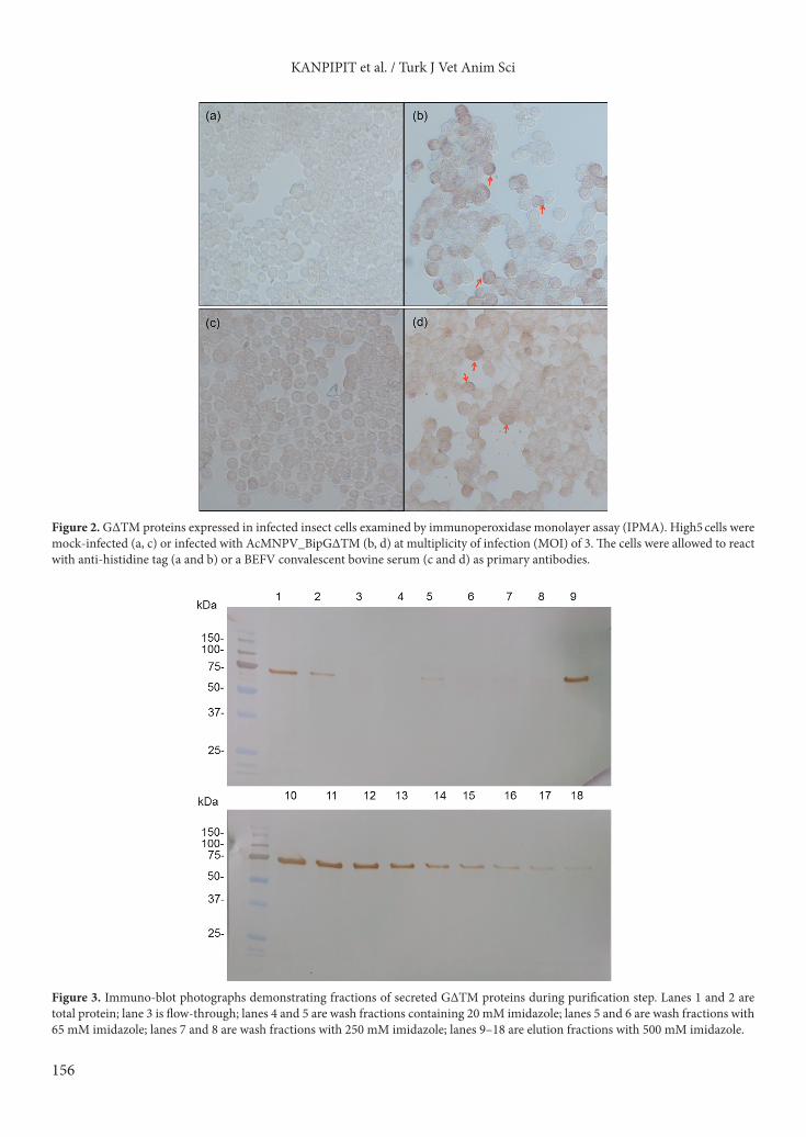
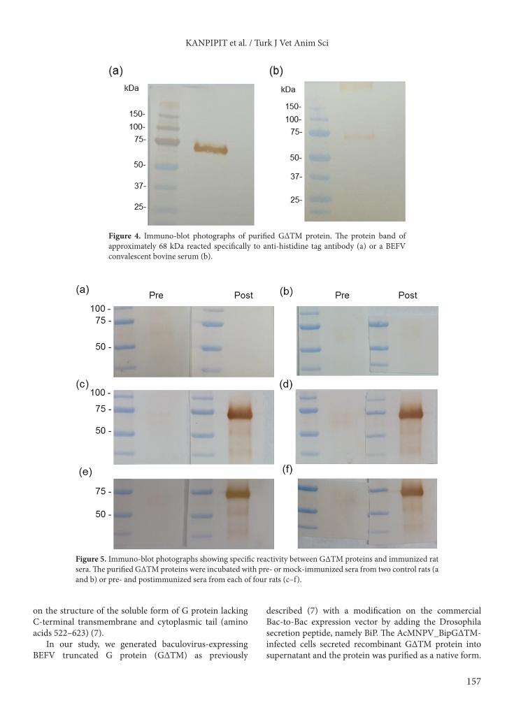
To study the expression of G∆TM in insect cells, AcMNPV_BipG∆TM was inoculated onto High5 cells at an MOI of 3 in a 96-well plate for 48 h. The presences of G∆TM protein in mock- and AcMNPV_BipG∆TM-infected cells were examined by IPMA using either anti-histidine tag or a convalescent bovine serum as primary antibodies. The results showed that the G∆TM protein appeared mostly at cell periphery and on the plasma membrane. The protein reacted well with both anti-histidine tag and a BEFV-convalescent bovine serum as shown in Figure 2. 3.2. Purification of G∆TM from cell supernatantThe secreted, soluble form of G∆TM proteins was purified from infected cell supernatant by PEG precipitation followed by affinity chromatography. Figure 3 demonstrates fractions of G∆TM proteins eluted from a nickel column by a step gradient method. The majority of G∆TM protein was eluted at 100% elution buffer containing 500 mM imidazole. The eluted fractions containing G∆TM proteins were pooled and concentrated before being analyzed by SDS-PAGE using 12% acrylamide gel. Specificity of the G∆TM protein was confirmed by western blot. The results showed that the protein reacted specifically to both anti 8X-his epitope tag antibody and a convalescent bovine serum as shown in Figure 4.
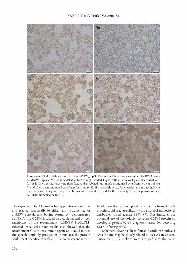
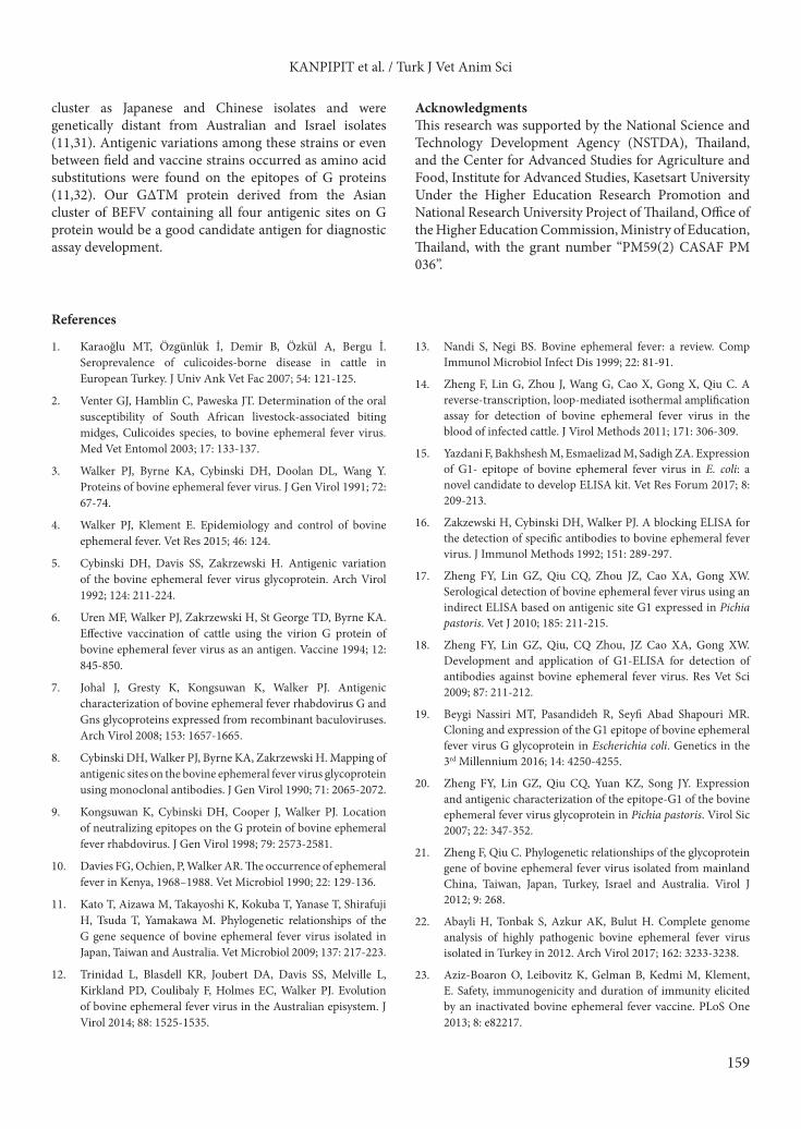
3.3. Immunogenicity of G∆TM proteinTo determine whether G∆TM protein could induce the specific immune response in animals, rats were subcutaneously injected with 40 µg of purified G∆TM protein. Rat serum samples were examined for the presence of BEFV G protein specific antibodies by western blot analysis and IPMA. The results showed that all serum samples from the immunized rats reacted specifically to the purified G∆TM protein. Sera from preimmunized rats and nonimmunized control rats did not react to the protein as shown in Figure 5. The immunized rat sera also bound specifically to the G protein presented in the positive cells as shown in Figure 6.
4. DiscussionBEF is one of the most important infectious diseases of livestock in tropical regions. Cattle infected with BEFV show clinical signs such as sudden onset of fever and lethargy, which become a serious management problem especially in large-scale farming. Vaccines have been developed and vaccination was applied as a successful strategy for disease prevention and control (5,23,26–29). BEFV glycoprotein (G) is an important protein to induce a protective immune response in cattle (28). Among the types of vaccines used for BEFV protection, the G protein expressed from recombinant vaccinia virus and partially purified virion had promising results in experimental and field studies (6,30). In fact, the G protein contains four antigenic sites (G1, G2, G3, and G4) with neutralizing activities (5,9). All four neutralizing epitopes remained
Figure 1. Soluble G∆TM protein collected from the supernatant of AcMNPV_BipG∆TM infected High5 cells. (a) The secreted G∆TM proteins were collected at 48 and 72 h postinfection (hpi). (b) The proteins were obtained from High5 cells infected with AcMNPV_BipG∆TM at MOIs of 3 or 5 for 48 hpi.
156
KANPIPIT et al. / Turk J Vet Anim Sci
Figure 2. G∆TM proteins expressed in infected insect cells examined by immunoperoxidase monolayer assay (IPMA). High5 cells were mock-infected (a, c) or infected with AcMNPV_BipG∆TM (b, d) at multiplicity of infection (MOI) of 3. The cells were allowed to react with anti-histidine tag (a and b) or a BEFV convalescent bovine serum (c and d) as primary antibodies.
Figure 3. Immuno-blot photographs demonstrating fractions of secreted G∆TM proteins during purification step. Lanes 1 and 2 are total protein; lane 3 is flow-through; lanes 4 and 5 are wash fractions containing 20 mM imidazole; lanes 5 and 6 are wash fractions with 65 mM imidazole; lanes 7 and 8 are wash fractions with 250 mM imidazole; lanes 9–18 are elution fractions with 500 mM imidazole.
157
KANPIPIT et al. / Turk J Vet Anim Sci
on the structure of the soluble form of G protein lacking C-terminal transmembrane and cytoplasmic tail (amino acids 522–623) (7).
In our study, we generated baculovirus-expressing BEFV truncated G protein (G∆TM) as previously
described (7) with a modification on the commercial Bac-to-Bac expression vector by adding the Drosophila secretion peptide, namely BiP. The AcMNPV_BipG∆TM-infected cells secreted recombinant G∆TM protein into supernatant and the protein was purified as a native form.
Figure 4. Immuno-blot photographs of purified G∆TM protein. The protein band of approximately 68 kDa reacted specifically to anti-histidine tag antibody (a) or a BEFV convalescent bovine serum (b).
Figure 5. Immuno-blot photographs showing specific reactivity between G∆TM proteins and immunized rat sera. The purified G∆TM proteins were incubated with pre- or mock-immunized sera from two control rats (a and b) or pre- and postimmunized sera from each of four rats (c–f).
158
KANPIPIT et al. / Turk J Vet Anim Sci
The expressed G∆TM protein has approximately 68 kDa and reacted specifically to either anti-histidine tag or a BEFV convalescent bovine serum. As demonstrated by IPMA, the G∆TM localized in cytoplasm and on cell membrane of the recombinant AcMNPV_BipG∆TM-infected insect cells. Our results also showed that the recombinant G∆TM was immunogenic as it could induce the specific antibody production in rats and the protein could react specifically with a BEFV convalescent serum.
In addition, it was shown previously that this form of the G protein could react specifically with a panel of monoclonal antibodies raised against BEFV (7). This indicates the potential use of the soluble, secreted G∆TM protein to develop a protein-based diagnostic assay for detecting BEFV infecting cattle.
Ephemeral fever has been found in cattle in Southeast Asia (4) and may be closely related to East Asian viruses. Taiwanese BEFV isolates were grouped into the same
Figure 6. G∆TM proteins expressed in AcMNPV_BipG∆TM infected insect cells examined by IPMA assay. AcMNPV_BipG∆TM was inoculated onto overnight seeded High5 cells in a 96-well plate at an MOI of 3 for 48 h. The infected cells were then fixed and incubated with mock-immunized sera from two control rats (a and b) or postimmunized sera form four rats (c–f). Horse-radish peroxidase-labeled anti-mouse IgG was used as a secondary antibody. The brown color was developed by the reactivity between peroxidase and 3,3’-diaminobenzidine (DAB).
159
KANPIPIT et al. / Turk J Vet Anim Sci
cluster as Japanese and Chinese isolates and were genetically distant from Australian and Israel isolates (11,31). Antigenic variations among these strains or even between field and vaccine strains occurred as amino acid substitutions were found on the epitopes of G proteins (11,32). Our G∆TM protein derived from the Asian cluster of BEFV containing all four antigenic sites on G protein would be a good candidate antigen for diagnostic assay development.
AcknowledgmentsThis research was supported by the National Science and Technology Development Agency (NSTDA), Thailand, and the Center for Advanced Studies for Agriculture and Food, Institute for Advanced Studies, Kasetsart University Under the Higher Education Research Promotion and National Research University Project of Thailand, Office of the Higher Education Commission, Ministry of Education, Thailand, with the grant number “PM59(2) CASAF PM 036”.
References
1. Karaoğlu MT, Özgünlük İ, Demir B, Özkül A, Bergu İ. Seroprevalence of culicoides-borne disease in cattle in European Turkey. J Univ Ank Vet Fac 2007; 54: 121-125.
2. Venter GJ, Hamblin C, Paweska JT. Determination of the oral susceptibility of South African livestock-associated biting midges, Culicoides species, to bovine ephemeral fever virus. Med Vet Entomol 2003; 17: 133-137.
3. Walker PJ, Byrne KA, Cybinski DH, Doolan DL, Wang Y. Proteins of bovine ephemeral fever virus. J Gen Virol 1991; 72: 67-74.
4. Walker PJ, Klement E. Epidemiology and control of bovine ephemeral fever. Vet Res 2015; 46: 124.
5. Cybinski DH, Davis SS, Zakrzewski H. Antigenic variation of the bovine ephemeral fever virus glycoprotein. Arch Virol 1992; 124: 211-224.
6. Uren MF, Walker PJ, Zakrzewski H, St George TD, Byrne KA. Effective vaccination of cattle using the virion G protein of bovine ephemeral fever virus as an antigen. Vaccine 1994; 12: 845-850.
7. Johal J, Gresty K, Kongsuwan K, Walker PJ. Antigenic characterization of bovine ephemeral fever rhabdovirus G and Gns glycoproteins expressed from recombinant baculoviruses. Arch Virol 2008; 153: 1657-1665.
8. Cybinski DH, Walker PJ, Byrne KA, Zakrzewski H. Mapping of antigenic sites on the bovine ephemeral fever virus glycoprotein using monoclonal antibodies. J Gen Virol 1990; 71: 2065-2072.
9. Kongsuwan K, Cybinski DH, Cooper J, Walker PJ. Location of neutralizing epitopes on the G protein of bovine ephemeral fever rhabdovirus. J Gen Virol 1998; 79: 2573-2581.
10. Davies FG, Ochien, P, Walker AR. The occurrence of ephemeral fever in Kenya, 1968–1988. Vet Microbiol 1990; 22: 129-136.
11. Kato T, Aizawa M, Takayoshi K, Kokuba T, Yanase T, Shirafuji H, Tsuda T, Yamakawa M. Phylogenetic relationships of the G gene sequence of bovine ephemeral fever virus isolated in Japan, Taiwan and Australia. Vet Microbiol 2009; 137: 217-223.
12. Trinidad L, Blasdell KR, Joubert DA, Davis SS, Melville L, Kirkland PD, Coulibaly F, Holmes EC, Walker PJ. Evolution of bovine ephemeral fever virus in the Australian episystem. J Virol 2014; 88: 1525-1535.
13. Nandi S, Negi BS. Bovine ephemeral fever: a review. Comp Immunol Microbiol Infect Dis 1999; 22: 81-91.
14. Zheng F, Lin G, Zhou J, Wang G, Cao X, Gong X, Qiu C. A reverse-transcription, loop-mediated isothermal amplification assay for detection of bovine ephemeral fever virus in the blood of infected cattle. J Virol Methods 2011; 171: 306-309.
15. Yazdani F, Bakhshesh M, Esmaelizad M, Sadigh ZA. Expression of G1- epitope of bovine ephemeral fever virus in E. coli: a novel candidate to develop ELISA kit. Vet Res Forum 2017; 8: 209-213.
16. Zakzewski H, Cybinski DH, Walker PJ. A blocking ELISA for the detection of specific antibodies to bovine ephemeral fever virus. J Immunol Methods 1992; 151: 289-297.
17. Zheng FY, Lin GZ, Qiu CQ, Zhou JZ, Cao XA, Gong XW. Serological detection of bovine ephemeral fever virus using an indirect ELISA based on antigenic site G1 expressed in Pichia pastoris. Vet J 2010; 185: 211-215.
18. Zheng FY, Lin GZ, Qiu, CQ Zhou, JZ Cao XA, Gong XW. Development and application of G1-ELISA for detection of antibodies against bovine ephemeral fever virus. Res Vet Sci 2009; 87: 211-212.
19. Beygi Nassiri MT, Pasandideh R, Seyfi Abad Shapouri MR. Cloning and expression of the G1 epitope of bovine ephemeral fever virus G glycoprotein in Escherichia coli. Genetics in the 3rd Millennium 2016; 14: 4250-4255.
20. Zheng FY, Lin GZ, Qiu CQ, Yuan KZ, Song JY. Expression and antigenic characterization of the epitope-G1 of the bovine ephemeral fever virus glycoprotein in Pichia pastoris. Virol Sic 2007; 22: 347-352.
21. Zheng F, Qiu C. Phylogenetic relationships of the glycoprotein gene of bovine ephemeral fever virus isolated from mainland China, Taiwan, Japan, Turkey, Israel and Australia. Virol J 2012; 9: 268.
22. Abayli H, Tonbak S, Azkur AK, Bulut H. Complete genome analysis of highly pathogenic bovine ephemeral fever virus isolated in Turkey in 2012. Arch Virol 2017; 162: 3233-3238.
23. Aziz-Boaron O, Leibovitz K, Gelman B, Kedmi M, Klement, E. Safety, immunogenicity and duration of immunity elicited by an inactivated bovine ephemeral fever vaccine. PLoS One 2013; 8: e82217.
160
KANPIPIT et al. / Turk J Vet Anim Sci
24. Srisombundit V, Tungthumniyom N, Linchongsubongkoch W, Lekcharoensuk C, Sariya L, Ramasoota P, Lekcharoensuk P. Development of an inactivated 3Cpro-3ABC (mu3ABC) ELISA to differentiate cattle infected with foot and mouth disease virus from vaccinated cattle. J Virol Methods 2013; 188: 161-167.
25. Lekcharoensuk P, Wiriyarat W, Petcharat N, Lekcharoensuk C, Auewarakul P, Richt JA. Cloned cDNA of A/swine/Iowa/15/1930 internal genes as a candidate backbone for reverse genetics vaccine against influenza A viruses. Vaccine 2012; 30: 1453-1459.
26. Inaba Y, Kurogi H, Takahash A, Sato K, Omori T, Goto Y, Hanaki T, Yamamoto M, Kishi S, Kodama K et al. Vaccination of cattle against bovine ephemeral fever with live attenuated virus followed by killed virus. Arch Gesamte Virusforsch 1974; 44: 121-132.
27. Vanselow BA, Walthall JC, Abetz I. Field trials of ephemeral fever vaccines. Vet Microbiol 1995; 46: 117-130.
28. Aziz-Boaron O, Gleser D, Yadin H, Gelman B, Kedmi M, Galon N, Klement E. The protective effectiveness of an inactivated bovine ephemeral fever virus vaccine. Vet Microbiol 2014; 173: 1-8.
29. Della-Porta AJ, Snowdon WA. Experimental inactivated virus vaccine against bovine ephemeral fever II. Do neutralizing antibodies protect against infection. Vet Microbiol 1979; 4: 197-208.
30. Hertig C, Pye AD, Hyatt AD, Davis SS, Mc William SM, Heine HG, Walker PJ, Boyle DB. Vaccinia virus-expressed bovine ephemeral fever virus G but not G(ns) glycoprotein induces neutralizing antibodies and protects against experimental infection. J Gen Virol 1996; 77 (Pt 4): 631-640.
31. Ting LJ, Lee MS, Lee SH, Tsai HJ, Lee F. Relationships of bovine ephemeral fever epizootics to population immunity and virus variation. Vet Microbiol 2014; 173: 241-248.
32. Hsieh YC, Chen SH, Chou CC, Ting LJ, Itakura C, Wang FI. Bovine ephemeral fever in Taiwan (2001-2002). J Vet Med Sci 2005; 67: 411-416.