Embed Size (px)
Citation preview
In situ visualization of telomere elongation patternsin human cellsMorgan E. Diolaiti1, Beth A. Cimini2, Robin Kageyama1, Florie A. Charles1 and
Bradley A. Stohr1,*
1Department of Pathology, University of California, San Francisco, CA 94143, USA and 2Department ofBiochemistry and Biophysics, University of California, San Francisco, CA 94158, USA
Received March 18, 2013; Revised June 29, 2013; Accepted July 15, 2013
ABSTRACT
The telomerase enzyme plays a critical role inhuman aging and cancer biology by maintainingtelomere length and extending the proliferativelifespan of most stem cells and cancer cells.Despite the importance of this enzyme, our under-standing of the mechanisms that regulate its activityand establish telomere length homeostasis in mam-malian cells is incomplete, in part because theperfect repetitive nature of telomeric sequencehampers in situ detection of telomere elongationpatterns. Here, we describe a novel assay using amutant telomerase that adds a well-tolerated varianttelomeric repeat sequence to telomere ends. Byspecifically detecting the addition of these variantrepeats, we can directly visualize telomere elong-ation events in human cells. We validate thisapproach by in situ mapping of telomere elongationpatterns within individual nuclei and across a popu-lation of cells.
INTRODUCTION
Human telomeres are composed of tandem arrays ofTTAGGG DNA repeats bound by a protective proteincomplex termed shelterin (1). These nucleoprotein struc-tures cap chromosome ends and prevent them frominitiating a DNA damage response. Telomeres shortenwith each cell division, and when telomeres becomecritically short, protection of chromosome ends is com-promised and cell proliferation is halted. Proliferativelifespan can be extended by the enzyme telomerase,which elongates telomeres by adding new TTAGGGrepeats. The telomerase enzyme minimally consists ofthe protein reverse transcriptase, TERT, and thetemplate-containing RNA, TER. Through its ability tocounteract replicative telomere shortening, the telomeraseenzyme plays an important role in maintaining stem cell
populations and extending the proliferative lifespan ofmost cancer cells. Understanding and modulating thefunction of telomerase is therefore of great interest inthe treatment of cancer and in the prevention of otheraging-associated diseases (2,3).Studies in yeast and mammalian cells have identified
numerous factors that can influence the activity of tel-omerase at individual telomeres, including telomerelength (4–9), telomeric repeat-containing RNA (10) andshelterin levels (11). To fully understand how suchfactors guide telomerase function and establish telomerelength homeostasis, it is necessary to directly identifywhich telomeres have been elongated by telomerase. InSaccharomyces cerevisiae, the telomeres consist of degen-erate TG1–3 repeats that allow for the detection of specifictelomere elongation events by sequencing (4). In contrast,the perfect repetitive nature of mammalian telomericrepeats has hindered such analysis, and as a result, theunderstanding of mammalian telomerase regulation haslagged that of yeast.Recently, an elegant method was developed that enables
the bulk detection of newly added telomeric sequence atthe lagging-strand ends of human telomeres (12). Usingthis method, the authors showed that most telomeres inHeLa cells are elongated by a single telomerase enzymeduring each cell cycle. This result suggests that, unlikeyeast telomerase, mammalian telomerase does not prefer-entially act at short telomeres (4,12). Further experimentsin HeLa cells with pre-shortened telomeres showed thatshort telomeres are sequentially elongated by multiple tel-omerase enzymes acting in a single cell cycle (13). In com-bination, these studies support a model for humantelomere homeostasis in which telomerase acts at all ormost telomeres, but with short telomeres elongatingmore rapidly owing to extension by multiple telomeraseenzymes (13). The assay used in these studies to identifythe newly added telomeric sequence requires degradationof the genome and purification of the lagging-strandtelomere ends, which precludes the ability to maptelomere elongation events to particular chromosomeends or to particular cells of a population (12).
*To whom correspondence should be addressed. Tel: +1 415 476 6729; Fax: +1 415 476 6206; Email: [email protected]
Published online 19 August 2013 Nucleic Acids Research, 2013, Vol. 41, No. 18 e176doi:10.1093/nar/gkt689
� The Author(s) 2013. Published by Oxford University Press.This is an Open Access article distributed under the terms of the Creative Commons Attribution Non-Commercial License (http://creativecommons.org/licenses/by-nc/3.0/), which permits non-commercial re-use, distribution, and reproduction in any medium, provided the original work is properly cited. For commercialre-use, please contact [email protected]
Dow
nloaded from https://academ
ic.oup.com/nar/article/41/18/e176/1036796 by guest on 26 January 2022
Here, we describe and validate a novel assay thatenables direct in situ visualization of telomere elongationpatterns within individual nuclei and across cell popula-tions. This assay provides a new approach for dissectingthe multiple overlapping regulatory mechanisms thatguide telomerase activity and ensure proper telomerelength homeostasis.
MATERIALS AND METHODS
Cell culture
Human LOX melanoma cells, MRC-5 fetal lung fibro-blasts and HeLa cervical carcinoma cells were grown inDulbecco’s modified Eagle’s medium (HeLa and MRC-5)or RPMI 1640 (LOX), supplemented with 10% (vol/vol)fetal bovine serum, 1% (vol/vol) GlutaMAX-1 (Gibco)and 1% (vol/vol) penicillin–streptomycin (Gibco). Cellswere grown at 37�C in 5% CO2. MRC-5-TERT andHeLa-TERT cells were produced by infecting MRC-5and HeLa cells with a retrovirus expressing TERT and aneomycin resistance gene. Infected cells were selected with500mg/ml G418 for 7 (HeLa-TERT) or 12 (MRC-5-TERT) days, which was sufficient to kill uninfectedcontrol cells treated in parallel.
Lentiviral plasmids and production
Lentivirus was produced as previously described (14,15)using a second-generation lentiviral system (16). TER-ex-pressing lentivectors included a wild-type or mutant TERgene driven by the IU1 promoter and a puromycin resist-ance gene driven by the CMV promoter. The wild-typeand mutant TER template sequences were as follows(with predicted telomeric repeat sequences in parentheses):wild-type – 30-CAAUCCCAAUC-50 (50-TTAGGG-30);TSQ1 – 30-CCAACGCCAAC-50 (50-GTTGCG-30); and47A – 30-CAAACCCAAAC-50 (50-TTTGGG-30). TERT-expressing lentivectors included a wild-type or mutantTERT gene driven by the CMV promoter, followed byan internal ribosome entry site and a puromycin resistancegene. Catalytically dead TERT D868A contains aninactivating mutation in the reverse transcriptase motif(17).
Lentiviral infections and growth curves
The day before infection, 1.2� 105 cells were seeded on a10-cm plate and allowed to attach overnight. On Day 0,approximately 75 transducing units of lentivirus per cellwas added to each plate. For LOX and HeLa cells, 8 mg/mlpolybrene was also added to the media at the start ofinfection. After 8 h, the virus-containing media werereplaced with fresh media. Puromycin selection(1.5 mg/ml) was initiated 24–36 h after the introductionof virus and lasted 2–3 days, by which time uninfectedcontrol cells treated with puromycin had died. Selectedcells were harvested at indicated time points for fluores-cence in situ hybridization (FISH) analysis. For growthcurves, cells were plated immediately following selectionin 6-well dishes at 2� 104 (LOX), 3� 104 (HeLa andHeLa-TERT) or 3.5� 104 (MRC-5-TERT) cells/well.
Cells were split as needed to maintain logarithmicgrowth, and viable cells were counted by hemocytometer.For experiments analysing TSQ1 incorporation after in-fection with TERT-expressing virus (Figures 1B and 4,and Supplementary Figure S4), MRC-5 cells were firstinfected with lentivirus expressing TSQ1 TER and sub-jected to puromycin selection as mentioned above. TheTSQ1-expressing MRC-5 cells were next infected withlentivirus expressing wild-type or catalytically deadTERT using the infection protocol outlined above. Cellswere harvested for FISH analysis 48 h after infection withTERT-expressing virus.
Metaphase spread preparation and FISH
Cells were treated with 0.1 mg/ml Colcemid for 2–4 hbefore the harvest. Collected cells underwent hypotonictreatment with 0.075M KCl for 10min at 37�C andwere subsequently fixed by 4–6 rounds of resuspensionin 3:1 methanol/acetic acid. Cells were dropped on micro-scope slides to prepare metaphase spreads, and the slideswere dried for at least 16 h before FISH. For FISH, thecells were rehydrated in 1� phosphate buffered saline(PBS) for 15min, followed by fixation with 4%paraformaldehyde (in 1� PBS) for 2min. After a washin 1� PBS, cells were treated with RNase A (Qiagencatalogue no. 158922, diluted 1:100 in PBS) for approxi-mately 2 h at room temperature. After three 1� PBSwashes, cells were treated with 0.1mg/mL pepsin(Sigma-Aldrich catalogue no. P7012, prepared fresh in0.01M HCl) at 37�C for 10min. The cells were washedtwice with 1� PBS, fixed in 4% formaldehyde (in 1� PBS)for 2min, and rinsed three times in 1� PBS. The cells weredehydrated in ethanol (2min each in 70% ethanol, 95%ethanol and 100% ethanol) and air dried. The FISH hy-bridization mix included 70% formamide, 10mM TrisHCl pH 7.2, 1% (weight/vol) blocking reagent (Rochecatalogue no. 11096176001) and 0.5mg/ml of eachpeptide nucleic acid (PNA) probe. The following PNAprobes (Panagene) were used: wild-type telomere probe(FAM-OO-ccctaaccctaaccctaa), TSQ1 telomere probe(Cy3-OO-ccgcaaccgcaaccgcaa), pan-centromere probe(Cy5-OO-cttcgttggaaacggga) and chromosome 9 centro-mere probe (Cy5-OO-aatcaacccgagtgcaat) (18). The hy-bridization mix was added to the cells under a coverslipand the cells were denatured on an 80�C heatblock for3min. After a 2 h hybridization at room temperature,the cells were washed twice for 15min in 70%formamide/Tris HCl pH 7.4 and three times for 5min in0.05M Tris HCl pH 7.4/0.15M NaCl/0.05% Tween-20.After ethanol dehydration as mentioned above, slideswere air dried and mounted in Prolong Gold with DAPI(Invitrogen catalogue no. P-36931).
Microscopy and image analysis
All images were obtained using a Deltavision deconvolu-tion microscope (Applied Precision) with a 60� 1.42 NAPlan Apo objective (Olympus). Deltavision images with Z-stacks were converted to projected Tagged Image FileFormat using the Fiji image processing package (www.fiji.sc), and TSQ1-positive telomeres were scored by
e176 Nucleic Acids Research, 2013, Vol. 41, No. 18 PAGE 2 OF 9
Dow
nloaded from https://academ
ic.oup.com/nar/article/41/18/e176/1036796 by guest on 26 January 2022
TSQ1 TER vector
LOX
MR
C-5
-TE
RT
A
B
47A TER
unin
fect
edW
T TE
RT
DN
TE
RT
Telomere PNA TSQ1 PNA merged
0% 97% 0%
0% 98% 0%
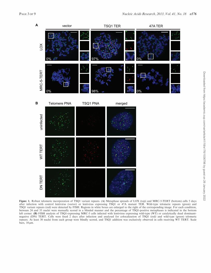
Figure 1. Robust telomeric incorporation of TSQ1 variant repeats. (A) Metaphase spreads of LOX (top) and MRC-5-TERT (bottom) cells 5 daysafter infection with control lentivirus (vector) or lentivirus expressing TSQ1 or 47A mutant TER. Wild-type telomeric repeats (green) andTSQ1 variant repeats (red) were detected by FISH. Regions in white boxes are enlarged to the right of the corresponding image. For each condition,between 24 and 35 nuclei were manually scored in a blinded manner and the percentage of TSQ1-positive metaphases is indicated in the bottomleft corner. (B) FISH analysis of TSQ1-expressing MRC-5 cells infected with lentivirus expressing wild-type (WT) or catalytically dead dominant-negative (DN) TERT. Cells were fixed 2 days after infection and analysed for colocalization of TSQ1 (red) and wild-type (green) telomericrepeats. At least 30 nuclei from each group were blindly scored, and TSQ1 addition was exclusively observed in cells receiving WT TERT. Scalebars, 10 mm.
PAGE 3 OF 9 Nucleic Acids Research, 2013, Vol. 41, No. 18 e176
Dow
nloaded from https://academ
ic.oup.com/nar/article/41/18/e176/1036796 by guest on 26 January 2022
manually counting TSQ1 foci that co-localize with wild-type telomere foci. Enumeration and intensity analysis ofwild-type telomere foci in intact nuclei were performedusing CellProfiler image analysis software (www.cellprofiler.org; analysis pipelines available on request).Chromosomal fusions on metaphase spreads weremanually counted in a blinded manner.
Real-time polymerase chain reaction (PCR)
TERT expression levels were determined using a one-stepBrilliant II QRT-PCR kit (Agilent Technologies) and thefollowing TERT and control primer sets: TERT (forward:cctgcactggctgatgagtgtg; reverse: gatgctgcctgacctctgctt);beta-2-microglobulin (forward: tcacgtcatccagcagagaatgga;reverse: cacacggcaggcatactcatcttt). TER expression levelswere determined using a two-step protocol. First, cDNAwas synthesized using SuperScript III reverse transcriptasewith random hexamer primers (Invitrogen). SubsequentQ-PCR was performed using the Brilliant II Q-PCR kit(Agilent Technologies) and the following TER and controlprimer sets: TER (forward: ttgcggagggtgggcct; reverse:cgggccagcagctgacatt); GAPDH (forward: catgttcgtcatgggtgtgaacca; reverse: atggcatggactgtggtcatgagt). AllPCR reactions were performed with the StepOnePlusreal-time PCR system (Applied Biosystems). Values werenormalized to beta-2-microglobulin or GAPDH and arereported as fold change over vector.
RESULTS
TSQ1 variant repeat incorporation is well tolerated inprimary and cancer cell lines
Our assay builds on work using mutant forms of TER thatcontain sequence changes in the template region. Whenoverexpressed in cells, mutant TER joins with wild-typeTERT to form mutant telomerase, which adds variantrepeats to telomere ends (19). Although detection ofincorporated variant repeat sequences could in principlebe used to map where telomerase has acted, incorporationof these sequences typically induces rapid telomere dys-function. Because variant repeats cannot properly recruitthe shelterin protein complexes that bind and protect wild-type telomeres (19), cells overexpressing mutant TERusually undergo rapid senescence or apoptosis, oftenaccompanied by telomere fusions (2,15,20). This cytotox-icity presents a significant barrier for using mutant TERsto map telomerase activity patterns.During a recent screen of mutant TER sequences, we
serendipitously identified a mutant TER that is welltolerated in human cells despite robust incorporation ofvariant telomeric repeats. This mutant TER, hereafterreferred to as TSQ1 for Tolerated Sequence 1, is designedto add GTTGCG variant repeats. Using PNA probes todetect wild-type (TTAGGG) and TSQ1 (GTTGCG)repeats, robust telomeric incorporation of TSQ1 repeatswas specifically detected in human LOX melanoma cellsoverexpressing TSQ1 (Figure 1A and SupplementaryFigure S1). Telomeric incorporation of TSQ1 variantrepeats was similarly observed in MRC-5 human diploidfibroblasts that overexpress TERT (MRC-5-TERT,
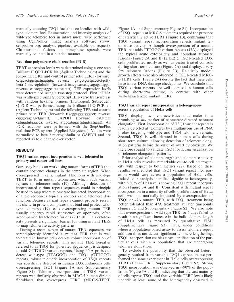
Figure 1A and Supplementary Figure S1). Incorporationof TSQ1 repeats at MRC-5 telomeres required the presenceof catalytically active TERT (Figure 1B), confirming thatTSQ1 variant repeat incorporation reflects mutant tel-omerase activity. Although overexpression of a mutantTER that adds TTTGGG variant repeats (47A) displayedthe typical acute cytotoxicity and abundant telomerefusions (Figure 2A and B) (2,15,21), TSQ1-treated LOXcells proliferated nearly as well as vector-treated controlsduring short-term culture (Figure 2A) and displayed veryfew telomere fusions (Figure 2B). Relatively modestgrowth effects were also observed in TSQ1-treated MRC-5-TERT cells (Figure 2A) despite the fact that these cellshave intact DNA damage checkpoints. We conclude thatTSQ1 variant repeats are well-tolerated in human cellsduring short-term culture, in contrast with otherwell-characterized mutant TERs.
TSQ1 variant repeat incorporation is heterogeneousacross a population of HeLa cells
TSQ1 displays two characteristics that make it apromising in situ marker of telomerase-directed telomereelongation. First, incorporation of TSQ1 variant repeats isreadily detected at telomeres by simultaneous use of PNAprobes targeting wild-type and TSQ1 telomeric repeats.Second, TSQ1 is well-tolerated in human cells duringshort-term culture, allowing detection of telomere elong-ation patterns before the onset of overt cytotoxicity. Wetherefore sought to validate TSQ1 for in situ visualizationof telomere elongation patterns.
Prior analysis of telomere length and telomerase activityin HeLa cells revealed remarkable cell-to-cell heterogen-eity with respect to both metrics (22). Based on theseresults, we predicted that TSQ1 variant repeat incorpor-ation would vary across a population of HeLa cells.Indeed, our analysis identified significant heterogeneity,with �30% of HeLa cells showing TSQ1 repeat incorpor-ation (Figure 3A and B). Consistent with mutant repeatincorporation in a minority of cells, proliferation of HeLacells was not markedly impacted by overexpression ofTSQ1 or 47A mutant TER, with TSQ1 treatment beingbetter tolerated than 47A treatment at later timepoints(Figure 3C and Supplementary Figure S2). We also notethat overexpression of wild-type TER for 6 days failed toresult in a significant increase in the bulk telomere lengthof HeLa cells as measured by quantitative FISH(Supplementary Figure S3). Thus, under conditionswhere a population-based assay to assess telomere repeataddition does not detect significant telomere lengthening,TSQ1 incorporation enables clear identification of the par-ticular cells within a population that are undergoingtelomere elongation.
To exclude the possibility that the observed hetero-geneity resulted from variable TSQ1 expression, we per-formed the same experiment in HeLa cells overexpressingTERT (HeLa–TERT, Supplementary Figure S2). StrongTSQ1 incorporation was observed in �90% of the popu-lation (Figure 3A and B), indicating that the vast majorityof cells express TSQ1 and that variable TERT levels likelyunderlie at least some of the heterogeneity observed in
e176 Nucleic Acids Research, 2013, Vol. 41, No. 18 PAGE 4 OF 9
Dow
nloaded from https://academ
ic.oup.com/nar/article/41/18/e176/1036796 by guest on 26 January 2022
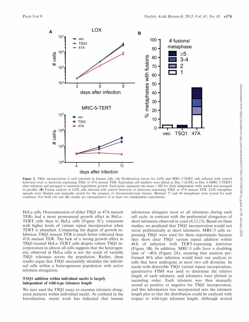
HeLa cells. Overexpression of either TSQ1 or 47A mutantTERs had a more pronounced growth effect in HeLa–TERT cells than in HeLa cells (Figure 3C), consistentwith higher levels of variant repeat incorporation whenTERT is abundant. Comparing the degree of growth in-hibition, TSQ1 mutant TER is much better tolerated than47A mutant TER. The lack of a strong growth effect inTSQ1-treated HeLa–TERT cells despite robust TSQ1 in-corporation in almost all cells suggests that the heterogen-eity observed in HeLa cells is not the result of variableTSQ1 tolerance across the population. Rather, theseresults argue that TSQ1 successfully identifies the individ-ual cells within a heterogeneous population with activetelomere elongation.
TSQ1 addition within individual nuclei is largelyindependent of wild-type telomere length
We next used the TSQ1 assay to examine telomere elong-ation patterns within individual nuclei. As outlined in theIntroduction, recent work has indicated that human
telomerase elongates most or all telomeres during eachcell cycle, in contrast with the preferential elongation ofshort telomeres observed in yeast (4,12,13). Based on thesestudies, we predicted that TSQ1 incorporation would notoccur preferentially at short telomeres. MRC-5 cells ex-pressing TSQ1 were used for these experiments becausethey show clear TSQ1 variant repeat addition within48 h of infection with TERT-expressing lentivirus(Figure 1B). In addition, MRC-5 cells have a doublingtime of �40 h (Figure 2A), ensuring that analysis per-formed 48 h after infection would limit our analysis tocells that have undergone at most two cell divisions. Innuclei with detectable TSQ1 variant repeat incorporation,quantitative FISH was used to determine the relativelength of each telomere, and telomeres were plotted inascending order. Each telomere was then manuallyscored as positive or negative for TSQ1 incorporation,and this information was incorporated into the telomerelength plot so that the distribution could be analysed withrespect to wild-type telomere length. Although several
A B
days after infection
# ce
lls
3 6 9104
105
106
107vecTSQ147A
% m
etap
hase
s w
ith fu
sion
s
vec TSQ1 47A0
10
20
30
40
50
60
70
80
90
100
123-4≥5
LOX
MRC-5-TERT
# fusions/metaphase
days aftff er infeff ction
#ce
lls
4 7 10104
105
106
107 vecTSQ147A
Figure 2. TSQ1 incorporation is well tolerated in human cells. (A) Proliferation curves for LOX and MRC-5-TERT cells infected with controllentivirus (vec) or lentivirus expressing TSQ1 or 47A mutant TER. Equivalent cell numbers were plated at Day 3 (LOX) or Day 4 (MRC-5-TERT)after infection and passaged to maintain logarithmic growth. Each point represents the mean±SD for three independent wells seeded and passagedin parallel. (B) Fusion analysis in LOX cells infected with control lentivirus or lentivirus expressing TSQ1 or 47A mutant TER. LOX metaphasespreads were blinded and manually scored for the presence of chromosome-type fusions. Between 37 and 44 metaphases were scored for eachcondition. For both (A) and (B), results are representative of at least two independent experiments.
PAGE 5 OF 9 Nucleic Acids Research, 2013, Vol. 41, No. 18 e176
Dow
nloaded from https://academ
ic.oup.com/nar/article/41/18/e176/1036796 by guest on 26 January 2022
TRET-aLeHaLeHC
days after infection
# ce
lls
3 6 9 13104
105
106
107
108
vecTSQ147A
days after infection3 6 9 13
104
105
106
107
vecTSQ147A
HeLa HeLa-TERT
B
A%
TSQ
-pos
itive
telo
mer
es
vec TSQ10
20
40
60
80
100
vec TSQ10
20
40
60
80
100TRET-aLeHaLeH
Figure 3. Variable TSQ1 incorporation across a population. (A) FISH analysis of HeLa and HeLa-TERT cells 6 days after infection with TSQ1lentivirus. Each image shows portions of two adjacent nuclei (outlined). Individual wild-type telomeric repeat (green) and TSQ1 variant repeat (red)channels are broken out to the right of each merged image. Both HeLa-TERT nuclei display abundant TSQ1-positive telomeres, whereas only one ofthe two HeLa cell nuclei displays abundant TSQ1-positive telomeres. Scale bars, 10 mm. (B) Plot showing the percentage of TSQ1-positive telomeresfor TSQ1-treated (purple) and vector-treated (green) HeLa and HeLa-TERT nuclei. Forty nuclei were scored for each condition, with each point onthe plot representing a single nucleus. The few telomeres scored as TSQ1-positive in vector-treated cells reflect the low-level background seen with theTSQ1-specific FISH probe. (C) Proliferation curves for HeLa and HeLa-TERT cells infected with control lentivirus (vec) or lentivirus expressingTSQ1 or 47A mutant TER. Equivalent cell numbers were plated 3 days after infection and passaged to maintain logarithmic growth. Each pointrepresents the mean±SD for three independent wells seeded and passaged in parallel. For (A–C), results are representative of at least two inde-pendent experiments.
e176 Nucleic Acids Research, 2013, Vol. 41, No. 18 PAGE 6 OF 9
Dow
nloaded from https://academ
ic.oup.com/nar/article/41/18/e176/1036796 by guest on 26 January 2022
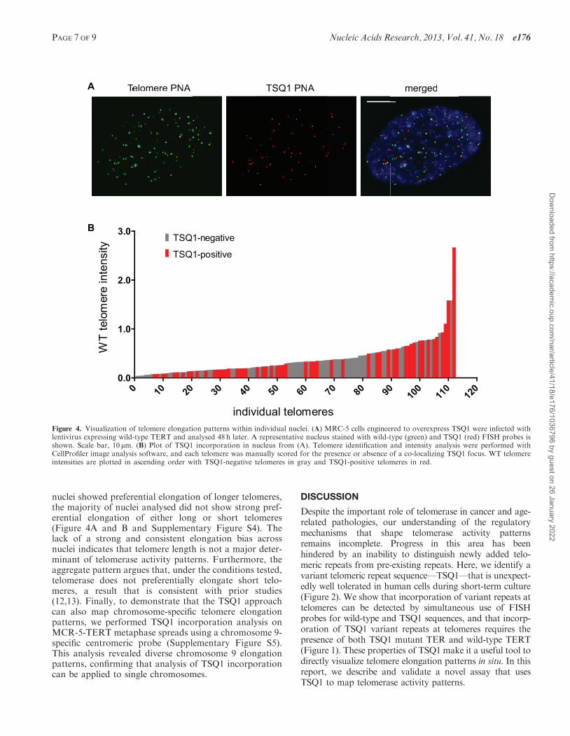
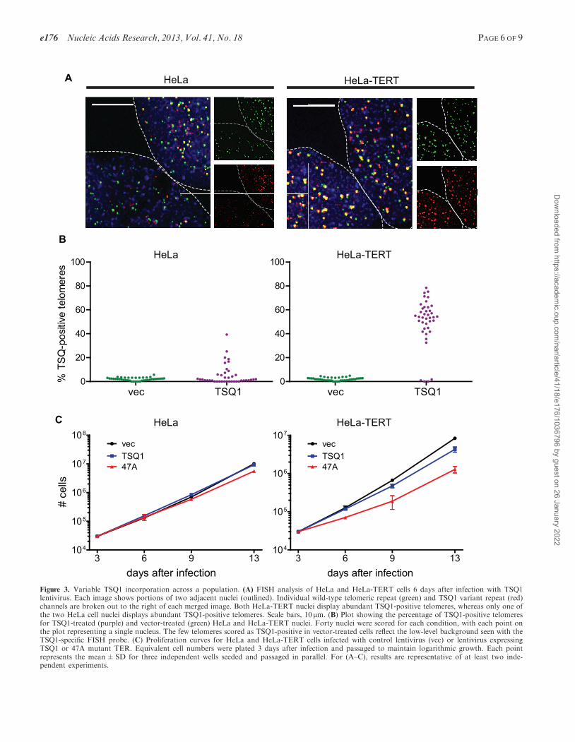
nuclei showed preferential elongation of longer telomeres,the majority of nuclei analysed did not show strong pref-erential elongation of either long or short telomeres(Figure 4A and B and Supplementary Figure S4). Thelack of a strong and consistent elongation bias acrossnuclei indicates that telomere length is not a major deter-minant of telomerase activity patterns. Furthermore, theaggregate pattern argues that, under the conditions tested,telomerase does not preferentially elongate short telo-meres, a result that is consistent with prior studies(12,13). Finally, to demonstrate that the TSQ1 approachcan also map chromosome-specific telomere elongationpatterns, we performed TSQ1 incorporation analysis onMCR-5-TERT metaphase spreads using a chromosome 9-specific centromeric probe (Supplementary Figure S5).This analysis revealed diverse chromosome 9 elongationpatterns, confirming that analysis of TSQ1 incorporationcan be applied to single chromosomes.
DISCUSSION
Despite the important role of telomerase in cancer and age-related pathologies, our understanding of the regulatorymechanisms that shape telomerase activity patternsremains incomplete. Progress in this area has beenhindered by an inability to distinguish newly added telo-meric repeats from pre-existing repeats. Here, we identify avariant telomeric repeat sequence—TSQ1—that is unexpect-edly well tolerated in human cells during short-term culture(Figure 2). We show that incorporation of variant repeats attelomeres can be detected by simultaneous use of FISHprobes for wild-type and TSQ1 sequences, and that incorp-oration of TSQ1 variant repeats at telomeres requires thepresence of both TSQ1 mutant TER and wild-type TERT(Figure 1). These properties of TSQ1 make it a useful tool todirectly visualize telomere elongation patterns in situ. In thisreport, we describe and validate a novel assay that usesTSQ1 to map telomerase activity patterns.
A
B
Telomere PNA TSQ1 PNA mergedM
RC
-5
TSQ1-negative
TSQ1-positive
TSQ1 PNAA
B
Telomere PNTT AM
RM
RM
RMMMMMMMMMMMMMMMMMMMMMM
C-
C-
C555
merged
individual telomeres
WT
telo
mer
e in
tens
ity
0 10 20 30 40 50 60 70 80 90 100
110
120
0.0
1.0
2.0
3.0
Figure 4. Visualization of telomere elongation patterns within individual nuclei. (A) MRC-5 cells engineered to overexpress TSQ1 were infected withlentivirus expressing wild-type TERT and analysed 48 h later. A representative nucleus stained with wild-type (green) and TSQ1 (red) FISH probes isshown. Scale bar, 10 mm. (B) Plot of TSQ1 incorporation in nucleus from (A). Telomere identification and intensity analysis were performed withCellProfiler image analysis software, and each telomere was manually scored for the presence or absence of a co-localizing TSQ1 focus. WT telomereintensities are plotted in ascending order with TSQ1-negative telomeres in gray and TSQ1-positive telomeres in red.
PAGE 7 OF 9 Nucleic Acids Research, 2013, Vol. 41, No. 18 e176
Dow
nloaded from https://academ
ic.oup.com/nar/article/41/18/e176/1036796 by guest on 26 January 2022
To validate the utility of TSQ1 as an in situ marker oftelomere elongation, we examined the pattern of TSQ1addition in individual nuclei. Although S. cerevisiae tel-omerase preferentially elongates the shortest telomeres (4),recent work in multiple cell lines indicates that humantelomerase acts at all or most telomeres during each cellcycle (12,13). In agreement with the recent studies, we findthat TSQ1 addition occurred without a strong, consistentbias for long or short telomeres in MRC-5 human fibro-blasts expressing exogenous TERT (Figure 4). A similarpattern was observed in HeLa cells treated with TSQ1(data not shown). Interestingly, we did not detect TSQ1addition at every telomere, even with simultaneousoverexpression of TSQ1 TER and TERT (Figure 4).There are several possible explanations for this observa-tion. First, TSQ1-treated cells still express wild-type tel-omerase, and our assay will not detect telomere elongationevents that add wild-type telomeric repeats. Second, aportion of TSQ1 elongation events may fall below thethreshold required for FISH detection. Finally, owing toits template mutations, TSQ1 mutant telomerase does notalign perfectly with the wild-type telomeric overhang, andthe resulting mismatches may serve as an initial barrier toelongation. Nevertheless, even if TSQ1 enables visualiza-tion of only a subset of elongation events, the aggregateanalysis of TSQ1 addition provides a snapshot of telomer-ase activity patterns. For example, if telomerase acted ex-clusively at short telomeres, we would expect detection ofTSQ1 addition events only at short telomeres. The factthat we do not observe this pattern provides strongevidence that, under the conditions tested, telomerasedoes not preferentially elongate short telomeres. In thefuture, it will be interesting to determine whether humantelomerase preferentially elongates short telomeres in thecontext of very low telomerase levels.In addition to analysing TSQ1 addition within individ-
ual cells, we also examined TSQ1 incorporation across apopulation of cells. To enable direct comparison betweenthe TSQ1-derived results and those obtained using otherapproaches, we focused our analyses on HeLa cellsbecause the telomerase and telomere dynamics of thiscell line have been relatively well characterized. A priorstudy of HeLa cell clones showed that only five of eightsingle-cell-derived clones were positive for telomeraseactivity when measured by the Telomere RepeatAmplification Protocol (TRAP) (22). Consistent withthis high degree of heterogeneity, we only detected TSQ1addition in approximately one-third of the HeLa cells(Figure 3). The lower percentage of telomerase-positivecells identified in our study may reflect differences inassay sensitivity. In particular, FISH detection ofincorporated TSQ1 sequence may miss cells with lowlevels of TSQ1 incorporation. Alternatively, the per-centage of telomerase-positive clones identified by TRAPin the earlier study may be inflated. Stochastic switching ofcells from a telomerase-negative to a telomerase-positivestate was reported in the prior study (22), and it is possiblesome clones that were initially telomerase-negative becamepositive during the 14 population doublings required toget sufficient outgrowth for TRAP. In either case, TSQ1incorporation allows for rapid and direct in situ
identification of the individual cells within a populationundergoing robust telomere elongation.
Heterogeneous telomerase activity and telomere elong-ation patterns may have significant implications for clonalevolution, genome instability, and the success of telomerase-targeted therapeutics, yet relatively little work has been doneto characterize this heterogeneity and define its causes. Inour HeLa cell experiments, heterogeneous TERT expressionmay be the major driver of variable telomere elongation.TERT overexpression indeed resulted in a more homogen-ous telomere elongation pattern across the HeLa cell popu-lation (Figure 3), consistent with prior work showing thatsimultaneous overexpression of TER and TERT inducesmassive telomere elongation (23). Alternatively, theabsence of detectable TSQ1 incorporation in many TSQ1-treated HeLa cells may stem from negative regulation oftelomere elongation. Numerous factors have been shownto inhibit telomerase activity at telomeres, including theshelterin protein POT1 (11) and telomeric repeat-containingRNA (24). We are now in a position to ask whether theexpression or localization of such negative regulatoryelements correlates with TSQ1 incorporation, and whetherexperimental manipulation of these factors alters the TSQ1incorporation patterns both within single cells and acrosspopulations.
There are several limitations to the TSQ1 approach thatbear mention. First, the TSQ1 assay requiresoverexpression of TSQ1 mutant TER and thereforecannot be used to assess the role of endogenous TER inestablishing telomerase activity patterns. Furthermore,because TER overexpression has been shown to increasetelomerase activity in many cell types including HeLa(23,25), this approach does not examine telomere elong-ation dynamics under homeostatic conditions. Despite therequirement for TER overexpression, the TSQ1 assay high-lights significant telomere elongation heterogeneity in theHeLa cell population and therefore enables analysis of themyriad other factors that establish that heterogeneity.
The addition of TSQ1 repeats at telomeres may alsointerfere with the sequence-specific binding of telomericproteins—including the POT1-TPP1 (11,26) and CST(27) complexes—that regulate telomerase function. As aresult, it is likely that TSQ1 incorporation disrupts thenormal feedback mechanisms that regulate telomeraserepeat addition processivity and thus the extent oftelomere elongation. For this reason, we have focusedour analysis on the overall pattern of TSQ1 additionrather than the extent of TSQ1 addition at any giventelomere. We also note that although TSQ1 variantrepeats are unusually well tolerated in human cells, theincorporation of variant repeats nevertheless induceslow-level telomere dysfunction that inhibits cell prolifer-ation over long-term culture (Figures 2 and 3). Becausesuch telomere dysfunction may ultimately induce telomerefusions and alternative lengthening of telomere-like telo-meric phenotypes (2,20,28,29), we have limited ouranalysis of TSQ1 addition in individual nuclei to veryshort-term culture (48 h in the case of the MRC-5 experi-ments described in Figure 4), and we have monitored forthe appearance of telomere fusions that might skew ourresults. Finally, although TSQ1 is better tolerated than
e176 Nucleic Acids Research, 2013, Vol. 41, No. 18 PAGE 8 OF 9
Dow
nloaded from https://academ
ic.oup.com/nar/article/41/18/e176/1036796 by guest on 26 January 2022
other mutant TERs in all of the cell types we have tested,we find that some cell types tolerate TSQ1 better thanothers (Figures 2 and 3 and data not shown). It will there-fore be necessary to evaluate the degree to which TSQ1induces proliferative inhibition and genome instability inother cell types before using this strategy in new contexts.
In conclusion, TSQ1 provides a technically simplemethod for in situ mapping of telomerase activitypatterns in many cell types. To our knowledge, it is theonly assay that can be used to track in situ elongation ofindividual telomeres. By providing a snapshot of recenttelomerase activity, TSQ1 will enable the exploration oftelomere elongation patterns and the dissection of themolecular mechanisms that shape those patterns.
SUPPLEMENTARY DATA
Supplementary Data are available at NAR Online.
ACKNOWLEDGEMENTS
We thank I. Listerman for critical review of the manuscript.
FUNDING
National Institute of Health grant [K08 CA134552]; theProgram for Breakthrough Biomedical Research, which isfunded in part by the Sandler Foundation; the Universityof California Cancer Research Coordinating Committee(CRCC). Funding for open access charge: University ofCalifornia San Francisco, Department of Pathology.
Conflict of interest statement. None declared.
REFERENCES
1. Palm,W. and de Lange,T. (2008) How shelterin protectsmammalian telomeres. Annu. Rev. Genet., 42, 301–334.
2. Stohr,B.A. and Blackburn,E.H. (2008) ATM mediates cytotoxicityof a mutant telomerase RNA in human cancer cells. Cancer Res.,68, 5309–5317.
3. Armanios,M. and Blackburn,E.H. (2012) The telomeresyndromes. Nat. Rev. Genet., 13, 693–704.
4. Teixeira,M.T., Arneric,M., Sperisen,P. and Lingner,J. (2004)Telomere length homeostasis is achieved via a switch betweentelomerase- extendible and -nonextendible states. Cell, 117,323–335.
5. Hemann,M.T., Strong,M.A., Hao,L.Y. and Greider,C.W. (2001)The shortest telomere, not average telomere length, is critical forcell viability and chromosome stability. Cell, 107, 67–77.
6. Zhu,L., Hathcock,K.S., Hande,P., Lansdorp,P.M., Seldin,M.F.and Hodes,R.J. (1998) Telomere length regulation in mice islinked to a novel chromosome locus. Proc. Natl Acad. Sci. USA,95, 8648–8653.
7. Ouellette,M.M., Liao,M., Herbert,B.S., Johnson,M., Holt,S.E.,Liss,H.S., Shay,J.W. and Wright,W.E. (2000) Subsenescenttelomere lengths in fibroblasts immortalized by limiting amountsof telomerase. J. Biol. Chem., 275, 10072–10076.
8. Britt-Compton,B., Capper,R., Rowson,J. and Baird,D.M. (2009)Short telomeres are preferentially elongated by telomerase inhuman cells. FEBS Lett., 583, 3076–3080.
9. Liu,Y., Kha,H., Ungrin,M., Robinson,M.O. and Harrington,L.(2002) Preferential maintenance of critically short telomeres inmammalian cells heterozygous for mTert. Proc. Natl Acad. Sci.USA, 99, 3597–3602.
10. Feuerhahn,S., Iglesias,N., Panza,A., Porro,A. and Lingner,J.(2010) TERRA biogenesis, turnover and implications for function.FEBS Lett., 584, 3812–3818.
11. Loayza,D. and De Lange,T. (2003) POT1 as a terminaltransducer of TRF1 telomere length control. Nature, 423,1013–1018.
12. Zhao,Y., Sfeir,A.J., Zou,Y., Buseman,C.M., Chow,T.T.,Shay,J.W. and Wright,W.E. (2009) Telomere extension occurs atmost chromosome ends and is uncoupled from fill-in in humancancer cells. Cell, 138, 463–475.
13. Zhao,Y., Abreu,E., Kim,J., Stadler,G., Eskiocak,U., Terns,M.P.,Terns,R.M., Shay,J.W. and Wright,W.E. (2011) Processive anddistributive extension of human telomeres by telomerase underhomeostatic and nonequilibrium conditions. Mol. Cell, 42,297–307.
14. Xu,L. and Blackburn,E.H. (2004) Human Rif1 protein bindsaberrant telomeres and aligns along anaphase midzonemicrotubules. J. Cell Biol., 167, 819–830.
15. Li,S., Rosenberg,J.E., Donjacour,A.A., Botchkina,I.L., Hom,Y.K.,Cunha,G.R. and Blackburn,E.H. (2004) Rapid inhibition ofcancer cell growth induced by lentiviral delivery and expression ofmutant-template telomerase RNA and anti-telomerase short-interfering RNA. Cancer Res., 64, 4833–4840.
16. Zufferey,R., Nagy,D., Mandel,R.J., Naldini,L. and Trono,D.(1997) Multiply attenuated lentiviral vector achieves efficient genedelivery in vivo. Nat. Biotechnol., 15, 871–875.
17. Nakayama,J., Tahara,H., Tahara,E., Saito,M., Ito,K.,Nakamura,H., Nakanishi,T., Ide,T. and Ishikawa,F. (1998)Telomerase activation by hTRT in human normal fibroblasts andhepatocellular carcinomas. Nat. Genet., 18, 65–68.
18. Chen,C., Wu,B., Wei,T., Egholm,M. and Strauss,W.M. (2000)Unique chromosome identification and sequence-specificstructural analysis with short PNA oligomers. Mamm. Genome,11, 384–391.
19. Marusic,L., Anton,M., Tidy,A., Wang,P., Villeponteau,B. andBacchetti,S. (1997) Reprogramming of telomerase by expressionof mutant telomerase RNA template in human cells leads toaltered telomeres that correlate with reduced cell viability. Mol.Cell. Biol., 17, 6394–6401.
20. Guiducci,C., Cerone,M.A. and Bacchetti,S. (2001) Expressionof mutant telomerase in immortal telomerase-negative humancells results in cell cycle deregulation, nuclear andchromosomal abnormalities and rapid loss of viability. Oncogene,20, 714–725.
21. Stohr,B.A., Xu,L. and Blackburn,E.H. (2010) The terminaltelomeric DNA sequence determines the mechanism ofdysfunctional telomere fusion. Mol. Cell, 39, 307–314.
22. Bryan,T.M., Englezou,A., Dunham,M.A. and Reddel,R.R. (1998)Telomere length dynamics in telomerase-positive immortal humancell populations. Exp. Cell Res., 239, 370–378.
23. Cristofari,G. and Lingner,J. (2006) Telomere length homeostasisrequires that telomerase levels are limiting. EMBO J., 25,565–574.
24. Redon,S., Reichenbach,P. and Lingner,J. (2010) The non-codingRNA TERRA is a natural ligand and direct inhibitor of humantelomerase. Nucleic Acids Res., 38, 5797–5806.
25. Pickett,H.A., Cesare,A.J., Johnston,R.L., Neumann,A.A. andReddel,R.R. (2009) Control of telomere length by a trimmingmechanism that involves generation of t-circles. EMBO J., 28,799–809.
26. Wang,F., Podell,E.R., Zaug,A.J., Yang,Y., Baciu,P., Cech,T.R.and Lei,M. (2007) The POT1-TPP1 telomere complex is atelomerase processivity factor. Nature, 445, 506–510.
27. Chen,L.Y., Redon,S. and Lingner,J. (2012) The human CSTcomplex is a terminator of telomerase activity. Nature, 488,540–544.
28. Conomos,D., Stutz,M.D., Hills,M., Neumann,A.A., Bryan,T.M.,Reddel,R.R. and Pickett,H.A. (2012) Variant repeats areinterspersed throughout the telomeres and recruit nuclearreceptors in ALT cells. J. Cell Biol., 199, 893–906.
29. Brault,M.E. and Autexier,C. (2011) Telomeric recombinationinduced by dysfunctional telomeres. Mol. Biol. Cell, 22, 179–188.
PAGE 9 OF 9 Nucleic Acids Research, 2013, Vol. 41, No. 18 e176
Dow
nloaded from https://academ
ic.oup.com/nar/article/41/18/e176/1036796 by guest on 26 January 2022


![Determination of Telomere Length by the Quantitative ... · Telomere intensity assessed by FISH using a PNA probe is known to correlate with telomere length [20]. Therefore, PNA probes](https://img.pdfslide.net/doc/110x75/5f2629add358ac5cd71a88d8/determination-of-telomere-length-by-the-quantitative-telomere-intensity-assessed.jpg)

![Intrarenal arteriosclerosis and telomere attrition ...€¦ · Telomere length is a well-established marker of biological age [4]. Although telomere length is partly heritable, there](https://img.pdfslide.net/doc/110x75/5f2629fb310cc83259516f06/intrarenal-arteriosclerosis-and-telomere-attrition-telomere-length-is-a-well-established.jpg)