Embed Size (px)
Citation preview

APPROVED: Michael Allen, Major Professor Aaron Roberts, Committee Member Gerard O’Donovan, Committee Member Art Goven, Chair of the Department of
Biology Mark Wardell, Dean of the Toulouse
Graduate School
CHANGES IN GENE EXPRESSION LEVELS OF THE ECF SIGMA FACTOR
BOV1605 UNDER PH SHIFT AND OXIDATIVE STRESS
IN THE SHEEP PATHOGEN Brucella ovis
Brittany Elaine Kiehler, BS
Thesis Prepared for the Degree of
MASTER OF SCIENCE
UNIVERSITY OF NORTH TEXAS
December 2012
Kiehler, Brittany Elaine. Changes in gene expression levels of the ECF sigma
factor bov1605 under pH shift and oxidative stress in the sheep pathogen Brucella ovis.
Master of Science (Molecular Biology), December 2012, 37 pp., 2 tables, 9 illustrations,
referenes, 89 titles.
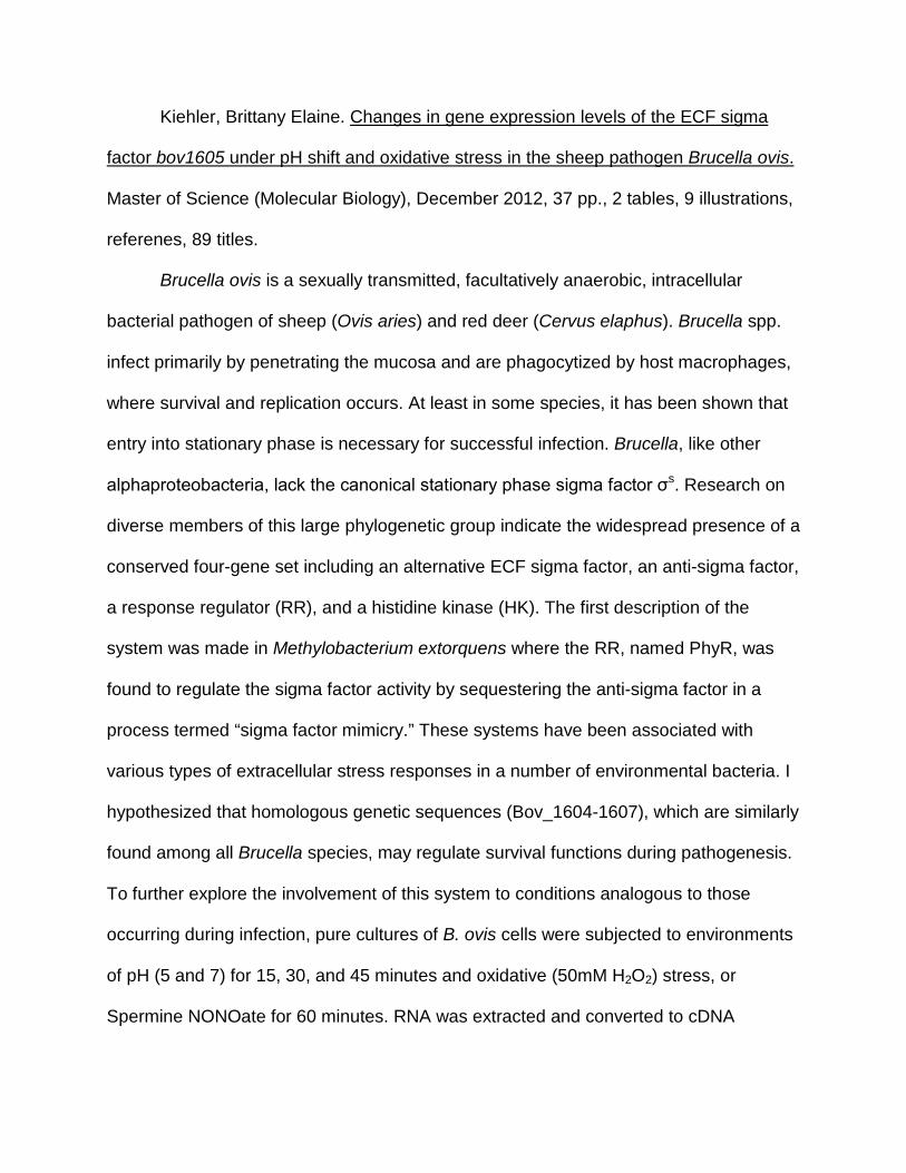
Brucella ovis is a sexually transmitted, facultatively anaerobic, intracellular
bacterial pathogen of sheep (Ovis aries) and red deer (Cervus elaphus). Brucella spp.
infect primarily by penetrating the mucosa and are phagocytized by host macrophages,
where survival and replication occurs. At least in some species, it has been shown that
entry into stationary phase is necessary for successful infection. Brucella, like other
alphaproteobacteria, lack the canonical stationary phase sigma factor σs. Research on
diverse members of this large phylogenetic group indicate the widespread presence of a
conserved four-gene set including an alternative ECF sigma factor, an anti-sigma factor,
a response regulator (RR), and a histidine kinase (HK). The first description of the
system was made in Methylobacterium extorquens where the RR, named PhyR, was
found to regulate the sigma factor activity by sequestering the anti-sigma factor in a
process termed “sigma factor mimicry.” These systems have been associated with
various types of extracellular stress responses in a number of environmental bacteria. I
hypothesized that homologous genetic sequences (Bov_1604-1607), which are similarly
found among all Brucella species, may regulate survival functions during pathogenesis.
To further explore the involvement of this system to conditions analogous to those
occurring during infection, pure cultures of B. ovis cells were subjected to environments
of pH (5 and 7) for 15, 30, and 45 minutes and oxidative (50mM H2O2) stress, or
Spermine NONOate for 60 minutes. RNA was extracted and converted to cDNA

andchanges in transcript levels of the sigma factor Bov1605 were measured using
qPCR. Preliminary results indicate that under the exposure to Spermine NONOate there
was little change in expression, but under oxidative stress expression of the sigma
factor Bov1605 was 4.68-fold higher than that expressed under normal conditions.
These results suggest that the sigma factor Bov1605 may be involved in oxidative
stress defense during infection. Under acid stress (pH5), Bov1605 was found to be
upregulated at 15 and 30 minutes, but after 45 and 60 minutes the time decreased.

ii
Copyright 2012
By
Brittany Elaine Kiehler

iii
ACKNOWLEDGMENTS
Special thanks to the University of North Texas, the University of North Texas
Office of Research for ROP grant funding to M.A., to my family for their encouragement
and support, Dr. Allen for his guidance during this project, Sarah Martinez and Leslie
Perry for their assistance with qPCR, and Dr. O’Donovan and Dr. Roberts for their
insight.

iv
TABLE OF CONTENTS
Page
ACKNOWLEDGEMENTS ............................................................................................... iii
LIST OF TABLES ............................................................................................................ vi
LIST OF FIGURES ......................................................................................................... vii
Chapter
I. INTRODUCTION ....................................................................................... 1 II. LITERATURE REVIEW ............................................................................. 3
Organism ........................................................................................ 3 Infection Pathway ............................................................................ 4 Environmental Stressors ................................................................. 6 Stationary Phase ............................................................................. 9 Intracellular Survival ...................................................................... 10 Genetics ........................................................................................ 11 Significance of Research and Objectives ...................................... 15
III. METHODOLOGY .................................................................................... 17
Cell Line ........................................................................................ 17 Artemis .......................................................................................... 17 PCR .............................................................................................. 17 qPCR ............................................................................................ 18 Analysis ......................................................................................... 20
IV. DATA ....................................................................................................... 21
PCR .............................................................................................. 21 qPCR ............................................................................................ 21
V. RESULTS: qPCR ..................................................................................... 24
Oxidative Stress ............................................................................ 24 Nitric Oxide Stress ........................................................................ 24 Acidic pH Stress ............................................................................ 25 Stationary Phase ........................................................................... 25

v
VI. DISCUSSION .......................................................................................... 28
REFERENCES .............................................................................................................. 31

vi
LIST OF TABLES
Page
1. BLAST results showing the percent similarity of the sigma factors of several Brucella spp. and R. palustris to that of bov1605 in Brucella ovis ...................... 13
2. Designed primer sets for sigma factor bov1605 and GAPDH bov1670 to be used in qPCR trials ...................................................................................................... 20

vii
LIST OF FIGURES
Page
1. The Infection Pathway of Brucella abortus. Wild type Brucella spp. activate the virB gene that allows for survival within the host phagosome ............................... 5
2. Interaction of the Haber-Weiss and Fenton Reactions ......................................... 9
3. NCBI gene maps showing location of the two component response system in B. ovis, B. abortus, B. melitensis, and R. palustris .................................................. 14
4. Example of the Two Component Response Regulator and Histidine Kinase ..... 14
5. Efficiency of bov1605 Primer Set 2 ..................................................................... 23
6. Efficiency of bov1670 Primer Set 3 ..................................................................... 23
7. Gene Expression Levels of bov1605 Under Oxidative and Nitric Oxide Stress .. 26
8. Gene Expression Levels at Interval Times Under Acidic Stress ......................... 27
9. Gene Expression Levels of bov1605 Entering and During Stationary Phase ..... 27

1
CHAPTER I
INTRODUCTION
Brucella ovis is a Gram negative, coccobacillus, facultative intracellular pathogen
that primarily infects rams and sheep (Ovis aries). Infection of red deer (Cervus
elaphus) has also been shown. B. ovis is transmitted by venereal transmission from ram
to ewe, resulting in impaired fertility in rams and weak offspring in ewes. This bacterium
can cause high economic loss in infected populations due to loss of offspring or the
inability to produce viable sperm that will effectively impregnate the female. [1]
The mechanism of infection identified in some Brucella sp. involves penetration
of the mucosa followed by being phagocytized by host macrophages.[2] In the
macrophage phagosomes, Brucella bacteria inhibit the phagosome-lysosome fusion
through rapid acidification of the phagosome following uptake. [3] [4] [5] Within the
phagosome, the invading cells must adapt to several changes in the environment
including pH change, nutrition deprivation, reactive oxidative intermediates (ROIs),
reactive nitrogen intermediates (RNIs) and lysosomal enzymes.
Genetic and immunological evidence suggest all members of the family
Brucellaceae are closely related. These bacteria are classified within the order
Rhizobiales. Also included within this order are many species of interest, both
pathogenic and environmental. Among these free-living bacteria, Rhodopseudomonas
palustris and Caulobacter crescentus have been extensively studied.[6] [7] Specifically
four genes including a two component response regulator, sigma factor, histidine-kinase
receptor, and a possible anti-sigma factor are thought to be involved in the response to
oxidative and pH stress in these and related organisms. However, no research has yet
2
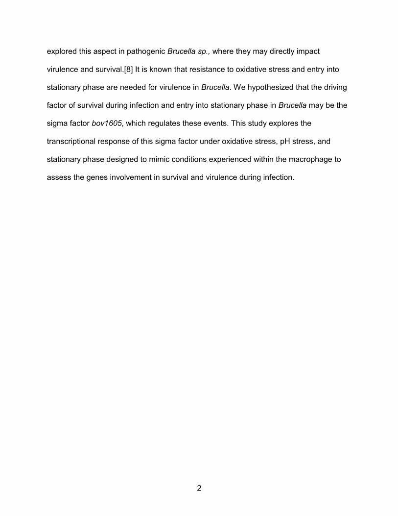
explored this aspect in pathogenic Brucella sp., where they may directly impact
virulence and survival.[8] It is known that resistance to oxidative stress and entry into
stationary phase are needed for virulence in Brucella. We hypothesized that the driving
factor of survival during infection and entry into stationary phase in Brucella may be the
sigma factor bov1605, which regulates these events. This study explores the
transcriptional response of this sigma factor under oxidative stress, pH stress, and
stationary phase designed to mimic conditions experienced within the macrophage to
assess the genes involvement in survival and virulence during infection.

3
CHAPTER II
LITERATURE REVIEW
Organism
Brucella ovis is a Gram negative coccobacillus, facultative intracellular pathogen
that primarily infects rams and sheep (Ovis aries). The organism has also been shown
to infect red deer (Cervus elaphus) following artificial inoculation.[1] While B. ovis has
not been shown to infect any other hosts, it is closely related to other species within the
Brucellaceae family, which consists of known pathogens that infect human hosts as well
as cattle and swine. Among these, Brucella abortus, Brucella suis, and Brucella
melitensis are known causative agents of human brucellosis, as well as the newly
discovered Brucella inopinata.[9] [10] In regards to human infection, brucellosis is
primarily an occupational hazard resulting from exposure to cattle, sheep, and pigs. In
humans, clinical treatment of brucellosis involves a combination of rifampicin and
doxycycline.[11] Brucella spp. are unusual in terms of pathogenic bacteria. They do not
exhibit the classic virulence factors seen in other pathogenic bacteria and rather survival
within the host depends on inhibition of programmed cell death by halting maturation of
the infected macrophage.[12]
Brucella ovis is transmitted by passive venereal transmission via ewes and non
venereal transmission via ram to ram. In cases of B. ovis infection, contaminated
pastures do not appear to be involved in spread of the disease.[13] In infected rams,
epididymitis and poor semen quality are noted, while in females impaired fertility,
placentitis, and weak offspring with high mortality occur. This bacterium can cause high
economic loss in infected populations due to loss of offspring or the inability to produce

4
viable sperm that will effectively impregnate the female.[14] [15] The disease, first
reported in New Zealand and Australia, has since been reported from other sheep-
raising areas of the world. Preventative measures, such as regular examinations of
rams before breeding, can help to reduce the spread of the disease. Rams infected by
B. ovis may display lesions or abnormalities of the scrotal tissues, mainly an enlarged
tail of the epididymis and a shrunken testicle.[16] For a definitive diagnosis, serological
tests or microscopic examination of stained semen smears may be necessary.
Susceptibility increases with age, and isolation of infected rams from the rest of the flock
is advised.[13] Though treatments of chlortetracycline and streptomycin are effective,
such measures are generally not economic as fertility in infected rams is likely
permanently impaired.[13] [17]
Infection Pathway
The general mechanism of infection for Brucella spp. involves penetration of the
mucosa of the nasal, oral, pharyngeal, or genital epithelium, where cells are
phagocytized by host macrophages.[18] [Figure 1] In the macrophage phagosome
Brucella bacteria inhibit the phagosome-lysosome fusion through rapid acidification of
the phagosome following uptake.[19] [20] [21] Normally, a host cell will use acidic pH,
oxidative stress, and nitric oxide stress to kill invading cells. A study by Porte et al. on
acidification of macrophages infected by B. suis described the survival of the bacteria
within the acidic compartments. In the murine macrophages, it was discovered that in
the phagosomes, the vacuoles containing live B.suis rapidly acidified to 4-4.5 pH. One
hypothesis proposed was that the bacteria required a low pH environment for survival
and multiplication within the macrophage and actively build a pathogen-specific niche

5
within the host cells. In B. suis, VirB, a virulence transcription gene, is induced under
acidic conditions and allows for the bacterium to reach certain bacterial compartments
where they are able to survive.[22] The acidic environment may be necessary to favor
a dissociation of iron from transferrin, a system described in the bacterium Francisella
tularensis.[23] Also, the lowered pH may act as an intracellular signal that regulates
genes involved in proliferation and survival within the host macrophage.
Figure 1: The Infection Pathway of Brucella abortus. Adapted from [24]. Wild type Brucella spp. activate the virB gene that allows for survival within the host phagosome.
Brucella
Phagocyte
Acquisition of early endosomal markers
Acquisition of late endosomal markers
Fusion with lysosomes
Mutant cells degraded Wild-type cell proliferation
6
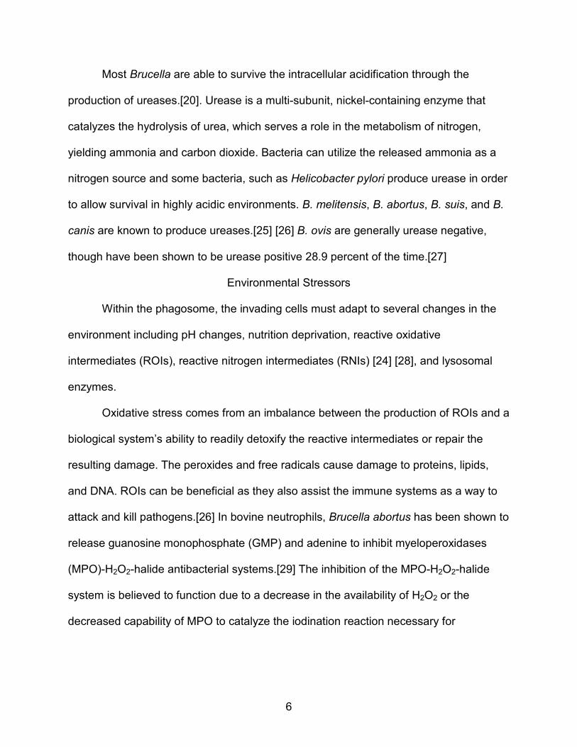
Most Brucella are able to survive the intracellular acidification through the
production of ureases.[20]. Urease is a multi-subunit, nickel-containing enzyme that
catalyzes the hydrolysis of urea, which serves a role in the metabolism of nitrogen,
yielding ammonia and carbon dioxide. Bacteria can utilize the released ammonia as a
nitrogen source and some bacteria, such as Helicobacter pylori produce urease in order
to allow survival in highly acidic environments. B. melitensis, B. abortus, B. suis, and B.
canis are known to produce ureases.[25] [26] B. ovis are generally urease negative,
though have been shown to be urease positive 28.9 percent of the time.[27]
Environmental Stressors
Within the phagosome, the invading cells must adapt to several changes in the
environment including pH changes, nutrition deprivation, reactive oxidative
intermediates (ROIs), reactive nitrogen intermediates (RNIs) [24] [28], and lysosomal
enzymes.
Oxidative stress comes from an imbalance between the production of ROIs and a
biological system’s ability to readily detoxify the reactive intermediates or repair the
resulting damage. The peroxides and free radicals cause damage to proteins, lipids,
and DNA. ROIs can be beneficial as they also assist the immune systems as a way to
attack and kill pathogens.[26] In bovine neutrophils, Brucella abortus has been shown to
release guanosine monophosphate (GMP) and adenine to inhibit myeloperoxidases
(MPO)-H2O2-halide antibacterial systems.[29] The inhibition of the MPO-H2O2-halide
system is believed to function due to a decrease in the availability of H2O2 or the
decreased capability of MPO to catalyze the iodination reaction necessary for

7
degranulation. Due to the small size of GMP, it is likely that the molecules escape
recognition of the immune system facilitating intracellular survival of Brucella.[30]
In the immune system, activated phagocytes produce both ROI and RNIs to fight
pathogenic invasion. These reactive compounds do cause some damage to host
tissues, as the oxidants destroy almost every part of the target cell due their
nonspecific, highly reactive nature. This generally prevents the pathogen from escaping
the immune response.[26] The bactericidal functions are induced by gamma interferon
(INF-γ) and tumor necrosis factor (TNF-α), which are associated with protective
immunity.[31] As described above, an increase of iron within the infected host cells
activated by INF-γ has been shown to increase decimation of intracellular Brucella. The
Haber Weiss reaction is catalyzed by the iron, confirming the importance of ROIs.[32]
[33] [34] [35] [Figure 2]
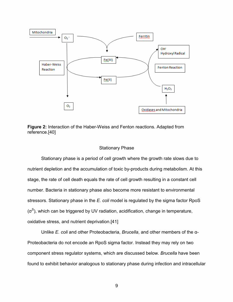
The Haber-Weiss reaction generates hydroxyl radicals from hydrogen peroxide
and superoxide resulting in oxidative stress. The reaction in catalyzed by iron, the first
step involving the reduction of iron in from the ferric to the ferrous states when reacted
with superoxide radicals. The second step involves the Fenton reaction, which reacts
the ferrous iron with hydrogen peroxide resulting in a hydroxyl radical and the cycling of
ferrous iron back to its ferric state. The accumulation of hydroxyl radicals result in
oxidative stress within a cell.[32]
RNIs are also present within the cell, albeit at lower levels than ROIs. Nitric oxide
(NO), a kind of RNI, is a free radical that is generated by the phagocytes and activated
by interferon- γ (INF-γ) and tumor necrosis factor (TNF). Nitric oxide induces DNA
damage and degrades iron sulfur centers.[33] [36] Many bacterial pathogens have

8
evolved to resist nitric oxide. Paracoccus denitrificans, for example, possesses a nitric
oxide reductase regulator (NNR) and a nitrite reductase. The bacterium uses
denitrification to synthesize NO from nitrite and then further reduces the NO down into
nitrous oxide. This is thought to be a strategy to prevent the accumulation of the toxic
NO. There is evidence that the process is also activated by high amounts of NO.[37]
[38]
A study of B. melitensis, B. abortus, and B. suis reported that the
lipopolyssacharride (LPS) and O-antigen of Brucella spp. may induce NO production.
They reported that while reactive oxygen intermediates play a major role in antibacterial
activities, the NO plays a relatively minor role. They also found that B. abortus and B.
melitensis smooth- lipopolyssacharride (S-LPS) induce NO synthesis in rat peritoneal
macrophages.[37] In murine models infected with B. suis, when NO was combined with
IFN-γ and antibrucella antibodies, the favored outcome was elimination of Brucella.[39]
Another study of NO stress in B. abortus reported that NO can accelerate the killing of
intracellular Brucella but not to completion during the first 24 hours of infection. The
Authors also showed that while NO can induce apoptosis in host cells, effectually
eliminating intracellular pathogens, B. abortus was found to prevent apoptosis. After the
24 hour mark, any surviving Brucella were able to replicate.[40] The data suggest that
there may be genetic sequences that link to survival during NO stress, perhaps similar
to that see in P. denitrificans.
9
Figure 2: Interaction of the Haber-Weiss and Fenton reactions. Adapted from reference.[40]
Stationary Phase
Stationary phase is a period of cell growth where the growth rate slows due to
nutrient depletion and the accumulation of toxic by-products during metabolism. At this
stage, the rate of cell death equals the rate of cell growth resulting in a constant cell
number. Bacteria in stationary phase also become more resistant to environmental
stressors. Stationary phase in the E. coli model is regulated by the sigma factor RpoS
(σS), which can be triggered by UV radiation, acidification, change in temperature,
oxidative stress, and nutrient deprivation.[41]
Unlike E. coli and other Proteobacteria, Brucella, and other members of the α-
Proteobacteria do not encode an RpoS sigma factor. Instead they may rely on two
component stress regulator systems, which are discussed below. Brucella have been
found to exhibit behavior analogous to stationary phase during infection and intracellular
10
survival. In murine models, the growth curves of Brucella melitensis and Brucella
abortus plateau, showing little growth after 2 weeks postinfection. It has also been found
that Brucella displaying stationary phase-like physiology are less affected by
antibacterial drug treatments.[42] These findings suggest that a stationary phase
physiology is necessary for intracellular survival within the host macrophage.
Intracellular Survival
Brucella spp., as stated before, infect primarily by penetrating the mucosa of the
nasal, oral, pharyngeal, or genital epithelium and are phagocytized by host
macrophages, where survival and subsequent replication occur. If present among
natural oxidative stressors, how do the Brucella survive to replicate?
One means of survival is the lipopolysaccharide (LPS) coat. The
lipopolysaccharide consists of a core polysaccharide and the O-antigen. The core
polysaccharide is formed by ketodeoxyoctonate, various heptoses, glucose, galactose,
glucose, rhamnose, mannose, and a few dideoxyhexoses. This core is then covalently
attached to the O-antigen.[43] The lipid A fatty acids of the lipopolysaccharide are
connected to the cell wall by the amine groups from a disaccharide composed of
glucosamine phosphate, and the disaccharide is connected to the core polysaccharide
by the ketodeoxyoctonate.[44] Griffith’s experiment, reported by Frederick Griffith
explored the LPS coat’s effect on virulence within a host and that bacteria are capable
of transferring genetic information through transformation. In his experiment, Griffith
used two strains of Streptococcus pneumonia, one the wild-type rough and the other the
virulent smooth. He found that the mice infected with the smooth pneumococcus or
those infected with both live rough and heat-killed smooth pneumococcus died, while

11
mice infected with the rough pneumococcus or heat-killed smooth pneumococcus
survived. This showed that the live rough bacteria could take in the genetic information
of the heat killed smooth bacteria and transform to be a smooth variant.[45]
B. ovis naturally generates a rough lipopolysaccharide (R-LPS) coat however,
LPS has been shown to be an important factor of other Brucella spp. virulence. In most
instances, smooth LPS coats are virulent and wild-type coats are much less virulent.[46]
This has been shown to be the case for B.ovis and B. canis, who are both rough yet still
virulent. However, these rough mutants have been shown to be more adherent and
invasive than smooth brucellae such as B. suis, B. abortus, and B. melitensis.[47] The
differentiation between a smooth and rough LPS designation depends on the presence
of O-antigen. Bacteria with a smooth LPS (such as B. abortus) display long chains of O-
antigen, while bacteria with a rough LPS display little to no O-antigen.[2] Rough mutants
are more susceptible to lysis, which is most likely the reason many rough mutants have
non virulent qualities in animal models. The capacity for rough mutants to replicate
intracellularily is not yet solved, though perhaps the answer lays within the genes that
code for virulence or in the difference in expression between the host and on agar.[2]
[48] [49] [50] [51] [52]
Genetics
Genetic and immunological evidence suggest all members of the family
Brucelleae are closely related. It is possible that similar linkage exists within the order
Rhizobiales, in which both Brucella species and Rhodopseudomonas species are
found.[53] [Table 1] [Figure 3] Specifically of interest are four primary stress response
genes (two component response regulator, sigma factor, histidine-kinase receptor, and
12
a possible anti-sigma factor) found in both Brucella spp. and Rhodopseudomonas spp,.
and their response to oxidative stress and the impact of that on the virulence of the
organism.[53] The first description of this system was made in Methylobacterium
extorquens, where two regulatory mechanisms controlled gene expression via
alternative sigma-antisigma factors and phosphorylation-dependent response
regulators.[54] [55]
The four genes in the system are the two component response regulator, sigma
factor, anti-sigma factor, and the sensor histidine kinase, as mentioned above. Each
part of the response regulator plays a part in gene expression. The key element in the
system is the membrane bound histidine kinase, which is responsible for the
autophosphorylation of the histidine with ATP, the phosphotransfer from the kinase to
an aspartate of the response regulation and the phosphotransfer from aspartyl
phosphate back to ADP or water. Once the aspartic acid residue is phosphorylated, the
conformation of the response regulator changes, activating an output domain which
leads to the expression of a target gene.[56] [57] [Figure 4] In this case, the activation of
the sigma factor would occur and the sigma factor would continue on to initiate the RNA
synthesis by the binding of the RNA polymerase to the gene promoters. The sigma
factor completes the RNA polymerase holoenzyme, and once transcription of the DNA
template strand commences, the sigma factor disassociates.[58] The anti-sigma factor
inhibits transcription by binding to the sigma factor. In this way, the anti-sigma factor
limits the ability of the sigma factor to direct expression of certain genes under stress or
another signal relieves its repression.[59]

13
Another factor in this system is the cyclases/histidine kinases associated sensory
extracellular (CHASE3) domain, located in extracellular or periplasmic locations
followed by an intracellular tail incorporating many enzymatic signaling domains such as
the histidine kinase. The CHASE3 domain binds ligands and mediates signal
transduction through the respective receptors.[60] [61]
In Rhodopseudomonas palustris, there is a gene network involving four
homologous [62] regulatory genes. The primary difference in pathogenic Brucella and
free living α-proteobacteria is the substitution of a CHASE domain in the histidine kinase
of the latter for the CHASE3 domain of the former. The otherwise highly conserved four-
gene system may then be expected to respond to different signals consistent with life as
a facultative intracellular parasite. Never-the-less, the driving factor of virulence and the
entry into stationary phase in Brucella may involve this highly conserved four-gene
system, which regulates the events. This study intends to observe the response of the
sigma factor bov1605 transcription upon exposure to conditions analogous to that
expected during the infection process and phagocytosis.
Table 1: BLAST results showing the percent similarity of the sigma factors of several Brucella spp. and R. palustris to that of bov1605 in Brucella ovis.
Organism Score Expect Identities Gaps Percent Match
B. canis 1059 0 573/573 0/573 100% B. suis 1059 0 573/573 0/573 100% B. melitensis 1053 0 572/573 0/573 99% B. abortus 1048 0 571/573 0/573 99% B. microti 1059 0 573/573 0/573 100% R. palustris 141 5.00E-38 289/424 4/424 68%

14
Figure 3: NCBI gene maps showing location of the conserved four-gene regulatoy system in B. ovis, B. abortus, B. melitensis, and R. palustris
Figure 4: Example of the Two Component Response Regulator and Histidine kinase.

15
Significance of Research and Objectives
The significance of this study is based on the genomic conservation among
Brucella species. While B.ovis does not have a dramatic impact on multiple host
species, B.ovis is closely related to B. abortus and B. melitensis, which are known
human pathogens.[63]
The major aim of this study is to determine the effect of stresses similar to those
that are expected to occur during infection and phagocytosis on transcription of the
sigma factor bov1605. In order to test this, four parameters were selected: oxidative
stress, nitric oxide stress, acidification, and conditions during stationary phase. Initially,
these parameters were to be compared to samples taken from infected rams, however
our collaborative lab at the Universidad Autonoma del Estado de Mexico, were not able
to produce the tissue samples in a timely manner.
To test the first parameter, B. ovis cultures were subjected individually to
oxidative stress using hydrogen peroxide. The RNA was extracted and converted to
cDNA for analysis by quantitative-Polymerase Chain Reaction (qPCR), the results of
which were analyzed to determine whether the gene was upregulated or down
regulated. To test the second parameter, the same process was used substituting
spermine NONOate, which releases controlled amounts of nitric oxide, as the source of
stress. The third parameter also uses the same process, however the samples were
subjected to acidic stress of pH 5 and were incubated at timed intervals of 15, 30, 45,
and 60 minutes, while for the first two parameters, the cells were incubated for 60
minutes each. For the final parameter unstressed cultures of B. ovis were incubated for
4, 7, and 9 days, then additional days to determine the onset of stationary phase-like

16
physiology prior to RNA extraction and processing as described. Our objective was to
test these parameters and determine if our gene of interest, bov1605, had any change
in regulation from what is seen under normal, unstressed conditions at pH 7 in mid-log
phase (OD600 = ~0.3)
We hypothesized that under the stressed conditions, we would see an
upregulation of the sigma factor bov1605. We further hypothesized that bov1605 was
similarly involved in entry into stationary phase. These parameters were tested and
analyzed using qPCR techniques and further described below.

17
CHAPTER III
METHODOLOGY
Cell Line
B. ovis cultures (strain designation 25840) was obtained from ATCC. Lyophilized
cells were reanimated and grown for 7 days (according to the previous literature) in 8%
CO2 at 37°C. ATCC recommends growth in 5% CO2, though previous literature shows
growth in 5-10% CO2 for Brucella ovis to be suitable.[62] [63]
Artemis
The Artemis [64] program was used to view chromosome I of B. ovis. From there,
gene sequences of interest were located, specifically the bov1604-1607 region
encompassing the two component response regulator at bov1604, sigma factor at
bov1605, possible anti-sigma factor at bov1606, and the sensor histidine kinase at
bov1607. These sequences were also used to perform genetic matches using the Basic
Local Alignment Search Tool (BLAST) software on the NCBI database.[65] [66]
PCR
A genomic prep of B. ovis was performed using predesigned primers selected for
the 1604-1607 region. The primers (forward 5°- AGGCACTTTT CTTTGAGGAAGCATA
reverse 5°-TACCCGCTT GTCATCGCCC) were designed using Primer3 [67] and
OligoCalc [68] web programs. PCR reaction was then performed to amplify that specific
sequence which contains a two component response regulator, sigma factor, anti-sigma
factor, and a sensor histidine kinase. The PCR profile used was 30 cycles of 30
seconds of template denaturation at 95°Celsius (C). 90 seconds of primer annealing at
54°C, and 90 seconds of primer extension at 72°C. The final extension was 6 minutes at

18
72°C, and then the sample was stored at 4°C.[69] [70] Gel electrophoresis was used to
separate the desired product, giving a band of 3252 bp. The DNA was extracted from
the gel and purified to be used in downstream applications. An attempt was made to
clone the sequence into E.coli competent cells, however the attempt failed.
qPCR
Primers for qPCR were designed using Primer 3 [67] and OligoCalc [68] for the
sigma factor bov1605 and GAPDH bov1670. [Table 2] In all, three primer sets were
designed for each target gene. Tests to determine the efficiencies of the primer sets
were performed in triplicate on dilutions of the genomic DNA sequence. Primer
efficiencies were determined using the equation found in the qPCR manual by
Trofimov.[71] [72]
𝐸𝑓𝑓𝑖𝑐𝑖𝑒𝑛𝑐𝑦 = �10(− 1𝑠𝑙𝑜𝑝𝑒) − 1� ∗ 100
Four forms of environmental stress were tested: acidic, oxidative, nitric oxide,
and stationary phase stress conditions. Pure B.ovis cell cultures were grown for 4 days
to an optical density at 600nm (OD600) of 0.3. The cultures were then pelleted to
concentrate the cells and the supernatant decanted. The cells were then resuspended
in fresh medium at pH 5, pH 7, or amended with 50mM H2O2, or 6mM Spermine
NONOate. The Spermine NONOate provides a steady-state concentration of NO that is
sufficient to overcome the tendency of NO to auto-oxidize in the presence of oxygen
[73] [74]and has been found to be an efficient inducer of NO stress.[75] [76] The
samples were incubated at 37°C shaking at r.p.m. with the exception of the 50mM H2O2
tubes, which were incubated stationary as the turbulent motion caused excessive
pressure to build in the tube. The pH samples were incubated at times of 15, 30, 45,
19
and 60 minutes. The 50mM H2O2 and 6mM Spermine NONOate samples were each
incubated for 60 minutes.[77] The cells were once again spun down and the
supernatant decanted. The cells were treated with RNAprotect [78] [79] then disrupted
following protocol 3 and 7 [79] for RNA extraction of the Qiagen RNeasy protocol. Once
the RNA was obtained, reverse transcription was performed to synthesize cDNA.
Resulting cDNA were stored at 0°C prior to use.
qPCR was performed to quantify the amount of sigma factor bov1605 transcript
expressed under each condition.[80] [81] [82]Tests were done in triplicate qPCR reads
of each sample (technical replicates) with triplicate qPCR reads of GAPDH as a
normalizer and no-RT RNA as a negative control. GAPDH, glyceraldehydes 3-
phosphate dehydrogenase, catalyzes he sixth step of glycolysis and serves to
metabolize glucose for energy and carbon molecules.[83] GAPDH is used here as a
normalizer to set a baseline to measure any activity of bov1605 against due to its
relative constant expression during cell growth.
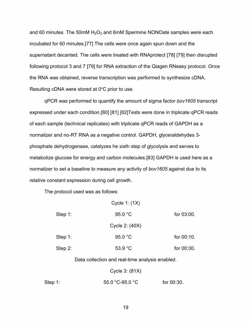
The protocol used was as follows:
Cycle 1: (1X)
Step 1: 95.0 °C for 03:00.
Cycle 2: (40X)
Step 1: 95.0 °C for 00:10.
Step 2: 53.9 °C for 00:30.
Data collection and real-time analysis enabled.
Cycle 3: (81X)
Step 1: 55.0 °C-95.0 °C for 00:30.

20
Analysis
Results were analyzed using a modified Livak method.[84] A simple equation
was used to generate the data:
∆∆𝐶𝑇 = ��CT(Target)� − �CT(GAP)�� − (∆CT)avg control
To generate the reference bar, the following equation was used:
∆∆𝐶𝑇 = ∆CT − ∆CTMean
The ΔΔCT value was used to calculate the expression level of the target gene, as
described by the Livak method. A mean and standard deviation was taken from the
ΔΔCT values generated and a standard error calculated to produce the error bars. One-
tailed T-tests were performed to generate p values. For the stationary phase time
course, ANOVA was performed to determine if there was significant change to the
bov1605 sigma factor during entry into and after stationary phase-like physiology was
reached.
Table 2: Designed Primer Sets for Sigma Factor bov1605 and GAPDH bov1670 to be used in qPCR trials.
Salt Adjusted PRIMER 1 (BOV1605)
PRIMER FWD: CCAAGCAGGAGTCCTTTGAA 58.4 PRIMER REV: TGAAGATCAAGCGTGCCATA 56.4
PRIMER 2 (BOV1605)
PRIMER FWD: GCAGGAGTCCTTTGAAGTGG 60.5 PRIMER REV: ATGAGGATGATTGCCTCACG 58.4
PRIMER 3 (BOV1605)
PRIMER FWD: GCAGGAGTCCTTTGAAAGTGG 61.2 PRIMER REV: CATATTGCGAGGGATGCAC 57.5
PRIMER 1 (BOV1670)
PRIMER FWD: TACGATCGATGTTGGCTACG 58.4 PRIMER REV: ACCAGATGGTCCTTCGTCAG 60.5
PRIMER 2 (BOV 1670)
PRIMER FWD: CGTGGAAGGAAGAAAACGTC 58.4 PRIMER REV: ACCAGATGGTCCTTCGTCAG 60.5
PRIMER 3 (BOV1670)
PRIMER FWD: TCTGCGTTATGACAGCGTTC 58.4 PRIMER REV: CAAGATGAAGTGCTGCCTTG 58.4

21
CHAPTER IV
DATA
PCR
The fragments generated via the PCR process were expected to be 3252 bp.
The first PCR profile setting resulted in two bands showing, one at the 3 kb region as
expected along with an equally bright band at the 1.5 kb region. The experiment was
run again at a higher annealing temperature to decrease the amount of 1.5 kb by-
product. 3 kb fragments were cut from the gel and DNA extraction was performed. The
four-gene sequence was to be cloned into competent E. coli cells, however the attempt
failed.
qPCR
Primer efficiency tests were performed to determine from three primer sets which
was the most efficient to use in the experimental tests. Using the qPCR primer
efficiency equation mentioned above in the methods, it was found that primer set 2 of
the designed bov1605 sigma primers [Figure 5] and primer set 3 of the designed
bov1670 GAPDH primers had the best efficiency. [Figure 6] Using the sigma primer 2
and GAPDH primer 3, the cDNA created from the stress induced pH, oxidative, and
nitric oxide samples were tested. The data were analyzed using the Livak method.[84]
[85]
The result of the Livak method calculations is a ratio of the target gene (CT(Target,
Test)) to the calibrator sample (CT(Target, Calibrator)) both normalized to the expression of the
same under a reference condition (CT(Referece, Test)). The sigma factor bov1605 was the

22
target gene, GAPDH bov1670 the calibrator, and unstressed sample at pH 7 was the
reference condition. [85] A simple equation was used to generate the data:
∆∆𝐶𝑇 = ��CT(Target)� − �CT(GAP)�� − (∆CT)avg control
To generate the reference bar, the following equation was used:
∆∆𝐶𝑇 = ∆CT − ∆CTMean
The ΔΔCT value was used to calculate the expression level of the target gene, as
described by the Livak method.
Using this method, the calculated data for the oxidative stress test showed an
upregulation 4.67-fold higher than that seen under normal conditions. The data for the
nitric oxide samples showed a very small down regulation 0.074-fold that seen under
normal conditions.
The data for the pH test showed a slight upregulation at 15 and 30 minutes at
1.04 and 1.62 respectively, with the optimal time being 30 minutes. This is however not
a significant value. At 45 and 60 minutes, decreases in transcripts were seen by 0.89-
fold at 45 minutes and 0.53-fold at 60 minutes. Stationary phase analysis showed a
decrease in transcripts at levels of 0.11-fold for 9 days, 0.09-fold for 10 days, 0.07-fold
for 11 days, 0.2-fold for 12 days, 0.11-fold for 13 days and 0.18-fold for 14 days.

23
Figure 5: Primer 2 of the bov1605 set. Efficiency 94.7%
Figure 6: Primer 3 of the bov1670 set. Efficiency 96.8%
y = -3.455x + 37.778
0
5
10
15
20
25
30
35
40
0 2 4 6 8
CT V
alue
Dilution
bov1605 Primer Set 2
y = -3.4x + 39.537
0
5
10
15
20
25
30
0 2 4 6 8
CT V
alue
Dilution by 10
bov1670 Primer Set 3

24
CHAPTER V
RESULTS: qPCR
Oxidative Stress
B. ovis cultures were grown for 4 days for an optical density of 0.3 at 600nm.
Individual samples from these cultures were taken and exposed to either 50mM H2O2 or
BHI broth at ph 7 for 60 minutes. Earlier tests incubated the samples for 30 minutes, but
levels of regulation did not yield results. RNA was extracted and cDNA synthesized as
described in the methodology. Samples were done in biological triplicate and analyzed
using qPCR with GAPDH bov1670 as the normalizer.
The results of the oxidative stress trial exhibited an upregulation of the sigma
factor gene 4.68-fold higher than that of the gene under normal conditions at pH 7 with
no stressors present. [Figure 7] A T-test was performed on the results giving a value of
p= 0.003. The values for the oxidative stress test were found to be statistically different.
Nitric Oxide Stress
B. ovis cultures were grown for 4 days to an optical density of 0.3 at 600nm.
Individual samples from these cultures were taken and exposed to either 6mM
Spermine NONOate or BHI broth at ph 7 for 60 minutes. RNA was extracted and cDNA
synthesized as described in the methodology. Samples were done in biological triplicate
and analyzed using qPCR with GAPDH bov1670 as the normalizer.
The results of the nitric oxide stress trial exhibited a minimal down regulation of
the sigma factor gene 0.074-fold that of the gene under normal conditions. [Figure 7] A
T-test was performed on these results giving values of p= 0.11. The results were not
statistically different.

25
Acidic pH Stress
B. ovis cultures were grown for 4 days for an optical density of 0.3 at 600nm.
Individual samples from these cultures were taken and exposed to either ph 5 or ph 7
for 15, 30, 45, or 60 minutes. An earlier test incubated the pH 5 samples for 60 minutes.
The results exhibited a down regulation of 0.53-fold. This test was to determine if a
certain time interval exhibited an upregulation of bov1605. RNA was extracted and
cDNA synthesized as described in the methodology. Samples were done in biological
triplicate and analyzed using qPCR with GAPDH bov1670 as the normalizer.
Cells incubated in broth of pH 5 showed down regulation of the sigma factor 0.59
times than that seen under normal conditions. Cells incubated in pH 5 for timed intervals
showed transcript numbers at the following levels: at 15 minutes an upregulation of
1.04-fold, at 30 minutes an upregulation of 1.62-fold, at 45 minutes a down regulation of
0.89-fold, and at 60 minutes a down regulation of 0.53-fold. [Figure 8] A T-test was
performed on these results giving values of p= 0.47 for the 15 minute interval, p= 0.16
for the 30 minute interval, p= 0.42 for the 45 minute interval, and p= 0.21 for the 60
minute interval. These values were not significant.
Stationary Phase
To determine whether bov1605 is involved in entry into a stationary phase
physiology, B. ovis cultures were grown up to 14 days in ph 7. Growth for 12 days
marked the beginning of a stationary phase growth. RNA was extracted and cDNA
synthesized as described in the methodology. Samples were done in biological triplicate
and analyzed using qPCR with GAPDH bov1670 as the normalizer.

26
Gene expression levels of bov1605 during stationary growth were tested at timed
intervals of 9 to 14 days with a control of 4 day growth. Transcript numbers were found
to be 0.11-fold at 9 days, 0.09-fold at 10 days, 0.07-fold at 11 days, 0.2-fold at 12 days,
0.11-fold at 13 days and 0.18-fold at 14 days. [Figure 9] These low values suggest that
during the stationary phase-like time period of the B. ovis growth curve, the bov1605
gene is down regulated. A t-test was performed comparing the results of the bov1605
target at 9-14 days with bov1605 reference at 4 days, giving values of p= 0.08 at 9
days, p= 0.08 at 10 days, p= 0.08 at 11 days, p= 0.10 at 12 days, p= 0.08 at 13 days,
and p= 0.09 at 14 days. ANOVA analysis across the 6 days gave a p value of 0.18. This
value was not statistically significant.
Figure 7: Gene expression levels of bov1605 under oxidative and nitric oxide stress.
-1
0
1
2
3
4
5
OX NO
2ΔΔC
T Va
lue
bov1605 Under Oxidative and Nitric Oxide Stress
Tested
Reference

27
Figure 8: Gene expression levels at interval times under acidic stress.
Figure 9: Normalized expression ratios of bov1605 over 9 to 14 days of incubation.
0
0.5
1
1.5
2
2.5
3
3.5
4
15 30 45 60
2ΔΔC
T Va
lue
Time (Minutes)
bov1605 Acidic pH Time Course
Tested
Reference
0
0.5
1
1.5
2
2.5
3
3.5
4
Day 9 Day 10 Day 11 Day 12 Day 13 Day 14
2ΔΔC
T Va
lue
bov1605 Stationary Phase-like Time Course
Tested
Reference
28
CHAPTER VI
DISCUSSION
This research has shown that under oxidative stress the regulation of the sigma
factor bov1605 is upregulated under laboratory conditions. It is not known what this
particular sigma factor influences, however we hypothesize that it has something to do
with intracellular survival and may assist in survival under oxidative conditions such as
may be expected in the phagosome of a host cell. In regards to the nitric oxide stress,
from the data we can conclude that it has very little effect on this particular gene. While
RNIs are present in the infected cell and used to attack a pathogenic organism invading
the host cell, it is clear that oxidative stress caused by hydroxyl radicals has far more
effect on this regulatory system. This supports previous research in that while nitric
oxide is present and may cause some stress to the invading bacteria, the main cause of
stress for the cell is caused by oxidative species.[86] Experiments with nitric oxide as a
main stress did not have as much effect on the B. ovis samples as reported in B.
abortus, B. suis, and B. melitensis.[37] [87] [88]
Brucella species acidify their environment during the infection and proliferation
process. Usually acidification would kill the cells; however, Brucella have found a way to
use this to their advantage. Our research has shown that sigma bov1605 is upregulated
during acid stress up to 30 minutes, after which the sigma factor is down regulated. It
may be that after that time the cell has produced proteins needed to adapt and survive
in the acidic conditions and more production is unnecessary. Other studies have shown
that the acidic environment turns on certain survival genes, such as virB in Brucella
suis.[89] B. ovis does possess a virB gene on chromosome II of the genome. While the
29
sigma factor studied shows little response to acidic stress, it may be that it does not
specifically have anything to do with survival under acidic conditions.
For Brucella, entry into the stationary phase physiology is thought to be
necessary for infection.[42] Our findings of the timed interval stationary phase test
showed low transcript numbers for bov1605. We had hypothesized that after 9 days,
there would be some regulation of the gene as it was left in conditions that normally
would be expected to trigger stationary phase physiology. It was shown that after 12
days, the growth curve of B. ovis had reached a plateau, signaling stationary phase
growth. Previous studies reported stationary phase-like physiology after 2 weeks
incubation within a murine host.[42] The difference in time to reach stationary phase
may be due to incubation in an animal host versus incubation in prepared media. Our
results showed low transcript numbers for the sigma factor bov1605 across all 6 days
displaying a down regulation of the sigma factor under these conditions. It is possible
this particular gene has little to do with entry into and survival during stationary phase.
Future research on intracellular survival of B. ovis would include a full genomic
analysis using RNA-seq and the analysis of the infected sheep tissues using qPCR.
The data presented here can further be used as a comparison to the infected
tissue when it ultimately arrives. The infected tissue will be processed using the same
techniques as before and analyzed using qPCR. Our hypothesis is that the oxidative
stress data gathered from the individual stress tests will correlate with the data from the
infected tissue.
RNA-seq will be utilized to view upregulation of genes across the entire genome
to determine not only the upregulation of the ECF-sigma factor bov1605 but any genes

30
that may be activated by the sigma factor similarly upregulated during infection. RNA-
seq is a novel method of measuring transcriptomic data experimentally. While there is
not a strong correlation between the abundance of mRNA and the related proteins,
mRNA concentration measurements are useful in that they give light to the gene
regulation of the cell during exposure to varying environmental conditions.[87] [88] The
use of RNA-seq to study gene regulation of the entire genome, will solidify our
hypotheses that sigma factor bov1605 plays a role in oxidative stress defense and acid
stress tolerance during infection, as well as give information in regards to the
upregulation of additional genes within the genome that respond to the stresses
encountered by Brucella during the infection of the host pathogen.

31
REFERENCES
1. The Center for Food Security and Public Health, I.S.U., Insitite for International Cooperation in Animal Biologics. Ovine Epididymitis: Brucella ovis. 2007 July 29, 2009 [cited; Available from: http://www.cfsph.iastate.edu/Factsheets/pdfs/brucellosis_ovis.pdf.
2. Gomes Cardoso, P., et al., Brucella spp noncanonical LPS: structure, biosynthesis, and interaction with host immune system. Microbial Cell Factories, 2006. 5(13).
3. Ko, J. and G.A. Splitter, Molecular Host-Pathogen Interaction in Brucellosis: Current Understanding and Future Approaches to Vaccine Development for Mice and Humans. Clinical Microbiology Reviews, 2003. 16(1): p. 65-78.
4. Kohler, S., et al., Secretion of Listeriolysin by Brucella suis Inhibits Its Intramacrophagic Replication. Infection and Immunity, 2001. 69(4): p. 2753-2756.
5. Porte, F., J.P. Liautard, and S. Köhler, Early Acidification of Phagosomes Containing Brucella suis Is Essential for Intracellular Survival in Murine Macrophages. Infection and Immunity, 1999. 67(8): p. 4041-4047.
6. Conlan, S., C. Lawrence, and L.A. McCue, Rhodopseudomonas palutris Regulons Detected by Cross-Species Analysis of Alphaproteobacterial Genomes. Appl. Environ. Microbiol, 2005. 71(11): p. 7442-7452.
7. Foreman, R., A. Fiebig, and S. Crosson, The LovK-LovR two-component system is a regulator of the general stress pathway in Caulobacter crescentus. Journal of Bacteriology, 2012. 194(16).
8. Lamontagne, J., et al., Intracellular Adaption of Brucella abortus. J Proteome Res, 2009. 8(3): p. 1594-1609.
9. Diagnosis and Management of Acute Brucellosis in Primary Care. Brucella Subgroup of the Northern Ireland Regional Zoonoses Group, 2004.
10. Scholz, H. and K. Nockler, Brucella inopinata sp. nov., isolated from a breast implant infection. International Journal of Systematic and Evolutionary Microbiology, 2010. 60: p. 801-808.
11. Robichaud, S., et al., Prevention of laboratory-acquired brucellosis. Clin Infect Dis., 2004. 38(12): p. 119-122.
12. Crasta, O.R., et al., Genome Sequence of Brucella abortus Vaccine Strain S19 Compared to Virulent Strains Yields Candidate Virulence Genes. PLoS ONE, 2008. 3(5).

32
13. Brucellosis in Sheep. The Merck Veterinary Manual 2011 [cited; Available from: http://www.merckvetmanual.com/mvm/index.jsp?cfile=htm/bc/110506.htm&word=brucella%2covis.
14. Lopez, G., et al., Use of Brucella canis antigen for detection of ovine serum antibodies against Brucella ovis. Veterinary Microbiology, 2005. 105: p. 181-187.
15. Chain, P.S.G., et al., Whole-Genome Analyses of Speciation Events in Pathogenic Brucellae. Infection and Immunity, 2005. 73(12): p. 8353-8361.
16. Plant, J.W. and J. Seaman. Ovine brucellosis. NSW DPI Primefacts 2007 [cited; Available from: http://www.dpi.nsw.gov.au/__data/assets/pdf_file/0006/145824/ovine-brucellosis.pdf.
17. Kohler, S., et al., The analysis of the intramacrophagic virulome of Brucella suis deciphers the enviroment encountered by the pathogen inside the macrophage host cell. Proc Natl Acad Sci USA, 2002. 99(24): p. 15711-15716.
18. Fortier, A., et al., Growth of Francisella tularensis LVS in Macrophages: the Acidic Intracellular Compartment Provides Essential Iron Required for Growth. Infection and Immunity, 1995. 63(4): p. 1478-1483.
19. Atluri, V.L., et al., Interactions of the Human Pathogenic Brucella species with Their Hosts. Annual Review Microbiology, 2011. 65: p. 523-541.
20. Paulsen, I.T., The Brucella suis genome reveals fundamental similarities between animal and plant pathogens and symbionts. PNAS, 2002. 99(20): p. 13148-13153.
21. Paixao, T.A., et al., Establishment of Systemic Brucella melitensis Infection through the Digestive Tract Requires Ureas, the Type IV Secretion System, and Lipopolysaccharide O Antigen. Infection and Immunity, 2009. 77(10): p. 4197-4208.
22. Sangari, F.J., et al., Characterization of the Urease Operon of Brucella abortus and Assessment of Its Role in Virulence of the Bacterium. Infection and Immunity, 2007. 75(2): p. 774-780.
23. Corbel, M.J. and D.M. Hendry, Urease activity of Brucella Species. Res Vet Sci., 1985. 38(2): p. 252-253.
24. Poole, R.K. and M.N. Hughes, New functions for the ancient globin family: bacterial responses to nitric oxide and nitrosative stress. Molecular Microbiology, 2000. 36(4): p. 775-783.

33
25. Hutchings, M.I., N. Mandhana, and S. Spiro, The NorR Protein of Eschericia coli Activates Expression of the Flavorubredoxin Gene norV in response to Reactive Nitrogen Species. Journal of Bacteriology, 2002. 184(16): p. 4640-4643.
26. Boelsterli, U.A., Mechanistic Toxicology. Second ed, ed. T.a.F. Group. 2007, Boca Raton: CRC Press.
27. Canning, P.C., et al., Isolation of Components of Brucella abortus Responsible for Inhibition of Function in Bovine Neutrophils. The Journal of Infectious Diseases, 1985. 152(5).
28. Spera, J.M., et al., A B Lymphocyte mitogen is a Brucella abortus virulence factor required for persistent infection. PNAS, 2006. 103(44): p. 16514-16519.
29. Jiang, X. and C. Baldwin, Iron augments macrophage-mediated killing of Brucella abortus alone and in conjuction with interferon-gamma. Cell Immunol, 1993. 148(2): p. 397-407.
30. Baldwin, C.L., X. Jiang, and D.M. Fernandes, Macrophage control of Brucella abortus: influence of cytokines and iron. Trends Microbiol, 1993. 1(3): p. 99-104.
31. Hentze, M.W. and L.C. Kuhn, Molecular control of vertebrate iron metabolism: mRNA-based regulatory circuits operated by iron, nitric oxide, and oxidative stress. Proc. Natl. Acad. Sci. USA, 1996. 93: p. 8175-8182.
32. Koppenol, W.H., The Haber-Weiss cycle - 70 Years Later. Redox Report, 2001. 6(4): p. 229-234.
33. Wink, D.A., DNA Deaminating Ability and Genotoxicity of Nitric Oxide and Its Progenitors. Science, 1991. 254(5034): p. 1001-1003.
34. Hibbs, J.B., et al., Nitric Oxide: A Cytotoxic Activated Macrophage Effector Molecule. Biochem Biophys Res Commun, 1988. 157(1): p. 87-94.
35. Lee, Y., N. Shearer, and S. Spiro, Transcription Factor NNR from Paracoccus Denitrificans is a Sensor of Both Nitric Oxide and Oxygen: Isolation of nnr Alleles Encoding Effector-Independent Proteins and Evidence for a Haem-Based Sensing Mechanism. Microbiology, 2005. 152: p. 1461-1470.
36. Hutchings, M.I., et al., Heterologous NNR-Mediated Nitric Oxide Signalling in Escherichia coli. Journal of Bacteriology, 2000. 182: p. 6434-6439.
37. Lopez-Urrutia, L., et al., Lipopolyssaccharides of Brucella abortus and Brucella melitensis Induce Nitric Oxide Synthesis in Rat Peritoneal Macrophages. Infection and Immunity, 2000. 68(3): p. 1740-1745.
38. Gross, A., et al., Expression and Bactericidal Activity of Nitric Oxide Synthase in Brucella suis-Infected Murine Macrophages. Infection and Immunity, 1998. 66(4).

34
39. Wang, M., et al., High Levels of Nitric Oxide Production Decrease Early But Increase Late Survival of Brucella abortus in Macrophages. Elsevier, 2001. 31(5): p. 221-230.
40. Kell, D.B., Iron Behaving Badly: Inappropriate Iron Chelation as a Major Contributor to the Aetiology of Vascular and Other Progressive Inflmmatory and Degenerative Diseases. BMC Medical Genomics, 2009. 2(2).
41. Hengge-Aronis, R., et al., Trehalose Systhesis Genes are Controlled by the Putative Sigma Factor Encoded by RpoS and are Involved in Stationary-Phase thermotolerance in Esherichia coli. Journal of Bacteriology, 1991. 173: p. 7918-7924.
42. Roop, R.M., et al., Brucella Stationary-Phase Gene Expression and Virulence Annual Review Microbiology, 2003. 57: p. 57-76.
43. Robbins, P.W., A. Wright, and M. Dankert, Polysaccharide Biosynthesis. J Gen Physiol, 1966. 49(6): p. 331-346.
44. Wang, X. and P.J. Quinn, Lipopolysaccharide: Biosynthetic pathway and structure modification. Progress in Lipid Research, 2010. 49: p. 97-107.
45. Griffith, F., The Significance of Pneumococcal Types. Journal of Hygiene, 1928. 27(2): p. 113-159.
46. Rittig, M.G., et al., Smooth and Rough Lipopolysaccharide phenotypes of Brucella induce different intracellular trafficking and cytokine/chemokine release in human monocytes Journal of Leukocyte Biology, 2003. 74(6): p. 1045-1055.
47. Detilleux, P.G., B.L. Deyoe, and N.F. Cheville, Penetration and Intracellular Growth of Bruella abortus in Nonphagocytic Cells In Vitro. Infection and Immunity, 1990. 58(7): p. 2320-2328.
48. Moriyon, I. and I. Lopez-Goni, Structure and Properties of the Outer Membranes of Brucella abortus and Brucella melitensis. Internatl Microbiol, 1998. 1: p. 19-26.
49. Gamazo, C., et al., Comaprative Analyses of Proteins Extracted by Hot Saline or Released Spontaneously into Outer Membrane Blebs from Field Strains of Brucella ovis and Brucella melitensis. Infection and Immunity, 1989. 57(5): p. 1419-1426.
50. Cloeckaert, A., et al., Nucelotide Sequence and Expression of the Gene Encoding the Major 25-Kilodalton Outer Membrane Protein of Brucella ovis: Evidence for Antigentic Shift, Compared with Other Brucella Species, due to a Deletion in the Gene. Infection and Immunity, 1996. 64(6): p. 2047-2055.
51. Holland, J.J. and M.J. Pickett, A Cellular Basis of Immunity in Experimental Brucella Infection. J. Exp. Med., 1958. 108(3): p. 343-360.
35
52. Mancilla, M., et al., Spontaneous Excision of the O-Polysaccharide wbkA Glycosyltransferase Gene is a Cause of Dissociation of Smooth to Rough Brucella Colonies. Journal of Bacteriology, 2012. 194(8): p. 1860-1867.
53. Francez-Charlot, A., et al., Sigma factor mimicry involved in regulationof general stress response. PNAS, 2009. 106(9): p. 3467-3472.
54. Gourion, B., A. Francez-Charlot, and J.A. Vorholt, PhyR is involved in the general stress response of Methylobacterium extorquens AM1. Journal of Bacteriology, 2008. 190(3): p. 1027-1035.
55. Stock, A.M., V.L. Robinson, and P.N. Goudreau, Two Component Signal Transduction. Annual Review of Biochemistry, 2000. 69: p. 183-215.
56. Gao, R., et al., Consituitive Activation of Two-Component Response Regulators: Characterization of VirG Activation in Agrobacterium tumefaciens. Journal of Bacteriology, 2006. 188(14): p. 5204-5211.
57. Gruber, T.M. and C.A. Gross, Multiple Sigma Subunits and the Partitioning of Bacterial Transcription Space. Annual Review Microbiology, 2003. 57: p. 441-466.
58. Hofmann, N., R. Wagner, and R. Wurm, The E. coli Anti-Sigma Factor Rsd: Studies on the Specificity and Regulation of Its Expression. PLoS ONE, 2011. 6(5).
59. Mougel, C. and I.B. Zhulin, CHASE: an extracellular sensing domain common to transmembrane receptors from prokaryotes, lower eukaryotes and plants. Trends Biochem. Sci., 2001. 26(10): p. 582-584.
60. Anantharaman, V. and L. Aravind, The CHASE Domain: A Predicted Ligand-Binding Module in Plant Cytokinin Receptors and Other Eukaryotic and Bacterial Receptors. Trends Biochem. Sci., 2001. 26(10): p. 579-582.
61. Billard, E., et al., High Susceptibility of Human Dendritic Cells to Invasion by the Intracelluar Pathogens Brucella suis, B. abortus, and B. melitensis. Infection and Immunity, 2005. 73(12): p. 8418-8424.
62. Schnurrenberger, P.R., et al., Brucella abortus in wildlife on selected cattle farms in Alabama. Journal of Wildlife Diseases, 1985. 21(2): p. 132-136.
63. Marin, C.M., J.L. Alabart, and J.M. Blasco, Effect of antibiotics contained in two Brucella selective media on growth of Brucella abortus, B. melitensis, and B. ovis. Journal of Clinical Microbiology, 1996. 34(2): p. 426-428.
64. Rutherford, K., et al., Artemis: Sequence Visualization and Annotation. Bioinformatics (Oxford, England), 2000. 16(10): p. 944-945.

36
65. Altschul, S.F., et al., Basic local alignment search tool. Journal of Molecular Biology, 1990. 215: p. 403-410.
66. NCBI. BLAST. [cited; Available from: http://blast.ncbi.nlm.nih.gov/Blast.cgi.
67. Rozen, S. and H.J. Skaletsky, Primer3 on the WWW for general users and for biological programmers. Bioinformatics Methods and Protocols: Methods in Molecular Biology, 2000: p. 365-386.
68. Kibbe, W.A., E. Buehler, and Q. Cao. OligoCalc. [cited; Available from: http://www.basic.northwestern.edu/biotools/oligocalc.html.
69. Navarro, E., et al., Comparison of three different PCR methods for detection of Brucella spp. in human blood samples. FEMS Immunology and Medical Microbiology, 2006. 34(2): p. 147-151.
70. Manterola, L., et al., Evaluation of a PCR test for the diagnosis of Brucella ovis infection in semen samples from rams. Veterinary Microbiology, 2002. 92: p. 65-72.
71. Rebrikov, D.V. and D.Y. Trofimov, Real-Time PCR: A Review of Approaches to Data Analysis. Applied Biochemistry and Microbiology, 2006. 42(5): p. 455-463.
72. Bustin, S.A., et al., The MIQE guidelines: minimum information for the publication of quantitative real-time PCR experiments. Clin Chem, 2009. 55(4): p. 611-622.
73. Baeumle, M., Sensor Dyes. Sigma-Aldrich Biofiles, 2011. 6.3(23).
74. Diodati, J.G., A.A. Quyyumi, and L.K. Keefer, Complexes of Nitric Oxide with Nucelophiles as Agents for the Controlled Biological Release of Nitric Oxide: Hemodynamic Effect in the Rabbit. Journal of Cardiovascular Pharmacology 1993. 22: p. 287-292.
75. Poole, R.K., et al., Nitric Oxide, Nitrite, and Fnr Regulation of hmp (Flavohemoglobin) Gene Expression in Eschericia coli K-12. Journal of Bacteriology, 1996. 178(18): p. 5487-5492.
76. Bodenmiller, D.M. and S. Spiro, the yjeB (nsrR) Gene of Escherichia coli Encodes a Nitric Oxide-Sensitive Transcriptional Regulator. Journal of Bacteriology, 2006. 188(3): p. 874-881.
77. Teixeira-Gomes, A.P., A. Cloeckaert, and M.S. Zygmunt, Characterization of Heat, Oxidative, and Acid Stress Responses in Brucella melitensis. Infection and Immunity, 2000. 68(5): p. 2954-2961.
78. Qiagen. [cited; Available from: www.qiagen.com.
79. Qiagen, RNAprotect® Bacteria Reagent Handbook. 2005.

37
80. Tomaso, H., et al., Comparison of commercial DNA preparation kits for the detection of Brucellae in tissue using quantitative real-time PCR. BMC Infectious Diseases, 2010. 10(100): p. 1-5.
81. Winchell, J.M., et al., Rapid Identification and Discrimination of Brucella Isolates by Use of Real-Time PCR and High-Resolution Melt Analysis. Journal of Clinical Microbiology, 2010. 48(3): p. 697-702.
82. Queipo-Ortuno, M.I., et al., Comparison of seven commercial DNA extraction kits for the recovery of Brucella DNA from spiked Human serum samples using real-time PCR. Eur J Clin Microbiol Infect Dis, 2008. 27: p. 109-114.
83. Tarze, A., et al., GAPDH, a Novel Regulator of the Pro-Apoptotic Mitochondrial Membrane permeabilization. Oncogene, 2007. 26(18): p. 2606-2620.
84. Livak, K.J. and T.D. Schmittgen, Analysis of Relative Gene Expression Data Using Real-Time Quantitative PCR and the 22DDCT Method. Methods, 2001. 25: p. 402-408.
85. BIO-RAD. Relative Quantification Normalized Against a Reference Gene. 2012 [cited; Available from: http://www3.bio-rad.com/B2B/vanity/gexp/content.do?language=English?language=English&ccatoid=-36539&pcatoid=-35468&com.broadvision.session.new=Yes&root=%2fProduct+Family%2fGX%2fHome&country=US#livak.
86. Jiang, X., et al., Macrophage Control of Brucella abortus: Role of Reactive Oxygen Intermediates and Nitric Oxide. Cellular Immunology, 1993. 151: p. 309-319.
87. Trapnell, C., et al., Differential gene and transcript expression analysis of RNA-seq experiments with TopHat and Cufflinks. Nature Protocols, 2012. 7(3): p. 562-578.
88. Greenbaum, D.O.V., et al., Comparing protein abundance and mRNA expression levels on a genomic scale. Genome Biology, 2003. 4(9).
89. Kohler S, F.V., Ouahrani-Bettache S, Bourg G, Teyssier J, Ramuz M, Liautard J.P., The analysis of the intramacrophagic virulome of Brucella suis deciphers the enviroment encountered by the pathogen inside the macrophage host cell. Proc Natl Acad Sci USA, 2002. 99(24): p. 15711-15716.