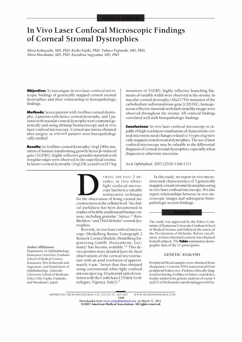
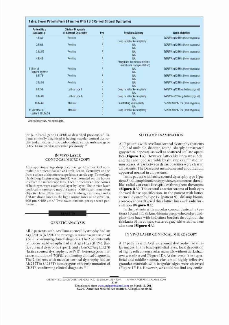
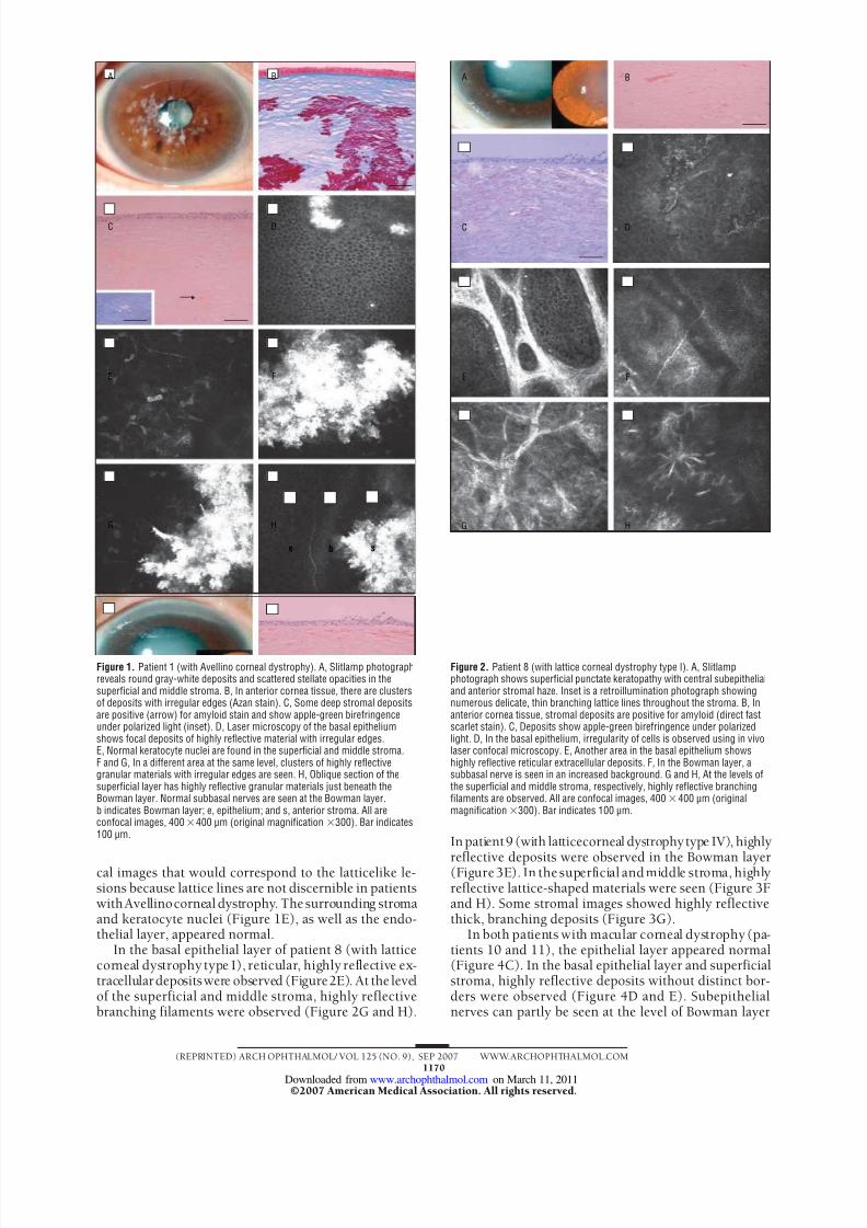
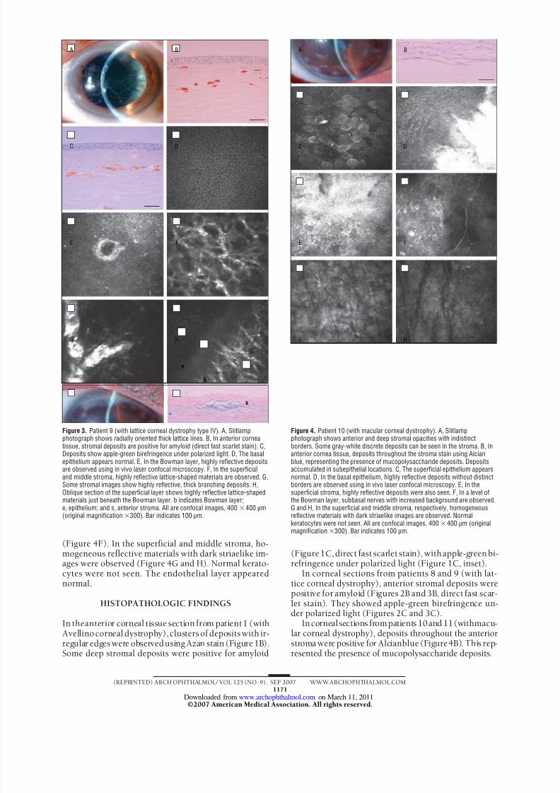
CLINICAL SCIENCES In Vivo Laser Confocal Microscopic Findings of Cornea l Stromal Dystr ophie s Akira Kobayashi, MD, PhD; Keiko Fujiki, PhD; Takuro Fujimaki, MD, PhD; Akira Murakami, MD, PhD; Kazuhisa Sugiyama, MD, PhD Objective: To investigate in vivo laser confocal micro- scopic findings of genetically mapped corneal stromal dystrophies and their relationship to histopathologic findings. Methods: Seven patients with Avellino corneal dystro- phy, 2 patients with lattice corneal dystrophy, and 2 pa- tients wit h macular cor nea l dys tro phy wer e examin ed ge- netically and using slitlamp biomicroscopy and in vivo laser confocal microscopy. Corneal specimens obtained after surgery in selec ted patie nts were histopatho logi - cally studied. Results: In Ave lli no corn eal dys trop hy (Arg 124 His mu- tation of human transforming growth factor –induced ge ne [TGFBI]), highl y refle ctive granu lar mate rials with irregular edges were observed in the superficial stroma. In latti ce corn eal dyst roph y (Arg 124 Cys and Leu527Arg mutations of TGFBI), highly reflective branching fila- ments of variable width were observed in the stroma. In macular corneal dystrophy (Ala217Thr mutation of the carbohydrate sulfotransferase gene [CHST6]), homoge- neou s reflectiv e mate rial s with dark stri aeli ke imag es were observed throughout the stroma. All confocal findings correlated well with histopathologic findings. Conclusions: In vivo laser confocal microscopy is ca- pab le of high -resolution visu aliz atio n of char acte rist ic cor- nea l mi crostruct ura l changes related to 3 types of gen eti - call y mapp ed corn eal stromaldystrop hies . The use of lase r confocal microscopy may be valuable in the differential diagn osis of cornea l stromal dystro phies, especi ally when diagnosis is otherwise uncertain. Arch Ophthalmol. 2007;125(9):1168-1173 D URING THE PAST 2 DE - cades, in vivo white- light confocal micros- copy has been a valuable noninvasive technique for the observation of living corneal mi- cros truct ures at the cell ular leve l. 1 Its clini- cal usefulness has been documented in stu di es of hea lth y anddise ase d hum an co r- neas, including granular, 2 lattice, 2,3 Reis- Bu ¨ ckle rs, 2 and Thiel- Behnke 4 corneal dys- trophies. Rec entl y, in vivo las er con foc al mic ros - copy (Heidelberg Retina Tomograph 2 Rostock Cornea Module; Heidelberg En- gineering GmbH, Dossenheim, Ger- many) has become available. 5,6 This de- vice permits more detailed layer-by-layer observations of the corneal microstruc- ture with an axial resolution of approxi- mately 4 µm, 7 better than that obtained using conventional white-light confocal micro sco pes (eg , 10-µm axi al opt ica l res o- lut ion wit h the Con foS can 2 [Ni dek Tech - nologies, Vigonza, Italy]). 8 In this study, we report in vivo micro- structural characteristics of 3 genetically mapp ed corneal strom al dystr ophi es using in viv o las er con foc al mi cro sco py. We als o report relationships between in vivo mi- croscopic images and subsequent histo- pathologic section findings. METHODS The study was approved by the Ethics Com- mitt ee of Kan azaw a Universi ty Grad uateSchool of Medical Science and followed the tenets of the Declaration of Helsinki. Before enroll- ment, written informed consent was obtained fromall subj ects. The Table summar izes demo- graphic data of the 11 participants. GENETIC ANALYSIS Peripheral blood samples were obtained from thepatie nts . Gen omi c DNA wasextracte d fro m peri pher al leu kocy tes. Pati ents cli nica lly diag- nos ed as havi ng Ave lli no or lat tic e cornea l dys- trophy underwent genetic analysis of exons 4 and12 of thehuma n transf orm inggrowth fac - Author Affiliations: Department of Ophthalmology, Kanazawa University Graduate School of Medical Science, Kanazawa (Drs Kobayashi and Sugiyama), and Department of Ophthalmology, Juntendo University School of Medicine, T okyo (Drs Fujiki, Fujimaki, and Murakami), Japan. (REPR INTED) ARCH OPHTHALMOL/ VOL 125 (NO. 9), SEP 2007 WWW.ARCHOPHT HALMOL. COM 1168 ©2007 American Medical Association. All rights reserved. on March 11, 2011 www.archophthalmol.com Downloaded from