Embed Size (px)
Citation preview
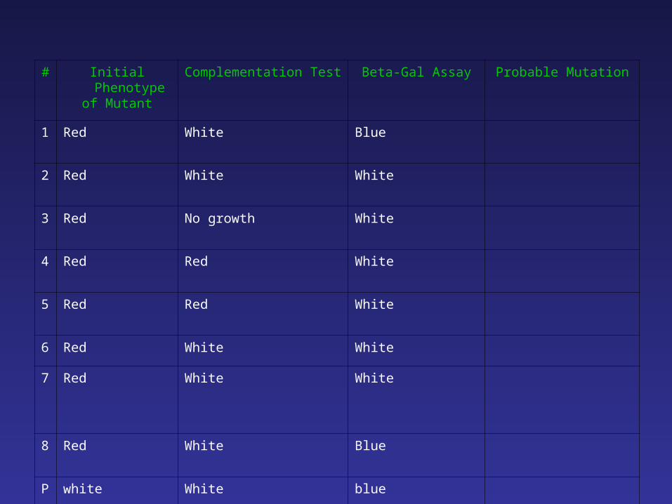
# Initial Phenotypeof Mutant
Complementation Test Beta-Gal Assay Probable Mutation
1 Red White Blue
2 Red White White
3 Red No growth White
4 Red Red White
5 Red Red White
6 Red White White
7 Red White White
8 Red White Blue
P white White blue
Techniques to know
Additional Techniques:
Northern BlotRNAse Protection Assay Immunoprecipitation and Western BlotEMSA (Electromobility Shift Assay)IHC (Immunohistochemistry)DNA microarray
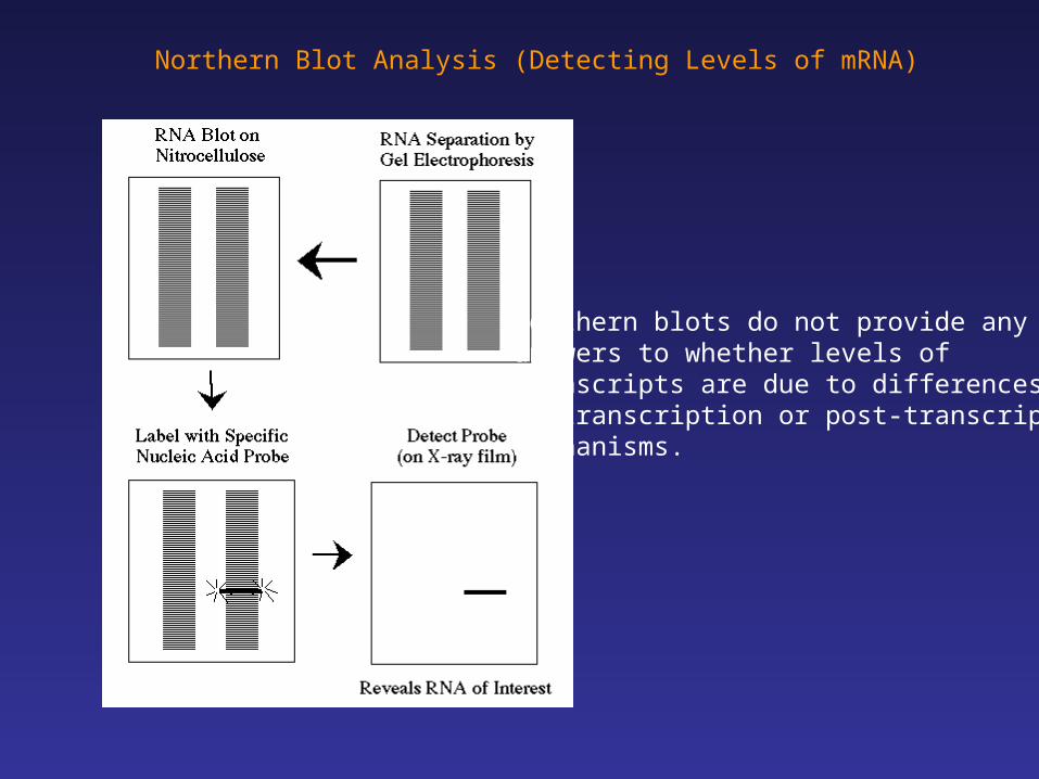
Northern Blot Analysis (Detecting Levels of mRNA)
Northern blots do not provide anyanswers to whether levels of transcripts are due to differences in transcription or post-transcriptionalmechanisms.
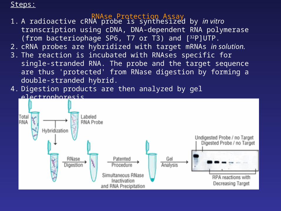
RNAse Protection AssaySteps:
1. A radioactive cRNA probe is synthesized by in vitro transcription using cDNA, DNA-dependent RNA polymerase (from bacteriophage SP6, T7 or T3) and [32P]UTP.
2. cRNA probes are hybridized with target mRNAs in solution.3. The reaction is incubated with RNAses specific for single-stranded RNA. The
probe and the target sequence are thus 'protected' from RNase digestion by forming a double-stranded hybrid.
4. Digestion products are then analyzed by gel electrophoresis
RPA methods are time-consuming but have advantages
1. At least ten-fold more sensitive than Northern Blots2. Multiple targets can be assayed simultaneously “multiplexing” 3. Very high specificity (mismatched RNA:probe hybrids are vulnerable to digestion
by RNases.4. Detect non-abundant and rare transcripts with high degree of accuracy
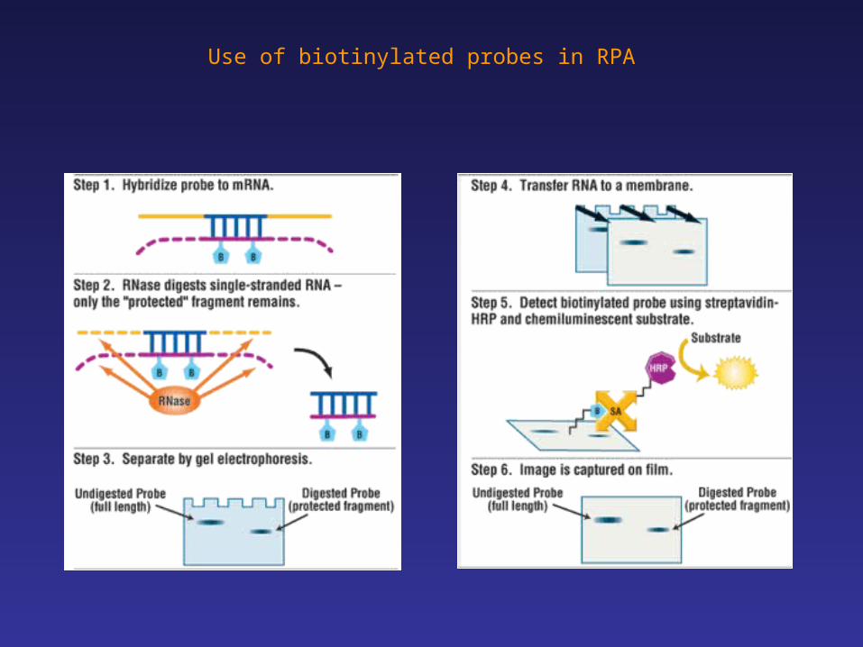
Use of biotinylated probes in RPA
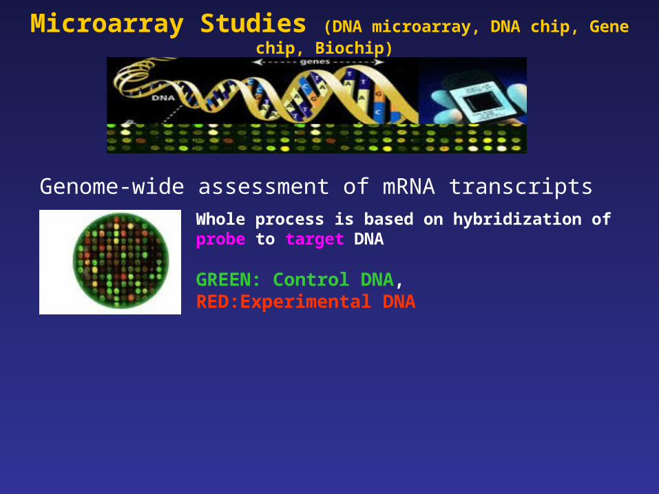
Genome-wide assessment of mRNA transcripts
Whole process is based on hybridization of probe to target DNA
GREEN: Control DNA, RED:Experimental DNA
Microarray Studies (DNA microarray, DNA chip, Gene chip, Biochip)
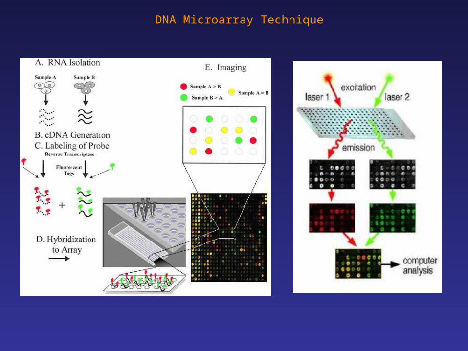
DNA Microarray Technique
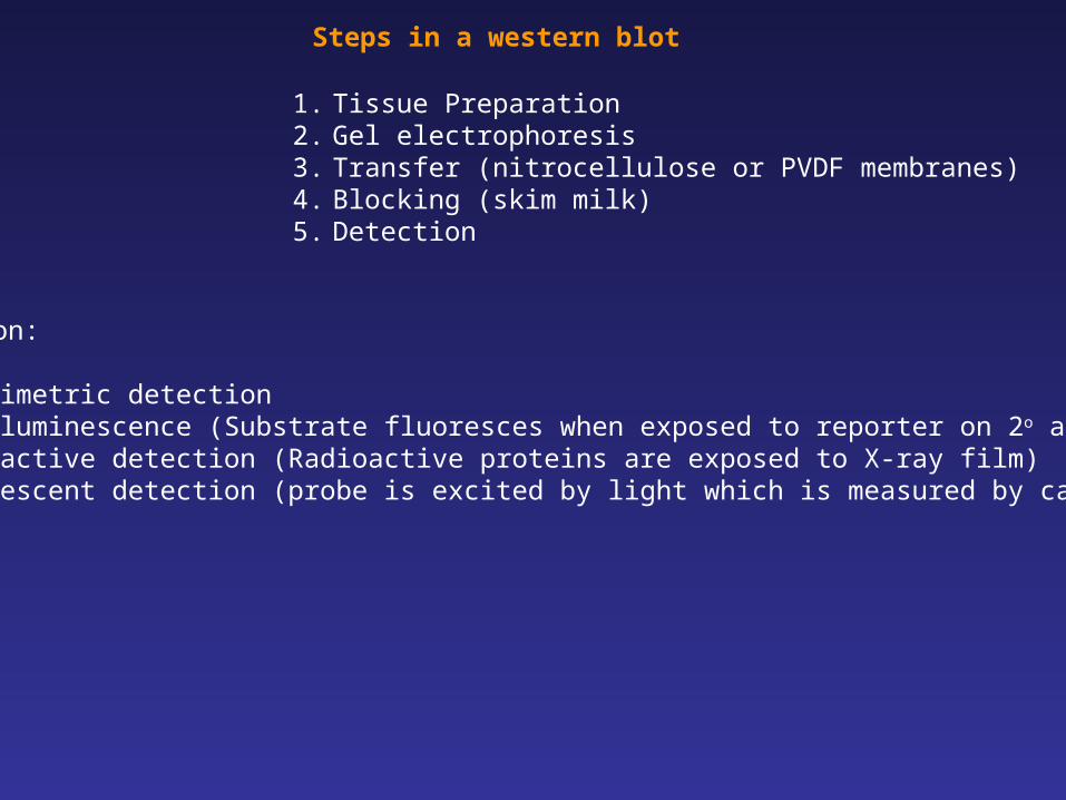
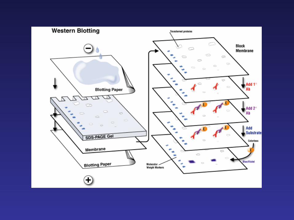
Steps in a western blot
1. Tissue Preparation2. Gel electrophoresis3. Transfer (nitrocellulose or PVDF membranes)4. Blocking (skim milk)5. Detection
Detection:
1. Colorimetric detection 2. Chemiluminescence (Substrate fluoresces when exposed to reporter on 2o antibody) 3. Radioactive detection (Radioactive proteins are exposed to X-ray film)4. Fluorescent detection (probe is excited by light which is measured by camera)
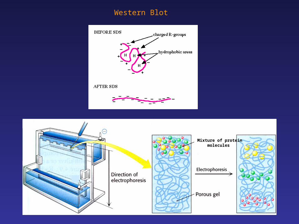
Western Blot
Mixture of protein molecules
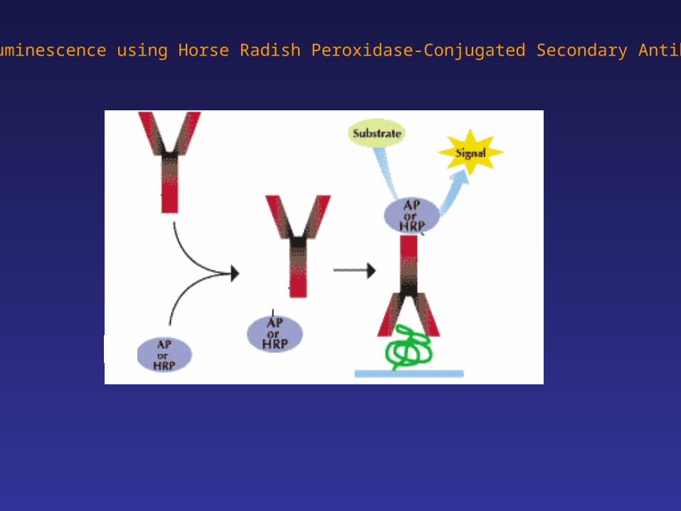
Chemiluminescence using Horse Radish Peroxidase-Conjugated Secondary Antibodies
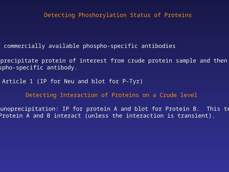
Detecting Phoshorylation Status of Proteins
1. Use of commercially available phospho-specific antibodies
2. Immunoprecipitate protein of interest from crude protein sample and then blot with phospho-specific antibody.
Example: Article 1 (IP for Neu and blot for P-Tyr)
Detecting Interaction of Proteins on a Crude level
3. Co-immunoprecipitation: IP for protein A and blot for Protein B. This tells you whether Protein A and B interact (unless the interaction is transient).
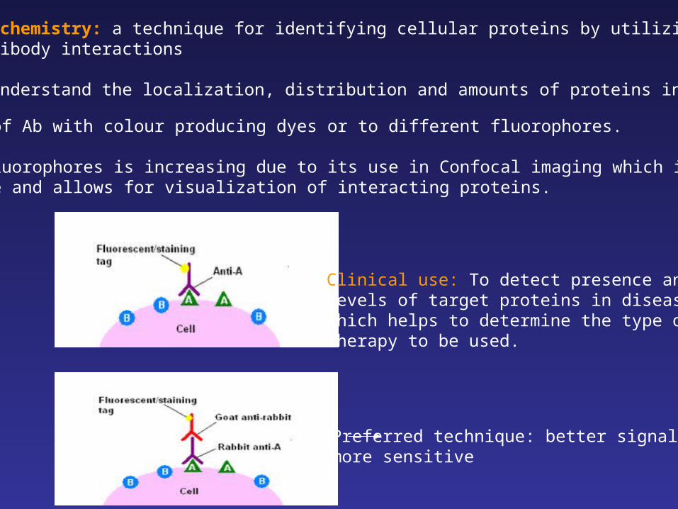
Immunohistochemistry: a technique for identifying cellular proteins by utilizing antigen-antibody interactions
To better understand the localization, distribution and amounts of proteins in the cells:
Tagging of Ab with colour producing dyes or to different fluorophores.
Use of fluorophores is increasing due to its use in Confocal imaging which is moresensitive and allows for visualization of interacting proteins.
Clinical use: To detect presence andlevels of target proteins in diseasewhich helps to determine the type of therapy to be used.
Preferred technique: better signal and more sensitive
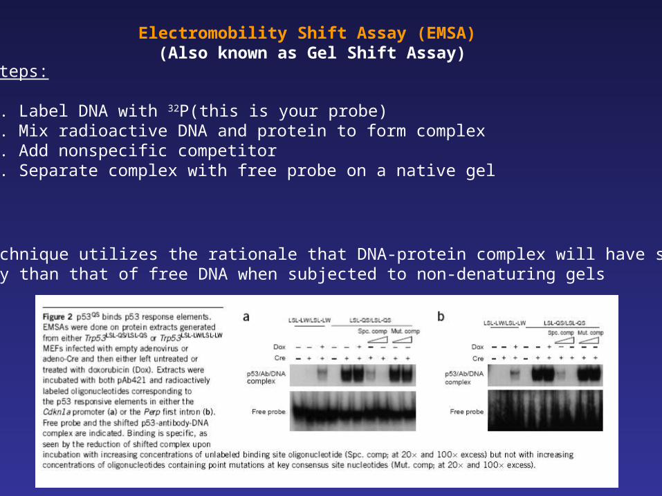
Steps:
1. Label DNA with 32P(this is your probe)2. Mix radioactive DNA and protein to form complex 3. Add nonspecific competitor 4. Separate complex with free probe on a native gel
Electromobility Shift Assay (EMSA) (Also known as Gel Shift Assay)
This technique utilizes the rationale that DNA-protein complex will have smaller mobility than that of free DNA when subjected to non-denaturing gels

The End