Embed Size (px)
Citation preview

Integumentary System
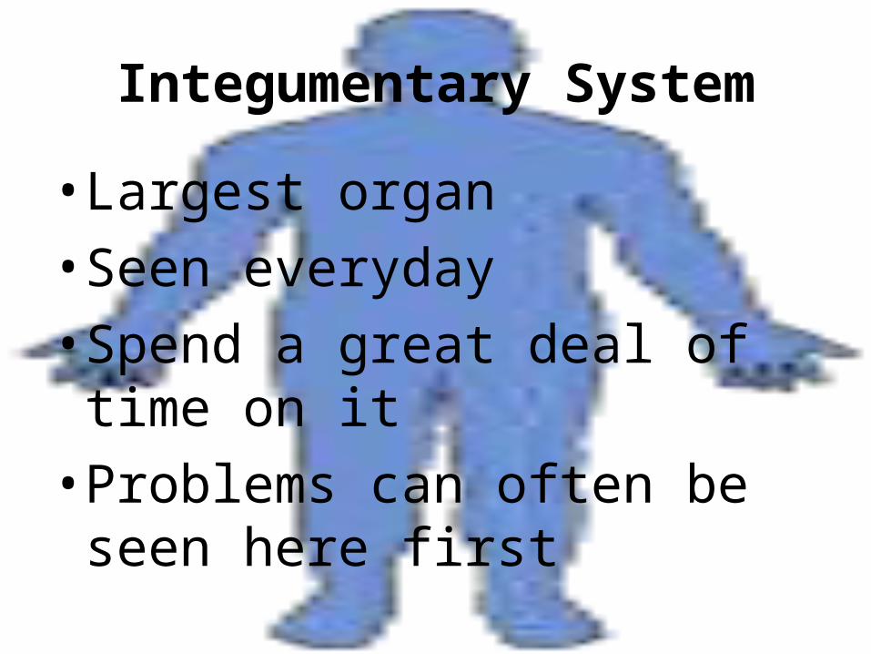
Integumentary System
• Largest organ
• Seen everyday
• Spend a great deal of time on it
• Problems can often be seen here first
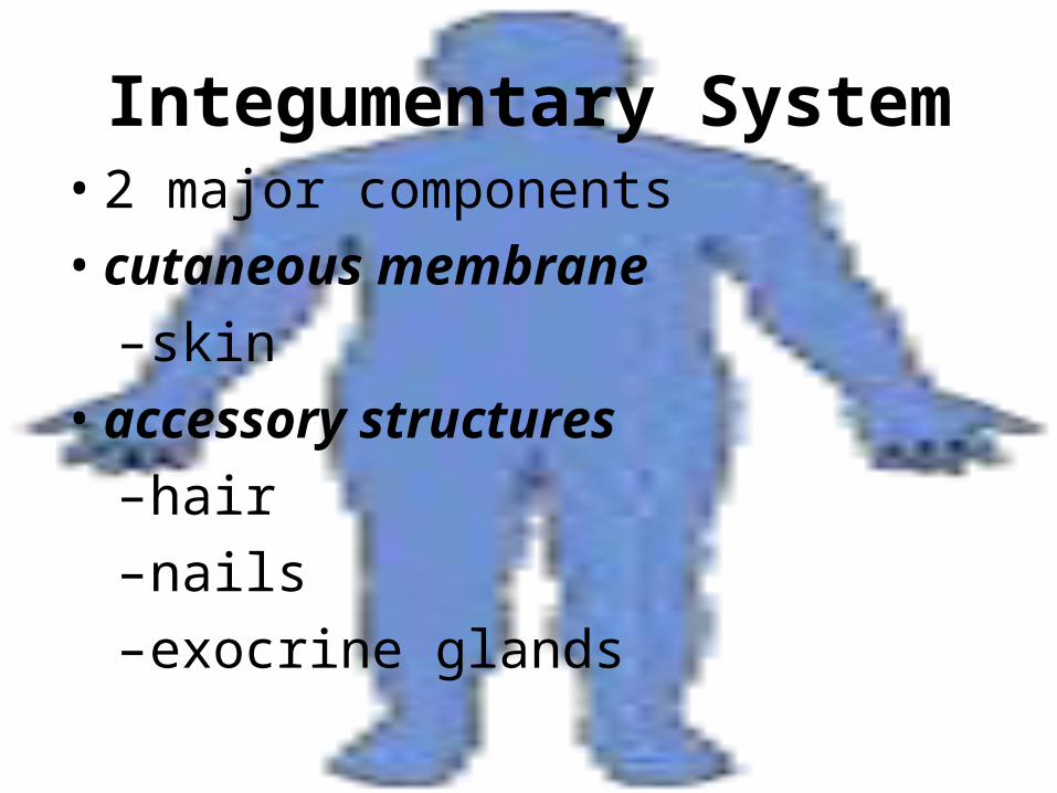
Integumentary System• 2 major components
• cutaneous membrane
–skin
• accessory structures
–hair
–nails
–exocrine glands
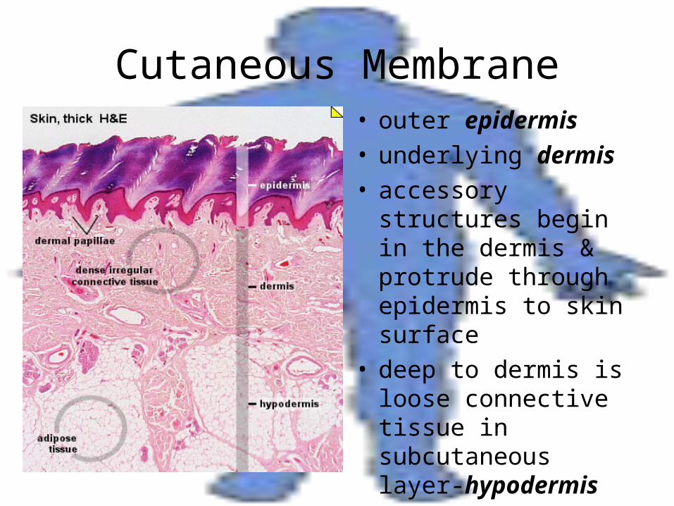
Cutaneous Membrane• outer epidermis• underlying dermis• accessory structures
begin in the dermis & protrude through epidermis to skin surface
• deep to dermis is loose connective tissue in subcutaneous layer-hypodermis
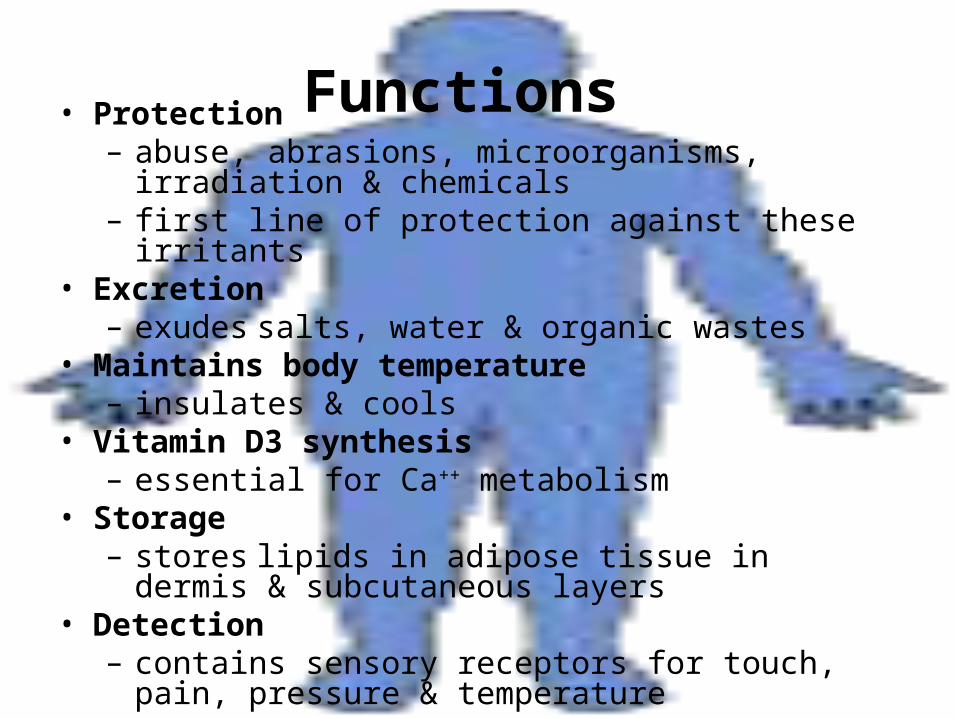
Functions • Protection– abuse, abrasions, microorganisms, irradiation &
chemicals– first line of protection against these irritants
• Excretion– exudes salts, water & organic wastes
• Maintains body temperature– insulates & cools
• Vitamin D3 synthesis– essential for Ca++ metabolism
• Storage – stores lipids in adipose tissue in dermis &
subcutaneous layers• Detection
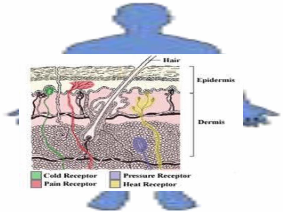
– contains sensory receptors for touch, pain, pressure & temperature
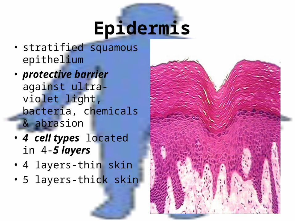
Epidermis • stratified squamous
epithelium• protective barrier
against ultra-violet light, bacteria, chemicals & abrasion
• 4 cell types located in 4-5 layers
• 4 layers-thin skin• 5 layers-thick skin
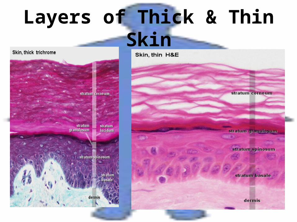
Layers of Thick & Thin Skin
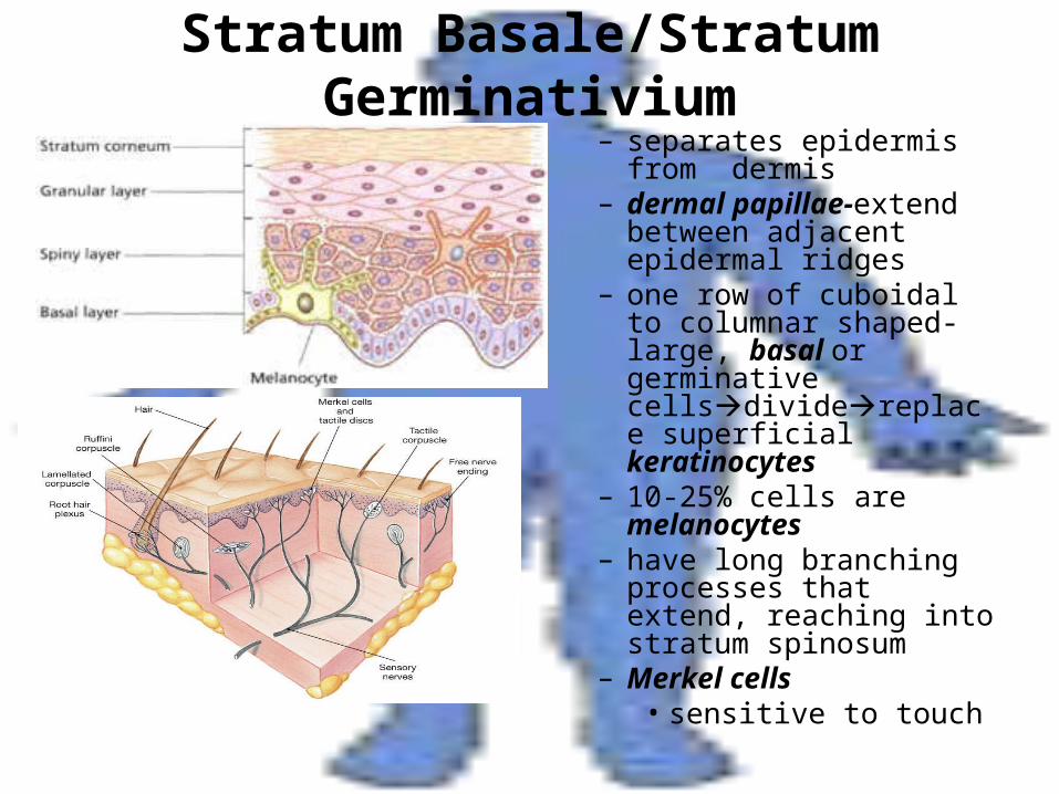
Stratum Basale/Stratum Germinativium
– separates epidermis from dermis
– dermal papillae-extend between adjacent epidermal ridges
– one row of cuboidal to columnar shaped-large, basal or germinative cellsdividereplace superficial keratinocytes
– 10-25% cells are melanocytes
– have long branching processes that extend, reaching into stratum spinosum
– Merkel cells• sensitive to touch
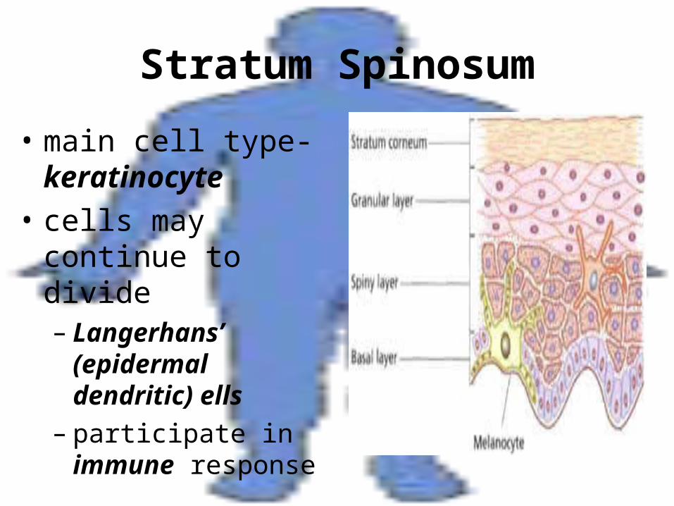
Stratum Spinosum
• main cell type-keratinocyte
• cells may continue to divide– Langerhans’
(epidermal dendritic) ells
– participate in immune response
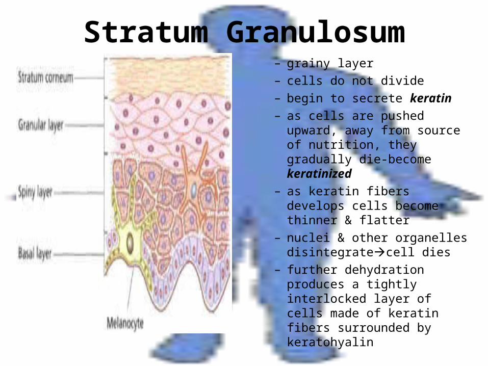
Stratum Granulosum– grainy layer– cells do not divide– begin to secrete keratin– as cells are pushed upward,
away from source of nutrition, they gradually die-become keratinized
– as keratin fibers develops cells become thinner & flatter
– nuclei & other organelles disintegratecell dies
– further dehydration produces a tightly interlocked layer of cells made of keratin fibers surrounded by keratohyalin
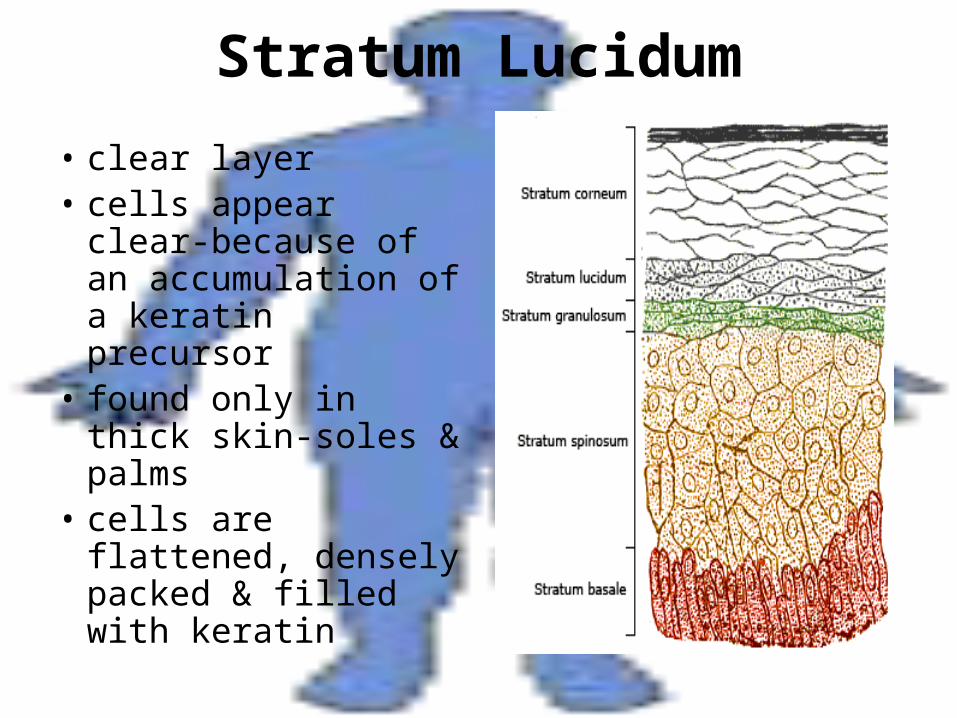
Stratum Lucidum
• clear layer• cells appear clear-
because of an accumulation of a keratin precursor
• found only in thick skin-soles & palms
• cells are flattened, densely packed & filled with keratin
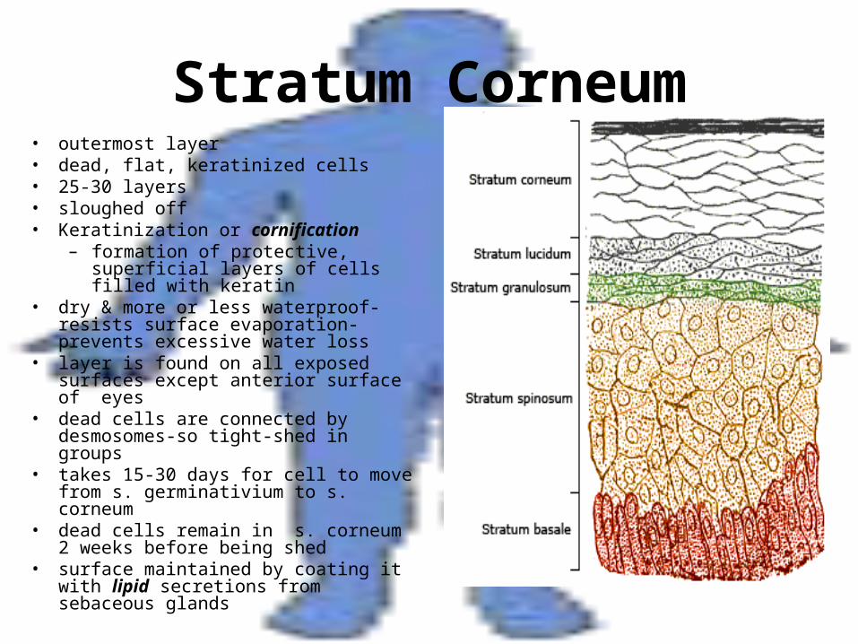
Stratum Corneum• outermost layer• dead, flat, keratinized cells• 25-30 layers• sloughed off• Keratinization or cornification
– formation of protective, superficial layers of cells filled with keratin
• dry & more or less waterproof-resists surface evaporation-prevents excessive water loss
• layer is found on all exposed surfaces except anterior surface of eyes
• dead cells are connected by desmosomes-so tight-shed in groups
• takes 15-30 days for cell to move from s. germinativium to s. corneum
• dead cells remain in s. corneum 2 weeks before being shed
• surface maintained by coating it with lipid secretions from sebaceous glands
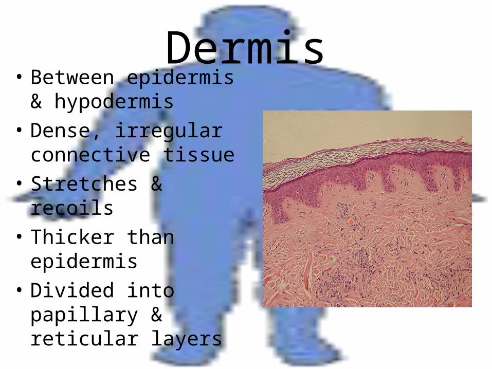
Dermis• Between epidermis &
hypodermis
• Dense, irregular connective tissue
• Stretches & recoils
• Thicker than epidermis
• Divided into papillary & reticular layers
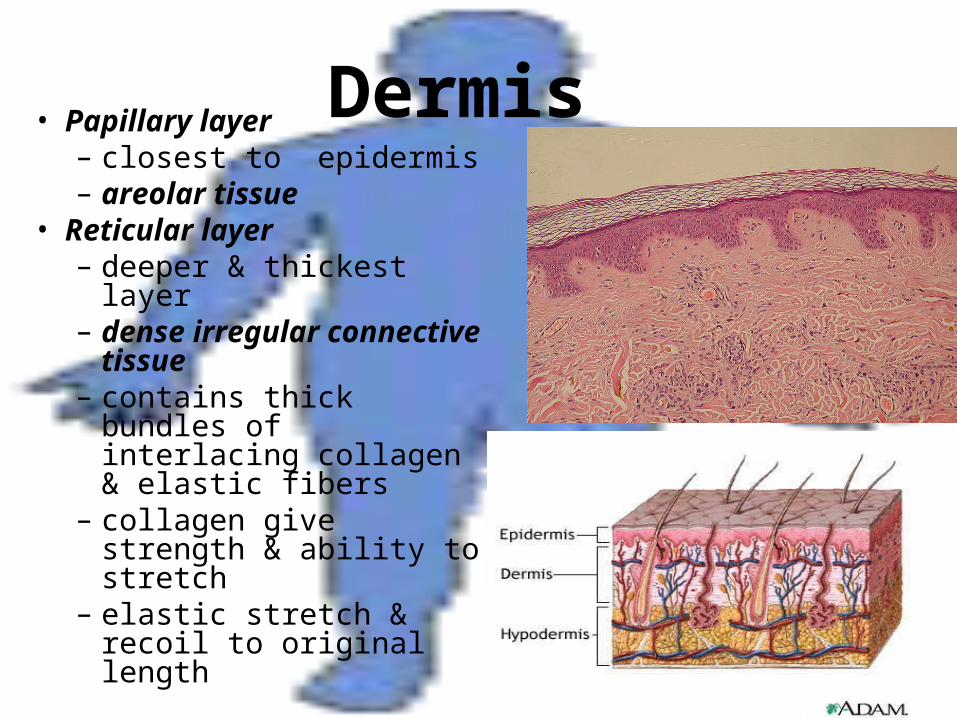
Dermis • Papillary layer– closest to epidermis – areolar tissue
• Reticular layer– deeper & thickest layer– dense irregular
connective tissue– contains thick bundles
of interlacing collagen & elastic fibers
– collagen give strength & ability to stretch
– elastic stretch & recoil to original length
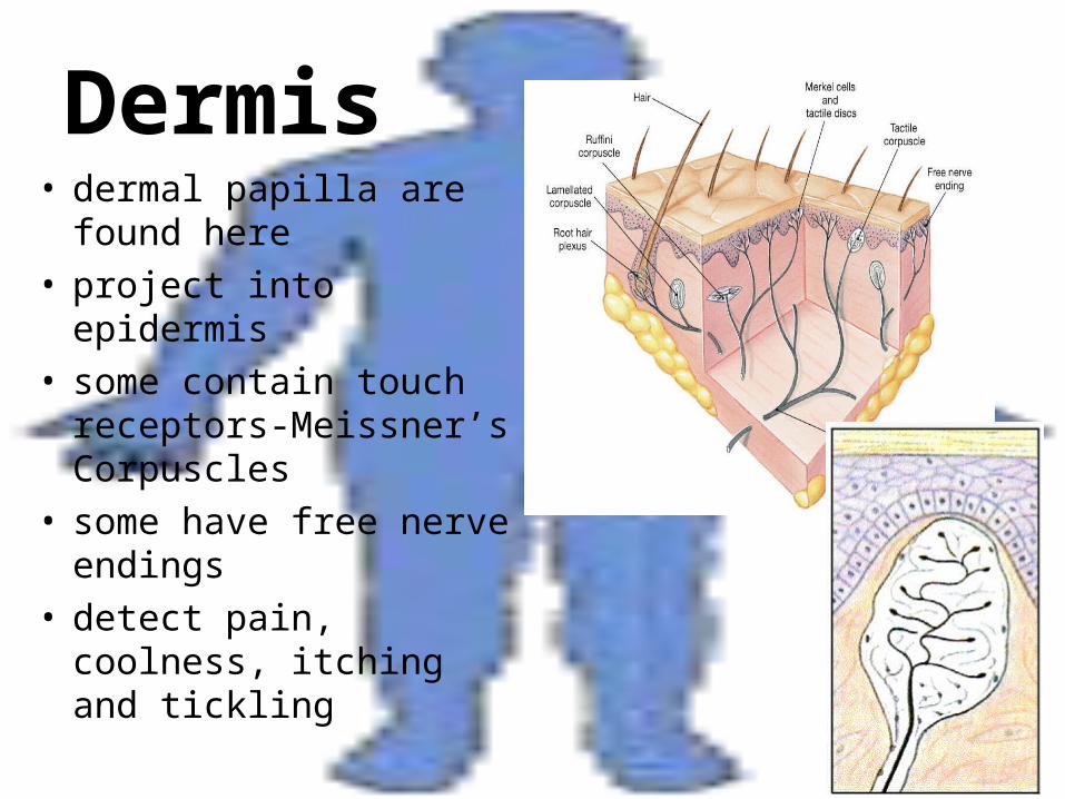
Dermis• dermal papilla are
found here• project into epidermis• some contain touch
receptors-Meissner’s Corpuscles
• some have free nerve endings
• detect pain, coolness, itching and tickling
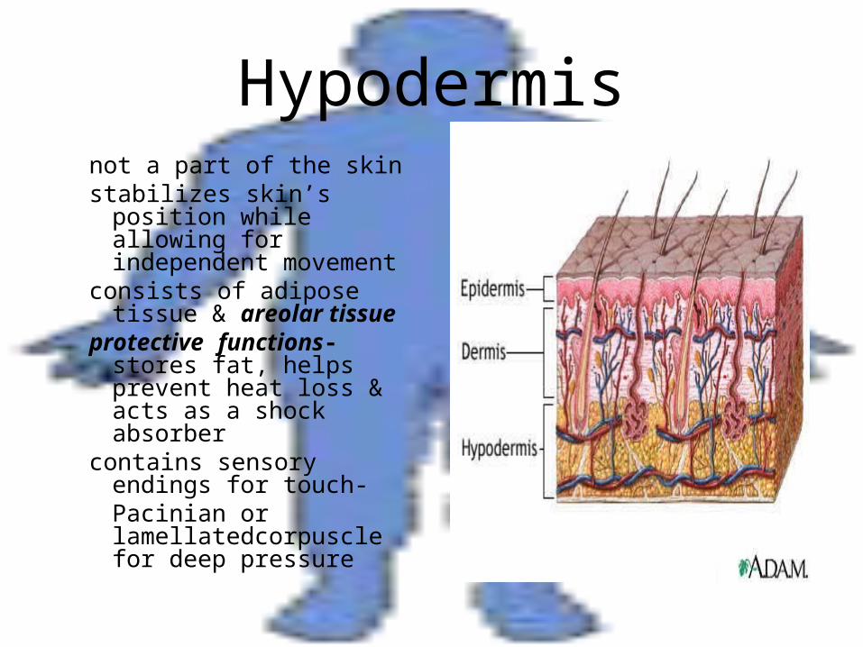
Hypodermisnot a part of the skinstabilizes skin’s position
while allowing for independent movement
consists of adipose tissue & areolar tissue
protective functions-stores fat, helps prevent heat loss & acts as a shock absorber
contains sensory endings for touch-
Pacinian or lamellatedcorpuscle for deep pressure
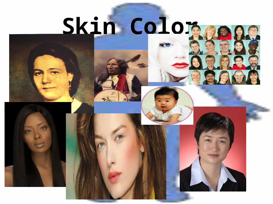
Skin Color
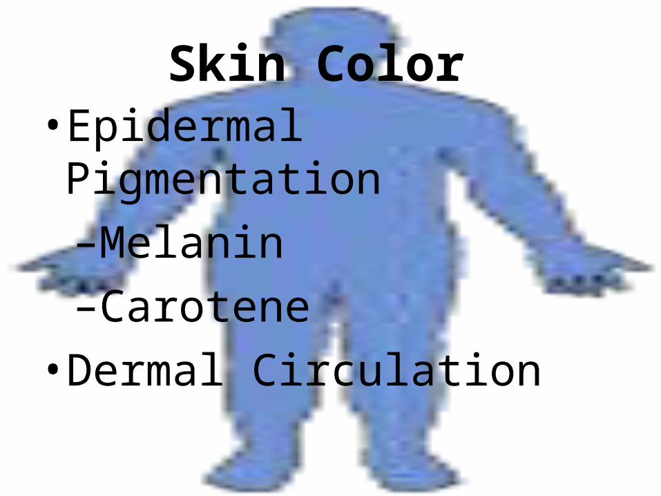
Skin Color • Epidermal Pigmentation
–Melanin
–Carotene
• Dermal Circulation
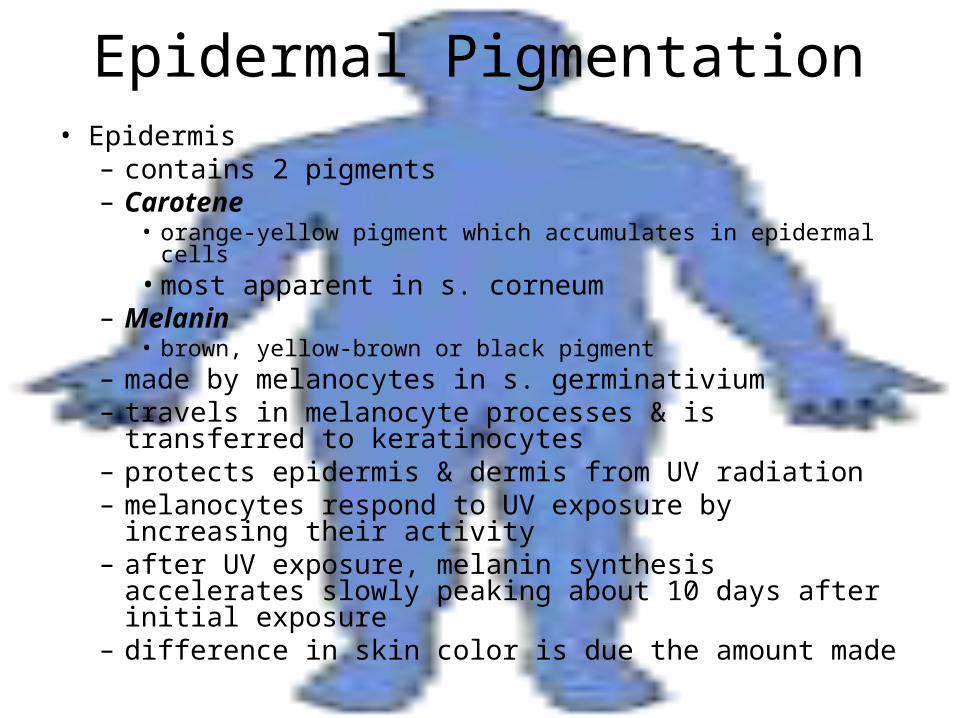
Epidermal Pigmentation• Epidermis
– contains 2 pigments– Carotene
• orange-yellow pigment which accumulates in epidermal cells• most apparent in s. corneum
– Melanin• brown, yellow-brown or black pigment
– made by melanocytes in s. germinativium– travels in melanocyte processes & is transferred to
keratinocytes– protects epidermis & dermis from UV radiation– melanocytes respond to UV exposure by increasing
their activity– after UV exposure, melanin synthesis accelerates
slowly peaking about 10 days after initial exposure– difference in skin color is due the amount made
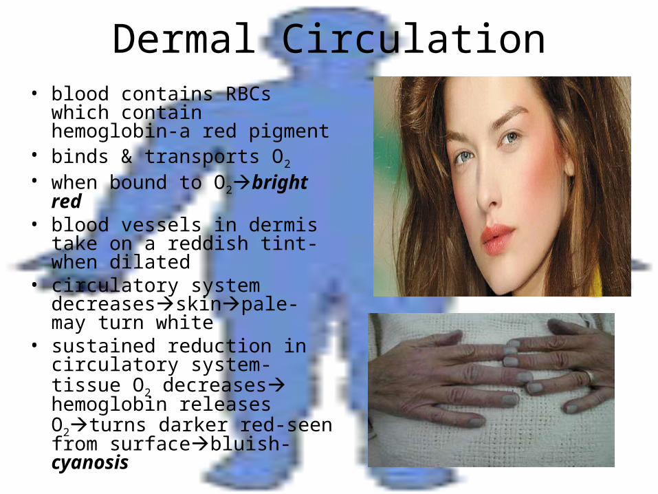
Dermal Circulation• blood contains RBCs which
contain hemoglobin-a red pigment
• binds & transports O2
• when bound to O2bright red
• blood vessels in dermis take on a reddish tint-when dilated
• circulatory system decreasesskinpale-may turn white
• sustained reduction in circulatory system-tissue O2 decreases hemoglobin releases O2turns darker red-seen from surfacebluish-cyanosis
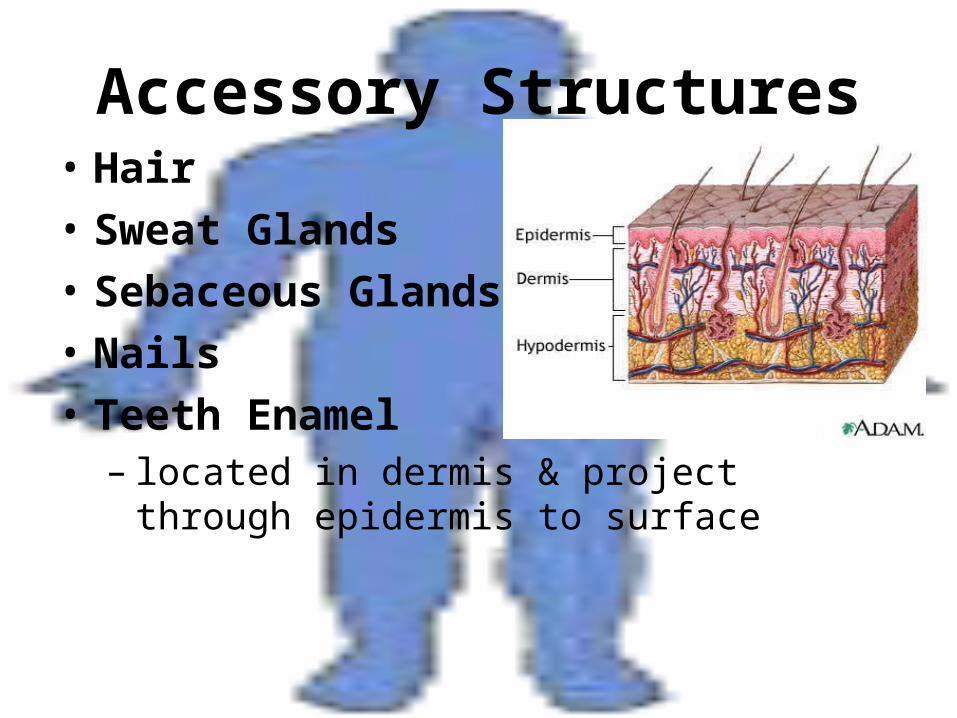
Accessory Structures• Hair
• Sweat Glands
• Sebaceous Glands
• Nails
• Teeth Enamel– located in dermis & project through epidermis
to surface
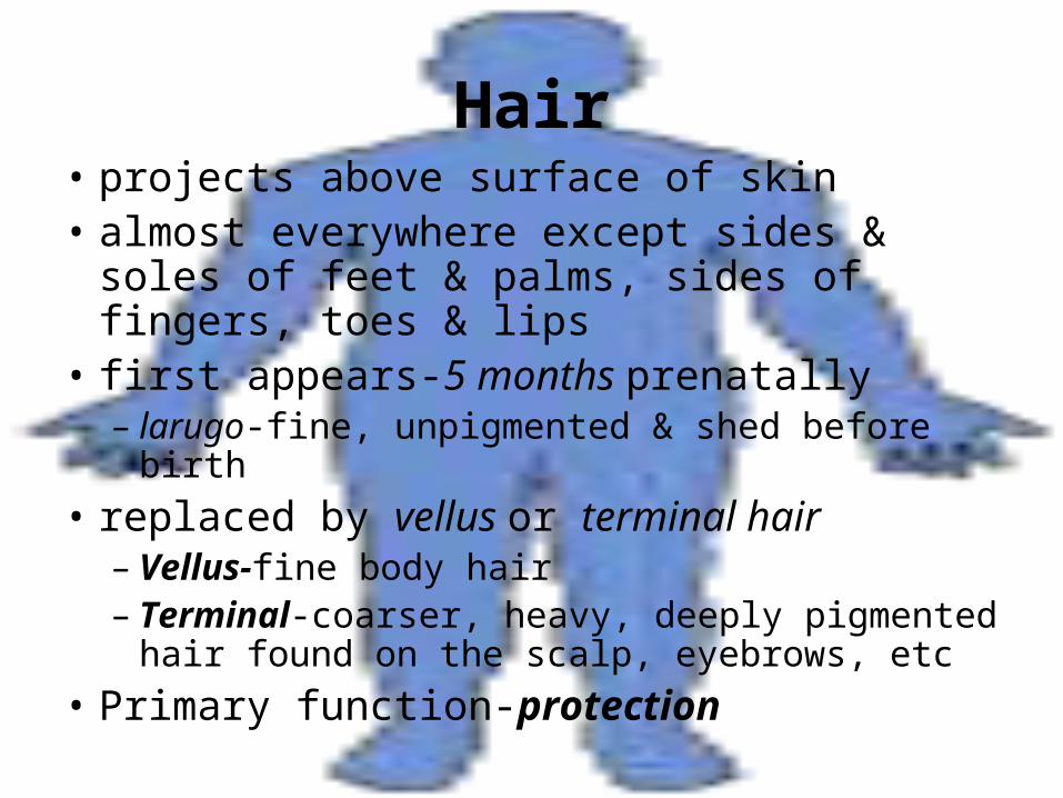
Hair• projects above surface of skin• almost everywhere except sides & soles of
feet & palms, sides of fingers, toes & lips• first appears-5 months prenatally
– larugo-fine, unpigmented & shed before birth
• replaced by vellus or terminal hair– Vellus-fine body hair– Terminal-coarser, heavy, deeply pigmented
hair found on the scalp, eyebrows, etc
• Primary function-protection
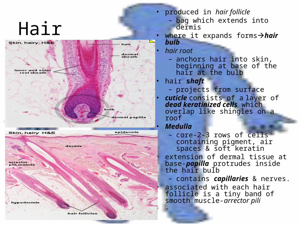
Hair• produced in hair follicle
– bag which extends into dermis• where it expands formshair bulb• hair root
– anchors hair into skin, beginning at base of the hair at the bulb
• hair shaft– projects from surface
• cuticle consists of a layer of dead keratinized cells which overlap like shingles on a roof
• Medulla – core-2-3 rows of cells
containing pigment, air spaces & soft keratin
• extension of dermal tissue at base-papilla protrudes inside the hair bulb– contains capillaries & nerves.
• associated with each hair follicle is a tiny band of smooth muscle-arrector pili
• -
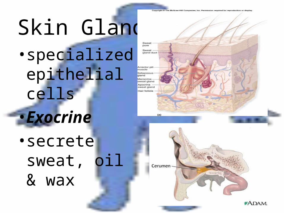
Skin Glands• specialized
epithelial cells• Exocrine• secrete
sweat, oil & wax
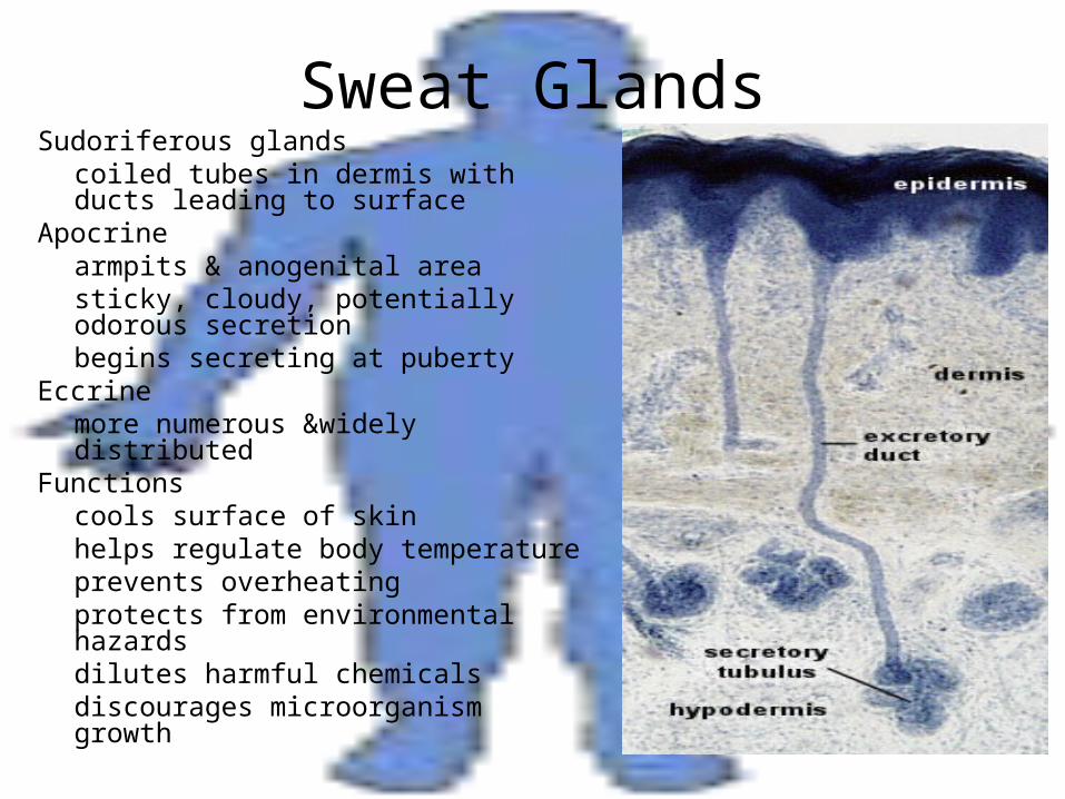
Sweat GlandsSudoriferous glands
coiled tubes in dermis with ducts leading to surface
Apocrinearmpits & anogenital areasticky, cloudy, potentially odorous secretionbegins secreting at puberty
Eccrinemore numerous &widely distributed
Functionscools surface of skinhelps regulate body temperatureprevents overheatingprotects from environmental hazardsdilutes harmful chemicalsdiscourages microorganism growth
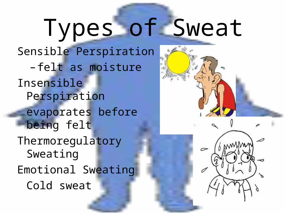
Types of SweatSensible Perspiration
– felt as moisture
Insensible Perspiration
evaporates before being felt
Thermoregulatory Sweating
Emotional Sweating
Cold sweat
Sebaceous Glands• oil glands• secrete sebum (seb = oil)
– mixture of triglycerides, cholesterol, proteins & electrolytes
– usually secreted into hair follicle in a few regions-lips & mammary papilla & directly secreted onto skin surface of face, back & chest
• Holocrine– entire gland dies when it secretes
• Functions– inhibits bacterial growth– lubricates– protects keratin– conditions skin