Embed Size (px)
Citation preview
C
ND
a
ARRA
KAAAMMP
1
i(o(cEabWbsst
sMb2ho
h1
International Journal of Paleopathology 10 (2015) 1–12
Contents lists available at ScienceDirect
International Journal of Paleopathology
j ourna l ho mepage: www.elsev ier .com/ locate / i jpp
ribra orbitalia in the ancient Nile Valley and its connection to malaria
icole E. Smith-Guzmán ∗
epartment of Anthropology, University of Arkansas, 330 Old Main, Fayetteville, AR 72701, United States
r t i c l e i n f o
rticle history:eceived 29 November 2014eceived in revised form 4 March 2015ccepted 11 March 2015
eywords:nemiancient Egyptncient Nubia
a b s t r a c t
Cribra orbitalia is a common skeletal lesion found on ancient human remains excavated from the NileValley. Recent etiological research implicates hemolytic anemia as a main factor leading to the formationof cribra orbitalia. Further, an association between the hemolytic anemia caused by malaria and cribraorbitalia has been demonstrated. The presence of malaria in the ancient Nile Valley has been verifieddirectly through genetic and immunologic studies of Egyptian mummies, but its prevalence and spreadremain unknown. As some models have pointed to the Nile Valley as the pathway of malarial dispersionduring the Egyptian Dynastic period, variability in cribra orbitalia rates should provide a way to trackthe disease spread. This study surveyed cribra orbitalia frequencies at 29 ancient Nile Valley sites, rep-
eta-analysisalaria
aleoepidemiology
resenting 4760 individuals ranging from prehistoric to Christian periods and situated between the 3rdCataract and Nile Delta. Results showed high cribra orbitalia rates, with an overall mean of 42.8% of thetotal population affected. Over time and space, the data showed no significant correlation, suggestinghigh levels of anemia affected individuals in the Nile Valley equally from late pre-dynastic to Christianperiods. These findings suggest widespread endemic malaria in the Nile Valley before Dynastic Egypt.
. Introduction
Cribra orbitalia is one of the most common skeletal lesions notedn ancient human skeletal remains excavated from the Nile ValleyHillson, 1980). Researchers have long explained this porous lesionf the eye orbits, along with the similar porous cranial vault lesionsporotic hyperostosis), as an expansion of the marrow space in theranial vault caused by iron-deficiency anemia (Carlson et al., 1974;l-Najjar et al., 1976, 1975; Lallo et al., 1977; Mensforth et al., 1978;mong others). This iron-deficiency anemia hypothesis has recentlyeen called into question by a number of researchers, includingalker and coworkers (2009), who maintain the depression of red
lood cell production in iron-deficiency anemia excludes the pos-ibility of its participation in the stimulation of increased marrowpace involved in porotic hyperostosis and cribra orbitalia forma-ion.
While Walker and coworkers’ (2009) etiological reappraisal istill being debated (Oxenham and Cavill, 2010; Rothschild, 2012;cIlvaine, 2013), other researchers have shown an association
etween cribra orbitalia and malarial infection (Rabino Massa et al.,
000; Nerlich et al., 2008; Gowland and Western, 2012). Malariaas been identified in the mummified tissue of ancient Egyptiansf various time periods, dating to as early as 3200 BCE using ancient∗ Tel.: +1 4792268256.E-mail address: [email protected]
ttp://dx.doi.org/10.1016/j.ijpp.2015.03.001879-9817/© 2015 Elsevier Inc. All rights reserved.
© 2015 Elsevier Inc. All rights reserved.
DNA (aDNA) sequencing and antigen evidence (Miller et al., 1994;Bianucci et al., 2008; Nerlich et al., 2008; Hawass et al., 2010). Thisdirect genetic and immunological evidence verifies the presence ofmalaria in antiquity, but leaves the prevalence and spread of thedisease unknown.
Although there are many factors that could have potentially con-tributed to the overall anemia seen in the human skeletal remainsof ancient Egypt, malarial infection has been shown to have a majorsynergistic effect with other factors to increase overall anemia lev-els; thus, would have arguably raised the overall frequencies ofcribra orbitalia (Nájera and Hempel, 1996; Gilles, 1997; Lusinguet al., 2004; Shanks et al., 2008). The present study surveys thetemporospatial variability in rates of cribra orbitalia reported atarchaeological sites along the Nile Valley in order to estimateancient prevalence and distribution of malaria in this region. Track-ing changes in cribra orbitalia in the Nile Valley provides not only amore holistic picture of ancient Egyptian anemia, but also a poten-tial way to test theoretical models of malaria’s spread out of Africa.
1.1. Porotic hyperostosis, cribra orbitalia, and anemia
Genetic conditions conferring resistance from malaria (e.g. tha-lassemia and sickle cell disorder) have been argued to cause skeletal
lesions such as porotic hyperostosis and cribra orbitalia in theancient Mediterranean and Near East (Angel, 1964, 1966, 1967,1972; Zaino, 1964). However, there is a discrepancy between thelow rates of these genetic disorders in modern endemic populations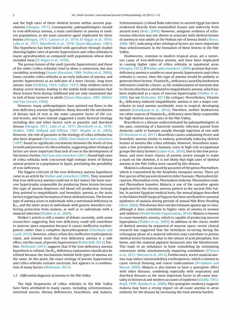
2 ourna
autelLTsmi
ovSpadtta
ionidWHa(iftoao
ctrttftBfm
rtpCcethrtat
1
hi
N.E. Smith-Guzmán / International J
nd the high rates of these skeletal lesions within ancient pop-lations (Hengen, 1971). Consequently, paleopathologists turnedo iron-deficiency anemia, a main contributor to anemia in mod-rn populations, as the main causative agent implicated for theseesions (Hengen, 1971; Carlson et al., 1974; El-Najjar et al., 1976;allo et al., 1977; Mensforth et al., 1978; Stuart-Macadam, 1987).his hypothesis has been linked with agriculture through studieshowing higher rates of porotic hyperostosis and cribra orbitalia inaize agriculturalists as compared with populations whose diets
ncluded meat (El-Najjar et al., 1976).The porous lesions of the vault (porotic hyperostosis) and those
f the orbits (cribra orbitalia) tend to show a connection, but alsoariability, in etiology (Stuart-Macadam, 1989; Walker et al., 2009).ome consider cribra orbitalia as an early indicator of anemia, andorotic hyperostosis as an indicator of a more chronic, long termnemic state (Hrdlicka, 1914; Caffey, 1937). Only children tend toisplay active lesions, leading to the widely held explanation thathese lesions form during childhood and are only maintained dueo lack of bone turnover in adults (Stuart-Macadam, 1985; Mittlernd Van Gerven, 1994).
However, many anthropologists have pointed out flaws in theron-deficiency anemia hypothesis. Many discredit the attributionf dietary lack of iron as the main causative factor of the cra-ial lesions, and have instead suggested a multi-factorial etiology
ncluding diet and other factors such as parasitic and diarrhealisease (Hengen, 1971; Lallo et al., 1977; Mensforth et al., 1978;alker, 1986; Holland and O’Brien, 1997; Wapler et al., 2004).
owever, the role of parasites in the etiology of cribra orbitalia haslso been disputed (DeGusta, 2009). Glen-Haduch and coworkers1997) found no significant correlations between the levels of ironn teeth and presence of cribra orbitalia, suggesting other etiologicalactors are more important than lack of iron in the development ofhis lesion. Further, McClure and coworkers (2011) found high ratesf cribra orbitalia with concurrent high isotopic levels of dietarynimal protein in a population in Spain, precluding the possibilityf iron-deficiency.
The biggest criticism of the iron-deficiency anemia hypothesisame in an article by Walker and coworkers (2009). They reasonedhat iron-deficiency anemia could not in fact induce the bone mar-ow hypertrophy responsible for producing these lesions becausehis type of anemia depresses red blood cell production. Instead,hey pointed to megaloblastic and hemolytic anemia as the mainactors triggering the formation of these skeletal lesions. The formerype of anemia arises in individuals with a nutritional deficiency in12, and the latter arises in individuals with genetic disorders con-
erring protection from malaria, as well as in individuals with aalarial infection (Walker et al., 2009).Walker’s article is still a matter of debate currently, with some
esearchers suggesting that iron deficiency could still contributeo marrow space expansion because it causes ineffective erythro-oiesis rather than a complete dyserythropoiesis (Oxenham andavill, 2010). However, others refute this ineffective erythropoiesislaim, and instead insist that iron deficiency anemia is a sideffect, not the cause, of porotic hyperostosis (Rothschild, 2012). Fur-her, McIlvaine (2013) suggests that if the iron-deficiency anemiaypothesis is refuted, the B12 deficiency explanation should also beefuted because the mechanisms behind both types of anemia arehe same. At this point, the exact etiology of porotic hyperostosisnd cribra orbitalia remains uncertain, but appears to a combina-ion of many factors (McIlvaine, 2013).
.2. Differential diagnosis of anemia in the Nile Valley
The high frequencies of cribra orbitalia in the Nile Valleyave been attributed to many causes, including schistosomiasis,
ntestinal worms, dietary deficiencies, brucellosis, and malaria.
l of Paleopathology 10 (2015) 1–12
Schistosomiasis (a blood fluke infection) in ancient Egypt has beenevidenced directly from mummified tissues and indirectly fromancient texts (Brier, 2004). However, antigenic evidence of schis-tosoma infection was not shown to associate with skeletal lesionsof anemia in non-adults at the Nubian site of Semna South (Alvrus,2006: 167), indicating other etiological factors are more importantthan schistosomiasis in the formation of these lesions in the NileValley.
Hookworms, common in modern tropical areas, are a notori-ous cause of iron-deficiency anemia, and have been implicatedin causing higher rates of cribra orbitalia in equatorial areas(Hengen, 1971). If Walker and coworkers’ (2009) position that iron-deficiency anemia is unable to cause porotic hyperostosis and cribraorbitalia is correct, then this type of anemia would be unlikely togenerate these lesions. Vitamin B12 deficiency caused by hookworminfestation could be a factor, as the malabsorption of nutrients dueto chronic diarrhea is attributed to megaloblastic anemia, which hasbeen implicated as a cause of marrow hypertrophy (Walker et al.,2009; but see McIlvaine, 2013 for critique). Nevertheless, VitaminB12 deficiency-induced megaloblastic anemia is not a major con-tributor to total anemia worldwide, even in tropical, developingnations (Kassebaum et al., 2014). Therefore, neither hookwormsnor other sources of Vitamin B12 deficiency were likely responsiblefor high skeletal anemia rates in the Nile Valley.
Brucellosis is a disease underestimated by paleopathologists inthe past, consisting of a bacterial zoonotic infection passed fromdomestic cattle to humans usually through ingestion of raw milk(D’Anastasio et al., 2011). Brucellosis causes undulating fevers andhemolytic anemia similar to malaria, potentially inducing skeletallesions of anemia like cribra orbitalia. However, brucellosis main-tains a low prevalence in humans, even in high-risk occupationalgroups like dairy farmers (Lopes et al., 2010). Due to this low preva-lence, and even lower chance of anemia severe enough to makea mark on the skeleton, it is not likely that high rates of skeletalanemia in the Nile Valley were caused by this disease.
Malaria is a disease caused by parasites of the genus Plasmodium,which is transmitted by the Anopheles mosquito vector. There arefive species of the parasite known to infect humans: Plasmodium fal-ciparum, Plasmodium vivax, Plasmodium malariae, Plasmodium ovale,and Plasmodium knowlesi. Malaria is one of the causative agentsimplicated for the chronic anemia pattern in the ancient Nile Val-ley. In ancient Egyptian medical texts, the annual plague describedin the Edwin Smith Surgical Papyrus has been attributed to seasonalepidemics of malaria during periods of annual Nile River flooding(Brier, 2004). This disease does not discriminate against age or class,although it does contribute to higher rates of anemia in womenand children (World Health Organization, 2014). Malaria is knownto cause hemolytic anemia, which is capable of producing marrowhyperplasia (Walker et al., 2009). In addition to the classic modelof skeletal anemia by expansion of marrow space, recent clinicalresearch has suggested that the hemolysis occurring during theschizogony phase of a malarial infection may contribute to porousskeletal lesion formation due to the release of acid phosphate, freeheme, and the malarial pigment hemozoin into the bloodstream.This leads to an imbalance in bone remodeling by stimulatingosteoclasts while simultaneously impairing osteoblasts (D’Souzaet al., 2011; Moreau et al., 2012). Furthermore, severe malarial ane-mia may induce extramedullary erythropoiesis, which is known tocause cortical thinning and coarse trabeculation (Al-Aabassi andMurad, 2005). Malaria is also known to have a synergistic effectwith other diseases, combining especially with respiratory anddiarrheal diseases as the most important factor in all-cause mor-
tality in historical and modern accounts of epidemics (Duffy, 1952;Boyd, 1999; Shanks et al., 2008). This synergistic tendency suggestsmalaria may have a strong impact on all-cause anemia in areasendemic for the disease by increasing the total anemia burden.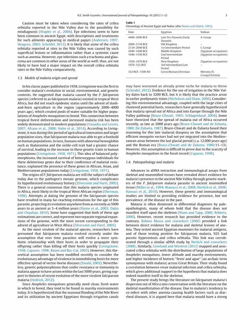
ournal of Paleopathology 10 (2015) 1–12 3
ombfHossclr
1
ccsAayltw2sposopmttM
tiTi1hpyaesa
pabo1oeelmpm
ita
Table 1Chronology of Ancient Egypt and Nubia (after Baines and Malek, 1983).
Date Egyptian Nubian
4400–2600 BCE Late Pre-Dynastic/EarlyDynastic
A-Group
2600–2134 BCE Old Kingdom –2134–2040 BCE 1st Intermediate Period C-Group2040–1640 BCE Middle Kingdom (Egyptian occupation)1640–1550 BCE 2nd Intermediate
Period(Egyptian occupation)
1550–1070 BCE New Kingdom (Egyptian occupation)1070–332 BCE 3rd Intermediate/Late
Period–
N.E. Smith-Guzmán / International J
Caution must be taken when considering the rates of cribrarbitalia reported in the Nile Valley due to the possibility forisdiagnosis (Wapler et al., 2004). Eye infections seem to have
een common in ancient Egypt, with descriptions and treatmentsor such ailments appearing in medical papyri (Andersen, 1997;eagren, 2003; Scheidel, 2012). It is likely that some of the cribrarbitalia reported at sites in the Nile Valley was caused by suchuperficial lesions or inflammation rather than a systemic causeuch as anemia. However, eye infections such a trachoma and glau-oma are common in other areas of the world as well; thus, are notikely to have had a major impact on the overall cribra orbitaliaates in the Nile Valley comparatively.
.3. Models of malaria origin and spread
In his classic paper published in 1958, Livingstone was the first toonsider malaria’s evolution in social, environmental, and geneticontexts. He suggested that malaria caused by the P. falciparumpecies (referred to as falciparum malaria) existed in tropical Westfrica, but did not reach epidemic status until the advent of slash-nd-burn agriculture in the region (approximately 2000–4000ears ago), which created pools of water suitable for higher popu-ations of Anopheles mosquitoes to breed. This connection betweenropical forest deforestation and increased malaria risk has beenidely demonstrated in modern populations (Yasuoka and Levins,
007; Afrane et al., 2008; Hahn et al., 2014). According to Living-tone, it was during this period of agricultural innovation and largeropulation sizes, that falciparum malaria was able to get a footholdn human populations. In turn, humans with abnormal hemoglobinuch as thalassemia and the sickle-cell trait had a greater chancef survival, leading to the increase in these genetic traits in humanopulations (Livingstone, 1958, 1971). This idea of balanced poly-orphisms, the increased survival of heterozygous individuals for
hese deleterious genes due to their conference of malarial resis-ance, explained the presence of these genes in African, Asian, and
editerranean populations today (Livingstone, 1958, 1971).The origins of P. falciparum malaria are still the subject of debate
oday due to the pathogen’s mosaic genome, which complicatests hypothesized evolutionary history (Zilversmit and Hartl, 2005).here is a general consensus that this malaria species originatedn Africa, most likely in the tropical West African region (Sherman,998). Attempts at dating the genome of the falciparum speciesave resulted in many far-reaching estimations for the age of thisarasite, projecting its evolution anywhere from as recently as 5000ears to as ancient as 3–4 million years (Hume et al., 2003; Dattand Chauhan, 2010). Some have suggested that both of these agestimations are correct, and represent two separate regional expan-ions of the genome, with the most recent corresponding to thedvent of agriculture in West Africa (Zilversmit and Hartl, 2005).
As the most virulent of the malarial species, researchers haveresumed that falciparum malaria evolved recently under thessumption that over time parasites will evolve a more sym-iotic relationship with their hosts in order to propagate theirffspring rather than killing off their hosts quickly (Livingstone,958; Capasso, 1998; Baum and Bar-Gal, 2003). However, this the-retical assumption has been modified recently to consider thevolutionary advantage of virulence in immobilizing hosts for moreffective spread of vector-borne diseases (Ewald, 2003). Neverthe-ess, genetic polymorphisms that confer resistance or immunity to
alaria appear to have arisen within the last 5000 years, giving sup-ort to theories of recent evolution of the more virulent falciparumalaria (Hedrick, 2012).
Since Anopheles mosquitoes generally need clean, fresh watern which to breed, they tend to be found in marshy environmentsoday. It is hypothesized that the seasonal flooding of the Nile Rivernd its utilization by ancient Egyptians through irrigation canals
332 BCE–1500 AD Greco/Roman/Christian Meroitic/X-Group/Christian
may have worsened an already prime niche for malaria to thrive(Scheidel, 2012). Evidence for the use of irrigation in the Nile Val-ley dates back to 3200 BCE, but it is likely that the practice arosein earlier predynastic times (Nicholson and Shaw, 2000). Consider-ing this environmental advantage, coupled with the large cities ofclustered potential hosts, researchers have generally hypothesizedthat malaria spread out of Africa and into Europe through the NileValley pathway (Bruce-Chwatt, 1965; Schlagenhauf, 2004). Somehave theorized that the spread of malaria out of Africa occurredrecently, as late as 2000 years ago (Bruce-Chwatt and de Zulueta,1980; De Zulueta, 1987). Bruce-Chwatt and de Zulueta based theirreasoning for this late malarial diaspora on the assumption thatefficient mosquito vectors had not yet migrated into the Mediter-ranean areas between the last glacial period (c.a. 12,000 years ago)and the Roman era (Bruce-Chwatt and de Zulueta, 1980:11–13).However, this assumption is difficult to prove due to the scarcity ofAnopheles mosquitoes in the fossil record (Capasso, 1998).
1.4. Paleopathology and malaria
Advances in aDNA extraction and immunological assays fromskeletal and mummified tissues have revealed direct evidence formalaria’s presence in the ancient Nile Valley through genetic mark-ers of the P. falciparum malaria parasite in ancient mummifiedtissue (Miller et al., 1994; Bianucci et al., 2008; Nerlich et al., 2008;Hawass et al., 2010). However, these genetic and immunologicalstudies are limited to providing evidence for presence, but notprevalence, of the disease in the past.
Malaria is often dismissed in differential diagnoses by pale-opathologists, many of whom hold that the disease does notmanifest itself upon the skeleton (Nunn and Tapp, 2000; Roberts,2000). However, recent research has provided evidence to thecontrary. Rabino Massa and coworkers (2000) provided a linkbetween direct evidence for malaria and skeletal lesions of ane-mia. They tested ancient Egyptian mummies for malarial antigens,and of those testing positive for falciparum malaria, 92% hadporotic hyperostosis and cribra orbitalia. This link was corrob-orated through a similar aDNA study by Nerlich and coworkers(2008). Similarly, Gowland and Western (2012) mapped and asso-ciated cribra orbitalia with the distribution of large populations ofAnopheles mosquitoes, lower altitude and marshy environments,and higher incidence of historic “fever and ague” (an archaic termsynonymous with malaria) across Great Britain. Their study founda correlation between vivax malarial infection and cribra orbitalia,which gives additional support to the hypothesis that malaria doesindeed manifest itself in the skeleton.
The present study brings the literature on falciparum malaria’s
dispersion out of Africa into conversation with the literature on theskeletal manifestation of the disease. Due to malaria’s tendency toco-infect with other anemia-causing health factors such as diar-rheal diseases, it is argued here that malaria would have a strong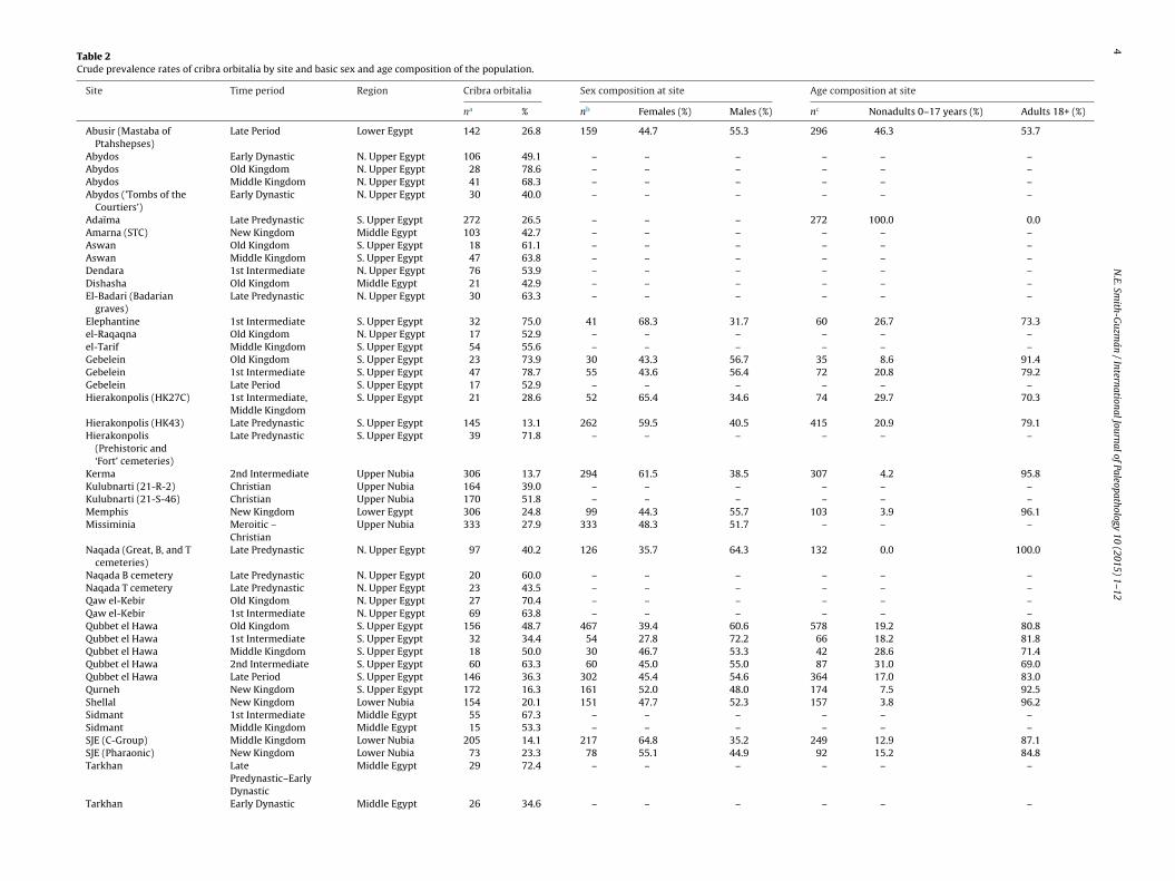
4
N.E.
Smith-G
uzmán
/ International
Journal of
Paleopathology 10
(2015) 1–12
Table 2Crude prevalence rates of cribra orbitalia by site and basic sex and age composition of the population.
Site Time period Region Cribra orbitalia Sex composition at site Age composition at site
na % nb Females (%) Males (%) nc Nonadults 0–17 years (%) Adults 18+ (%)
Abusir (Mastaba ofPtahshepses)
Late Period Lower Egypt 142 26.8 159 44.7 55.3 296 46.3 53.7
Abydos Early Dynastic N. Upper Egypt 106 49.1 – – – – – –Abydos Old Kingdom N. Upper Egypt 28 78.6 – – – – – –Abydos Middle Kingdom N. Upper Egypt 41 68.3 – – – – – –Abydos (’Tombs of the
Courtiers’)Early Dynastic N. Upper Egypt 30 40.0 – – – – – –
Adaïma Late Predynastic S. Upper Egypt 272 26.5 – – – 272 100.0 0.0Amarna (STC) New Kingdom Middle Egypt 103 42.7 – – – – – –Aswan Old Kingdom S. Upper Egypt 18 61.1 – – – – – –Aswan Middle Kingdom S. Upper Egypt 47 63.8 – – – – – –Dendara 1st Intermediate N. Upper Egypt 76 53.9 – – – – – –Dishasha Old Kingdom Middle Egypt 21 42.9 – – – – – –El-Badari (Badarian
graves)Late Predynastic N. Upper Egypt 30 63.3 – – – – – –
Elephantine 1st Intermediate S. Upper Egypt 32 75.0 41 68.3 31.7 60 26.7 73.3el-Raqaqna Old Kingdom N. Upper Egypt 17 52.9 – – – – – –el-Tarif Middle Kingdom S. Upper Egypt 54 55.6 – – – – – –Gebelein Old Kingdom S. Upper Egypt 23 73.9 30 43.3 56.7 35 8.6 91.4Gebelein 1st Intermediate S. Upper Egypt 47 78.7 55 43.6 56.4 72 20.8 79.2Gebelein Late Period S. Upper Egypt 17 52.9 – – – – – –Hierakonpolis (HK27C) 1st Intermediate,
Middle KingdomS. Upper Egypt 21 28.6 52 65.4 34.6 74 29.7 70.3
Hierakonpolis (HK43) Late Predynastic S. Upper Egypt 145 13.1 262 59.5 40.5 415 20.9 79.1Hierakonpolis
(Prehistoric and‘Fort’ cemeteries)
Late Predynastic S. Upper Egypt 39 71.8 – – – – – –
Kerma 2nd Intermediate Upper Nubia 306 13.7 294 61.5 38.5 307 4.2 95.8Kulubnarti (21-R-2) Christian Upper Nubia 164 39.0 – – – – – –Kulubnarti (21-S-46) Christian Upper Nubia 170 51.8 – – – – – –Memphis New Kingdom Lower Egypt 306 24.8 99 44.3 55.7 103 3.9 96.1Missiminia Meroitic –
ChristianUpper Nubia 333 27.9 333 48.3 51.7 – – –
Naqada (Great, B, and Tcemeteries)
Late Predynastic N. Upper Egypt 97 40.2 126 35.7 64.3 132 0.0 100.0
Naqada B cemetery Late Predynastic N. Upper Egypt 20 60.0 – – – – – –Naqada T cemetery Late Predynastic N. Upper Egypt 23 43.5 – – – – – –Qaw el-Kebir Old Kingdom N. Upper Egypt 27 70.4 – – – – – –Qaw el-Kebir 1st Intermediate N. Upper Egypt 69 63.8 – – – – – –Qubbet el Hawa Old Kingdom S. Upper Egypt 156 48.7 467 39.4 60.6 578 19.2 80.8Qubbet el Hawa 1st Intermediate S. Upper Egypt 32 34.4 54 27.8 72.2 66 18.2 81.8Qubbet el Hawa Middle Kingdom S. Upper Egypt 18 50.0 30 46.7 53.3 42 28.6 71.4Qubbet el Hawa 2nd Intermediate S. Upper Egypt 60 63.3 60 45.0 55.0 87 31.0 69.0Qubbet el Hawa Late Period S. Upper Egypt 146 36.3 302 45.4 54.6 364 17.0 83.0Qurneh New Kingdom S. Upper Egypt 172 16.3 161 52.0 48.0 174 7.5 92.5Shellal New Kingdom Lower Nubia 154 20.1 151 47.7 52.3 157 3.8 96.2Sidmant 1st Intermediate Middle Egypt 55 67.3 – – – – – –Sidmant Middle Kingdom Middle Egypt 15 53.3 – – – – – –SJE (C-Group) Middle Kingdom Lower Nubia 205 14.1 217 64.8 35.2 249 12.9 87.1SJE (Pharaonic) New Kingdom Lower Nubia 73 23.3 78 55.1 44.9 92 15.2 84.8Tarkhan Late
Predynastic–EarlyDynastic
Middle Egypt 29 72.4 – – – – – –
Tarkhan Early Dynastic Middle Egypt 26 34.6 – – – – – –
N.E. Smith-Guzmán / International Journa
Tabl
e
2
(Con
tinu
ed)
Site
Tim
e
per
iod
Reg
ion
Cri
bra
orbi
tali
aSe
x
com
pos
itio
n
at
site
Age
com
pos
itio
n
at
site
na%
nbFe
mal
es
(%)
Mal
es
(%)
ncN
onad
ult
s
0–17
year
s
(%)
Ad
ult
s
18+
(%)
Tell
el-D
ab’a
2nd
Inte
rmed
iate
Low
er
Egyp
t41
26.8
120
40.8
59.2
257
48.1
51.9
Theb
es-W
est
New
Kin
gdom
–La
te
Peri
odS.
Up
per
Egyp
t
168
29.2
187
45.5
54.5
167
20.2
79.8
Theb
es-W
est
(Val
ley
ofth
e
Qu
een
s)R
oman
S.
Up
per
Egyp
t21
2
18.4
288
48.0
52.0
1070
19.2
80.8
Tom
bos
New
Kin
gdom
Up
per
Nu
bia
83
10.8
85
59.5
40.5
100
15.0
85.0
Wad
i Hal
fa
(24I
3)
X-G
rou
p
Up
per
Nu
bia
45
26.7
30
50.0
50.0
54
29.6
70.4
Wad
i Hal
fa
(6B
13)
Ch
rist
ian
Up
per
Nu
bia
28
14.3
–
–
–
37
32.4
67.6
Wad
i Hal
fa
(6B
16)
Mer
oiti
c
Up
per
Nu
bia
62
11.3
48
58.3
41.7
129
17.1
82.9
Wad
i Hal
fa
(6G
8)C
hri
stia
n
Up
per
Nu
bia
29
13.8
–
–
–
33
39.4
60.6
Wad
i Hal
fa
(NA
X)
X-G
rou
p
Up
per
Nu
bia
127
26.7
106
56.6
43.4
164
14.1
85.9
aTo
tal i
nd
ivid
ual
s
at
the
site
wit
h
obse
rvab
le
orbi
ts.
bTo
tal a
du
lt
ind
ivid
ual
s
at
the
site
wh
ose
sex
was
det
erm
ined
.c
Tota
l in
div
idu
als
at
the
site
wh
ose
age
was
det
erm
ined
.
l of Paleopathology 10 (2015) 1–12 5
impact on cribra orbitalia formation in the Nile Valley. Using thevariability in cribra orbitalia frequencies among ancient Egyptianand Nubian remains as a proxy for malarial infection, this studytests the theoretical Dynastic Egyptian time frame for the spread ofmalaria up the Nile Valley and out of Africa. If malaria did spreadinto Egypt during the Dynastic period, an increasing trend in cribraorbitalia frequency over time from South to North in the Nile Valleywas predicted.
2. Materials and methods
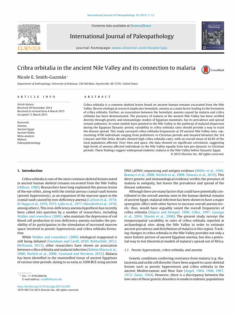
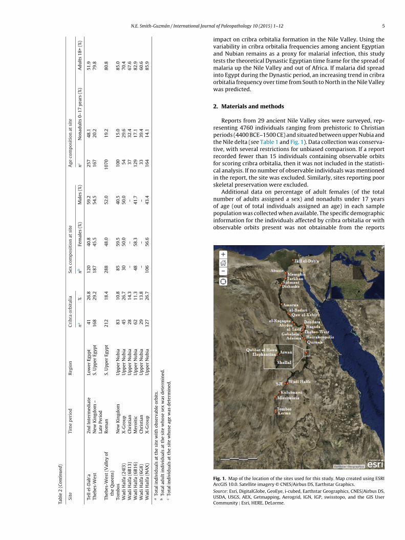
Reports from 29 ancient Nile Valley sites were surveyed, rep-resenting 4760 individuals ranging from prehistoric to Christianperiods (4400 BCE–1500 CE) and situated between upper Nubia andthe Nile delta (see Table 1 and Fig. 1). Data collection was conserva-tive, with several restrictions for unbiased comparison. If a reportrecorded fewer than 15 individuals containing observable orbitsfor scoring cribra orbitalia, then it was not included in the statisti-cal analysis. If no number of observable individuals was mentionedin the report, the site was excluded. Similarly, sites reporting poorskeletal preservation were excluded.
Additional data on percentage of adult females (of the totalnumber of adults assigned a sex) and nonadults under 17 years
of age (out of total individuals assigned an age) in each samplepopulation was collected when available. The specific demographicinformation for the individuals affected by cribra orbitalia or withobservable orbits present was not obtainable from the reportsFig. 1. Map of the location of the sites used for this study. Map created using ESRIArcGIS 10.0. Satellite imagery © CNES/Airbus DS, Earthstar Graphics.
Source: Esri, DigitalGlobe, GeoEye, i-cubed, Earthstar Geographics, CNES/Airbus DS,USDA, USGS, AEX, Getmapping, Aerogrid, IGN, IGP, swisstopo, and the GIS UserCommunity | Esri, HERE, DeLorme.
6 N.E. Smith-Guzmán / International Journal of Paleopathology 10 (2015) 1–12
F with tv
sttt
FE
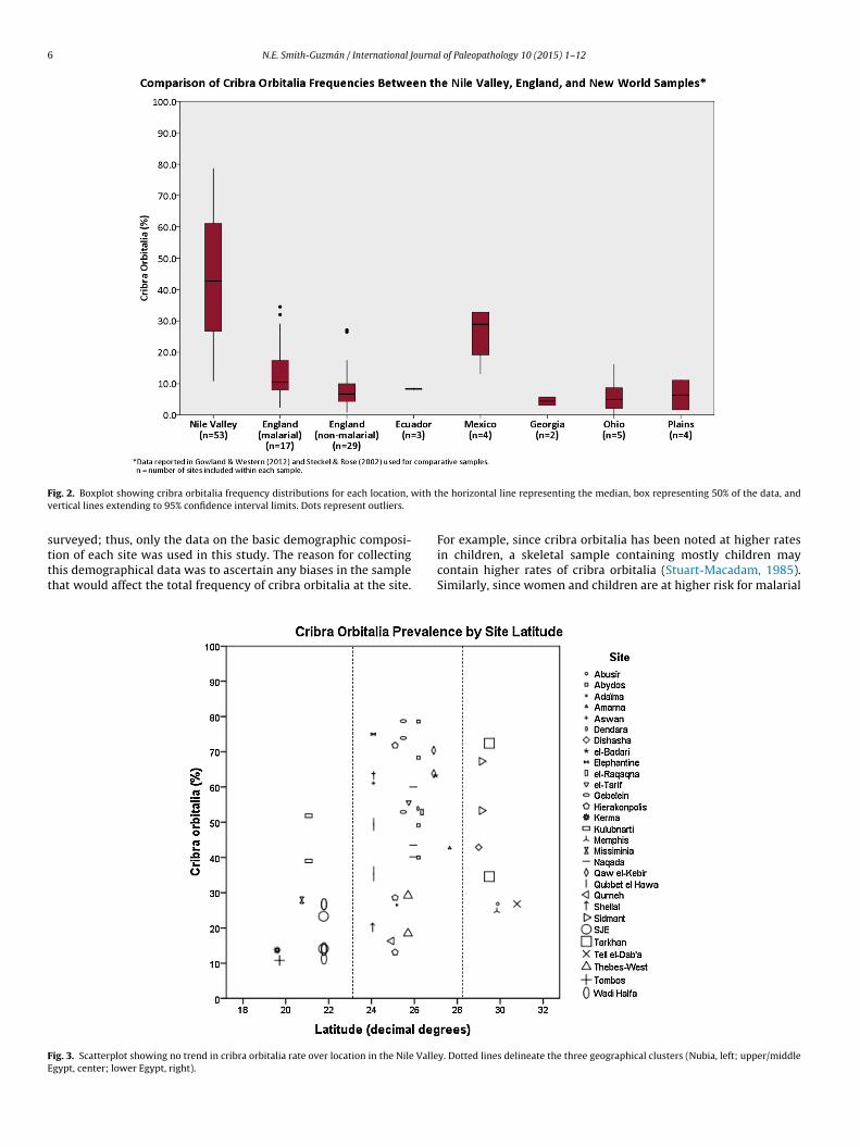
ig. 2. Boxplot showing cribra orbitalia frequency distributions for each location,
ertical lines extending to 95% confidence interval limits. Dots represent outliers.
urveyed; thus, only the data on the basic demographic composi-
ion of each site was used in this study. The reason for collectinghis demographical data was to ascertain any biases in the samplehat would affect the total frequency of cribra orbitalia at the site.ig. 3. Scatterplot showing no trend in cribra orbitalia rate over location in the Nile Vallegypt, center; lower Egypt, right).
he horizontal line representing the median, box representing 50% of the data, and
For example, since cribra orbitalia has been noted at higher rates
in children, a skeletal sample containing mostly children maycontain higher rates of cribra orbitalia (Stuart-Macadam, 1985).Similarly, since women and children are at higher risk for malarialy. Dotted lines delineate the three geographical clusters (Nubia, left; upper/middle
N.E. Smith-Guzmán / International Journal of Paleopathology 10 (2015) 1–12 7
e in t
ieapBoc
iawf
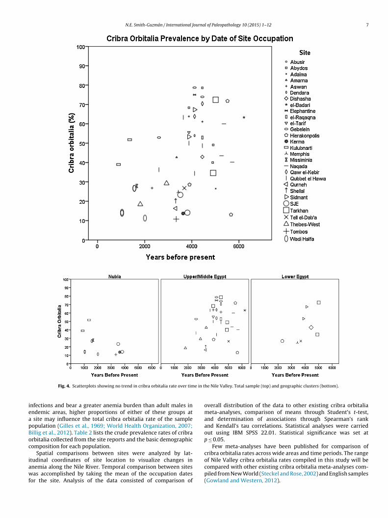
Fig. 4. Scatterplots showing no trend in cribra orbitalia rate over tim
nfections and bear a greater anemia burden than adult males inndemic areas, higher proportions of either of these groups at
site may influence the total cribra orbitalia rate of the sampleopulation (Gilles et al., 1969; World Health Organization, 2007;illig et al., 2012). Table 2 lists the crude prevalence rates of cribrarbitalia collected from the site reports and the basic demographicomposition for each population.
Spatial comparisons between sites were analyzed by lat-
tudinal coordinates of site location to visualize changes innemia along the Nile River. Temporal comparison between sitesas accomplished by taking the mean of the occupation datesor the site. Analysis of the data consisted of comparison of
he Nile Valley. Total sample (top) and geographic clusters (bottom).
overall distribution of the data to other existing cribra orbitaliameta-analyses, comparison of means through Student’s t-test,and determination of associations through Spearman’s rankand Kendall’s tau correlations. Statistical analyses were carriedout using IBM SPSS 22.01. Statistical significance was set atp ≤ 0.05.
Few meta-analyses have been published for comparison ofcribra orbitalia rates across wide areas and time periods. The range
of Nile Valley cribra orbitalia rates compiled in this study will becompared with other existing cribra orbitalia meta-analyses com-piled from New World (Steckel and Rose, 2002) and English samples(Gowland and Western, 2012).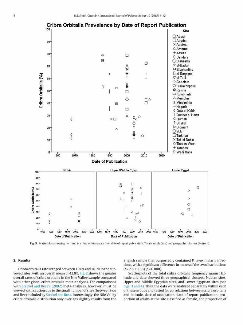
8 N.E. Smith-Guzmán / International Journal of Paleopathology 10 (2015) 1–12
of rep
3
vowwvac
Fig. 5. Scatterplots showing no trend in cribra orbitalia rate over date
. Results
Cribra orbitalia rates ranged between 10.8% and 78.7% in the sur-eyed sites, with an overall mean of 42.8%. Fig. 2 shows the greaterverall rates of cribra orbitalia in the Nile Valley sample comparedith other global cribra orbitalia meta-analyses. The comparisonsith Steckel and Rose’s (2002) meta-analyses, however, must be
iewed with caution due to the small number of sites (between twond five) included by Steckel and Rose. Interestingly, the Nile Valleyribra orbitalia distribution only overlaps slightly results from the
ort publication. Total sample (top) and geographic clusters (bottom).
English sample that purportedly contained P. vivax malaria infec-tions, with a significant difference in means of the two distributions(t = 7.898 (58), p = 0.000).
Scatterplots of the total cribra orbitalia frequency against lat-itude and date showed three geographical clusters: Nubian sites,Upper and Middle Egyptian sites, and Lower Egyptian sites (seeFigs. 3 and 4). Thus, the data were analyzed separately within each
of these groups and tested for correlations between cribra orbitaliaand latitude, date of occupation, date of report publication, pro-portion of adults at the site classified as female, and proportion of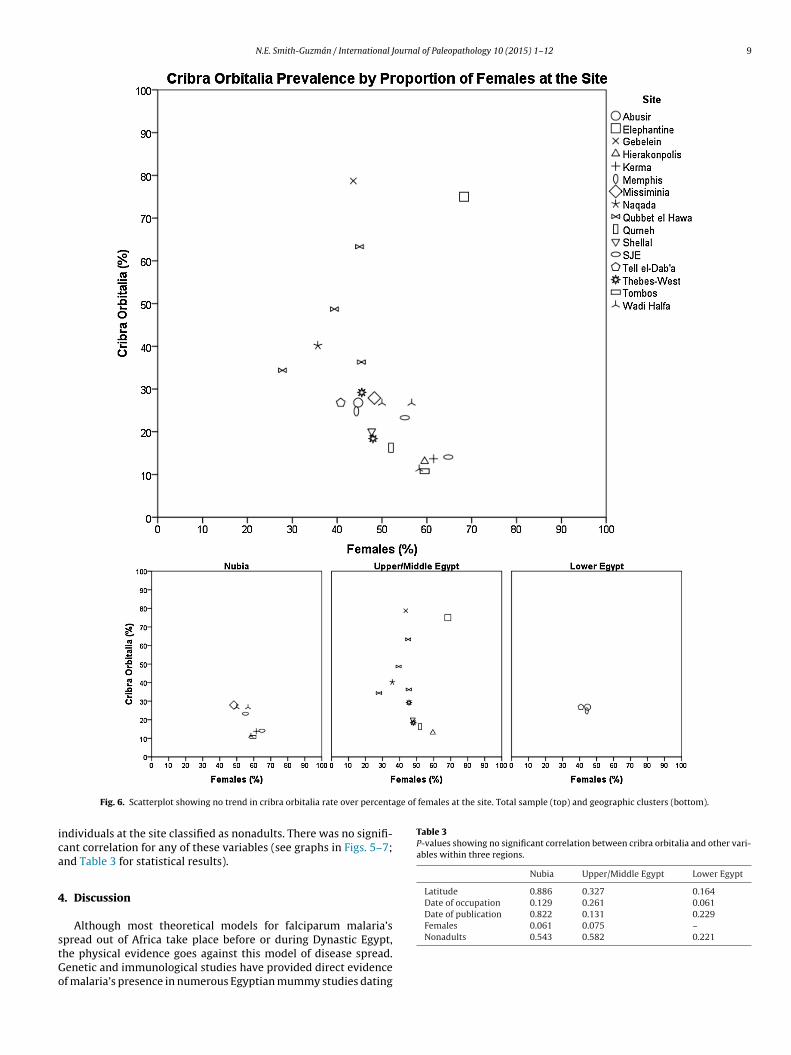
N.E. Smith-Guzmán / International Journal of Paleopathology 10 (2015) 1–12 9
ge of females at the site. Total sample (top) and geographic clusters (bottom).
ica
4
stGo
Table 3P-values showing no significant correlation between cribra orbitalia and other vari-ables within three regions.
Nubia Upper/Middle Egypt Lower Egypt
Latitude 0.886 0.327 0.164Date of occupation 0.129 0.261 0.061Date of publication 0.822 0.131 0.229Females 0.061 0.075 –
Fig. 6. Scatterplot showing no trend in cribra orbitalia rate over percenta
ndividuals at the site classified as nonadults. There was no signifi-ant correlation for any of these variables (see graphs in Figs. 5–7;nd Table 3 for statistical results).
. Discussion
Although most theoretical models for falciparum malaria’s
pread out of Africa take place before or during Dynastic Egypt,he physical evidence goes against this model of disease spread.enetic and immunological studies have provided direct evidencef malaria’s presence in numerous Egyptian mummy studies datingNonadults 0.543 0.582 0.221
10 N.E. Smith-Guzmán / International Journal of Paleopathology 10 (2015) 1–12
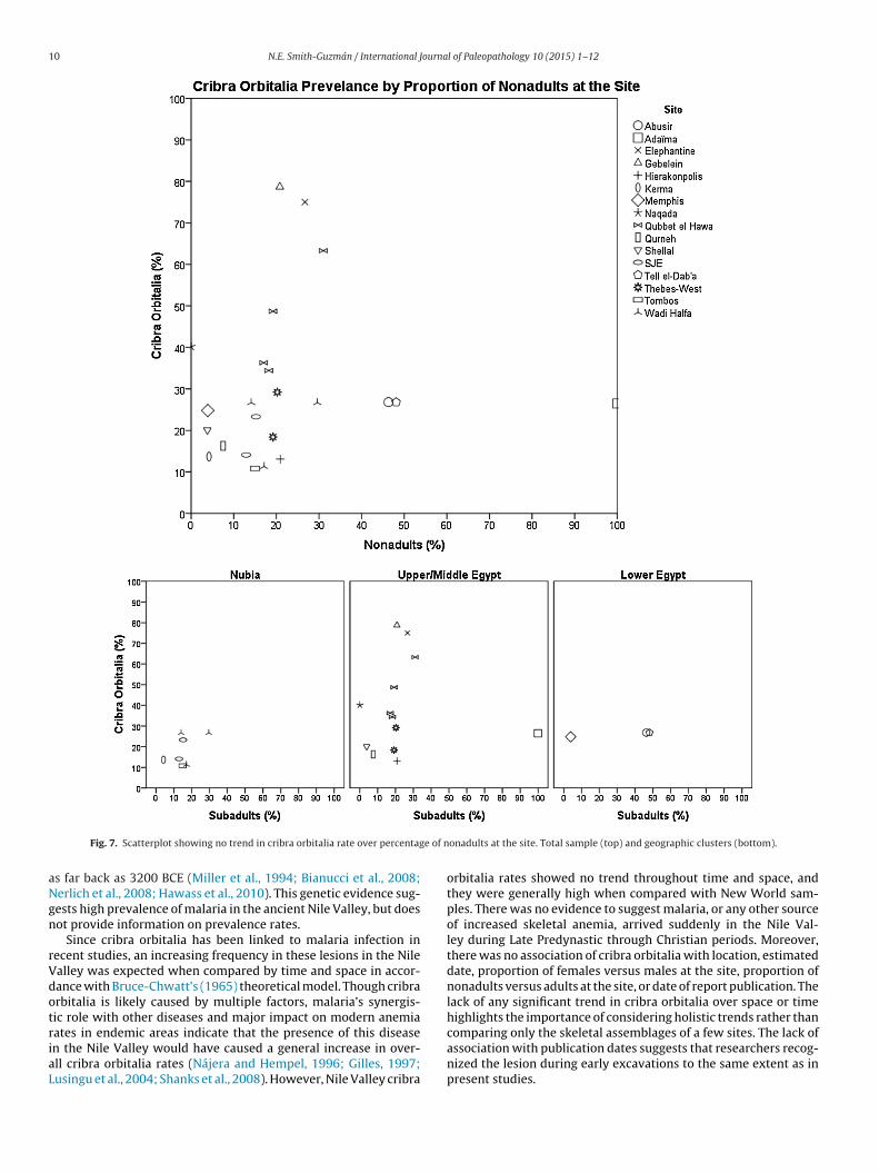
e of n
aNgn
rVdotriaL
Fig. 7. Scatterplot showing no trend in cribra orbitalia rate over percentag
s far back as 3200 BCE (Miller et al., 1994; Bianucci et al., 2008;erlich et al., 2008; Hawass et al., 2010). This genetic evidence sug-ests high prevalence of malaria in the ancient Nile Valley, but doesot provide information on prevalence rates.
Since cribra orbitalia has been linked to malaria infection inecent studies, an increasing frequency in these lesions in the Nilealley was expected when compared by time and space in accor-ance with Bruce-Chwatt’s (1965) theoretical model. Though cribrarbitalia is likely caused by multiple factors, malaria’s synergis-ic role with other diseases and major impact on modern anemiaates in endemic areas indicate that the presence of this disease
n the Nile Valley would have caused a general increase in over-ll cribra orbitalia rates (Nájera and Hempel, 1996; Gilles, 1997;usingu et al., 2004; Shanks et al., 2008). However, Nile Valley cribraonadults at the site. Total sample (top) and geographic clusters (bottom).
orbitalia rates showed no trend throughout time and space, andthey were generally high when compared with New World sam-ples. There was no evidence to suggest malaria, or any other sourceof increased skeletal anemia, arrived suddenly in the Nile Val-ley during Late Predynastic through Christian periods. Moreover,there was no association of cribra orbitalia with location, estimateddate, proportion of females versus males at the site, proportion ofnonadults versus adults at the site, or date of report publication. Thelack of any significant trend in cribra orbitalia over space or timehighlights the importance of considering holistic trends rather thancomparing only the skeletal assemblages of a few sites. The lack of
association with publication dates suggests that researchers recog-nized the lesion during early excavations to the same extent as inpresent studies.
ourna
otprltaiTolawia
m(caEpFthrtNBhti
coiTEhaDaaw
oioartctitlno
5
EV
N.E. Smith-Guzmán / International J
This study has three implications for interpreting the etiologyf cribra orbitalia and health in the Nile Valley. First, the failureo correlate cribra orbitalia frequency with site-specific age pro-ortions suggests that the main cause of the high cribra orbitaliaates is not age-specific. Cribra orbitalia is generally considered aesion formed in childhood, due to the principal location of ery-hropoiesis in the cranium, thinner cranial bones, weaning stresses,nd perhaps inadequate vitamin intake necessary for their grow-ng bodies (Mittler and Van Gerven, 1994; Walker et al., 2009).he results of this study counter this assumption, as the amountf nonadults at the site did not affect cribra orbitalia rates. Thisack of age-controlled prevalence suggests childhood factors suchs diet, exposure to parasitic worms, or nutritional stress caused byeaning did not have a large effect on the formation of this lesion
n the Nile Valley. Instead, the main contributing factor seems to ben infectious cause that affects all age groups indiscriminately.
Second, assuming that cribra orbitalia is indeed indicative ofalarial infection (as suggested by Rabino Massa and coworkers
2000) and Gowland and Western (2012)), the ubiquity of highribra orbitalia rates shown in this study suggest this disease had
general high prevalence in the Nile Valley long before Dynasticgypt. This implication is supported by the aDNA evidence, and sup-orts earlier theoretical timelines for malaria’s spread out of Africa.rom the differential diagnosis of the potential causes of anemia inhe Nile Valley, it seems reasonable to assume that malaria wouldave had a great impact on the frequencies of cribra orbitalia in theegion. Thus, if the high cribra orbitalia rates in the Nile Valley areantamount to high malaria rates, malaria must have spread up theile Valley and out of Africa before the Badarian period (4400–4000CE), which is the earliest date used in this study. Alternatively, thisigher anemia burden in the Nile Valley sites could simply reflecthe multitude of factors combining to cause and aggregate anemian this region.
Third, Gowland and Western (2012) showed an association ofribra orbitalia with P. vivax malaria infection in their meta-analysisf English sites, while the sites used in this study would havencluded individuals infected with the P. falciparum malaria species.he mean rates of the cribra orbitalia frequencies found in thenglish study and this Nile Valley study differed significantly, per-aps reflecting the higher levels of severe malarial anemia generallyssociated with P. falciparum infections (Billig et al., 2012; Botez andoughty, 2014). This finding is important because it suggests thatlthough P. vivax infections tend to involve a chronic, but less severenemia than P. falciparum infections, the latter species is associatedith higher rates of skeletal responses to infection.
One of the main limitations of this study involved the clusteringf many dates and locations of sites, leading to a greater variabil-ty of cribra orbitalia frequencies in these clusters simply becausef the greater number of sites. This limitation forced the statisticalnalysis to follow the clustering by separation into three groups byegional position. This study was also limited by the many siteshat had to be excluded because they reported the presence ofribra orbitalia and porotic hyperostosis together, combined underhe name porotic hyperostosis. Nevertheless, the great variationn cribra orbitalia rates of sites included in this study (i.e. some ofhe lowest and some of the highest rates found within very similaratitudes and time periods) is such that including more sites willot change the absence of a significant association between cribrarbitalia rates and date or latitude.
. Conclusion
This study tested a method of identifying malaria in the Nearast, and shed new light on the patterns of health in the ancient Nilealley by providing a holistic view of anemia present throughout
l of Paleopathology 10 (2015) 1–12 11
time and space. In compiling cribra orbitalia rates from sites alongthe Nile Valley from various time periods, no significant associationwas shown between cribra orbitalia rates and date or latitude. Fur-thermore, cribra orbitalia rates were not affected by the proportionof females or nonadults in the sample, or by the date of site reportpublication. These results support the notion of a major infectiouscausative factor for cribra orbitalia in the ancient Nile Valley, andadd credence to previous studies associating cribra orbitalia withmalaria. With Gowland and Western’s (2012) English malarial sam-ple, this study provided the first interspecific (P. vivax versus P.falciparum) malaria comparison through large-scale cribra orbitaliafrequency comparisons across many sites.
The interpretations of this study rely on the assumption thatthe hemolytic anemia caused by malaria is responsible for highcribra orbitalia rates, but they do not account for additional skeletallesions that may also be caused by malarial infection. To identifythese potential additional skeletal lesions of malaria, future studiesare planned involving a clinical comparison in a modern skeletalcollection from an endemic malarial area, which will provide betterdiagnostic criteria for malaria.
Acknowledgments
I thank Jerry Rose for his assistance in the designing this projectand for providing a large number of the site reports from whichthe data for this study were gleaned. Additional thanks go to MikePlavcan for advice regarding the statistical analysis. I am gratefulto the IJPP Associate Editor and two anonymous reviewers for theirhelpful comments.
References
Afrane, Y.A., Little, T.J., Lawson, B.W., Githeko, A.K., Yan, G., 2008. Deforestation andvectorial capacity of Anopheles gambiae Giles mosquitoes in malaria transmis-sion, Kenya. Emerg. Infect. Dis. 14, 1533–1538.
Al-Aabassi, A., Murad, B.A., 2005. Presacral extramedullary hematopoiesis: a diag-nostic confusion concerning a rare presentation. Med. Princ. Pract. 14, 358–362.
Alvrus, A.B., (Unpublished Ph.D. dissertation) 2006. The Conqueror Worm: Schisto-somiasis in Ancient Nubia. University of Arizona.
Andersen, S.R., 1997. The eye and its diseases in Ancient Egypt. Acta Ophthalmol.Scand. 75, 338–344.
Angel, J.L., 1964. Osteoporosis: thalassemia? Am. J. Phys. Anthropol. 22, 369–373.Angel, J.L., 1966. Porotic hyperostosis, anemias, malarias, and marshes in the pre-
historic eastern Mediterranean. Science 153, 760–763.Angel, J.L., 1967. Porotic hyperostosis or osteoporosis symmetrica. In: Brothwell,
D.R., Sandison, A.T. (Eds.), Disease in Antiquity. Charles C Thomas, Springfield,Illinois, pp. 378–389.
Angel, J.L., 1972. Ecology and population in the eastern Mediterranean. WorldArchaeol. 4, 88–105.
Baines, J., Malek, J., 1983. Atlas of Ancient Egypt. Facts on File Publications, New York.Baum, J., Bar-Gal, G.K., 2003. The emergence and co-evolution of human pathogens.
In: Greenblatt, C.L., Spigelman, M. (Eds.), Emerging Pathogens: the Archaeology,Ecology, and Evolution of Infectious Disease. Oxford University Press, Oxford,pp. 378–389.
Bianucci, R., Mattutino, G., Lallo, R., Charlier, P., Jouin-Spriet, H., Peluso, A., Higham,T., Torre, C., Rabino Massa, E., 2008. Immunological evidence of Plasmodium fal-ciparum infection in an Egyptian child mummy from the Early Dynastic Period.J. Archaeol. Sci. 35, 1880–1885.
Billig, E.M.W., O’Meara, W.P., Riley, E.M., McKenzie, F.E., 2012. Developmental allom-etry and paediatric malaria. Malar. J. 11, 1–13.
Botez, G.I., Doughty, L., 2014. Life threatening tropical infections. In: Wheeler, D.S.(Ed.), Pediatric Critical Care Medicine. Springer, London, pp. 577–605.
Boyd, R., 1999. The Coming of the Spirit of Pestilence: Introduced Infectious Diseasesand Population Decline Among Northwest Coast Indians, 1774–1874. Universityof Washington Press, Seattle.
Brier, B., 2004. Infectious diseases in ancient Egypt. Infect. Dis. Clin. N. Am. 18, 17–27.Bruce-Chwatt, L.J., de Zulueta, J., 1980. The Rise and Fall of Malaria in Europe: A
Historico-Epidemiological Study. Oxford University Press, Oxford, UK.Bruce-Chwatt, L.J., 1965. Paleogenesis and paleo-epidemiology of primate malaria.
Bull. World Health Org. 32, 363–387.Caffey, J., 1937. Skeletal changes in the chronic hemolytic anemias (erythroblastic
anemia, sickle cell anemia and chronic hemolytic icterus). Am. J. Roentgenol. 37,293–334.
Capasso, L., 1998. The origin of human malaria. Int. J. Anthropol. 13, 165–175.Carlson, D.S., Armelagos, G.J., van Gerven, D.P., 1974. Factors influencing the etiology
of cribra orbitalia in prehistoric Nubia. J. Hum. Evol. 3, 405–410.
1 ourna
D
D
D
D
D
D
E
E
E
G
G
G
G
H
H
H
H
H
HH
H
H
K
L
L
L
L
L
M
450–460.
2 N.E. Smith-Guzmán / International J
’Anastasio, R., Staniscia, T., Milia, M.L., Manzoli, L., Capasso, L., 2011. Origin, evolu-tion and paleoepidemiology of brucellosis. Epidemiol. Infect 139, 149–156.
’Souza, B., Parthasarathy, R., Sreekantha D’Souza, V., 2011. Acid phosphatase as amarker in malaria. Indian J. Clin. Biochem. 26, 396–399.
atta, N., Chauhan, V.S., 2010. Origin and evolution of human malaria parasite, P.falciparum and P. vivax. In: Sharma, V.P. (Ed.), Nature at Work: The Ongoing Sagaof Evolution. Springer, New Delhi, pp. 307–317.
eGusta, D., 2009. Cribra orbitalia: a non-human primate perspective. Int. J. Osteoar-chaeol. 20, 597–602.
e Zulueta, J., 1987. Changes in the geographical distribution of malaria throughouthistory. Parassitologia 29, 193–203.
uffy, J., 1952. Eighteenth-Century Carolina health conditions. J. South. Hist. 18,289–302.
l-Najjar, M.Y., Lozoff, B., Ryan, D.J., 1975. The paleoepidemiology of porotic hyper-ostosis in the American Southwest: radiological and ecological considerations.Am. J. Roentgenol. Radium Ther. Nucl. Med. 125, 918–924.
l-Najjar, M.Y., Ryan, D.J., Turner, C.G., Lozoff, B., 1976. The etiology of porotic hyper-ostosis among the prehistoric and historic Anasazi Indians of southwesternUnited States. Am. J. Phys. Anthropol. 44, 477–487.
wald, P.W., 2003. Evolution and ancient diseases: the roles of genes, germs,and transmission modes. In: Greenblatt, C.L., Spigelman, M. (Eds.), Emerg-ing Pathogens: The Archaeology, Ecology, and Evolution of Infectious Disease.Oxford University Press, Oxford.
illes, H.M., Lawson, J.B., Sibelas, M., Voller, A., Allan, N., 1969. Malaria, anaemia andpregnancy. Ann. Trop. Med. Parasitol. 63, 245–263.
illes, H.M., 1997. Pathology of malaria. In: Carosi, G., Castelli, F. (Eds.), Handbookof Malaria Infection in the Tropics. Associazione Italiana Amici di R. Follereau,Bologna, Italy.
len-Haduch, E., Szostek, K., Głab, H., 1997. Cribra orbitalia and trace element con-tent in human teeth from Neolithic and Early Bronze Age graves in SouthernPoland. Am. J. Phys. Anthropol. 103, 201–207.
owland, R.L., Western, A.G., 2012. Morbidity in the marshes: using spatial epidemi-ology to investigate skeletal evidence for malaria in Anglo-Saxon England (AD410-1050). Am. J. Phys. Anthropol. 147, 301–311.
ahn, M.B., Gangnon, R.E., Barcellos, C., Asner, G.P., Patz, J.A., 2014. Influence ofdeforestation, logging, and fire on malaria in the Brazilian Amazon. PLOS ONE 9,e85725.
awass, Z., Gad, Y.Z., Ismail, S., Khairat, R., Fathalla, D., Hasan, N., Ahmed, A., Elleithy,H., Ball, M., Gaballah, F., Wasef, S., Fateen, M., Amer, H., Gostner, P., Selim, A., Zink,A., Pusch, C.M., 2010. Ancestry and pathology in King Tutankhamun’s family.JAMA 303, 638–647.
eagren, B., 2003. Water related diseases in Ancient Egypt. In: L’acqua nell’anticoEgitto: vita, rigenerazione, incantesimo, medicamento: Proceedings of the FirstInternational Conference for Young Egyptologists, October 15–18, Italy, Chian-ciano Terme, L’Erma di Bretschneider, pp. 151–157.
edrick, P.W., 2012. Resistance to malaria in humans: the impact of strong, recentselection. Malar. J. 11, 1–7.
engen, O.P., 1971. Cribra Orbitalia: Pathogenesis and Probable Etiology. Homo, USA,pp. 57–75.
illson, S.W., 1980. Chronic anaemias in the Nile Valley. Masca J. 1, 172–174.olland, T.D., O’Brien, M.J., 1997. Parasites, porotic hyperostosis, and the implica-
tions of changing perspectives. Am. Antiq. 62, 183–193.rdlicka, A., 1914. Anthropological work in Peru in 1913, with notes on the pathology
of the Ancient Peruvians. Smithsonian Miscellaneous Collections, vol. 61., pp.1–69.
ume, J.C.C., Lyons, E.J., Day, K.P., 2003. Malaria in antiquity: a genetics perspective.World Archaeol. 35, 180–192.
assebaum, N.J., Jasrasaria, R., Naghavi, M., Wulf, S.K., Johns, N., Lozano, R., Regan,M., Weatherall, D., Chou, D.P., Eisele, T.P., Flaxman, S.R., Pullan, R.L., Brooker, S.J.,Murray, C.J.L., 2014. A systematic analysis of global anemia burden from 1990to 2010. Blood 123, 615–624.
allo, J.W., Armelagos, G.J., Mensforth, R.P., 1977. The role of diet, disease, and phys-iology in the origin of porotic hyperostosis. Hum. Biol. 49, 471–483.
ivingstone, F.B., 1958. The distribution of the sickle cell gene in Liberia. Am. J. HumanGenet. 10, 33–41.
ivingstone, F.B., 1971. Malaria and human polymorphisms. Ann. Rev. Genet. 5,33–64.
opes, L., Nicolino, R., Haddad, J., 2010. Brucellosis- risk factors and prevalence: areview. The Open Veterinary Science Journal 4, 72–84.
usingu, J.P.A., Vestergaard, L.S., Mmbando, B.P., Drakeley, C.J., Jones, C., Akida, J.,Savaeli, Z.X., Kitua, A.Y., Lemnge, M.M., Theander, T.G., 2004. Malaria morbidity
and immunity among residents of villages with different Plasmodium falciparumtransmission intensity in North-Eastern Tanzania. Malar. J. 3, 26.cClure, S.B., García, O., Roca de Togores, C., Culleton, B.J., Kennett, D.J., 2011.Osteological and paleodietary investigation of burials from Cova de la Pastora,Alicante, Spain. J. Archaeol. Sci. 38, 420–428.
l of Paleopathology 10 (2015) 1–12
McIlvaine, B.K., 2013. Implications of reappraising the iron-deficiency anemiahypothesis. Int. J. Osteoarchaeol., http://dx.doi.org/10.1002/oa.2383.
Mensforth, R.P., Lovejoy, C.O., Lallo, J.W., Armelagos, G.J., 1978. The role of constitu-tional factors, diet, and infectious disease in the etiology of porotic hyperostosisand periosteal reactions in prehistoric infants and children. Med. Anthropol. 2,1–59.
Miller, R.L., Ikram, S., Armelagos, G.J., Walker, R., Harer, W.B., Shiff, C.J., Baggett, D.,Carrigan, M., Maret, S.M., 1994. Diagnosis of Plasmodium falciparum infections inmummies using the rapid manual ParaSightTM-F test. Trans. R. Soc. Trop. Med.Hyg. 88, 31–32.
Mittler, D.M., Van Gerven, D.P., 1994. Developmental, diachronic, and demographicanalysis of cribra orbitalia in the medieval Christian populations of Kulubnarti.Am. J. Phys. Anthropol. 93, 287–297.
Moreau, R., Tshikudi Malu, D., Dumais, M., Dalko, E., Gaudreault, V., Roméro, H.,Martineau, C., Kevorkova, O., Dardon, J.S., Dodd, E.L., Bohle, D.S., Scorza, T., 2012.Alterations in bone and erythropoiesis in hemolytic anemia: comparative studyin bled, phenylhydrazine-treated and Plasmodium-infected mice. PLoS ONE 7,e46101.
Nájera, J.A., Hempel, J., 1996. The burden of malaria. In: Div. of Control of Trop-ical Disease, Malaria Unit. World Health Organization, Geneva (Report No.CTD/MAL/96.10).
Nerlich, A.G., Schraut, B., Dittrich, S., Jelinek, T., Zink, A.R., 2008. Plasmodium falci-parum in Ancient Egypt. Emerg. Infect. Dis. 14, 1317–1319.
Nicholson, P.T., Shaw, I., 2000. Ancient Egyptian Materials and Technology. Cam-bridge University Press, Cambridge.
Nunn, J.F., Tapp, E., 2000. Tropical diseases in Ancient Egypt. Trans. R. Soc. Trop. Med.Hyg. 94, 147–153.
Oxenham, M.F., Cavill, I., 2010. Porotic hyperostosis and cribra orbitalia: the ery-thropoietic response to iron-deficiency anaemia. Anthropol. Sci. 118, 199–200.
Rabino Massa, E., Cerutti, N., Marin, A., Savoia, D., 2000. Malaria in Ancient Egypt:paleoimmunological investigation on Predynastic mummified remains. Chun-gará 32, 7–9.
Roberts, C.A., 2000. Infectious disease in biocultural perspective: past, presentand future work in Britain. In: Cox, M., Mays, S. (Eds.), Human Osteology:In Archaeology and Forensic Science. Cambridge University Press, Cambridge,pp. 145–162.
Rothschild, B., 2012. Extirpolation of the mythology that porotic hyperostosis iscaused by iron deficiency secondary to dietary shift to maize. Adv. Anthropol. 2,157–160.
Scheidel, W., 2012. Age and health. In: Riggs, C. (Ed.), The Oxford Handbook of RomanEgypt. Oxford University Press, Oxford, UK, pp. 305–316.
Schlagenhauf, P., 2004. Malaria: from prehistory to present. Infect. Dis. Clin. N. Am.18, 189–205.
Shanks, G.D., Hay, S.I., Bradley, D.J., 2008. Malaria’s indirect contribution to all-causemortality in the Andaman Islands during the colonial era. Lancet Infect. Dis. 8,564–570.
Sherman, I.W. (Ed.), 1998. Malaria: Parasite Biology, Pathogenesis and Protection.ASM Press, Washington, DC.
Steckel, R.H., Rose, J.C. (Eds.), 2002. The Backbone of History: Health and Nutritionin the Western Hemisphere, vol. 2. Cambridge University Press, Cambridge.
Stuart-Macadam, P., 1985. Porotic hyperostosis: representative of a childhood con-dition. Am. J. Phys. Anthropol. 66, 391–398.
Stuart-Macadam, P., 1987. Porotic hyperostosis: new evidence to support the anemiatheory. Am. J. Phys. Anthropol. 74, 521–526.
Stuart-Macadam, P., 1989. Porotic hyperostosis: relationship between orbital andvault lesions. Am. J. Phys. Anthropol. 80, 187–193.
Walker, P.L., Bathurst, R.R., Richman, R., Gjerdrum, T., Andrushko, V.A., 2009. Thecauses of porotic hyperostosis and cribra orbitalia: a reappraisal of the iron-deficiency-anemia hypothesis. Am. J. Phys. Anthropol. 139, 109–125.
Walker, P.L., 1986. Porotic hyperostosis in a marine-dependent California Indianpopulation. Am. J. Phys. Anthropol. 69, 345–354.
Wapler, U., Crubézy, E., Schultz, M., 2004. Is cribra orbitalia synonymous with ane-mia? Analysis and interpretation of cranial pathology in Sudan. Am. J. Phys.Anthropol. 123, 333–339.
World Health Organization, 2007. Gender, Health and Malaria. World Health Orga-nization, Geneva, Switzerland.
World Health Organization, 2014. World Malaria Report 2014. World Health Orga-nization, Geneva, Switzerland.
Yasuoka, J., Levins, R., 2007. Impact of deforestation and agricultural developmenton Anopheline ecology and malaria epidemiology. Am. J. Trop. Med. Hyg. 76,
Zaino, E.C., 1964. Paleontologic thalassemia. Ann. N Y Acad. Sci. 119, 402–412.Zilversmit, M., Hartl, D.L., 2005. Evolutionary history and population genetics of
human malaria parasites. In: Sherman, I.W. (Ed.), Molecular Approaches toMalaria. American Society for Microbiology Press, Washington, DC.