Embed Size (px)
Citation preview
10
Intracranial Arterial Collateralization: Relevance in Neuro-Endovascular Procedures
Peng R. Chen1, Adnan H. Siddiqui2 and Peng Roc Chen3
1Department of Neurosurgery, University of Texas Medical School at Houston, Houston, Texas;
2Department of Neurosurgery, School of Medicine and Biomedical Sciences, State University of New York, University at Buffalo, Buffalo, New York,
3Department of Neurosurgery University of Texas Medical School at Houston, USA
1. Introduction
Endovascular strategies for addressing intracranial and extracranial diseases continue to gain momentum. These techniques are limited principally by technology and imagination. As newer devices and implements are introduced to the endovascular surgeon, more diseases previously construed to be the realm of open surgery or untreatable are becoming amenable to endovascular interventions. Because of the nature of endovascular procedures, with liquid agents, flow-directed therapies, and embolic materials, it is critical for a neuro-interventionalist to be aware of the collaterals that exist between the vessels being embolized and other critical collaterally connected vessels, occlusion of which may result in undesirable outcomes. Similarly, for other occasions, such collaterals may provide unique conduits that may afford access in novel ways to the intracranial or extracranial circulation. The understanding of these collaterals is best undertaken with an initial understanding of the development of the cranial vasculature. The rich anastomotic connections and interlinked development shed great light and provide a firm basis for understanding the cranial collaterals. Secondly, the collateral circulation may be divided by collaterals between extracranial and intracranial systems on one hand and collaterals between the internal carotid and vertebrobasilar (VB) systems on the other. In this chapter, we endeavor to provide a brief overview of cranial vascular development, followed by specific clinically relevant examples of extracranial and intracranial anastomoses and the internal carotid artery (ICA) and vertebrobasilar (VB) anastomoses, intracranial and intracranial anastomoses.
2. Cranial vascular embryology
The cranial vasculature begins with the development of a vascular supply to the paired
pharyngeal arches. This supply develops as vascular arches that emanate from the ventral
aortic sac connect with the paired dorsal aortae. Each pharyngeal arch gets its own vascular
arch. These vascular arches then develop and regress in rostrocaudal fashion. The
pharyngeal arches become apparent at approximately 3 to 4 weeks’ gestation. The
pharyngeal arches develop plexiform vascular channels that ultimately connect the ventral
aortic sac with the paired dorsal aortae, forming the vascular arch. The first arch gives rise to
www.intechopen.com
Neuroimaging for Clinicians – Combining Research and Practice
176
the primitive stapedial artery, whereas the second gives rise to the hyoid artery. These
arches then regress and coalesce to form the primitive hyoidostapedial artery. These vessels
are critical to the vascular development of the skull base. This primitive branch follows the
three divisions of the trigeminal nerve such that one trunk that develops along the
mandibular division becomes the adult internal maxillary artery; the superior trunk
becomes the middle meningeal artery and contributes to the ophthalmic artery. The
primitive maxillary artery develops as the meningohypophyseal trunk, whereas the third
division becomes the corticotympanic branch, which communicates with the ICA in the
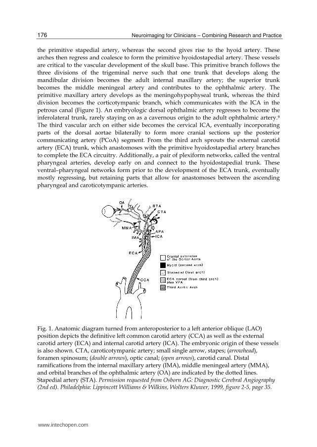
petrous canal (Figure 1). An embryologic dorsal ophthalmic artery regresses to become the
inferolateral trunk, rarely staying on as a cavernous origin to the adult ophthalmic artery.9
The third vascular arch on either side becomes the cervical ICA, eventually incorporating
parts of the dorsal aortae bilaterally to form more cranial sections up the posterior
communicating artery (PCoA) segment. From the third arch sprouts the external carotid
artery (ECA) trunk, which anastomoses with the primitive hyoidostapedial artery branches
to complete the ECA circuitry. Additionally, a pair of plexiform networks, called the ventral
pharyngeal arteries, develop early on and connect to the hyoidostapedial trunk. These
ventral–pharyngeal networks form prior to the development of the ECA trunk, eventually
mostly regressing, but retaining parts that allow for anastomoses between the ascending
pharyngeal and caroticotympanic arteries.
Fig. 1. Anatomic diagram turned from anteroposterior to a left anterior oblique (LAO) position depicts the definitive left common carotid artery (CCA) as well as the external carotid artery (ECA) and internal carotid artery (ICA). The embryonic origin of these vessels is also shown. CTA, caroticotympanic artery; small single arrow, stapes; (arrowhead), foramen spinosum; (double arrows), optic canal; (open arrows), carotid canal. Distal ramifications from the internal maxillary artery (IMA), middle meningeal artery (MMA), and orbital branches of the ophthalmic artery (OA) are indicated by the dotted lines. Stapedial artery (STA). Permission requested from Osborn AG: Diagnostic Cerebral Angiography (2nd ed). Philadelphia: Lippincott Williams & Wilkins, Wolters Kluwer, 1999, figure 2-5, page 35.
www.intechopen.com
Intracranial Arterial Collateralization: Relevance in Neuro-Endovascular Procedures
177
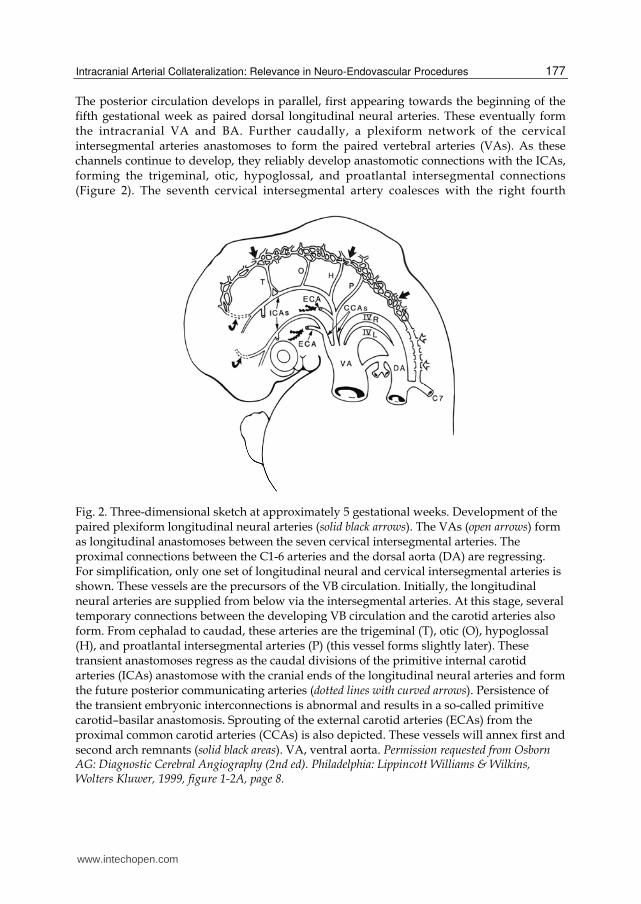
The posterior circulation develops in parallel, first appearing towards the beginning of the fifth gestational week as paired dorsal longitudinal neural arteries. These eventually form the intracranial VA and BA. Further caudally, a plexiform network of the cervical intersegmental arteries anastomoses to form the paired vertebral arteries (VAs). As these channels continue to develop, they reliably develop anastomotic connections with the ICAs, forming the trigeminal, otic, hypoglossal, and proatlantal intersegmental connections (Figure 2). The seventh cervical intersegmental artery coalesces with the right fourth
Fig. 2. Three-dimensional sketch at approximately 5 gestational weeks. Development of the paired plexiform longitudinal neural arteries (solid black arrows). The VAs (open arrows) form as longitudinal anastomoses between the seven cervical intersegmental arteries. The proximal connections between the C1-6 arteries and the dorsal aorta (DA) are regressing. For simplification, only one set of longitudinal neural and cervical intersegmental arteries is shown. These vessels are the precursors of the VB circulation. Initially, the longitudinal neural arteries are supplied from below via the intersegmental arteries. At this stage, several temporary connections between the developing VB circulation and the carotid arteries also form. From cephalad to caudad, these arteries are the trigeminal (T), otic (O), hypoglossal (H), and proatlantal intersegmental arteries (P) (this vessel forms slightly later). These transient anastomoses regress as the caudal divisions of the primitive internal carotid arteries (ICAs) anastomose with the cranial ends of the longitudinal neural arteries and form the future posterior communicating arteries (dotted lines with curved arrows). Persistence of the transient embryonic interconnections is abnormal and results in a so-called primitive carotid–basilar anastomosis. Sprouting of the external carotid arteries (ECAs) from the proximal common carotid arteries (CCAs) is also depicted. These vessels will annex first and second arch remnants (solid black areas). VA, ventral aorta. Permission requested from Osborn AG: Diagnostic Cerebral Angiography (2nd ed). Philadelphia: Lippincott Williams & Wilkins, Wolters Kluwer, 1999, figure 1-2A, page 8.
www.intechopen.com
Neuroimaging for Clinicians – Combining Research and Practice
178
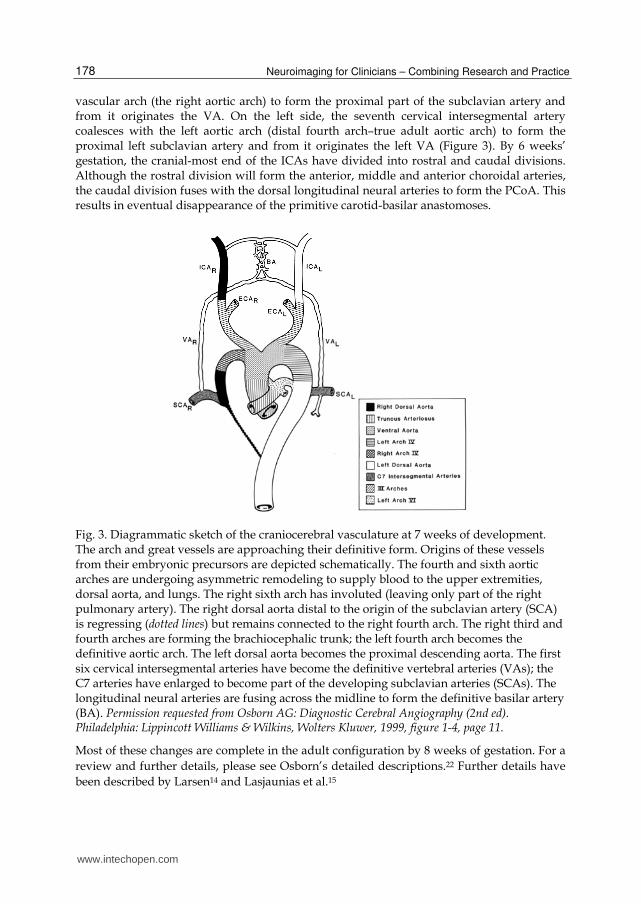
vascular arch (the right aortic arch) to form the proximal part of the subclavian artery and from it originates the VA. On the left side, the seventh cervical intersegmental artery coalesces with the left aortic arch (distal fourth arch–true adult aortic arch) to form the proximal left subclavian artery and from it originates the left VA (Figure 3). By 6 weeks’ gestation, the cranial-most end of the ICAs have divided into rostral and caudal divisions. Although the rostral division will form the anterior, middle and anterior choroidal arteries, the caudal division fuses with the dorsal longitudinal neural arteries to form the PCoA. This results in eventual disappearance of the primitive carotid-basilar anastomoses.
Fig. 3. Diagrammatic sketch of the craniocerebral vasculature at 7 weeks of development. The arch and great vessels are approaching their definitive form. Origins of these vessels from their embryonic precursors are depicted schematically. The fourth and sixth aortic arches are undergoing asymmetric remodeling to supply blood to the upper extremities, dorsal aorta, and lungs. The right sixth arch has involuted (leaving only part of the right pulmonary artery). The right dorsal aorta distal to the origin of the subclavian artery (SCA) is regressing (dotted lines) but remains connected to the right fourth arch. The right third and fourth arches are forming the brachiocephalic trunk; the left fourth arch becomes the definitive aortic arch. The left dorsal aorta becomes the proximal descending aorta. The first six cervical intersegmental arteries have become the definitive vertebral arteries (VAs); the C7 arteries have enlarged to become part of the developing subclavian arteries (SCAs). The longitudinal neural arteries are fusing across the midline to form the definitive basilar artery (BA). Permission requested from Osborn AG: Diagnostic Cerebral Angiography (2nd ed). Philadelphia: Lippincott Williams & Wilkins, Wolters Kluwer, 1999, figure 1-4, page 11.
Most of these changes are complete in the adult configuration by 8 weeks of gestation. For a
review and further details, please see Osborn’s detailed descriptions.22 Further details have
been described by Larsen14 and Lasjaunias et al.15
www.intechopen.com
Intracranial Arterial Collateralization: Relevance in Neuro-Endovascular Procedures
179
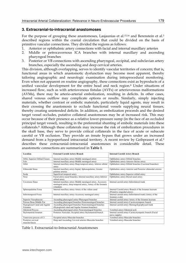
3. Extracranial-to-intracranial anastomoses
For the purpose of grouping these anastomoses, Lasjaunias et al.15,16 and Berenstein et al.3 described regions within the cranial circulation that could be divided on the basis of primitive vascular connections. They divided the regions as follows: 1. Anterior or ophthalmic artery connections with facial and internal maxillary arteries 2. Middle or petrocavernous ICA branches with internal maxillary and ascending
pharyngeal branches 3. Posterior or VB connections with ascending pharyngeal, occipital, and subclavian artery
branches, especially the ascending and deep cervical arteries. This division, although overlapping, serves to identify vascular territories of concern; that is,
functional areas in which anastomotic dysfunction may become most apparent, thereby tailoring angiographic and neurologic examination during intraprocedural monitoring. Even when not apparent on routine angiography, these connections exist as byproducts of a unified vascular development for the entire head and neck region.8 Under situations of increased flow, such as with arteriovenous fistulas (AVFs) or arteriovenous malformations (AVMs), there may be arterio-arterial embolization, resulting in deficits. In other cases, shared venous outflow may complicate options or results. Similarly, simply injecting materials, whether contrast or embolic materials, particularly liquid agents, may result in their crossing the anastomosis to occlude functional vessels supplying neural tissues, thereby creating unintended deficits. In addition, as embolization proceeds and the desired target vessel occludes, putative collateral anastomoses may be at increased risk. This may occur because of their presence as a relative lower-pressure sump (in the face of an occluded principal target vessel), resulting in the preferential shunting of embolic materials into these collaterals.5 Although these collaterals may increase the risk of embolization procedures in the skull base, they serve to provide critical collaterals in the face of acute or subacute carotid or VB occlusion. They provide an innate bypass that grows under an increased demand from a hypoperfused intracranial territory. A recent review by Geibprasert et al.9 describes these extracranial–intracranial anastomoses in considerable detail. These anastomotic connections are summarized in Table 1.
Table 1. Extracranial-to-Intracranial Anastomoses
www.intechopen.com
Neuroimaging for Clinicians – Combining Research and Practice
180
3.1 Ophthalmic artery anastomoses The ophthalmic artery is the principal vascular supply for contents in the orbit.13 It is the principal supply to the central retinal artery, which in turn, supplies the retina and choroid. Occlusion of this vessel will result in monocular blindness. Therefore, visualization of a choroid blush with an ECA injection should raise alarms about potential anatomic variations and dangerous collaterals.25 The ophthalmic artery originates from the ICA; however, during its development, it necessarily develops connections with other sources of supply to the contents of the orbit. The primitive stapedial artery contributes to the middle meningeal artery, and this develops some connections to the ophthalmic artery through the superior orbital fissure.26 The magnitude of this connection may vary; rarely, the middle meningeal and internal maxillary arteries completely assume the principal source of supply to the ophthalmic artery. In cases in which the ophthalmic artery is not visualized, such variation should be suspected.
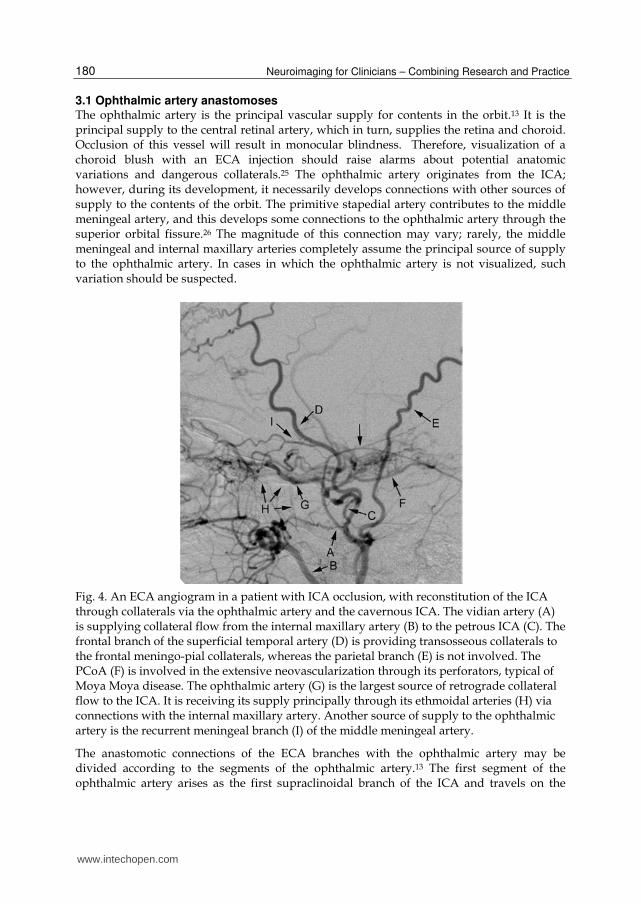
Fig. 4. An ECA angiogram in a patient with ICA occlusion, with reconstitution of the ICA through collaterals via the ophthalmic artery and the cavernous ICA. The vidian artery (A) is supplying collateral flow from the internal maxillary artery (B) to the petrous ICA (C). The frontal branch of the superficial temporal artery (D) is providing transosseous collaterals to the frontal meningo-pial collaterals, whereas the parietal branch (E) is not involved. The PCoA (F) is involved in the extensive neovascularization through its perforators, typical of Moya Moya disease. The ophthalmic artery (G) is the largest source of retrograde collateral flow to the ICA. It is receiving its supply principally through its ethmoidal arteries (H) via connections with the internal maxillary artery. Another source of supply to the ophthalmic artery is the recurrent meningeal branch (I) of the middle meningeal artery.
The anastomotic connections of the ECA branches with the ophthalmic artery may be divided according to the segments of the ophthalmic artery.13 The first segment of the ophthalmic artery arises as the first supraclinoidal branch of the ICA and travels on the
www.intechopen.com
Intracranial Arterial Collateralization: Relevance in Neuro-Endovascular Procedures
181
underside of the optic nerve in the optic canal. Upon entering the orbit, it maintains its close relationship with the optic nerve traveling towards the posterior globe. The recurrent meningeal artery is a reliable branch often noted by microvascular surgeons along the lateral aspect of the superior orbital fissure and is a branch of the middle meningeal artery. This supplies the contents of the superior orbital fissure and then anastomoses with the second segment of the ophthalmic artery in the orbit. This anastomosis is of particular importance when embolizing the middle meningeal artery branches, particularly for convexity meningiomas. The recurrent meningeal artery can often be visualized as the middle meningeal artery crosses the sphenoid wing. In cases in which embolization is desirable, it is best to obtain distal access close to or in the tumor proper and be vigilant to reflux to this more proximal branch point. In other cases where a branch is not noted, an intraarterial injection of sodium amytal and lidocaine may allow Wada testing32 of the anastomosis, which may not be apparent (Figures 4 and 5).
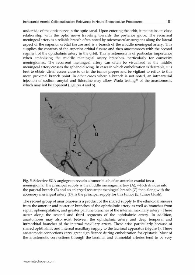
Fig. 5. Selective ECA angiogram reveals a tumor blush of an anterior cranial fossa meningioma. The principal supply is the middle meningeal artery (A), which divides into the parietal branch (B) and an enlarged recurrent meningeal branch (C) that, along with the accessory meningeal artery (D), is the principal supply for this tumor (E, tumor blush).
The second group of anastomoses is a product of the shared supply to the ethmoidal sinuses from the anterior and posterior branches of the ophthalmic artery as well as branches from septal, sphenopalatine, and greater palatine branches of the internal maxillary artery.1 These occur along the second and third segments of the ophthalmic artery. In addition, anastomoses may also exist between the ophthalmic artery and deep temporal and infraorbital branches of the internal maxillary artery. These arise particularly because of shared ophthalmic and internal maxillary supply to the lacrimal apparatus (Figure 4). These anastomotic connections carry great significance during embolization for epistaxis. Most of the anastomotic connections through the lacrimal and ethmoidal arteries tend to be very
www.intechopen.com
Neuroimaging for Clinicians – Combining Research and Practice
182
small-caliber vessels (less than 80 microns). If embolization is desired, particles chosen should be greater than 150 microns. This prevents inadvertent passage of these particles into the ICA or ophthalmic artery through these collaterals. As noted above, one needs to identify an aberrant ophthalmic artery origin by always performing a cerebral angiogram to identify the origin of the ophthalmic artery from the ICA. Embolization of anterior fossa-based tumors, such as olfactory groove meningiomas, is considered high risk because the principal supply to these tumors is from the ethmoidal arteries, and transinternal maxillary or transophthalmic embolization of these branches may have an inordinate increased risk for some of the material refluxing into the central retinal artery or retrograde into the ICA (Figure 5).1 Another consideration is with falcine meningiomas in which the anterior falcine artery, a branch of the ophthalmic artery, may anastomose with frontal branches of the middle meningeal artery, with retrograde flux of embolization materials into the ophthalmic artery from a middle meningeal artery branch embolization. Other situations in which these anastomoses need to be considered are during embolization for tumors in the head and neck region, particularly juvenile angiofibromas. Cavernous sinus DAVFs are particularly challenging because their inherent incorporation of these varied collateral channels may create significant risk for transarterial embolization (Figure 6). The third group of anastomoses occurs along the third or terminal segments of the ophthalmic artery and its terminal branches, the dorsal nasal artery and superior orbital
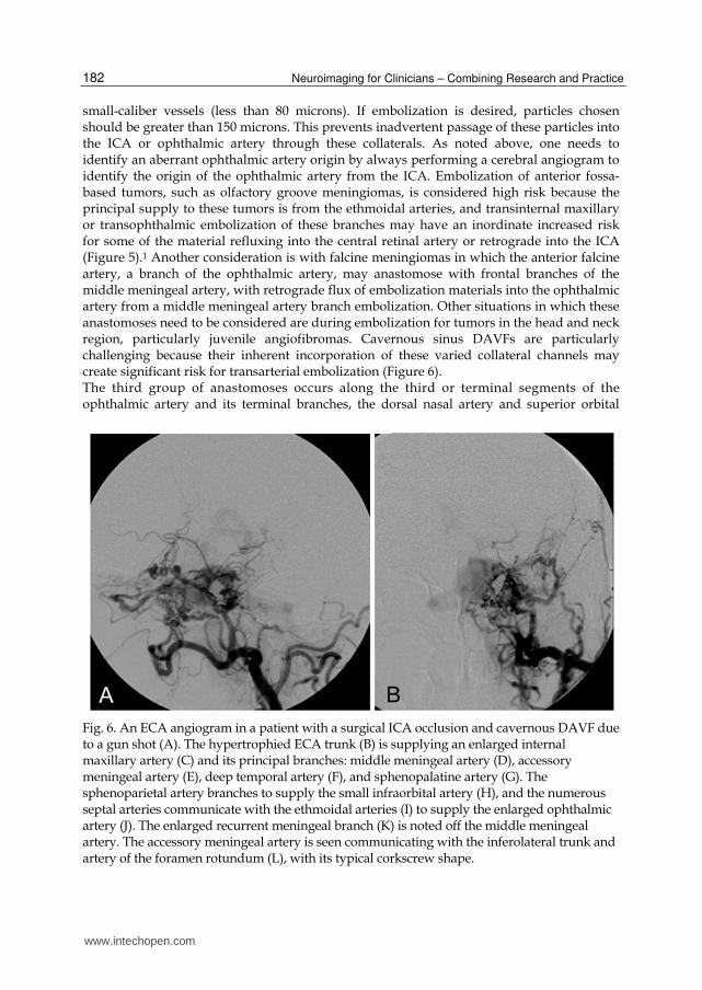
Fig. 6. An ECA angiogram in a patient with a surgical ICA occlusion and cavernous DAVF due to a gun shot (A). The hypertrophied ECA trunk (B) is supplying an enlarged internal maxillary artery (C) and its principal branches: middle meningeal artery (D), accessory meningeal artery (E), deep temporal artery (F), and sphenopalatine artery (G). The sphenoparietal artery branches to supply the small infraorbital artery (H), and the numerous septal arteries communicate with the ethmoidal arteries (I) to supply the enlarged ophthalmic artery (J). The enlarged recurrent meningeal branch (K) is noted off the middle meningeal artery. The accessory meningeal artery is seen communicating with the inferolateral trunk and artery of the foramen rotundum (L), with its typical corkscrew shape.
www.intechopen.com
Intracranial Arterial Collateralization: Relevance in Neuro-Endovascular Procedures
183
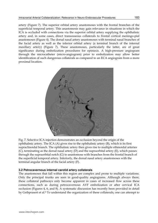
artery (Figure 7). The superior orbital artery anastomoses with the frontal branches of the superficial temporal artery. This anastomosis may gain relevance in situations in which the ICA is occluded with connections via the superior orbital artery supplying the ophthalmic artery and, in some cases, direct transosseous collaterals to frontal cortical meningo-pial anastomoses (Figure 4). The dorsal nasal artery anastomoses with terminal nasal branches of the facial artery as well as the inferior orbital artery (a terminal branch of the internal maxillary artery) (Figure 7). These anastomoses, particularly the latter, are of great significance during embolization procedures for epistaxis. A high-pressure angiogram through the microcatheter (micro-angiogram) prior to embolization may allow better identification of such dangerous collaterals as compared to an ECA angiogram from a more proximal location.
Fig. 7. Selective ICA injection demonstrates an occlusion beyond the origin of the ophthalmic artery. The ICA (A) gives rise to the ophthalmic artery (B), which is its first supraclinoidal branch. The ophthalmic artery then gives rise to multiple ethmoidal arteries (C), terminating as the dorsal nasal artery (D) and the supraorbital artery (E), which passes through the supraorbital notch (G) to anastomose with branches from the frontal branch of the superficial temporal artery. Inferiorly, the dorsal nasal artery anastomoses with the terminal angular branch of the facial artery (F).
3.2 Petrocavernous internal carotid artery collaterals The anastomoses that fall within this region are complex and prone to multiple variations.
Only the principal trunks are seen in good-quality angiograms. Although always there,
these collateral pathways only become apparent in cases of increased flow across these
connections, such as during petrocavernous AVF embolization or after cervical ICA
occlusion (Figures 4, 6, and 8). A systematic discussion has recently been provided in detail
by Geibprasert et al.9 To understand the organization of these collaterals, one can attempt to
www.intechopen.com
Neuroimaging for Clinicians – Combining Research and Practice
184
look at these putative connections in terms of branches of the ICA, which are involved in the
anastomosis. The first branch traveling rostrocaudal or distal to proximal along the ICA is
the inferolateral trunk off the ICA in the cavernous sinus.
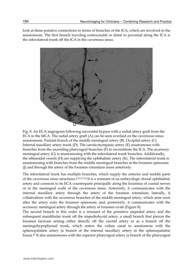
Fig. 8. An ECA angiogram following successful bypass with a radial artery graft from the ECA to the MCA. The radial artery graft (A) can be seen overlaid on the cavernous sinus anastomosis. Parietal branch of the middle meningeal artery (B). Occipital artery (C). Internal maxillary artery trunk (D). The caroticotympanic artery (E) anastomoses with branches from the ascending pharyngeal branches (F) to reconstitute the ICA. The accessory meningeal artery (G) is anastomosing with the inferolateral trunk branches. Additionally, the ethmoidal vessels (H) are supplying the ophthalmic artery (K). The inferolateral trunk is anastomosing with branches from the middle meningeal branches at the foramen spinosum (J) and through the artery of the foramen rotundum more anteriorly.
The inferolateral trunk has multiple branches, which supply the anterior and middle parts
of the cavernous sinus structures.4,15,17,30 It is a remnant of an embryologic dorsal ophthalmic
artery and connects to its ECA counterparts principally along the foramina of cranial nerves
or in the meningeal walls of the cavernous sinus. Anteriorly, it communicates with the
internal maxillary artery through the artery of the foramen rotundum; laterally, it
collateralizes with the cavernous branches of the middle meningeal artery, which arise soon
after the artery exits the foramen spinosum; and, posteriorly, it communicates with the
accessory meningeal artery through the artery of foramen ovale (Figure 8).
The second branch in this order is a remnant of the primitive stapedial artery and the
subsequent mandibular trunk off the stapediohyoid artery, a small branch that pierces the
foramen lacerum arising either directly off the carotid artery or as a branch off the
meningohypophyseal trunk, which enters the vidian canal to anastomose with the
sphenopalatine artery (a branch of the internal maxillary artery in the sphenopalatine
fossa).29 It also anastomoses with the superior pharyngeal artery (a branch of the pharyngeal
www.intechopen.com
Intracranial Arterial Collateralization: Relevance in Neuro-Endovascular Procedures
185
trunk of the ascending pharyngeal artery) and the accessory meningeal artery to provide a
branch to the pterygovaginal canal, which ends by anastomosing with internal maxillary
artery branches (Figure 9).
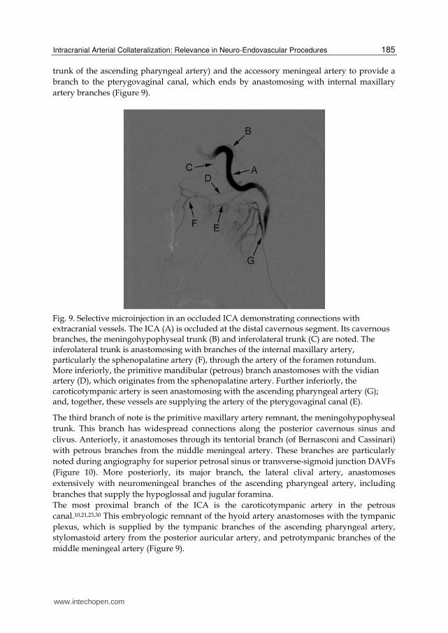
Fig. 9. Selective microinjection in an occluded ICA demonstrating connections with extracranial vessels. The ICA (A) is occluded at the distal cavernous segment. Its cavernous branches, the meningohypophyseal trunk (B) and inferolateral trunk (C) are noted. The inferolateral trunk is anastomosing with branches of the internal maxillary artery, particularly the sphenopalatine artery (F), through the artery of the foramen rotundum. More inferiorly, the primitive mandibular (petrous) branch anastomoses with the vidian artery (D), which originates from the sphenopalatine artery. Further inferiorly, the caroticotympanic artery is seen anastomosing with the ascending pharyngeal artery (G); and, together, these vessels are supplying the artery of the pterygovaginal canal (E).
The third branch of note is the primitive maxillary artery remnant, the meningohypophyseal
trunk. This branch has widespread connections along the posterior cavernous sinus and
clivus. Anteriorly, it anastomoses through its tentorial branch (of Bernasconi and Cassinari)
with petrous branches from the middle meningeal artery. These branches are particularly
noted during angiography for superior petrosal sinus or transverse-sigmoid junction DAVFs
(Figure 10). More posteriorly, its major branch, the lateral clival artery, anastomoses
extensively with neuromeningeal branches of the ascending pharyngeal artery, including
branches that supply the hypoglossal and jugular foramina.
The most proximal branch of the ICA is the caroticotympanic artery in the petrous
canal.10,21,23,30 This embryologic remnant of the hyoid artery anastomoses with the tympanic
plexus, which is supplied by the tympanic branches of the ascending pharyngeal artery,
stylomastoid artery from the posterior auricular artery, and petrotympanic branches of the
middle meningeal artery (Figure 9).
www.intechopen.com
Neuroimaging for Clinicians – Combining Research and Practice
186
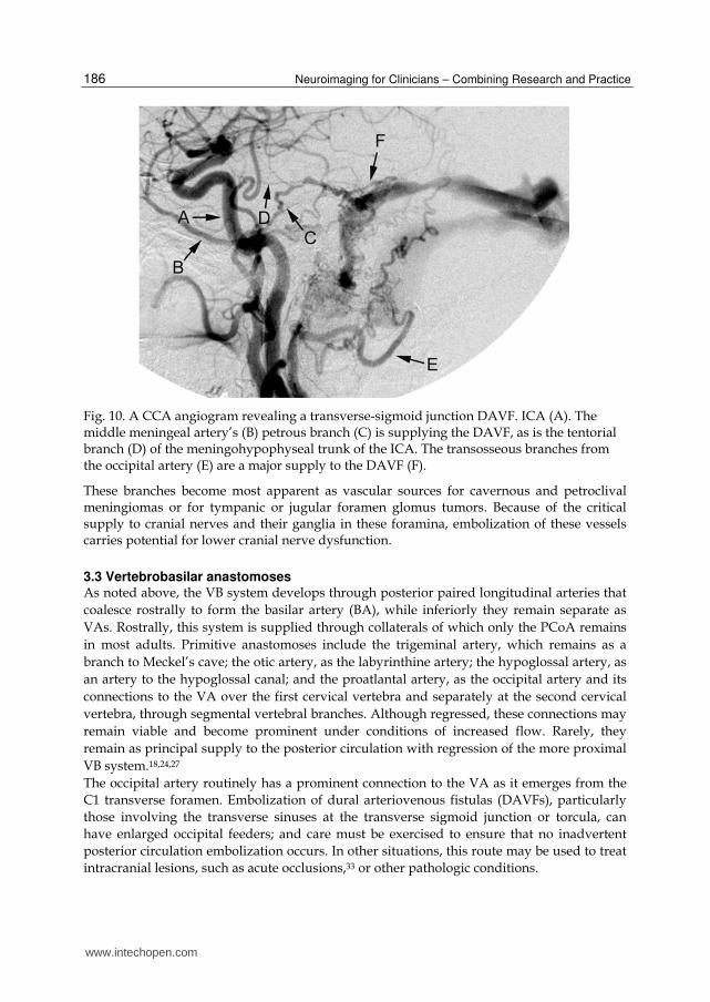
Fig. 10. A CCA angiogram revealing a transverse-sigmoid junction DAVF. ICA (A). The middle meningeal artery’s (B) petrous branch (C) is supplying the DAVF, as is the tentorial branch (D) of the meningohypophyseal trunk of the ICA. The transosseous branches from the occipital artery (E) are a major supply to the DAVF (F).
These branches become most apparent as vascular sources for cavernous and petroclival meningiomas or for tympanic or jugular foramen glomus tumors. Because of the critical supply to cranial nerves and their ganglia in these foramina, embolization of these vessels carries potential for lower cranial nerve dysfunction.
3.3 Vertebrobasilar anastomoses As noted above, the VB system develops through posterior paired longitudinal arteries that
coalesce rostrally to form the basilar artery (BA), while inferiorly they remain separate as
VAs. Rostrally, this system is supplied through collaterals of which only the PCoA remains
in most adults. Primitive anastomoses include the trigeminal artery, which remains as a
branch to Meckel’s cave; the otic artery, as the labyrinthine artery; the hypoglossal artery, as
an artery to the hypoglossal canal; and the proatlantal artery, as the occipital artery and its
connections to the VA over the first cervical vertebra and separately at the second cervical
vertebra, through segmental vertebral branches. Although regressed, these connections may
remain viable and become prominent under conditions of increased flow. Rarely, they
remain as principal supply to the posterior circulation with regression of the more proximal
VB system.18,24,27
The occipital artery routinely has a prominent connection to the VA as it emerges from the
C1 transverse foramen. Embolization of dural arteriovenous fistulas (DAVFs), particularly
those involving the transverse sinuses at the transverse sigmoid junction or torcula, can
have enlarged occipital feeders; and care must be exercised to ensure that no inadvertent
posterior circulation embolization occurs. In other situations, this route may be used to treat
intracranial lesions, such as acute occlusions,33 or other pathologic conditions.
www.intechopen.com
Intracranial Arterial Collateralization: Relevance in Neuro-Endovascular Procedures
187
The neuromeningeal trunk of the ascending pharyngeal artery anastomoses with the VA, through connections at C3 through muscular collaterals, and more rostrally, as the remnant of the primitive hypoglossal artery with the odontoid arch vascular system.11,19 More caudally, the cervical branches of the subclavian artery anastomose with proximal sections of the VAs (C3-7). The ascending cervical artery arises from the thyrocervical trunk, while the deep cervical artery arises from the costocervical trunk. Both these branches form connections with the segmental vertebral branches through muscular collaterals. They are commonly noted to reconstitute the distal VA after proximal occlusion.
3.4 Extracranial to intracranial collateralization and complication avoidance in neurointervention procedures First and foremost, one needs to be aware of the potential for collateral anastomotic
channels. Such knowledge facilitates angiographic visualization. Even if the initial proximal
external carotid or subclavian angiograms do not result in the visualization of these
collaterals, we recommend that selective microcatheter angiograms be performed in as distal
a position as is reasonably attainable immediately prior to embolization. These higher
pressure selective injections are more likely to reveal these putative connections. If there is
no visualization, a second element to further bolster confidence is to perform a Wada test
with sodium amytal (75 mg) and lidocaine (30 mg) intraarterially through the microcatheter
from the position planned for embolization and then immediately after injection to test for
loss of appropriate neural function including that of the cranial nerves. Thirdly, if
embolization is desired, polyvinyl alcohol particles, which considerably exceed the size (150
microns or greater) of most of these non-angiographically visualized collaterals (50-80
microns), should be used. Liquid embolics, such as glue (N-butyl cyanoacrylate; Trufill,
Codman Neurovascular, Raynham, MA) and Onyx (EV3, Irvine, California), provide
superior visualization; however, they are not discriminatory with respect to vessel size and,
therefore, their use may be associated with a higher likelihood of collateral vessel occlusion.
Angiographic visualization does not preclude embolization. One may attempt to attain
distal purchase beyond the collateral communication and pay great attention to reflux
during the embolization procedure. If such positioning is not attainable, pre-embolization
occlusion of the collateral channel is another strategy. Typically, coil embolization of the
collateral channel will not result in ischemic injury because of the multiple sources of
vascular supply at the skull base; however, subsequent embolization of the intended vessel
will prevent inadvertent passage of embolic materials through the occluded vessel to critical
neural structures.
4. Intracranial anastomoses
In contrast to the extracranial-to-intracranial collateral anastomoses described above, which may be less obvious on non-superselective angiography, many of the ICA-to-ICA or ICA-to-VB anastomoses are rather easily identified and commonly seen. These common known collateral anastomoses as parts of the circle of Willis include the following: 1) anterior communicating artery (ACoA) connecting bilateral anterior cerebral arteries (ACAs); and 2) PCoA connecting the ipsilateral ICA to the posterior cerebral artery (PCA). Less obvious anastomoses occur among the various terminal cortical branches from each of the major vascular territories. The middle cerebral artery (MCA) branches anastomose with branches
www.intechopen.com
Neuroimaging for Clinicians – Combining Research and Practice
188
of the PCA and ACA. The pericallosal branches of the ACA connect to splenial branches of PCA. Distal branches of the superior cerebellar artery (SCA) connect to branches from posterior inferior cerebellar artery (PICA), or to branches from anterior inferior cerebellar artery (AICA). Branches of AICA could also have connection with PICA. These anastomoses not only provide collaterals to preserve potentially affected brain tissue in the case of an occlusion event but also provide crucial collateral supply in the situation if carotid artery or VA sacrifice becomes necessary; also, potentially, the collaterals may be utilized as alternative access routes to the target during interventional procedures. Typically, these distal branch anastomoses are not visible with standard angiogram runs. In order to delineate the present of the anastomoses, a superselective balloon test occlusion is often required.
4.1 Intracranial collateralization in neurointerventional procedures 4.1.1 Flow replacement by collateral vessels
Intracranial large artery sacrifice
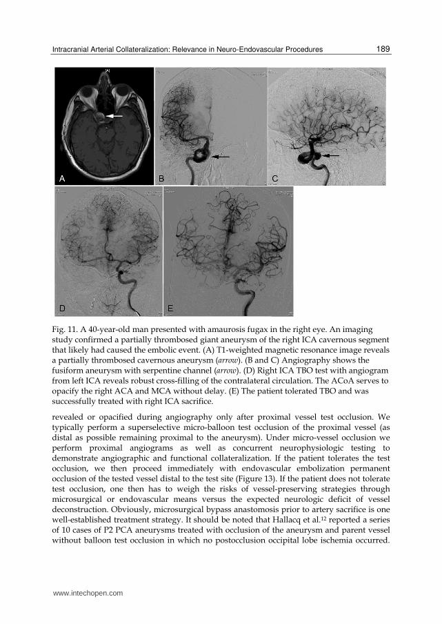
In patients with fusiform or giant aneurysms of the cavernous or supraclinoidal segment of the carotid artery or supraclinoidal carotid artery dissection with hemorrhage, sacrifice of the ipsilateral ICA can be a simple therapeutic solution with low risk if the ACoA and PCoA provide sufficient cross filling to the affected side. This is especially true if both angiographic and neurophysiologic tests are completed during a temporary balloon occlusion (TBO) test6,28,31 (Figure 11). It should be noted that endovascular strategies for vessel preservation during treatment continue to gradually erode options for vessel
sacrifice, e.g., flow diversion technique, (Pipeline stent by EV3) in treating carotid artery giant aneurysms, which previously required carotid sacrifice. Sacrifice of the VA is an effective strategy for the management of pathologic conditions of
the VA, such as dissection with hemorrhage or endovascularly unmanageable aneurysms,
simply because most patients are endowed with two vessels; and, after the sacrifice of one,
unabated flow typically continues through the other. It is, however, important that the TBO
test be performed before a vessel deconstruction procedure is carried out (Figure 12). There
are other more complex situations involving the distal VAs, their junction, or the BA in
which neither VA can be preserved, so flow reversal is desired. These situations include
dissections with hemorrhage and complex, enlarging, symptomatic, or ruptured fusiform
aneurysms.2,20 The presence of a robust PCoA and a large-caliber ipsilateral PCA P1 segment
may allow sacrifice of both VAs, thereby creating flow reversal with a diminution of flow,
enough to reduce hemodynamic stresses on the diseased vessel and allowing it to heal. It
should be borne in mind that, before such deconstruction, which is undoubtedly high risk, a
comprehensive angiographic evaluation, including a VA angiogram with transient bilateral
manual carotid artery compression, could be very useful to identify bilateral PCoA. Further
hemodynamic and neurophysiologic testing should be completed prior to vessel
deconstruction.
Intracranial distal end artery sacrifice
Occasionally, one may encounter dissecting, mycotic, or ruptured broad-based aneurysms involving the distal ACA, MCA or PCA and considered higher risk for surgical or endovascular reconstruction. Because of well-developed cortical collaterals from ipsilateral ACA to PCA or MCA to PCA or ACA to MCA territories, these collaterals typically are
www.intechopen.com
Intracranial Arterial Collateralization: Relevance in Neuro-Endovascular Procedures
189
Fig. 11. A 40-year-old man presented with amaurosis fugax in the right eye. An imaging study confirmed a partially thrombosed giant aneurysm of the right ICA cavernous segment that likely had caused the embolic event. (A) T1-weighted magnetic resonance image reveals a partially thrombosed cavernous aneurysm (arrow). (B and C) Angiography shows the fusiform aneurysm with serpentine channel (arrow). (D) Right ICA TBO test with angiogram from left ICA reveals robust cross-filling of the contralateral circulation. The ACoA serves to opacify the right ACA and MCA without delay. (E) The patient tolerated TBO and was successfully treated with right ICA sacrifice.
revealed or opacified during angiography only after proximal vessel test occlusion. We typically perform a superselective micro-balloon test occlusion of the proximal vessel (as distal as possible remaining proximal to the aneurysm). Under micro-vessel occlusion we perform proximal angiograms as well as concurrent neurophysiologic testing to demonstrate angiographic and functional collateralization. If the patient tolerates the test occlusion, we then proceed immediately with endovascular embolization permanent occlusion of the tested vessel distal to the test site (Figure 13). If the patient does not tolerate test occlusion, one then has to weigh the risks of vessel-preserving strategies through microsurgical or endovascular means versus the expected neurologic deficit of vessel deconstruction. Obviously, microsurgical bypass anastomosis prior to artery sacrifice is one well-established treatment strategy. It should be noted that Hallacq et al.12 reported a series of 10 cases of P2 PCA aneurysms treated with occlusion of the aneurysm and parent vessel without balloon test occlusion in which no postocclusion occipital lobe ischemia occurred.
www.intechopen.com
Neuroimaging for Clinicians – Combining Research and Practice
190
Similar patterns of distal arterial collateralization can often be expected in PICA, AICA and SCA circulations. In cases of PICA ruptured dissections or dissecting aneurysms, proximal PICA can be occluded without needs of microsurgical bypass anastomosis. (Figure 21).
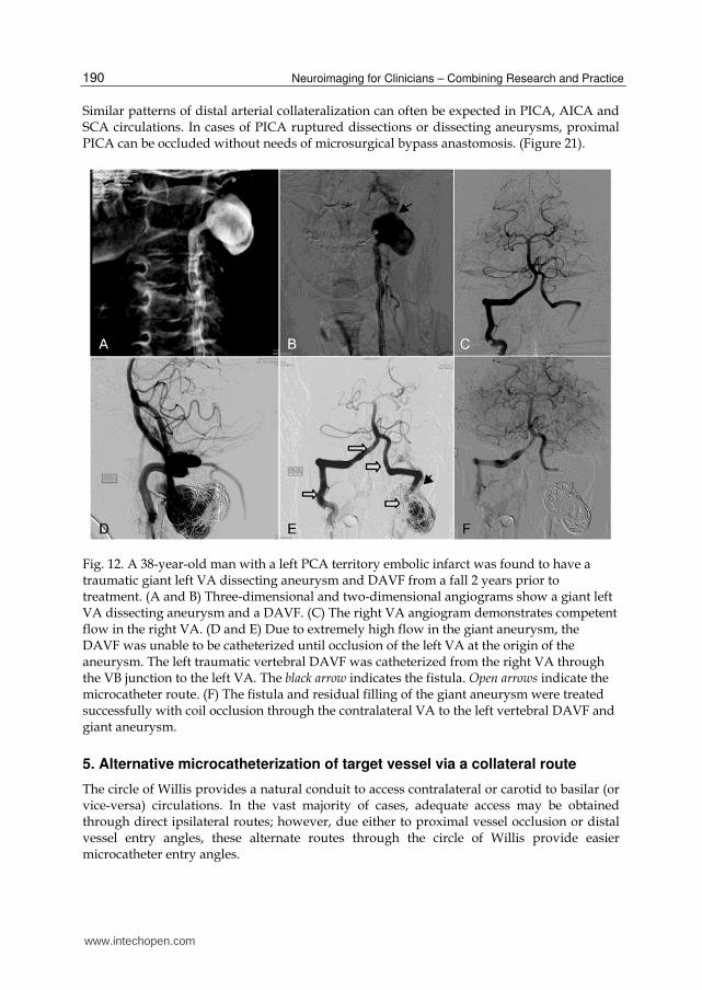
Fig. 12. A 38-year-old man with a left PCA territory embolic infarct was found to have a traumatic giant left VA dissecting aneurysm and DAVF from a fall 2 years prior to treatment. (A and B) Three-dimensional and two-dimensional angiograms show a giant left VA dissecting aneurysm and a DAVF. (C) The right VA angiogram demonstrates competent flow in the right VA. (D and E) Due to extremely high flow in the giant aneurysm, the DAVF was unable to be catheterized until occlusion of the left VA at the origin of the aneurysm. The left traumatic vertebral DAVF was catheterized from the right VA through the VB junction to the left VA. The black arrow indicates the fistula. Open arrows indicate the microcatheter route. (F) The fistula and residual filling of the giant aneurysm were treated successfully with coil occlusion through the contralateral VA to the left vertebral DAVF and giant aneurysm.
5. Alternative microcatheterization of target vessel via a collateral route
The circle of Willis provides a natural conduit to access contralateral or carotid to basilar (or vice-versa) circulations. In the vast majority of cases, adequate access may be obtained through direct ipsilateral routes; however, due either to proximal vessel occlusion or distal vessel entry angles, these alternate routes through the circle of Willis provide easier microcatheter entry angles.
www.intechopen.com
Intracranial Arterial Collateralization: Relevance in Neuro-Endovascular Procedures
191
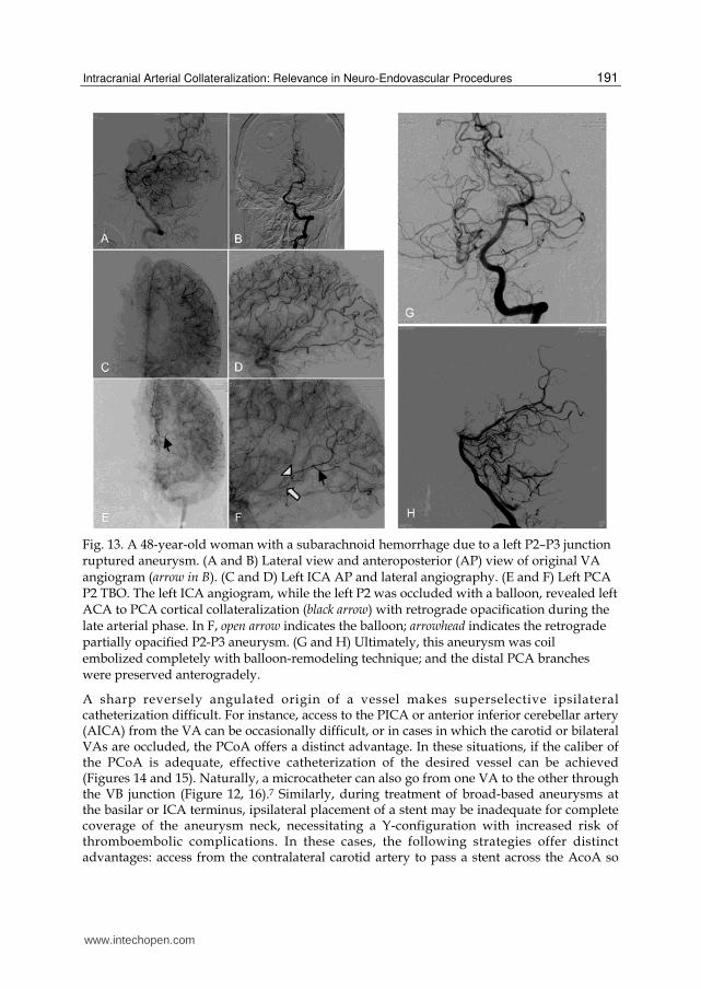
Fig. 13. A 48-year-old woman with a subarachnoid hemorrhage due to a left P2–P3 junction ruptured aneurysm. (A and B) Lateral view and anteroposterior (AP) view of original VA angiogram (arrow in B). (C and D) Left ICA AP and lateral angiography. (E and F) Left PCA P2 TBO. The left ICA angiogram, while the left P2 was occluded with a balloon, revealed left ACA to PCA cortical collateralization (black arrow) with retrograde opacification during the late arterial phase. In F, open arrow indicates the balloon; arrowhead indicates the retrograde partially opacified P2-P3 aneurysm. (G and H) Ultimately, this aneurysm was coil embolized completely with balloon-remodeling technique; and the distal PCA branches were preserved anterogradely.
A sharp reversely angulated origin of a vessel makes superselective ipsilateral catheterization difficult. For instance, access to the PICA or anterior inferior cerebellar artery (AICA) from the VA can be occasionally difficult, or in cases in which the carotid or bilateral VAs are occluded, the PCoA offers a distinct advantage. In these situations, if the caliber of the PCoA is adequate, effective catheterization of the desired vessel can be achieved (Figures 14 and 15). Naturally, a microcatheter can also go from one VA to the other through the VB junction (Figure 12, 16).7 Similarly, during treatment of broad-based aneurysms at the basilar or ICA terminus, ipsilateral placement of a stent may be inadequate for complete coverage of the aneurysm neck, necessitating a Y-configuration with increased risk of thromboembolic complications. In these cases, the following strategies offer distinct advantages: access from the contralateral carotid artery to pass a stent across the AcoA so
www.intechopen.com
Neuroimaging for Clinicians – Combining Research and Practice
192
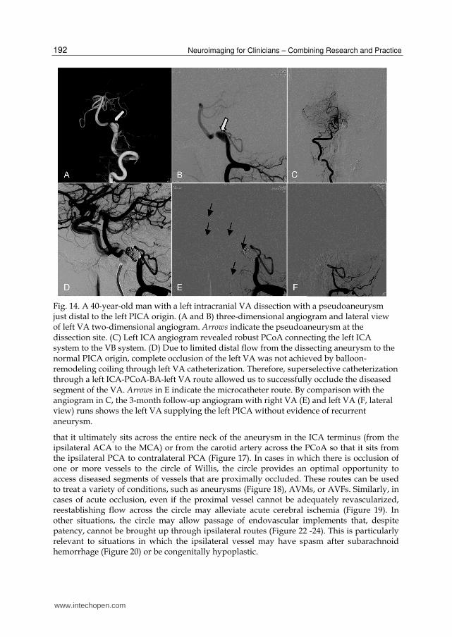
Fig. 14. A 40-year-old man with a left intracranial VA dissection with a pseudoaneurysm just distal to the left PICA origin. (A and B) three-dimensional angiogram and lateral view of left VA two-dimensional angiogram. Arrows indicate the pseudoaneurysm at the dissection site. (C) Left ICA angiogram revealed robust PCoA connecting the left ICA system to the VB system. (D) Due to limited distal flow from the dissecting aneurysm to the normal PICA origin, complete occlusion of the left VA was not achieved by balloon-remodeling coiling through left VA catheterization. Therefore, superselective catheterization through a left ICA-PCoA-BA-left VA route allowed us to successfully occlude the diseased segment of the VA. Arrows in E indicate the microcatheter route. By comparison with the angiogram in C, the 3-month follow-up angiogram with right VA (E) and left VA (F, lateral view) runs shows the left VA supplying the left PICA without evidence of recurrent aneurysm.
that it ultimately sits across the entire neck of the aneurysm in the ICA terminus (from the ipsilateral ACA to the MCA) or from the carotid artery across the PCoA so that it sits from the ipsilateral PCA to contralateral PCA (Figure 17). In cases in which there is occlusion of one or more vessels to the circle of Willis, the circle provides an optimal opportunity to access diseased segments of vessels that are proximally occluded. These routes can be used to treat a variety of conditions, such as aneurysms (Figure 18), AVMs, or AVFs. Similarly, in cases of acute occlusion, even if the proximal vessel cannot be adequately revascularized, reestablishing flow across the circle may alleviate acute cerebral ischemia (Figure 19). In other situations, the circle may allow passage of endovascular implements that, despite patency, cannot be brought up through ipsilateral routes (Figure 22 -24). This is particularly relevant to situations in which the ipsilateral vessel may have spasm after subarachnoid hemorrhage (Figure 20) or be congenitally hypoplastic.
www.intechopen.com
Intracranial Arterial Collateralization: Relevance in Neuro-Endovascular Procedures
193
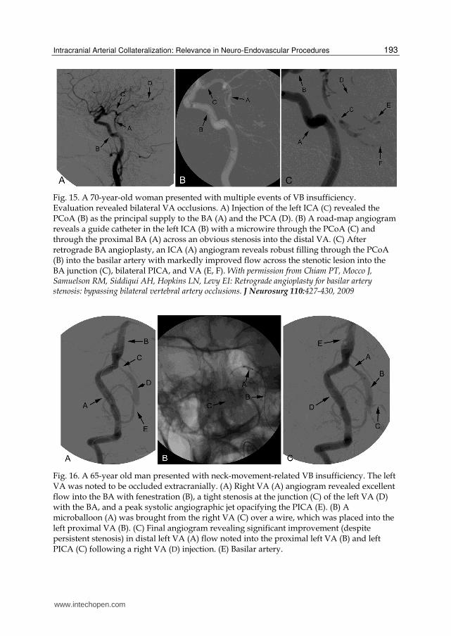
Fig. 15. A 70-year-old woman presented with multiple events of VB insufficiency. Evaluation revealed bilateral VA occlusions. A) Injection of the left ICA (C) revealed the PCoA (B) as the principal supply to the BA (A) and the PCA (D). (B) A road-map angiogram reveals a guide catheter in the left ICA (B) with a microwire through the PCoA (C) and through the proximal BA (A) across an obvious stenosis into the distal VA. (C) After retrograde BA angioplasty, an ICA (A) angiogram reveals robust filling through the PCoA (B) into the basilar artery with markedly improved flow across the stenotic lesion into the BA junction (C), bilateral PICA, and VA (E, F). With permission from Chiam PT, Mocco J, Samuelson RM, Siddiqui AH, Hopkins LN, Levy EI: Retrograde angioplasty for basilar artery stenosis: bypassing bilateral vertebral artery occlusions. J Neurosurg 110:427-430, 2009
Fig. 16. A 65-year old man presented with neck-movement-related VB insufficiency. The left VA was noted to be occluded extracranially. (A) Right VA (A) angiogram revealed excellent flow into the BA with fenestration (B), a tight stenosis at the junction (C) of the left VA (D) with the BA, and a peak systolic angiographic jet opacifying the PICA (E). (B) A microballoon (A) was brought from the right VA (C) over a wire, which was placed into the left proximal VA (B). (C) Final angiogram revealing significant improvement (despite persistent stenosis) in distal left VA (A) flow noted into the proximal left VA (B) and left PICA (C) following a right VA (D) injection. (E) Basilar artery.
www.intechopen.com
Neuroimaging for Clinicians – Combining Research and Practice
194
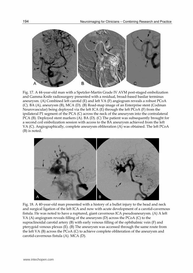
Fig. 17. A 44-year-old man with a Spetzler-Martin Grade IV AVM post-staged embolization and Gamma Knife radiosurgery presented with a residual, broad-based basilar terminus aneurysm. (A) Combined left carotid (E) and left VA (F) angiogram reveals a robust PCoA (C). BA (A), aneurysm (B), MCA (D). (B) Road-map image of an Enterprise stent (Codman Neurovascular) being deployed via the left ICA (E) through the left PCoA (F) from the ipsilateral P1 segment of the PCA (C) across the neck of the aneurysm into the contralateral PCA (B). Deployed stent markers (A). BA (D). (C) The patient was subsequently brought for a second coil embolization session with access to the BA aneurysm achieved from the left VA (C). Angiographically, complete aneurysm obliteration (A) was obtained. The left PCoA (B) is noted.
Fig. 18. A 40-year-old man presented with a history of a bullet injury to the head and neck and surgical ligation of the left ICA and now with acute development of a carotid-cavernous fistula. He was noted to have a ruptured, giant cavernous ICA pseudoaneurysm. (A) A left VA (A) angiogram reveals filling of the aneurysm (D) across the PCoA (C) to the supraclinoidal carotid artery (B) with early venous filling of the ophthalmic vein (F) and pterygoid venous plexus (E). (B) The aneurysm was accessed through the same route from the left VA (B) across the PCoA (C) to achieve complete obliteration of the aneurysm and carotid-cavernous fistula (A). MCA (D).
www.intechopen.com
Intracranial Arterial Collateralization: Relevance in Neuro-Endovascular Procedures
195
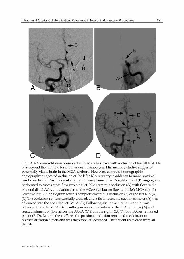
Fig. 19. A 65-year-old man presented with an acute stroke with occlusion of his left ICA. He was beyond the window for intravenous thrombolysis. His ancillary studies suggested potentially viable brain in the MCA territory. However, computed tomographic angiography suggested occlusion of the left MCA territory in addition to more proximal carotid occlusion. An emergent angiogram was planned. (A) A right carotid (D) angiogram
performed to assess cross-flow reveals a left ICA terminus occlusion (A) with flow to the
bilateral distal ACA circulation across the ACoA (C) but no flow to the left MCA (B). (B)
Selective left ICA angiogram reveals complete cavernous occlusion (B) of the left ICA (A).
(C) The occlusion (B) was carefully crossed, and a thrombectomy suction catheter (A) was
advanced into the occluded left MCA. (D) Following suction aspiration, the clot was retrieved from the MCA (B), resulting in revascularization of the ICA terminus (A) and reestablishment of flow across the ACoA (C) from the right ICA (F). Both ACAs remained patent (E, D). Despite these efforts, the proximal occlusion remained recalcitrant to revascularization efforts and was therefore left occluded. The patient recovered from all deficits.
www.intechopen.com
Neuroimaging for Clinicians – Combining Research and Practice
196
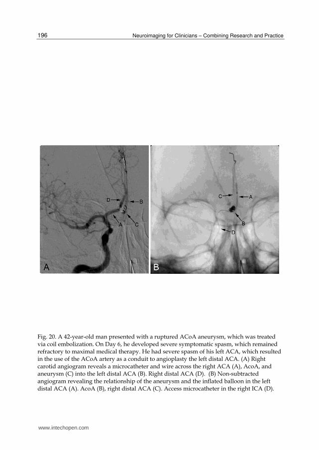
Fig. 20. A 42-year-old man presented with a ruptured ACoA aneurysm, which was treated via coil embolization. On Day 6, he developed severe symptomatic spasm, which remained refractory to maximal medical therapy. He had severe spasm of his left ACA, which resulted in the use of the ACoA artery as a conduit to angioplasty the left distal ACA. (A) Right carotid angiogram reveals a microcatheter and wire across the right ACA (A), AcoA, and aneurysm (C) into the left distal ACA (B). Right distal ACA (D). (B) Non-subtracted angiogram revealing the relationship of the aneurysm and the inflated balloon in the left distal ACA (A). AcoA (B), right distal ACA (C). Access microcatheter in the right ICA (D).
www.intechopen.com
Intracranial Arterial Collateralization: Relevance in Neuro-Endovascular Procedures
197
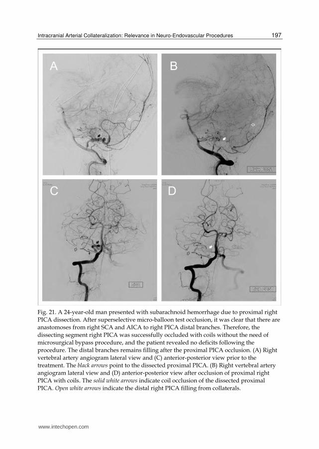
Fig. 21. A 24-year-old man presented with subarachnoid hemorrhage due to proximal right
PICA dissection. After superselective micro-balloon test occlusion, it was clear that there are
anastomoses from right SCA and AICA to right PICA distal branches. Therefore, the
dissecting segment right PICA was successfully occluded with coils without the need of
microsurgical bypass procedure, and the patient revealed no deficits following the
procedure. The distal branches remains filling after the proximal PICA occlusion. (A) Right
vertebral artery angiogram lateral view and (C) anterior-posterior view prior to the
treatment. The black arrows point to the dissected proximal PICA. (B) Right vertebral artery
angiogram lateral view and (D) anterior-posterior view after occlusion of proximal right
PICA with coils. The solid white arrows indicate coil occlusion of the dissected proximal
PICA. Open white arrows indicate the distal right PICA filling from collaterals.
www.intechopen.com
Neuroimaging for Clinicians – Combining Research and Practice
198
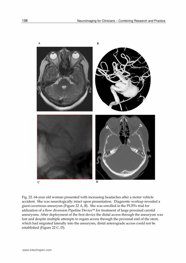
Fig. 22. 64-year old woman presented with increasing headaches after a motor vehicle accident. She was neurologically intact upon presentation. Diagnostic workup revealed a giant cavernous aneurysm (Figure 22 A, B). She was enrolled in the PUFFs trial for
utilization of a flow diversion Pipeline Device for treatment of large proximal carotid aneurysms. After deployment of the first device the distal access through the aneurysm was lost and despite multiple attempts to regain access through the proximal end of the stent, which had migrated laterally into the aneurysm, distal anterograde access could not be established (Figure 22 C, D).
www.intechopen.com
Intracranial Arterial Collateralization: Relevance in Neuro-Endovascular Procedures
199
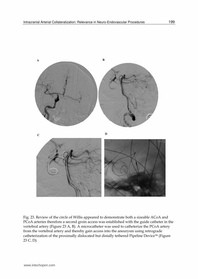
Fig. 23. Review of the circle of Willis appeared to demonstrate both a sizeable ACoA and PCoA arteries therefore a second groin access was established with the guide catheter in the vertebral artery (Figure 23 A, B). A microcatheter was used to catheterize the PCoA artery from the vertebral artery and thereby gain access into the aneurysm using retrograde
catheterization of the proximally dislocated but distally tethered Pipeline Device (Figure 23 C, D).
www.intechopen.com

Neuroimaging for Clinicians – Combining Research and Practice
200
Fig. 24. Once the microwire was in the aneurysm a Snare was deployed to grab the microwire and the Marksman microcatheter was advanced using the snare over the microwire into the Pipeline device by pulling the microwire back out through the PCoA (Figure 24 A, B). Once distal access was reestablished additional Pipeline Devices were used to complete the endovascular reconstruction of the aneurysm. The aneurysm appeared almost completely obliterated at the end of the procedure (Figure 24 C) and remained obliterated at the 6-month follow-up (Figure 24 D).
6. Conclusion
An in-depth knowledge of intracranial and extracranial collateral anastomoses, overt or hidden, is crucial for a neurointerventionist to devise optimal endovascular strategies to manage a host of pathological conditions; to ascertain potential pitfalls; and ultimately, to
www.intechopen.com
Intracranial Arterial Collateralization: Relevance in Neuro-Endovascular Procedures
201
avoid complications that could have been prevented by a better understanding of underlying vascular anatomy. As the scope and extent of endovascular interventions for cerebrovascular and cranial disease continues to expand, the recognition of these putative anastomoses will continue to become a larger part of diagnostic evaluation and interventional design.
7. References
[1] Agid R, Terbrugge K, Rodesch G, Andersson T, Soderman M: Management strategies for anterior cranial fossa (ethmoidal) dural arteriovenous fistulas with an emphasis on endovascular treatment. J Neurosurg 110:79-84, 2009
[2] Albuquerque FC, Fiorella DJ, Han PP, Deshmukh VR, Kim LJ, McDougall CG: Endovascular management of intracranial vertebral artery dissecting aneurysms. Neurosurg Focus 18:E3, 2005
[3] Berenstein A, Lasjaunias P, Kricheff, II: Functional anatomy of the facial vasculature in pathologic conditions and its therapeutic application. AJNR Am J Neuroradiol 4:149-153, 1983
[4] Capo H, Kupersmith MJ, Berenstein A, Choi IS, Diamond GA: The clinical importance of the inferolateral trunk of the internal carotid artery. Neurosurgery 28:733-738, 1991
[5] Casasco A, Houdart E, Biondi A, Jhaveri HS, Herbreteau D, Aymard A, et al: Major complications of percutaneous embolization of skull-base tumors. AJNR Am J Neuroradiol 20:179-181, 1999
[6] Chen PR, Ortiz R, Page JH, Siddiqui AH, Veznedaroglu E, Rosenwasser RH: Spontaneous systolic blood pressure elevation during temporary balloon occlusion increases the risk of ischemic events after carotid artery occlusion. Neurosurgery 63:256-265, 2008
[7] Chiam PT, Mocco J, Samuelson RM, Siddiqui AH, Hopkins LN, Levy EI: Retrograde angioplasty for basilar artery stenosis: bypassing bilateral vertebral artery occlusions. J Neurosurg 110:427-430, 2009
[8] Countee RW, Vijayanathan T: External carotid artery in internal carotid artery occlusion. Angiographic, therapeutic, and prognostic considerations. Stroke 10:450-460, 1979
[9] Geibprasert S, Pongpech S, Armstrong D, Krings T: Dangerous extracranial-intracranial anastomoses and supply to the cranial nerves: vessels the neurointerventionalist needs to nnow. AJNR Am J Neuroradiol doi 10.3174/ajnr.A1500; epub March 11, 2009
[10] Hacein-Bey L, Daniels DL, Ulmer JL, Mark LP, Smith MM, Strottmann JM, et al: The ascending pharyngeal artery: branches, anastomoses, and clinical significance. AJNR Am J Neuroradiol 23:1246-1256, 2002
[11] Haffajee MR: A contribution by the ascending pharyngeal artery to the arterial supply of the odontoid process of the axis vertebra. Clin Anat 10:14-18, 1997
[12] Hallacq P, Piotin M, Moret J: Endovascular occlusion of the posterior cerebral artery for the treatment of p2 segment aneurysms: retrospective review of a 10-year series. AJNR Am J Neuroradiol 23:1128-1136, 2002
[13] Hayreh SS: Orbital vascular anatomy. Eye 20:1130-1144, 2006 [14] Larsen WJ: Development of Vasculature. New York (NY): Churchill Livingstone, 1997 [15] Lasjaunias P, Berenstein A, ter Brugge K: Surgical Neuroangiography: 1 Clinical
Vascular Anatomy and Variations. Berlin: Springer-Verlag, 2001
www.intechopen.com
Neuroimaging for Clinicians – Combining Research and Practice
202
[16] Lasjaunias P, Berenstein A, ter Brugge K: Surgical Neuroangiography: 2 Clinical and Endovascular Treatment Aspects in Adults. Berlin: Springer-Verlag, 2004
[17] Lasjaunias P, Moret J, Mink J: The anatomy of the inferolateral trunk (ILT) of the internal carotid artery. Neuroradiology 13:215-220, 1977
[18] Miyachi S, Negoro M, Sugita K: The occipital-vertebral anastomosis as a collateral pathway: hemodynamic patterns--case report. Surg Neurol 32:350-355, 1989
[19] Nakamura M, Kobayashi S, Yoshida T, Kamagata M, Sasaki T: Persistent external carotid-vertebrobasilar anastomosis via the hypoglossal canal. Neuroradiology 42:821-823, 2000
[20] O'Shaughnessy BA, Getch CC, Bendok BR, Batjer HH: Late morphological progression of a dissecting basilar artery aneurysm after staged bilateral vertebral artery occlusion: case report. Surg Neurol 63:236-243, 2005
[21] Osawa S, Rhoton AL, Jr., Tanriover N, Shimizu S, Fujii K: Microsurgical anatomy and surgical exposure of the petrous segment of the internal carotid artery. Neurosurgery 63:210-239, 2008
[22] Osborn AG: Diagnostic Cerebral Angiography (2nd ed). Philadelphia: Lippincott Williams & Wilkins, Wolters Kluwer, 1999
[23] Osborn AG: The vidian artery: normal and pathologic anatomy. Radiology 136:373-378, 1980
[24] Papon X, Pasco A, Fournier HD, Mercier P, Cronier P, Pillet J: Anastomosis between the internal carotid and vertebral artery in the neck. Surg Radiol Anat 17:335-337, 1995
[25] Perrini P, Cardia A, Fraser K, Lanzino G: A microsurgical study of the anatomy and course of the ophthalmic artery and its possibly dangerous anastomoses. J Neurosurg 106:142-150, 2007
[26] Silbergleit R, Quint DJ, Mehta BA, Patel SC, Metes JJ, Noujaim SE: The persistent stapedial artery. AJNR Am J Neuroradiol 21:572-577, 2000
[27] Spetzler RF, Modic M, Bonstelle C: Spontaneous opening of large occipital-vertebral artery anastomosis during embolization. Case report. J Neurosurg 53:849-850, 1980
[28] Standard SC, Ahuja A, Guterman LR, Chavis TD, Gibbons KJ, Barth AP, et al: Balloon test occlusion of the internal carotid artery with hypotensive challenge. AJNR Am J Neuroradiol 16:1453-1458, 1995
[29] Takeuchi M, Kuwayama N, Kubo M, Umemura K, Hirashima Y, Endo S: Vidian artery as a collateral channel between the external and occluded internal carotid arteries--case report. Neurol Med Chir (Tokyo) 45:470-471, 2005
[30] Tubbs RS, Hansasuta A, Loukas M, Louis RG, Jr., Shoja MM, Salter EG, et al: Branches of the petrous and cavernous segments of the internal carotid artery. Clin Anat 20:596-601, 2007
[31] van Rooij WJ, Sluzewski M, Slob MJ, Rinkel GJ: Predictive value of angiographic testing for tolerance to therapeutic occlusion of the carotid artery. AJNR Am J Neuroradiol 26:175-178, 2005
[32] Wada J: A new method for determination of the side of cerebral speech dominance. A preliminary report of the intra-carotid injection of sodium amytal in man. Igaku Seibutsugaki 14:221-222, 1949
[33] Wang H, Fraser K, Wang D, Alvernia J, Lanzino G: Successful intra-arterial basilar artery thrombolysis in a patient with bilateral vertebral artery occlusion: technical case report. Neurosurgery 57:E398, 2005
www.intechopen.com
Neuroimaging for Clinicians - Combining Research and PracticeEdited by Dr. Julio F. P. Peres
ISBN 978-953-307-450-4Hard cover, 424 pagesPublisher InTechPublished online 09, December, 2011Published in print edition December, 2011
InTech EuropeUniversity Campus STeP Ri Slavka Krautzeka 83/A 51000 Rijeka, Croatia Phone: +385 (51) 770 447 Fax: +385 (51) 686 166www.intechopen.com
InTech ChinaUnit 405, Office Block, Hotel Equatorial Shanghai No.65, Yan An Road (West), Shanghai, 200040, China
Phone: +86-21-62489820 Fax: +86-21-62489821
Neuroimaging for clinicians sourced 19 chapters from some of the world's top brain-imaging researchers andclinicians to provide a timely review of the state of the art in neuroimaging, covering radiology, neurology,psychiatry, psychology, and geriatrics. Contributors from China, Brazil, France, Germany, Italy, Japan,Macedonia, Poland, Spain, South Africa, and the United States of America have collaborated enthusiasticallyand efficiently to create this reader-friendly but comprehensive work covering the diagnosis, pathophysiology,and effective treatment of several common health conditions, with many explanatory figures, tables and boxesto enhance legibility and make the book clinically useful. Countless hours have gone into writing thesechapters, and our profound appreciation is in order for their consistent advice on the use of neuroimaging indiagnostic work-ups for conditions such as acute stroke, cell biology, ciliopathies, cognitive integration,dementia and other amnestic disorders, Post-Traumatic Stress Disorder, and many more
How to referenceIn order to correctly reference this scholarly work, feel free to copy and paste the following:
Peng R. Chen, Adnan H. Siddiqui and Peng Roc Chen (2011). Intracranial Arterial Collateralization: Relevancein Neuro-Endovascular Procedures, Neuroimaging for Clinicians - Combining Research and Practice, Dr. JulioF. P. Peres (Ed.), ISBN: 978-953-307-450-4, InTech, Available from:http://www.intechopen.com/books/neuroimaging-for-clinicians-combining-research-and-practice/intracranial-arterial-collateralization-relevance-in-neuro-endovascular-procedures

© 2011 The Author(s). Licensee IntechOpen. This is an open access articledistributed under the terms of the Creative Commons Attribution 3.0License, which permits unrestricted use, distribution, and reproduction inany medium, provided the original work is properly cited.