Embed Size (px)
Citation preview
614
Intradural Paraganglioma of the Thoracic Spine Andrew M. Silverstein,1·
2 Douglas J. Quint,1 and Paul E. McKeever
Extraadrenal paragangliomas are usually located in the head and neck region, with 90% arising in the carotid body or glomus jugulare (1) . These tumors have also been located in a number of other areas, including, rarely , the spinal canal (1-7). Previously reported intraspinal paragangliomas have all been located in the intradural compartment in the region of the cauda equina, with the exception of three epidural thoracic lesions (1 , 2). We now report what we believe is the first case of a purely intradural thoracic region paraganglioma.
Case Report
A 50-year-old man presented with a history of bilateral leg and buttock pain for several years and periodic episodes of profuse sweating over the past 25 years. Radiographic evaluation revealed L4-L5 disk herniation, and a lumbar laminectomy was performed; however, the patient 's symptoms persisted and progressed. Two years later, he presented with paresthesias of the lower extremities and buttocks, bilateral trunk pain, urinary and bowel incontinence, and impotence. There was bilateral lower extremity weakness, greater on the left side, as well as sensory deficits localized to T6. Radiographs of the thoracic spine showed no bone destruction or remodeling. Myelography (Fig. 1) and postmyelography CT (Fig. 2) revealed a high-grade incomplete obstruction to the flow of intrathecal contrast at the T4-T5 level resulting from a 2.5 x 1.5 em intradural , extramedullary mass. Also noted were prominent tortuous intradural structures resembling dilated vessels. Spinal angiography (Fig. 3) revealed a vascular mass at the level of T4-T5 supplied from an ascending branch of the left sixth intercostal artery. The descending limb of the artery of Adamkiewicz was also noted to be unusually tortuous . No arteriovenous malformation or early draining veins were identified. At surgery, an approximately 2-cm intradural, extramedullary paraganglioma was resected . Postoperatively , the patient's deficits improved .
Microscopic examination revealed a well-differentiated neoplasm containing "Zellballen" (clusters of granulated epithelioid cells surrounded by thin fibrovascular stroma). Round to oval nuclei with finely granulated chromatin and prominent nucleoli occupied the center of the cells. Binucleated cells were rare. Pyknotic nuclei and mitoses were extremely rare (Fig . 4). Round 0.15-0.251'm diameter neurosecretory granules occupied the granular cytoplasm of neoplastic cells along with prominence of the rough and smooth endoplasmic reticulum, Golgi structures, and mitochondria. Some cells contained over
Received June 5, 1989; accepted August 1, 1989.
50 neurosecretory granules. The structural features of the neoplasm were indicative of a paraganglioma.
Discussion
Paragangliomas (also known as chemodectomas) are neoplasms arising in the paraganglia, which are accessory organs of the peripheral nervous system [2]. These neoplasms havr been classified into chromaffinic (pheochromocytomas) and nonchromaffinic forms on the basis of their affinity for dichromic ions, which stain cytoplasmic granules containing biogenic amines, such as epinephrine and norepinephrine. It should be noted, however, that this differentiation is now considered somewhat artifical as newer diagnostic techniques, such as electron microscopy, can detect very small numbers of these cytoplasmic granules, which are present in both forms of this neoplasms (8] .
The most common sites of chromaffinic paragangliomas are the adrenal glands and the sympathetic ganglia. The most common locations of nonchromaffinic paragangliomas are in the head and neck region, and include the carotid body and jugular bulb regions (constituting 90% of the total) as well as the middle ear cavity . However, these tumors have also been described in the retroperitoneum [9) , prostate [1 0) , aortic bifurcation [11 ), urinary bladder (12) , gall bladder [13), intrathoracic region [14) , duodenum [15], larynx (16], and spinal canal. Spinal paragangliomas are much less common, and most represent epidural extension of primary paraspinal or retroperitoneal tumors. A review of the literature revealed 55 cases of purely intradural paragangliomas, all of which were located in the cauda equina region, and also three cases of primary epidural paragangliomas in the thoracic spine (1-7]. Our patient represents what we believe to be the first reported case of a primary intradural thoracic paraganglioma.
Current imaging evaluation of patients with spinal canal paragangliomas is nonspecific and similar to the evaluation for other intraspinal mass lesions. However, this may change in the future as an early report of the MR appearance of paragangliomas (of the head and neck region) suggests a more specific appearance [17).
' Department of Radiology, Division of Neuroradiology, University of Michigan Hospitals, Room B1 D530H, Box 0030, 1500 E. Medical Center Dr., Ann Arbor, Ml 48109-0030. Address reprint requests to D. J. Quint .
2 Present address: Section of Radiology, Crawford Long Hospital, 550 Peachtree St. , N.E., Atlanta, GA 30365. 3 Department of Pathology, Division of Neuropathology, University of Michigan School of Medicine, Ann Arbor , Ml 48109.
AJNR 11:614-616, May/ June 1990 0195- 6108/90/ 1103-0614 © American Society of Neuroradiology
AJNR :11 , MayfJune 1990 INTRADURAL PARAGANGLIOMA 615
Fig. 1.-Myelogram (frontal view) shows an intradural , extramedullary mass (arrows) at T4-TS level producing high-grade but incomplete obstruction. The spinal cord is slightly displaced to the right.
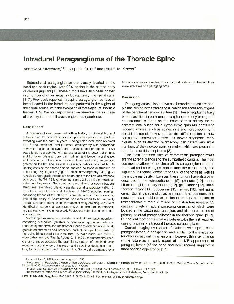
Fig. 2.-A and B, Postmyelography CT scans show an intradural, extramedullary soft-tissue mass (arrows in A) located anterior and to the left of the spinal cord (c) , displacing the cord to the right.
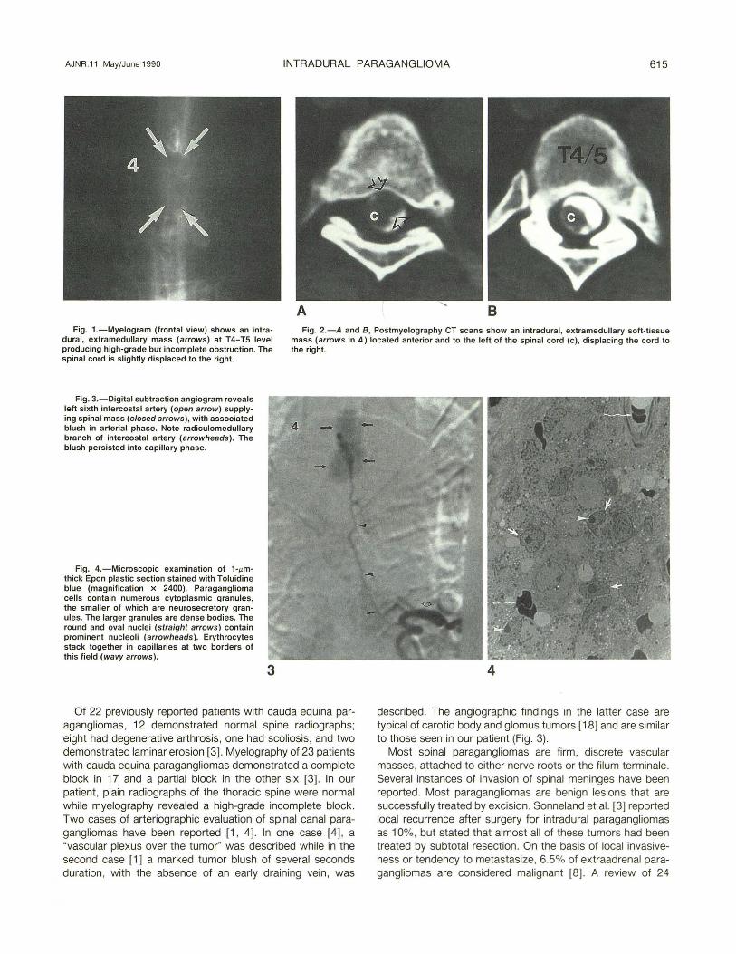
Fig. 3.-Digital subtraction angiogram reveals left sixth intercostal artery (open arrow) supplying spinal mass (closed arrows), with associated blush in arterial phase. Note radiculomedullary branch of intercostal artery (arrowheads). The blush persisted into capillary phase.
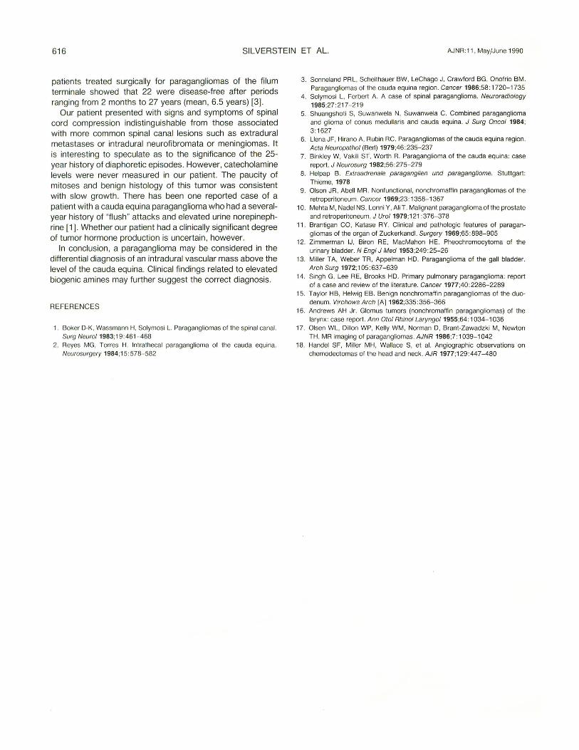
Fig. 4.-Microscopic examination of 1-11mthick Epon plastic section stained with Toluidine blue (magnification x 2400). Paraganglioma cells contain numerous cytoplasmic granules, the smaller of which are neurosecretory granules. The larger granules are dense bodies. The round and oval nuclei (straight arrows) contain prominent nucleoli (arrowheads). Erythrocytes stack together in capillaries at two borders of this field (wavy arrows).
3
Of 22 previously reported patients with cauda equina paragangliomas, 12 demonstrated normal spine radiographs; eight had degenerative arthrosis, one had scoliosis, and two demonstrated laminar erosion [3]. Myelography of 23 patients with cauda equina paragangliomas demonstrated a complete block in 17 and a partial block in the other six [3]. In our patient, plain radiographs of the thoracic spine were normal while myelography revealed a high-grade incomplete block. Two cases of arteriographic evaluation of spinal canal paragangliomas have been reported [1 , 4]. In one case [4] , a "vascular plexus over the tumor" was described while in the second case [1] a marked tumor blush of several seconds duration , with the absence of an early draining vein , was
4
described. The angiographic findings in the latter case are typical of carotid body and glomus tumors [18] and are similar to those seen in our patient (Fig . 3).
Most spinal paragangliomas are firm , discrete vascular masses, attached to either nerve roots or the filum terminale. Several instances of invasion of spinal meninges have been reported. Most paragangliomas are benign lesions that are successfully treated by excision. Sonneland et al. [3] reported local recurrence after surgery for intradural paragangliomas as 1 0%, but stated that almost all of these tumors had been treated by subtotal resection . On the basis of local invasiveness or tendency to metastasize, 6.5% of extraadrenal paragangliomas are considered malignant [8]. A review of 24
616 SILVERSTEIN ET AL. AJNR :11 , May/June 1990
patients treated surgically for paragangliomas of the filum terminale showed that 22 were disease-free after periods ranging from 2 months to 27 years (mean , 6.5 years) [3).
Our patient presented with signs and symptoms of spinal cord compression indistinguishable from those associated with more common spinal canal lesions such as extradural metastases or intradural neurofibromata or meningiomas. It is interesting to speculate as to the significance of the 25-year history of diaphoretic episodes. However, catecholamine levels were never measured in our patient. The paucity of mitoses and benign histology of this tumor was consistent with slow growth. There has been one reported case of a patient with a cauda equina paraganglioma who had a severalyear history of "flush " attacks and elevated urine norepinephrine (1 ). Whether our patient had a clinically significant degree of tumor hormone production is uncertain , however.
In conclusion , a paraganglioma may be considered in the differential diagnosis of an intradural vascular mass above the level of the cauda equina. Clinical findings related to elevated biogenic amines may further suggest the correct diagnosis .
REFERENCES
1. Boker D-K. Wassmann H, Solymosi L. Paragangliomas of the spinal canal. Surg Neuro/ 1983;19:461-468
2. Reyes MG, Torres H. Intrathecal paraganglioma of the cauda equina. Neurosurgery 1984; 15:578-582
3. Sonneland PRL, Scheithauer BW, LeChago J, Crawford BG, Onofrio BM. Paragangliomas of the cauda equina region . Cancer 1986;58: 1720-1735
4. Solymosi L, Ferber! A. A case of spinal paraganglioma. Neuroradiology 1985;27:217-219
5. Shuangshoti S, Suwanwela N, Suwanwela C. Combined paraganglioma and glioma of conus medullaris and cauda equina. J Surg Oneal 1984; 3: 1627
6. Uena JF, Hirano A, Rubin RC . Paragangliomas of the cauda equina region . Acta Neuropathol (Berl) 1979;46 :235-237
7. Binkley W, Vakili ST, Worth R. Paraganglioma of the cauda equina: case report . J Neurosurg 1982;56:275-279
8. Helpap B. Extraadrenale paragang/ien und paragang/iome. Stuttgart: Thieme, 1978
9. Olson JR, Abell MR. Nonfunctional, nonchromaffin paragangliomas of the retroperitoneum . Cancer 1969;23: 1358-1367
10. Mehta M, Nadel NS, Lenni Y, AliT. Malignant paraganglioma of the prostate and retroperitoneum. J Uro/1979;121 :376-378
11 . Brantigan CO, Katase R Y. Clinical and pathologic features of paragangliomas of the organ of Zuckerkandl. Surgery 1969;65 :898-905
12. Zimmerman IJ, Biron RE , MacMahon HE. Pheochromocytoma of the urinary bladder. N Eng/ J Med 1953;249 :25- 26
13. Miller TA, Weber TR , Appelman HD. Paraganglioma of the gall bladder. Arch Surg 1972;105 :637- 639
14. Singh G, Lee RE, Brooks HD. Primary pulmonary paraganglioma: report of a case and review of the literature. Cancer 1977;40 :2286-2289
15. Taylor HB, Helwig EB. Benign nonchromaffin paragangliomas of the duodenum. Virchows Arch (A]1962;335:356-366
16. Andrews AH Jr. Glomus tumors (nonchromaffin paragangliomas) of the larynx: case report . Ann Otol Rhino/ Laryngo/1955 ;64 : 1034- 1036
17. Olsen WL, Dillon WP, Kelly WM , NormanD, Brant-Zawadzki M, Newton TH . MR imaging of paragangliomas. AJNR 1986;7 :1 039-1042
18. Handel SF, Miller MH, Wallace S, et al. Angiographic observations on chemodectomas of the head and neck. AJR 1977;129:447-480