Embed Size (px)
Citation preview
47Egy Spine J - Volume 37 - January 2021
The
EGYPTIAN SPINEJournal
Online ISSN : 2314-8969Print ISSN: 2314-8950www.esj.journals.ekb.eg
CLINICAL ARTICLE EgySpineJ 37:47-55, 2021
Address correspondence and reprint requests: Ramy AE Teama, MD.Neurosurgery Department, Faculty of Medicine, Benha University, Benha, Egypt.Email: [email protected]
Submitted: October 25th, 2020. Accepted: December 11th, 2020. Published: January 2021.
The article does not contain information about medical device(s)/drug(s).No funds were received in support of this work.The authors report no conflict of interest.
Intradural Cauda Equina Tumors: Early Surgical Experience of a Small Neurosurgical Team
Mohamed Adawi, MD., Mohamed Elhawary, MD., Ramy Teama, MD.Neurosurgery Department, Faculty of Medicine, Benha University, Benha, Egypt.
ABSTRACTBackground Data: Cauda equina syndrome (CES) is a rare situation and is one of the few surgical emergencies in neurosurgery. While L4-L5 disc is the most common cause of CES, ependymoma and schwannoma are the most common tumors affecting cauda equina.Purpose: To present our experience and outcome of management of cauda equina tumors.Study Design: A retrospective clinical case study.Patients and Methods: We operated upon 22 cases with known cauda equina tumors at our institution in the period between October 2016 and April 2020. All patients were subjected to detailed general, neurological, and radiological evaluation both preoperatively and postoperatively according to our follow-up protocol. Moreover, the modified McCormick scale (MMS) has been used for pre- and postoperative functional evaluation. Patients underwent operation using the posterior midline approach, with neuromonitoring applied in 50% of patients.Results: The mean age was 44 ± 12.5 years. Nine (41%) patients were female and 13 (59%) were male. Ninety-one percent of patients presented with radiculopathy. Fifty percent of cases presented with schwannoma. Growth total tumor resection was achieved in 20 cases (91%). The last follow-up showed marked improvement in radiculopathy, motor power deficit, and MMS compared to preoperative values. Immediate postoperative motor power deterioration was reported in two cases. Recurrence was reported in one case of ependymoma at a two-year follow-up visit that underwent operation with no further recurrence after 1-year follow-up.Conclusion: The data of this study may suggest that most cauda equina tumors are benign and favorable outcome could be achieved in small-sized lesions with a short history and good preoperative clinical status. (2020ESJ220)Keywords: Cauda equina, Schwannoma, Ependymoma, Radiculopathy
DOI: 10.21608/ESJ.2021.58068.116

48 Egy Spine J - Volume 37 - January 2021
The
EGYPTIAN SPINEJournal
INTRODUCTION
Cauda equina syndrome (CES) can be defined as a disordered function of the cauda equina composed of lumbar, sacral, and coccygeal nerve roots surrounding the filum terminale.3 It is a rare clinical situation and is one of the few surgical emergencies in neurosurgical practice.7 The clinical presentation of CES includes saddle sensory deficit with motor, bowel, bladder, and sexual disorders.17 MRI is a very efficient tool for diagnosis and differential diagnosis of CES etiology and is characterized by being highly sensitive in detecting the lesions, their site, and size and being simple and noninvasive.15
CES can occur due to compression of cauda equina rootlets, with the most common cause being a large central L4-L5 and L5-S1 lumbar disc herniation.7,12 Less common causes are spinal injury, postmanipulation, spinal anesthesia, space-occupying lesions such as hematoma, abscess, schwannomas, ependymomas, lipomas, teratomas, arteriovenous malformations, and metastasis, ischemic injury, and inflammatory disorders.7,14 The prognosis in cases with CES is influenced by many clinical variables such as duration of symptoms, the timing of surgery, and severity of symptoms.8
In this study, we present our experience and clinical outcome in managing intradural cauda equina tumors in our neurosurgical unit.
PATIENTS AND METHODS
We operated upon 22 patients with known intradural cauda equina tumors at our university hospitals between October 2016 and April 2020. We performed this retrospective study to evaluate the surgical outcome of this group of patients. Inclusion criteria included any patient with low back pain and/or sciatica with or without neurological deficit proved to have intradural cauda equine tumor with full clinical and
radiological data, complete follow-up, and contact data. Exclusion criteria included other causes of CES and contraindication to general anesthesia. Our institution’s medical records were reviewed through this period and only 22 patients were eligible according to our inclusion criteria, while nine patients were excluded due to incomplete contact, clinical, radiological, or follow-up data. The study was approved by the ethical review board at our institution, and all patients formally consented before surgery.All patients were subjected to detailed general and neurological examination and contrast-enhanced magnetic resonance imaging (MRI) of the lumbar spine. Tumor characteristics, tumor level, and tumor length measured in mm were reported. A whole spine MRI was performed if drop metastasis of an ependymoma was suspected. Routine laboratory investigations were done for all cases. The neurological status of patients was recorded carefully, the motor power was graded from 0 to 5 on the motor power scale, and the duration of complaint was recorded. Operative details were reported with special attention given to the type of anesthesia, duration of surgery, operative blood loss, the degree of tumor resection (total, subtotal, and partial), and intraoperative complications if present. Intraoperative neurophysiological monitoring was used in eleven patients. The clinical outcome has been assessed using the modified McCormick scale (MMS) for grading the neurological function of spinal cord conditions.Surgical Technique:Surgery was performed in a prone position under general anesthesia. A midline skin incision was done at the desired levels guided by fluoroscopy in some cases. A subfascial muscle dissection followed by careful laminectomies of the planned levels to avoid injury of the dura, especially in large tumors. The laminectomy is planned to reveal the whole tumor extending above and below the tumor margins. A midline dural incision centered over the tumor and extending beyond the tumor poles was made, followed by dural hatching using Vicryl 0.3; under the operative microscope, a
49Egy Spine J - Volume 37 - January 2021
The
EGYPTIAN SPINEJournal
gentle exploration of the tumor circumference was carried out to know the tumor attachment, usually directed from the poles to the center, to achieve the goal of total removal. Bipolar coagulation of the tumor under continuous irrigation was conducted, followed by tumor removal as one piece in 17 patients or piecemeal in 5 patients. The tumor attachment was released using sharp dissection using microscissors and/or arachnoid knife and microdissectors. However, in 5 patients with schwannoma, the tumor dissection was not possible, so we had to cut the nerve rootlets (3 patients) or to leave a small sheet of the tumor over the nerve rootlets (2 patients). Closure of the dura using Vicryl 0.3 in a watertight fashion was performed and the wound was tightly closed in layers over a suction drain that was removed two days after surgery.Intraoperative Neurophysiological Monitoring:Combined motor and sensory monitoring were used in 8 cases, whereas motor monitoring was used alone in only 3 cases. Multimodality spinal cord monitoring was carried out using free-run electromyography (EMG), motor evoked potential (MEP), and continuous somatosensory evoked potential (SSEP) monitoring. The alarming signals for evoked potential were more than 50% reduction of motor nerve amplitude compared to the baseline MEP, while prolongation of sensory latency of more than 10% of the recorded baseline is considered significant. If significant changes occur during surgery, surgery is stopped for a while until evoked potential changes return to normal.Postoperative Care:Detailed neurological examination after recovery from anesthesia was performed by the attendant surgeon daily till discharge from hospital. Patients were followed up at the outpatient’s clinic after 1, 3, and 6 months, then on a yearly basis. Follow-up contrast MRI was performed 3 and 6 months after surgery, then on a yearly basis whenever possible. At each visit, the following parameters were reported: motor power, new emerging symptoms, MMS, and tumor regrowth and or recurrence.
RESULTS
Twenty-two patients were recruited for this study with different types of intradural cauda equina tumors. Nine patients (41%) were females and 13 (59%) were males. The mean age was 44 ± 12.5 (23–65) years. The mean duration of complaint was 10.1 ± 5.56 (range, 2–24) months. The most common presenting symptom was radiculopathy in 91% (N = 20) of the cases, of which 63.6% (N = 14) were bilateral. Low back pain was present in 15 patients (68.1%). Low back pain was common among patients with ependymoma (7 out of 9), while radiculopathy was common among patients with schwannoma (10 out of 11). Sphincteric dysfunctions were observed in 4 cases (18.1%); 3 of them were ependymomas. Motor deficit was observed in 7 patients (31.8%) and was at the foot level and it was of grade III or IV in most cases (4 were grade IV and 2 were grade III). Moreover, the motor deficit was bilateral in 5 patients and it was common in patients with ependymoma (6 out of 7). Sensory disturbances were reported in 63.6% of cases (N = 14) with saddle anesthesia or hypoesthesia in 3 cases; they are often bilateral at L5 and S1 (N = 8) and less common at higher levels. Preoperative functional evaluation of the spinal cord according to MMS showed that 14 patients were grade I (neurologically intact), 5 patients were grade II (motor weakness grade IV and/or saddle hypoesthesia), 2 patients were grade III (motor weakness grade III and saddle anesthesia in one patient), and 1 patient was grade IV (motor weakness grade II). We had no patients graded as grade V (Table 1).According to MRI, the reported tumor levels were as follows: L1-L2 in 3 cases, L2-L3 in 4 cases, L3-L4 in 9 cases, and L4-L5 in 6 cases. The mean tumor length was 38.77 ± 10.52 (range, 20–60) mm. Operative data showed that operative time was 160 ± 0.48 hours (range, 90–27) minutes and the operative blood loss was 275 ± 130 (range, 330–650) ml. The mean hospital stay was 3.5 ± 1.97 (range, 2–7 days). All specimens of patients
50 Egy Spine J - Volume 37 - January 2021
The
EGYPTIAN SPINEJournal
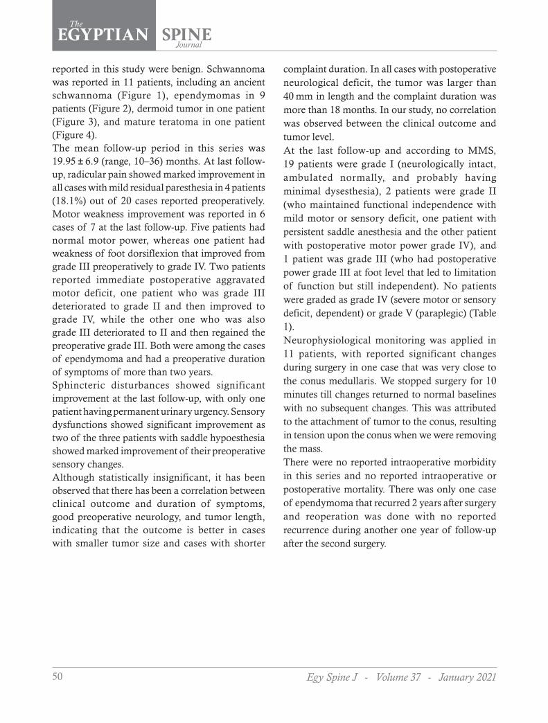
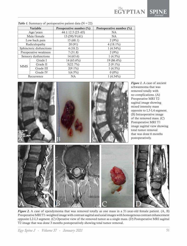
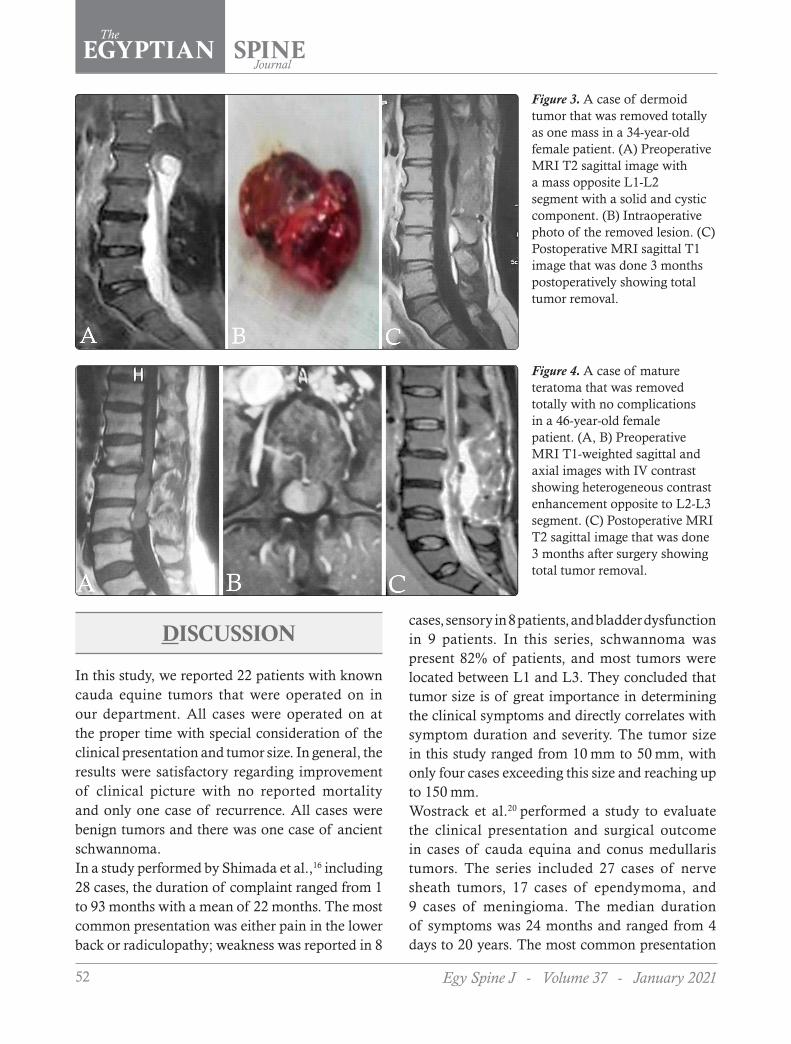
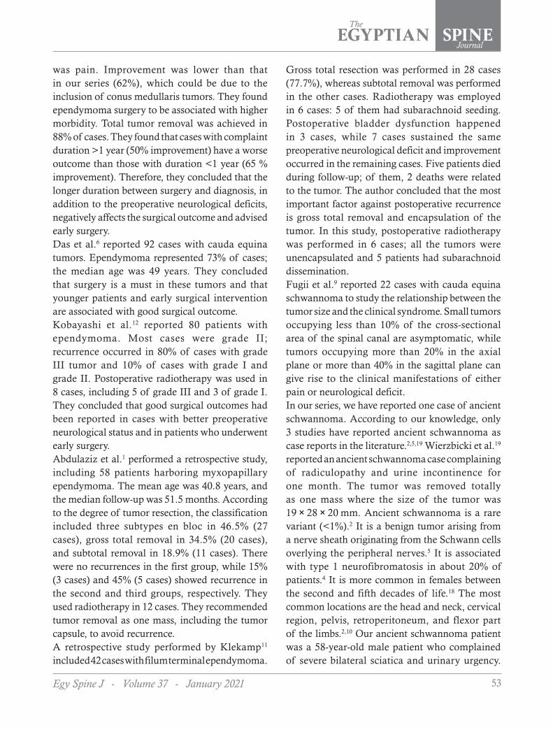
reported in this study were benign. Schwannoma was reported in 11 patients, including an ancient schwannoma (Figure 1), ependymomas in 9 patients (Figure 2), dermoid tumor in one patient (Figure 3), and mature teratoma in one patient (Figure 4).The mean follow-up period in this series was 19.95 ± 6.9 (range, 10–36) months. At last follow-up, radicular pain showed marked improvement in all cases with mild residual paresthesia in 4 patients (18.1%) out of 20 cases reported preoperatively. Motor weakness improvement was reported in 6 cases of 7 at the last follow-up. Five patients had normal motor power, whereas one patient had weakness of foot dorsiflexion that improved from grade III preoperatively to grade IV. Two patients reported immediate postoperative aggravated motor deficit, one patient who was grade III deteriorated to grade II and then improved to grade IV, while the other one who was also grade III deteriorated to II and then regained the preoperative grade III. Both were among the cases of ependymoma and had a preoperative duration of symptoms of more than two years.Sphincteric disturbances showed significant improvement at the last follow-up, with only one patient having permanent urinary urgency. Sensory dysfunctions showed significant improvement as two of the three patients with saddle hypoesthesia showed marked improvement of their preoperative sensory changes.Although statistically insignificant, it has been observed that there has been a correlation between clinical outcome and duration of symptoms, good preoperative neurology, and tumor length, indicating that the outcome is better in cases with smaller tumor size and cases with shorter
complaint duration. In all cases with postoperative neurological deficit, the tumor was larger than 40 mm in length and the complaint duration was more than 18 months. In our study, no correlation was observed between the clinical outcome and tumor level.At the last follow-up and according to MMS, 19 patients were grade I (neurologically intact, ambulated normally, and probably having minimal dysesthesia), 2 patients were grade II (who maintained functional independence with mild motor or sensory deficit, one patient with persistent saddle anesthesia and the other patient with postoperative motor power grade IV), and 1 patient was grade III (who had postoperative power grade III at foot level that led to limitation of function but still independent). No patients were graded as grade IV (severe motor or sensory deficit, dependent) or grade V (paraplegic) (Table 1).Neurophysiological monitoring was applied in 11 patients, with reported significant changes during surgery in one case that was very close to the conus medullaris. We stopped surgery for 10 minutes till changes returned to normal baselines with no subsequent changes. This was attributed to the attachment of tumor to the conus, resulting in tension upon the conus when we were removing the mass.There were no reported intraoperative morbidity in this series and no reported intraoperative or postoperative mortality. There was only one case of ependymoma that recurred 2 years after surgery and reoperation was done with no reported recurrence during another one year of follow-up after the second surgery.
51Egy Spine J - Volume 37 - January 2021
The
EGYPTIAN SPINEJournal
Figure 1. A case of ancient schwannoma that was removed totally with no complications. (A) Preoperative MRI T2 sagittal image showing mixed intensity mass opposite to L3-L4 segment. (B) Intraoperative image of the removed mass. (C) Postoperative MRI T1 image sagittal view showing total tumor removal that was done 6 months postoperatively.
Table 1. Summary of perioperative patient data (N = 22)
Variable Preoperative number (%) Postoperative number (%)Age/years 44 ± 12.5 (23–65) NA
Male/female 13 (59)/9 (41) NALow back pain 15 (68.1) 2 (9%)Radiculopathy 20 (91) 4 (18.1%)
Sphincteric disfunctions 4 (18.2) 1 (4.54%)Preoperative weakness 7 (31.8) 2 (9%)Sensory dysfunctions 14 (63.6) 1 (4.5%)
MMS
Grade I 14 (63.6%) 19 (86.4%)Grade II 5(22.7%) 2 (9.1%)Grade III 2(9.1%) 1 (4.5%)Grade IV 1(4.5%) 0 (0%)
Recurrence NA 1 (4.54%)
Figure 2. A case of ependymoma that was removed totally as one mass in a 51-year-old female patient. (A, B) Preoperative MRI T1-weighted image with contrast sagittal and axial images with homogenous contrast enhancement opposite L2-L3 segment. (C) Operative view of the removed tumor as a single mass. (D) Postoperative MRI sagittal T2 image that was done 3 months postoperatively showing total tumor removal.
52 Egy Spine J - Volume 37 - January 2021
The
EGYPTIAN SPINEJournal
Figure 4. A case of mature teratoma that was removed totally with no complications in a 46-year-old female patient. (A, B) Preoperative MRI T1-weighted sagittal and axial images with IV contrast showing heterogeneous contrast enhancement opposite to L2-L3 segment. (C) Postoperative MRI T2 sagittal image that was done 3 months after surgery showing total tumor removal.
Figure 3. A case of dermoid tumor that was removed totally as one mass in a 34-year-old female patient. (A) Preoperative MRI T2 sagittal image with a mass opposite L1-L2 segment with a solid and cystic component. (B) Intraoperative photo of the removed lesion. (C) Postoperative MRI sagittal T1 image that was done 3 months postoperatively showing total tumor removal.
DISCUSSION
In this study, we reported 22 patients with known cauda equine tumors that were operated on in our department. All cases were operated on at the proper time with special consideration of the clinical presentation and tumor size. In general, the results were satisfactory regarding improvement of clinical picture with no reported mortality and only one case of recurrence. All cases were benign tumors and there was one case of ancient schwannoma.In a study performed by Shimada et al.,16 including 28 cases, the duration of complaint ranged from 1 to 93 months with a mean of 22 months. The most common presentation was either pain in the lower back or radiculopathy; weakness was reported in 8
cases, sensory in 8 patients, and bladder dysfunction in 9 patients. In this series, schwannoma was present 82% of patients, and most tumors were located between L1 and L3. They concluded that tumor size is of great importance in determining the clinical symptoms and directly correlates with symptom duration and severity. The tumor size in this study ranged from 10 mm to 50 mm, with only four cases exceeding this size and reaching up to 150 mm.Wostrack et al.20 performed a study to evaluate the clinical presentation and surgical outcome in cases of cauda equina and conus medullaris tumors. The series included 27 cases of nerve sheath tumors, 17 cases of ependymoma, and 9 cases of meningioma. The median duration of symptoms was 24 months and ranged from 4 days to 20 years. The most common presentation
53Egy Spine J - Volume 37 - January 2021
The
EGYPTIAN SPINEJournal
was pain. Improvement was lower than that in our series (62%), which could be due to the inclusion of conus medullaris tumors. They found ependymoma surgery to be associated with higher morbidity. Total tumor removal was achieved in 88% of cases. They found that cases with complaint duration >1 year (50% improvement) have a worse outcome than those with duration <1 year (65 % improvement). Therefore, they concluded that the longer duration between surgery and diagnosis, in addition to the preoperative neurological deficits, negatively affects the surgical outcome and advised early surgery.Das et al.6 reported 92 cases with cauda equina tumors. Ependymoma represented 73% of cases; the median age was 49 years. They concluded that surgery is a must in these tumors and that younger patients and early surgical intervention are associated with good surgical outcome.Kobayashi et al.12 reported 80 patients with ependymoma. Most cases were grade II; recurrence occurred in 80% of cases with grade III tumor and 10% of cases with grade I and grade II. Postoperative radiotherapy was used in 8 cases, including 5 of grade III and 3 of grade I. They concluded that good surgical outcomes had been reported in cases with better preoperative neurological status and in patients who underwent early surgery.Abdulaziz et al.1 performed a retrospective study, including 58 patients harboring myxopapillary ependymoma. The mean age was 40.8 years, and the median follow-up was 51.5 months. According to the degree of tumor resection, the classification included three subtypes en bloc in 46.5% (27 cases), gross total removal in 34.5% (20 cases), and subtotal removal in 18.9% (11 cases). There were no recurrences in the first group, while 15% (3 cases) and 45% (5 cases) showed recurrence in the second and third groups, respectively. They used radiotherapy in 12 cases. They recommended tumor removal as one mass, including the tumor capsule, to avoid recurrence.A retrospective study performed by Klekamp11 included 42 cases with filum terminal ependymoma.
Gross total resection was performed in 28 cases (77.7%), whereas subtotal removal was performed in the other cases. Radiotherapy was employed in 6 cases: 5 of them had subarachnoid seeding. Postoperative bladder dysfunction happened in 3 cases, while 7 cases sustained the same preoperative neurological deficit and improvement occurred in the remaining cases. Five patients died during follow-up; of them, 2 deaths were related to the tumor. The author concluded that the most important factor against postoperative recurrence is gross total removal and encapsulation of the tumor. In this study, postoperative radiotherapy was performed in 6 cases; all the tumors were unencapsulated and 5 patients had subarachnoid dissemination.Fugii et al.9 reported 22 cases with cauda equina schwannoma to study the relationship between the tumor size and the clinical syndrome. Small tumors occupying less than 10% of the cross-sectional area of the spinal canal are asymptomatic, while tumors occupying more than 20% in the axial plane or more than 40% in the sagittal plane can give rise to the clinical manifestations of either pain or neurological deficit.In our series, we have reported one case of ancient schwannoma. According to our knowledge, only 3 studies have reported ancient schwannoma as case reports in the literature.2,5,19 Wierzbicki et al.19 reported an ancient schwannoma case complaining of radiculopathy and urine incontinence for one month. The tumor was removed totally as one mass where the size of the tumor was 19 × 28 × 20 mm. Ancient schwannoma is a rare variant (<1%).2 It is a benign tumor arising from a nerve sheath originating from the Schwann cells overlying the peripheral nerves.5 It is associated with type 1 neurofibromatosis in about 20% of patients.4 It is more common in females between the second and fifth decades of life.18 The most common locations are the head and neck, cervical region, pelvis, retroperitoneum, and flexor part of the limbs.2,10 Our ancient schwannoma patient was a 58-year-old male patient who complained of severe bilateral sciatica and urinary urgency.
54 Egy Spine J - Volume 37 - January 2021
The
EGYPTIAN SPINEJournal
He had a large mass opposite L3-L4 (Figure 1)
that was removed totally with no complications
and the patient was greatly improved with mild
postoperative dysesthesia.
This study has some limitations, including the
retrospective nature, the small sample size,
the short-term follow-up, and the shortages of
detailed histopathological data and adjuvant
therapy. Further prospective multicenter studies
with a large sample size and long-term follow-up
are highly recommended.
CONCLUSION
The data of this study may suggest that most cauda
equina tumors are benign and favorable outcomes
could be achieved in small-sized lesions with a
short history and good preoperative clinical status.
REFERENCES
1. Abdulaziz M, Mallory GW, Bydon M, Ramos
RD, Ellis JA, Laack NN, et al: Outcomes
following myxopapillary ependymoma
resection: the importance of capsule integrity.
Neurosurg Focus 39 (2):E8, 2015
2. Azli SM, Abd Rahman IG, Salzihan MSM.
Ancient schwannoma of the conus medullaris.
Medical Journal of Malaysia 62(3):256–258,
2007
3. Bennett SJ, Katzman GL, Roos RP, Mehta AS,
Ali S: Neoplastic cauda equina syndrome: a
neuroimaging-based review. Pract Neurology
16(1):35–41, 2016
4. Bhatia RK, Banerjea A, Ram M, Lovett BE:
Benign ancient Schwannoma of the abdominal
wall: an unwanted birthday present, BMC
Surgery 10: article 1, 2010
5. Darwish BS, Balakrishnan V, Maitra R:
Intramedullary ancient schwannoma of the
cervical spinal cord: case report and review
of literature. Journal of Clinical Neuroscience
9(3):321–323, 2002
6. Das S, Ugiliweneza B, Burton E: Tumors in
the cauda equina: a SEER analysis of tumor
types and predicators of outcome. Neuro-
Oncology 6:82–89, 2018
7. Deniz K, Pararajasingham J: Cauda equina
syndrome, InnovAit 4 (10):551–555, 2011
8. Elsawaf A, Gallhom A, Khattab M: Peri-
operative radiological factors predicting
outcome of discogenic cauda equina
syndrome. Egy Spine J 25:54–64, 2017
9. Fujii K, Sakane M, Abe T, Nakagawa T, Sakai
S, Tatsumura M, et al: Tumor occupation in
the spinal canal and clinical symptoms of
cauda equina schwannoma: An analysis of 22
cases. Asian Spine J 10(6):1079–1084, 2016
10. Isobe K, Shimizu T, Akahane T, Kato H:
Imaging of ancient schwannoma. American
Journal of Roentgenology 183(2):331–336,
2004
11. Klekamp J: Spinal ependymomas. Part
2: Ependymomas of the filum terminale.
Neurosurg Focus 39(2):E7, 2015
12. Kobayashi K, Ando K, Kato F, Kanemura
T, Sato K, Kamiya M, et al: Surgical outcomes
of spinal cord and cauda equina ependymoma:
Postoperative motor status and recurrence for
each WHO grade in a multicenter study, J
Orthop Sci 23(4):614–621, 2018
13. Lavy C, James A, Wilson-MacDonald J,
Fairbank J: Cauda equina syndrome. BMJ
338:881–884, 2009
14. Orendacova J, Cizkova D, Kafka J, Lukacova
N, Marsala M, Sulla I, et al: Cauda equina
syndrome. Prog Neurobiology 64:613–637,
2001
55Egy Spine J - Volume 37 - January 2021
The
EGYPTIAN SPINEJournal
15. Sedeek KH, Aboualfotouh K, Hassanein SM, Osman NM, Shalaby MH: Role of MRI evaluation in acute secondary inability to walk in children. Egyptian Journal of Radiology and Nuclear Medicine 52:37, 2021
16. Shimada Y, Miyakoshi N, Kasukawa Y, Hongo M, Ando S, Itoi E: Clinical features of cauda equina tumors requiring surgical treatment. Tohoku J Exp Med 209(1):1–6, 2006
17. Tharmabala M, LaBrash D& Kanthan R: Acute cauda equina syndrome secondary to lumbar chordoma: case report and literature review. The Spine Journal 13:35–43, 2013
18. White W, Shiu MH, Rosenblum MK, Erlandson RA, Woodruff JM: Cellular
schwannoma. Aclinicopathologic study of 57 patients and 58 tumors. Cancer 66(6):1266–1275, 1990
19. Wierzbicki V, Pesce A, Marrocco L, Piccione E, Frati A, Caruso R, et al: Ancient Schwannoma of the Cauda Equina: Our Experience and Review of the Literature (case report). Case Reports in Surgery Article ID 7930521, 4 pages, 2016
20. Wostrack M1, Shiban E, Obermueller T, Gempt J, Meyer B, Ringel F: Conus medullaris and cauda equina tumors: clinical presentation, prognosis, and outcome after surgical treatment: clinical article. J Neurosurg Spine 20(3):335–343, 2014
الملخص العربى
اورام شبيه ذنب الفرس داخل الام الجافية في الانسان خبرة جراحية لفريق صغير من الجراحين بجامعة بنهاالبيانـات الخلفيـة: متلازمـة ذيـل الفـرس هـي حالـة نـادرة وهـي واحـدة مـن حـالات الطـوارئ الجراحيـة الصغيـرة فـي جراحـة الأعصـاب. فـي حيـن أن القـرص L4-L5 هـو السـبب الأكثـر شـيوعًا لــمتلازمة ذيـل الفـرس ، فـإن الـورم البطاني
العصبي والورم الشفاني هما أكثر الأورام شيوعًا التي تصيب ذيل الفرس.الغرض: تقديم خبرتنا ونتائج علاج أورام ذيل الفرس.
تصميم الدراسة: دراسة حالة سريرية بأثر رجعي.المرضـى والطـرق: قمنـا بإجـراء 22 حالـة مـع أورام ذنـب الفـرس المعروفـة فـي مؤسسـتنا فـي الفتـرة مـا بيـن أكتوبر 2016 وأبريـل 2020. خضـع جميـع المرضـى لتقييـم مفصـل عـام وعصبـي وأشـعاعي علـى حـد سـواء قبـل الجراحـة وبعدهـا وفقًـا لبروتوكـول المتابعـة الخـاص بنـا. تـم اسـتخدام مقياس ماكورميـك المعـدل (MMS) للتقييم الوظيفي قبل وبعد العملية الجراحية أيضًا. تم تشـغيل المرضى من خلال نهج خط الوسـط الخلفي مع المراقبة العصبية في
50 ٪ من المرضى.النتائج: كان متوسط العمر 44 ± 12.5 سنة. تسعة )41 ٪( من المرضى كانوا من الإناث و 13 (59 ٪( من الذكور. واحـد وتسـعون فـي المئـة مـن المرضـى يعانـون مـن اعتـلال الجـذور. تـم عـرض خمسـين بالمائـة من الحـالات المصابة بورم شفاني. تم تحقيق استئصال الورم الكلي للنمو في 20 حالة )91٪(. أظهرت المتابعة الأخيرة تحسنًا ملحوظًا فـي كل مـن اعتـلال الجـذور ، وعجـز القـوة الحركيـة ، والرسـائل متعـددة الوسـائط مقارنـةً بما قبـل الجراحة. تم الإبلاغ عن تدهور فوري في قوة المحرك بعد الجراحة في حالتين. تم الإبلاغ عن ارتجاع في حالة واحدة من الورم البطاني
العصبي في سنتان من المتابعة وتم إجراء الجراحة دون تكرار آخر بعد سنة واحده من المتابعة.الخلاصـة: قـد تشـير بيانـات هـذه الدراسـة إلـى أن معظـم أورام ذيـل الفـرس حميـدة ويمكن تحقيق نتائـج إيجابية في
الآورام صغيرة الحجم ذات التاريخ القصير والحالة السريرية الجيدة قبل الجراحة.