Embed Size (px)
Citation preview
Invited Review
The conceptual basis for a new classification of the coccidia
Astrid M. Tentera,*, John R. Bartab, Ian Beveridgec, Donald W. Duszynskid, Heinz Mehlhorne,David A. Morrisonf, R.C. Andrew Thompsong, Patricia A. Conradh
aInstitut fur Parasitologie, Tierarztliche Hochschule Hannover, Bunteweg 17, D-30559 Hannover, GermanybDepartment of Pathobiology, University of Guelph, Guelph, Ontario, Canada N1G 2W1
cDepartment of Veterinary Science, University of Melbourne, 250 Princes Highway, Werribee, VIC 3030, AustraliadDepartment of Biology, The University of New Mexico, Albuquerque, NM 87131, USA
eInstitut fur Zoomorphologie, Zellbiologie und Parasitologie, Heinrich-Heine-Universitat, Universitatsstraße 1, D-40225 Dusseldorf, GermanyfMolecular Parasitology Unit, University of Technology, Sydney, Westbourne Street, Gore Hill, NSW 2065, Australia
gDivision of Veterinary and Biomedical Sciences, Murdoch University, WA 6150, AustraliahDepartment of Pathology, Microbiology and Immunology, School of Veterinary Medicine, University of California Davis, One Shields Avenue,
Davis, CA 95616-8739, USA
Received 24 December 2001; received in revised form 12 February 2002; accepted 18 February 2002
Abstract
At the joint meeting of the 8th International Coccidiosis Conference and the Annual Scientific Meeting of the Australian Society for
Parasitology in Palm Cove, Australia, in July 2001, a Controversial Roundtable was held on ‘New classification of coccidia’. The aim of this
Roundtable was to stimulate and encourage discussion and debate on current classification schemes for the group of parasitic protozoa known
as the eimeriid coccidia. In the past, such classifications have been based only on phenotypic characters such as morphology, ultrastructure,
life cycles, and host specificity. However, over the past 10–15 years, molecular phylogenetic studies on taxa of the eimeriid coccidia have
revealed that several of the families, subfamilies, and genera that have been erected based on non-molecular characters are paraphyletic.
Therefore, this Roundtable was an important forum for initial discussions on how a new and more comprehensive classification of the
eimeriid coccidia, which takes into consideration both phenotypic and molecular characters, can be devised. The stimulus came from invited
speakers who gave introductions into selected areas of taxonomy and classification. Following these introductions, a more general discussion
with the audience addressed potential steps that may be taken in future work. This review is the immediate outcome of the Roundtable. It
describes advantages and disadvantages of the use of phenotypic or molecular characters as the base for taxonomic schemes for eimeriid
coccidia. It gives specific examples for drawbacks of current classifications based only on phenotypic characters as well as potential pitfalls
associated with the use of only molecular phylogenies. It addresses current controversies as well as rules of taxonomy and nomenclature
relevant for the eimeriid coccidia. Finally, it recommends the establishment of an international group of scientists to meet on a regular basis,
stimulate further discussions, and give direction on how the final goal, i.e. a proposal for a revised, and widely accepted, classification of the
eimeriid coccidia, may be achieved. q 2002 Published by Elsevier Science Ltd. on behalf of Australian Society for Parasitology Inc.
Keywords: Coccidia; Classification; Taxonomy; Phylogeny; Ultrastructure; Ribosomal RNA
1. Introduction
The coccidia are a diverse group of parasitic protozoa.
Some species of coccidia are homoxenous and strictly host
specific, other species have complex heteroxenous life
cycles that involve a broad range of different host species.
Since their first description in the 19th century, coccidia
have been found in almost every animal examined, includ-
ing humans. Thus, while the oldest species and genera of
coccidia have now been known for more than a century,
others have been described and named only recently.
During the 19th and early 20th centuries, classifications
of protozoa were based mainly on organelles of locomotion,
but with increasing knowledge on their morphology, biol-
ogy, life cycle, and host specificity, a broad range of pheno-
typic characters have been used to classify protozoa into
different taxonomic groups (reviewed in Cox, 1991).
Thus, several schemes for the classification of protozoa,
including the coccidia, have been proposed during the
second half of the 20th century. The first major reclassifica-
tion was undertaken by an international ‘Committee on
Taxonomy and Taxonomic Problems’ which was set up
International Journal for Parasitology 32 (2002) 595–616
0020-7519/02/$20.00 q 2002 Published by Elsevier Science Ltd. on behalf of Australian Society for Parasitology Inc.
PII: S0020-7519(02)00021-8
www.parasitology-online.com
* Corresponding author. Tel.: 149-511-953-8717; fax: 149-511-953-
8870.
E-mail address: [email protected] (A.M. Tenter).
by the Society of Protozoologists in 1954 and published a
revised classification of the phylum Protozoa 10 years later
(Honigberg et al., 1964). This classification was based on
phenotypic characters of about 48,000 species of protozoa
recorded at the time and defined 140 taxa at suprafamilial
levels. However, it incorporated only few ultrastructural
data, which became available after the advent of electron
microscopy in the 1950s and 1960s, and was thus
confounded by incomplete knowledge on heteroxenous
life cycles such as those of the tissue cyst-forming coccidia
that were elucidated only in the 1970s (reviewed in Tenter
and Johnson, 1997). Therefore, although the classification
by Honigberg et al. (1964) was a major advance over the
variety of traditional classification schemes that had been in
use until the 1960s, it already appeared to be out of date by
the end of the 1970s.
Consequently, in 1980 the ‘Committee on Systematics
and Evolution’ of the Society of Protozoologists, again
consisting of an international group of experts, published
another revised classification of the protozoa that was
based on knowledge of more than 65,000 species, of
which more than half were extant and about 10,000 were
parasitic, and incorporated information gained from ultra-
structural data (Levine et al., 1980). This committee classi-
fied the Protozoa, then a subkingdom, into seven phyla, gave
descriptions for 234 higher taxa down to the level of subor-
der, and gave examples of representative genera of each.
The classification by Levine et al. (1980) was widely
adopted by authors of zoological and biological textbooks,
and most groups of protozoa defined in it were generally
accepted by many protozoologists until the early 1990s.
However, a major drawback of this classification was that
only few molecular data from protozoa were available when
it was produced. Over the past three decades, molecular data
have been increasingly used to infer phylogenetic relation-
ships among various protozoa. The results of such molecular
phylogenetic studies supported the monophyly of some taxa
defined by Levine et al. (1980), but several other taxa
described in this classification have been found to be para-
phyletic or even polyphyletic. This has led to the recently
revised ‘interim’ classifications of unicellular organisms by
Cavalier-Smith (1993) and Corliss (1994). However, these
classifications vary greatly from each other. In fact, they
probably represent two extremes with respect to the number
of taxonomic categories that have been used in modern
classifications of protozoa (Fig. 1), and neither has been
widely accepted.
Thus, at the beginning of the 21st century, it appears
appropriate to undertake another major revision of the clas-
sification of protozoa that, for the first time, will be based on
both phenotypic and molecular characters. Such a classifi-
cation should reflect the phylogeny of the taxonomic groups
described in it as accurately as possible to ensure its stability
for a reasonable number of years, and again should be
produced by an international group of experts to encourage
wide acceptance. Considering the number of new protozoan
species described since 1980, which may now have
exceeded well over 100,000, and the large amount of ultra-
structural, biochemical, and molecular biological data that
have been generated for a broad range of protozoa, such a
revision will be an enormous task. A revised classification
of the protozoa at higher taxonomic levels is currently being
deliberated by another committee of the Society of Proto-
zoologists and is hoped to become available within the next
few years.
To contribute to the current efforts of producing a more
accurate and stable classification of protozoa, a Controver-
sial Roundtable on ‘New classification of coccidia’ was held
during the joint meeting of the 8th International Coccidiosis
Conference and the Annual Scientific Meeting of the
Australian Society for Parasitology at Palm Cove, Australia,
in July 2001. This Roundtable focussed on one group of
coccidia, i.e. the eimeriid coccidia which are one of the
largest groups of parasitic protozoa and comprise many
species of veterinary and medical importance. While
changes at higher taxonomic levels usually have no effect
on applied areas of parasitology, taxonomic changes at
subfamilial levels may change the names of the organisms
involved and thus may be less acceptable to a wide commu-
nity of general parasitologists, practitioners, and clinicians
working in applied fields of veterinary and human medicine.
However, although stability of nomenclature is greatly
desired to aid communication and avoid confusion among
protozoologists and non-protozoologists, an accurate classi-
fication of unicellular organisms that reflects their natural
history also has impact on practical applications such as in
the areas of differential diagnosis and drug development. It
is also important for the elucidation of unknown life cycles
and epidemiological investigations, in particular on emer-
ging diseases. For example, the recognition of the human
pathogen Pneumocystis carinii, previously classified with
the protozoa, as a fungus increased knowledge and enabled
new experiments to improve treatment of pneumocystic
pneumonia (Edman et al., 1988).
The eimeriid coccidia are one of the more controversial
groups of protozoa, and their taxonomy and classification
have been debated for more than 50 years (reviewed in
Cox, 1991, 1994; Tenter and Johnson, 1997). Therefore,
the aim of the Roundtable held at Palm Cove was to initiate
discussion on how a new and more comprehensive classifica-
tion of eimeriid coccidia can be devised. The stimulus came
from invited speakers who gave introductions into selected
areas relevant to the taxonomy and classification of coccidia.
Following these introductions to specific topics, a more
general discussion with the audience, which consisted of
more than 50 participants with interest in taxonomy, general
biology, immunology, or molecular biology of coccidia as
well as applied areas of coccidiosis, took place. This publica-
tion describes the immediate outcome of the Roundtable and
subsequent discussions that continued over the following
days of the conference. Individual authors have contributed
various sections (taxonomy, Donald W. Duszynski; ultra-
A.M. Tenter et al. / International Journal for Parasitology 32 (2002) 595–616596

structure, Heinz Mehlhorn; molecular characters, John R.
Barta; Cryptosporidium, R.C. Andrew Thompson;
phenetic/phylogenetic classifications, Ian Beveridge; future
steps, David A. Morrison and Patricia A. Conrad) as well as
contributions throughout the manuscript. It should be noted
that this publication reflects the cooperative efforts of the
authors, aided by the opinions of other participants of the
Roundtable, and thus does not necessarily represent the
views of any one single author. It is not complete and does
not cover all aspects relevant to the taxonomy and classifica-
tion of the coccidia. Rather, it should be taken as a stimulus
for further discussion, preferably in conjunction with other
conferences of interest to scientists and practitioners working
on coccidia and the diseases caused by them.
A.M. Tenter et al. / International Journal for Parasitology 32 (2002) 595–616 597
Fig. 1. Comparison of classifications of protozoa at suprafamilial levels proposed during the second half of the 20th century. Classifications have been based on
phenotypic characters only, except for the classifications of Cavalier-Smith (1993) and Corliss (1994) who included information derived from phylogenetic
reconstruction based on 18S rRNA gene sequences. Only those parts relevant to coccidia and their closest sister-taxa (gregarines, haemosporids, and
piroplasms) are shown. Total numbers of taxa at the next lower hierarchic level, if not listed here, are given in brackets. Taxa containing eimeriid coccidia
of medical and/or veterinary importance are shaded; *, only taxa containing parasitic protozoa were listed in the classification; ** in this classification the genus
Cryptosporidium is classified with the adeleid coccidia. For cross-referencing and retrieval purposes, Margulis et al. (1990) is mentioned here in the text.

2. Traditional classifications of coccidia
The phylum Apicomplexa consists of a diverse group of
parasitic protozoa that are characterised by an apical
complex consisting of special organelles at the anterior
end of their invasive life-cycle stages (Levine, 1970). This
apical complex facilitates the entry of the parasites into their
host cells (Soldati et al., 2001; Tomley et al., 2001). Except
for the, still disputed, addition of the genus Perkinsus to this
phylum, the whole group remains essentially identical to the
phylum Sporozoa which was erected more than a century
ago (Leuckart, 1879). The largest group of organisms in the
phylum Apicomplexa are the coccidia which contain some
of the most advanced sporozoa (reviewed in Levine, 1988;
Cox, 1994; Lee et al., 2001). They are usually classified into
this phylum as a class or subclass (Fig. 1) and are further
divided into lower taxa based on phenotypic characters such
as morphology and the pattern of their life cycles that alter-
nate between asexual and sexual phases of reproduction
(reviewed in Levine, 1985, 1988; Vivier and Desportes,
1990; Tenter and Johnson, 1997; Lee et al., 2001). While
some authors include the haemosporids, the piroplasms, or
both in the Coccidia (Honigberg et al., 1964; Levine et al.,
1980; Levine, 1985; Kreier and Baker, 1987; Mehlhorn and
Walldorf, 1988; Bush et al., 2001; Mehlhorn, 2001), the
coccidia sensu stricto contain two subgroups, i.e. the adeleid
and eimeriid coccidia (Cavalier-Smith, 1993; Cox, 1994).
The eimeriid coccidia (order Eimeriida or suborder
Eimeri(or)ina, see Figs. 1 and 2) comprise members with
a typical coccidian life cycle consisting of three phases: one
or more generations of asexual multiplication by merogony,
sexual reproduction by gamogony, and asexual reproduction
by sporogony. However, unlike other coccidia, gamogony
in the eimeriid coccidia is characterised by the independent
development of macrogametes (female) and microgametes
(male), with the latter being motile and often produced in
large numbers. In addition, sporozoites are typically
enclosed in sporocysts that form within oocysts which are
passed into the environment as a resistant stage (Levine et
al., 1980; Levine, 1985, 1988; Kreier and Baker, 1987; Cox,
1994; Hausmann and Hulsmann, 1996; Lee et al., 2001).
Most species of eimeriid coccidia are homoxenous.
However, the tissue cyst-forming coccidia are obligately
or facultatively heteroxenous, with an asexual phase
(merogony) of the life cycle leading to the formation of
tissue cysts in various tissues of an intermediate host and
the sexual phase (gamogony) to the formation of oocysts in
the intestine of a definitive host (reviewed in Dubey, 1993;
Tenter and Johnson, 1997). While the taxonomic position of
eimeriid coccidia at suprafamilial levels is relatively consis-
tent in traditional classifications of protozoa (Fig. 1), their
classification into families, subfamilies, and genera has been
inconsistent for many years (reviewed in Tenter and John-
son, 1997; Fig. 2). In fact, it has been considered ‘one of the
most controversial areas of parasitic protozoology’ (Cox,
1991). Therefore, a stable classification of the eimeriid
coccidia, which comprise many important parasites of
humans and animals, at the subordinal level is urgently
needed.
There are several pre-requisites to achieve this goal such
as general agreement of the scientific community to follow
the same rules of taxonomy and nomenclature, in particular
when describing new species, consistency of nomenclature,
agreement on what weight to assign to descriptions of indi-
vidual stages of parasites with unknown life cycles, agree-
ment on what weight to assign to molecular characters
versus phenotypic characters, and which molecular charac-
ters to use for meaningful inference of phylogenetic rela-
tionships.
3. Current taxonomy and taxonomic problems ofeimeriid coccidia
3.1. Defining the terms
Taxonomy is the most basic activity in biology because it
involves the discovery, analysis of variation (quantitatively
or qualitatively), naming (nomenclature), ordering (classifi-
cation/systematics), and communication (publication) of the
patterns of life-forms. It makes these life-forms (species)
historical, temporal, and spatial entities that are the essential
elements of biodiversity, i.e. the genealogical packages that
store and transmit the information that leads to the interac-
tions within complex ecosystems. Species are the atoms of
biology, they comprise our periodic table.
Thus, taxonomy involves a number of important compo-
nents, many of which have been done poorly or ignored by
taxonomists of coccidia, even to the present day. Here, we
briefly address the primary components of taxonomy
(collection, identification, nomenclature, systematics) and
comment on the status of these interrelated facets as they
concern the coccidia.
3.1.1. Collection
Parasitologists are unique among their colleagues who
study plants, animals, or microbes because they must collect
not only the organism, but also the host(s) in/on which the
organism resides. As a general rule, collections must
include: (1) locality data, (2) reliably identified host speci-
men(s), if possible a statistically significant series, and (3)
the parasite (in its different stages).
(1) Obligately intracellular protozoa such as the coccidia
are associated intimately with their host taxon. Answers
to important questions about both the host and its coccidia
depend heavily on good locality data. Thus, data on the
parasite should always include the collection locality,
which is the locality of the individual host from which
the type (parasite) specimen was obtained. Such data give
valuable insight to the relationships among geography
and host and parasite phylogeny that, if available, could
A.M. Tenter et al. / International Journal for Parasitology 32 (2002) 595–616598
A.M. Tenter et al. / International Journal for Parasitology 32 (2002) 595–616 599
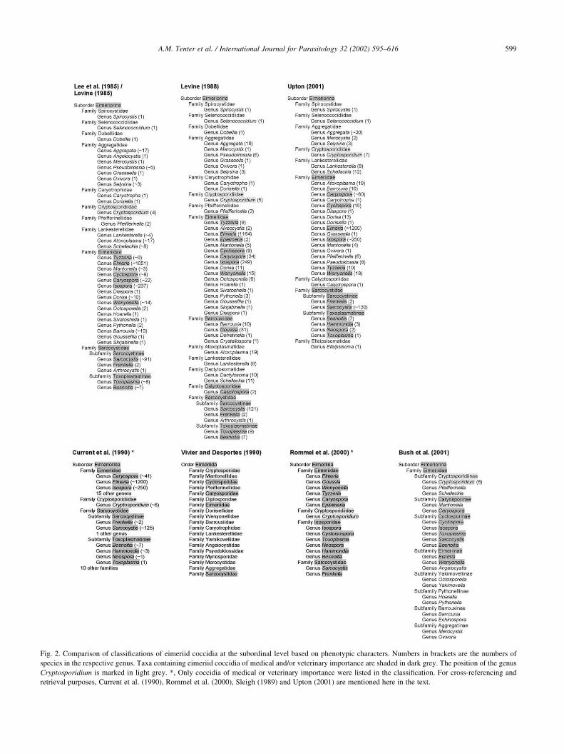
Fig. 2. Comparison of classifications of eimeriid coccidia at the subordinal level based on phenotypic characters. Numbers in brackets are the numbers of
species in the respective genus. Taxa containing eimeriid coccidia of medical and/or veterinary importance are shaded in dark grey. The position of the genus
Cryptosporidium is marked in light grey. *, Only coccidia of medical or veterinary importance were listed in the classification. For cross-referencing and
retrieval purposes, Current et al. (1990), Rommel et al. (2000), Sleigh (1989) and Upton (2001) are mentioned here in the text.

revolutionise our understanding of the historical biogeo-
graphy of these organisms. With good locality data, we
can gain new perspectives on both the distribution and
diversity of the coccidia in their host populations.
(2) Frey et al. (1992) first recommended that the host
from which the ‘type’ of a new parasite species is
described be designated as the ‘symbiotype’ host. They
argued that the accurate identification of a host is an
important component in the taxonomic recognition of a
new parasite species (especially for coccidia) and that
curatorial management and safekeeping of the symbio-
type host was desirable. Brooks (1993) concurred,
emphasising that parasite evolutionary biology necessa-
rily involves the host(s) of the parasite and, therefore,
requires ‘the best possible estimates of the host species
involved’, whether one is discussing host specificity in
parasite evolution, host switching in parasite speciation,
patterns or processes of parasite–host coevolution, or
differentiation of evolutionary or ecological components
of community evolution. To emphasise his point, Brooks
(1993) noted that prior to the last quarter of the 20th
century, herpetologists believed that only one species of
leopard frog, Rana pipiens, ranged from near the Arctic
Circle to Panama and that this species hosted dozens of
parasite species of amazing diversity. However, by the
beginning of the 21st century, herpetologists recognised
that leopard frogs represent a clade of 27 (or more) extant
and recently extinct species. With the exception of two
studies by Brooks (1976, 1979), no other specimen of
leopard frog is known to have been deposited in museum
collections. Thus, there is no way to determine the speci-
fic identity of the ranid hosts reported in all other surveys
to harbour helminth, arthropod, and protozoan parasites.
Therefore, archiving hosts from which parasites are
collected and described is critical to preserving their
true identity in perpetuity.
(3) There are three critical elements when working with
coccidia or other parasites once they have been collected
from the host. First, they need to be isolated, handled,
fixed, and stored properly. Second, as much qualitative
and quantitative parasite data as possible should be
recorded during observation to be able to write the most
accurate, detailed description possible. Guidelines cover-
ing these techniques for homoxenous eimeriid coccidia
have been provided by Duszynski and Wilber (1997).
However, there are currently no such guidelines for
heteroxenous coccidia, and a significant amount of the
confusion that exists about the taxonomy and nomencla-
ture of heteroxenous coccidia such as the tissue cyst-
forming coccidia has come from incomplete species
descriptions. Prior to the 1970s these were based either
on the oocyst or sporocyst excreted by the definitive host,
without knowledge of any intermediate host, or only on
life-cycle stages found in an intermediate host, without
knowledge of the definitive host (reviewed in Tenter and
Johnson, 1997). Unfortunately, even today many authors
describe and assign names to tissue cyst-forming coccidia
on the basis of only individual stages found in an inter-
mediate host, and the complete life cycles of many of
these species are still unknown. Finally, the appropriate
stage(s) of the parasite must be archived in an accredited
museum from which they can be loaned to interested
workers. Bandoni and Duszynski (1988) provided both
philosophical and practical arguments why archiving
type specimens of coccidia is important and suggested a
template for archiving photomicrographs of sporulated
oocysts of eimeriid coccidia. Duszynski (1999) reviewed
the name-bearing ‘types’ for photomicrographs of proto-
zoa and suggested that ‘photosyntype’ may be the best
term to use when a series of several photomicrographs of
different sporulated oocysts, representing the same new
species, is submitted to a museum as part of the original
publication and naming process. However, this is not
sufficient for polyxenous coccidia which use a wide
range of different host species, or for the tissue cyst-form-
ing coccidia which often have a very similar oocyst
morphology, but differ by their intermediate host specifi-
city and the type and location of life-cycle stages that are
formed in the intermediate host(s).
3.1.2. Identification
Identification is intimately entwined with collection. The
locality, the host, and the parasite all must be identified
accurately. Global Positioning Systems data, archiving the
symbiotype host (Bandoni and Duszynski, 1988; Brooks,
1993), and photosyntypes of sporulated oocysts (Duszynski,
1999) are tools for accurate identification. Host-specificity
issues dissolve without accurate host and parasite identifica-
tions. Therefore, it is important to note as many of the
morphological details of sporulated oocysts as can be iden-
tified when describing eimeriid coccidia (Fig. 3). Life-cycle
details and cross-transmission studies (Hnida and
Duszynski, 1999a) can greatly aid one’s decisions in making
specific identifications of morphologically similar sporu-
lated oocysts from closely related hosts, and are also essen-
tial for identification of the potential natural host range of
polyxenous coccidia (Tenter and Johnson, 1997; Mugridge
et al., 1999b; Tenter et al., 2000). Finally, if possible, pure
samples of oocysts and/or tissue cysts should be preserved
in absolute ethanol so that molecular data may be obtained
later, i.e. when resources and new, more sophisticated meth-
ods are available, to support or refute previous identifica-
tions based solely on morphology (Hnida and Duszynski,
1999b). This will also enable phylogenetic relationships to
be viewed in entirely new ways, in particular, where geno-
types are associated with morphological characters. For
example, it has recently been found that the presence or
absence of a Stieda body or an oocyst residuum is associated
with different genetic lineages within the current genera
Isospora and Eimeria (Barta et al., 1997; Zhao and
Duszynski, 2001a).
A.M. Tenter et al. / International Journal for Parasitology 32 (2002) 595–616600

3.1.3. Nomenclature
The relationship between naming an organism and the
type specimen cannot be overemphasised. Names are
important because they provide an unambiguous label for
each species. The type specimen serves as the anchor for
this name and, to some extent, it is the name (Mayr et al.,
1953). Rules for guiding zoologists to arrive at names for
taxa that are correct under given circumstances are
presented in the International Code of Zoological Nomen-
clature (Ride et al., 1999; see Section 7).
3.1.4. Systematics
Initially, the ‘ordering’ of organisms into groups is based
on their perceived similarities and differences, starting with
species as the smallest unit. These are then classified into
successively larger groups, i.e. species within genera within
families within orders, etc. (Figs. 1 and 2). This ‘nested
hierarchy’ implies a single branching tree with a common
base that branches into ever finer divisions and thus embo-
dies the causality of Darwinian evolution (Gould, 2000).
The ordering of these groups changes as more rigorous
and accurate methods become available and are used to
infer more precisely the phylogenetic relationships among
the different taxonomic groups. Most systematists strive to
make classifications mirror evolutionary history by recog-
nising only monophyletic groups as natural ones.
3.2. Where do we stand today?
Having defined the components of, and the connections
among, taxonomy, nomenclature, and systematics, where do
we stand in regard to our comprehension and interpretation
of these relationships within the coccidia? Barraclough and
Nee (2001) emphasised the critical importance of sampling
to inferring meaningful species-level phylogenies from
molecular data: “To obtain an accurate view of speciation
in a higher group, nearly all the species from that group
should be sampled. Missing species reduce the sample
size of reconstructed speciation events available, and can
introduce bias … in particular, the ability to consider the
effects of other processes, such as extinction, on the
observed patterns relies crucially on a very complete sample
of species”. It has been estimated that within the protists
(unicellular algae, moulds, and protozoa), in general, we are
ignorant of the number of species at least to the nearest order
of magnitude (Kelly, 2000). Within the eimeriid coccidia,
our ignorance may be even greater, as will be explained by
five examples with which we are most familiar:
(1) Rodents comprise the most speciose group of
mammals with 2,015 extant species in 443 genera and
29 families (Wilson and Reeder, 1993). However, only
about 15% (300/2,015) of the species of rodents in 34%
(150/443) of the genera and 52% (15/29) of the families
have been examined for coccidia, with the description of
more than 500 named species of coccidia that have been
A.M. Tenter et al. / International Journal for Parasitology 32 (2002) 595–616 601
Fig. 3. Some morphological characters of oocysts of eimeriid coccidia. (A)
Composite sporulated oocyst of an Eimeria species, drawn in optical cross-
section: ol, length of the oocyst; or, residual body of oocyst; ow, width of
the oocyst; pg, polar granule; row, rough outer wall. (B) The top of a
hypothetical oocyst that has a micropyle, micropyle cap, and a smooth
wall: mcd, depth ( ¼ height) of the micropyle cap; mcw, width of the
micropyle cap; mw, width of the micropyle; sow, smooth outer wall. (C)
Composite sporocyst of an eimerian-type oocyst: psb, parastieda body; sb,
Stieda body; sl, length of the sporocyst; sp, sporozoite; sr, residual body of
sporocyst; srb, sporozoite refractile body; ssb, substieda body; sw, width of
the sporocyst. For further information and other characters see Duszynski
and Wilber (1997).
classified into about 10 genera in two families (Eimerii-
dae and Sarcocystidae). These estimates do not include
Cryptosporidiidae since molecular evidence suggests that
these are not closely related to the eimeriid coccidia (see
Section 6). Some rodent species are known to have at
least five species of coccidia that are unique to them. If
we estimate, conservatively, that each rodent species may
be parasitised by three unique species of coccidia, then
we currently know only about 8% (500/6,045) of the
coccidian species that inhabit rodents. In addition, in
most cases, we know only the name and a brief descrip-
tion of the sporulated oocyst, but nothing more.
(2) The Chiroptera (bats) are the second most speciose
order of mammals with 925 species, 177 genera, and 17
families (Wilson and Reeder, 1993); yet, only 9% (86/
925) of chiropteran species in 24% (43/177) of the genera
and 59% (10/17) of the families have been examined for
coccidia. What is even more surprising, given the ubiqui-
tous nature of bats, is that only 2,114 individual bats have
ever been examined for coccidia (Duszynski, 2002), and
only 33 species of coccidia in three genera have been
recorded. Only a few bat species have had large numbers
(.30) of individuals examined and from some of these
species at least two unique species of coccidia are known.
Thus, if we assume that each bat species may be a host for
two unique species of coccidia, we currently know less
than 2% (33/1,850) of the coccidia harboured by bats on
Earth.
(3) Insectivora (eg., shrews and moles) is the third largest
order of mammals with 428 species, 66 genera, and seven
families (Wilson and Reeder, 1993). However, only 9%
(37/428) of the species of insectivores in 29% (19/66) of
the genera and 57% (4/7) of the families have been exam-
ined for coccidia, and 75 species of coccidia in three
genera of Eimeriidae have been found (Duszynski and
Upton, 2000). The best studied genera of insectivores,
Sorex and Talpa, have 10 and 11 coccidian species,
respectively, that are unique to them. Assuming, again
conservatively, that each species of insectivores may be
a host for three unique species of coccidia, we know less
than 6% (75/1,284) of the coccidia of insectivores.
(4) Primates, the fifth most speciose order of mammals,
comprise 233 species, 60 genera, and 17 families (Wilson
and Reeder, 1993). To date, only 8% (18/233) of prima-
tial species in 29% (14/60) of the genera in 54% (7/17) of
the families have been examined for coccidia, but only
four of the 18 primate species examined were wild
animals, while the other 14 species were humans, zoo,
or captive breeding colony animals (Duszynski et al.,
1999). To date, only 18 species of coccidia in only four
genera in two families have been found in primates.
Conservatively estimating that there may be two coccidia
that are unique to each species of primates, we know less
than 4% (18/466) of the coccidia in the mammals most
closely related to ourselves.
(5) Finally, within the Reptilia, there are 2,973 species of
snakes in 483 genera and 18 families (http://www.embl-
heidelberg.de/~uetz/Reptiles.html). To date, less than 5%
(138/2,973) of snake species in 15% (71/483) of the
genera and 22% (4/18) of the families have been exam-
ined for coccidia, and 131 coccidian species have been
described in seven genera in two families. Most snake
species examined to date have three or more unique
species of coccidia, often representing several genera. If
we conservatively estimate that each snake species may
be parasitised by three unique species of coccidia, we
know less than 1.5% (131/8,919) of the coccidia of snakes
(Duszynski and Upton, unpublished data).
If the above examples give a reasonable estimate of how
little we know about the coccidia of vertebrates, then it is
certainly premature to make sweeping changes within the
taxonomy of the eimeriid coccidia when we know only
about 4% (757/18,564 in the above examples) of the species
that exist in this lineage of intracellular protozoan parasites.
Imagine trying to do calculations in chemistry or physics
knowing only 4% of the periodic table: any conclusions
drawn would be so far-fetched and ignorant they would be
laughable. Yet, we are trying to do taxonomic and systema-
tic biology knowing only a fraction of the extant species.
The eimeriid coccidia are only one of the many extant
groups of parasites capable of living on or in animals and
plants and we know as little about some of those groups as
we do about the coccidia. Thus, what these numbers do tell
us is that we are deeply ignorant about biodiversity, not only
within the coccidia, but of life on Earth. In addition, most of
what we believe we know about the relationships of the
eimeriid coccidia among each other and to other groups of
parasitic protozoa has been based only on phenotypic char-
acters of the limited number of species that have been
described so far, and only few molecular data have been
collected for an even smaller number of species, i.e. mainly
those of medical or veterinary importance.
4. Phenotypic characters used for classifications ofeimeriid coccidia
Phenotypic characters traditionally used for the classifi-
cation of eimeriid coccidia include the morphology of avail-
able parasite stages and host specificity (Table 1). One
problem with the use of these characters for classification
is that in most cases only the oocyst stage and the ‘host’ by
which it was shed had been known when a new species was
named. Such incomplete species descriptions are
confounded by the fact that the ‘host’ described for the
new species may not be its true natural host, because
many animals may passage oocysts through their intestine
without being a host for these parasites. For example, hunt-
ing dogs frequently shed oocysts of Eimeria in their faeces
although no valid Eimeria species is known to infect canids.
These oocysts may often originate from lagomorphs on
A.M. Tenter et al. / International Journal for Parasitology 32 (2002) 595–616602

A.M. Tenter et al. / International Journal for Parasitology 32 (2002) 595–616 603
Table 1
Some phenotypic characters used for the classification of, and discrimination among, eimeriid coccidia at the subordinal level
Character Character states
All eimeriid coccidia
Oocysta
Number of sporocysts 0, 1, 2, 4, 8, 16, or .16
Size of sporulated oocyst Several statesa
Morphology of oocyst wall Several statesa
Micropyle Present or absent
Micropyle cap Present or absent
Polar granule(s) Present or absent
Residual body of oocyst Present or absent
Sporocysta
Number of sporozoites 1, 2, 3, 4, 8, 12, 16, or .16
Size of sporocyst Several statesa
Morphology of sporocyst wall Several statesa
Stieda body Present or absent
Substieda body Present or absent
Parastieda body Present or absent
Residual body of sporocyst Present or absent
Sporozoitea
Refractile body(ies) Present or absent, numbers, position
Life-cycle data
Type of life cycle Homoxenous or heteroxenous
Merogony Intestinal or extraintestinal (several statesb)
Gamogony Intestinal or extraintestinal (several statesb)
Extraintestinal dormozoites Present or absent
Host data
Specificity Mammals, birds, reptiles, amphibia, fishes, invertebrates (several statesb)
Tissue cyst-forming coccidia only
Definitive host
Host specificity Carnivores or omnivores (several statesb)
Degree of host specificity Species-specific or variable (several statesb)
Intestinal phase of merogony Present or absent
Extraintestinal stages Present or absent
Location of zygote Epithelium or lamina propria
Sporogony Endogenous or exogenous
Period of patency Days, weeks, or months
Intermediate host
Host specificity Herbivores or omnivores (several statesb)
Degree of host specificity Species-specific or variable (several statesb)
Type of pre-cystic merogony Endodyogeny or endopolygeny
Location of pre-cystic merogony Vascular endothelial cells, hepatocytes, lymphoid cells, neural cells, or many types of host cells (several statesb)
Shape of merozoites Elongate or spherical
Tissue cystc
Location of host nucleus Outside of primary tissue cyst wall
Location of tissue cyst Central nervous system, striated muscles, fibroblasts, or many types of host cells (several statesb)
Shape of tissue cyst Spherical, subspherical, cylindrical, lobulated, long stretched or variable (several statesb)
Morphology of tissue cyst wall Thin, thick, or variable (several statesb)
Ultrastructure of tissue cyst wall Several statesb
Number of zoites Monozoic or polyzoic
Number of stages within tissue cyst Monomorphic (only bradyzoites) or dimorphic (metrocytes and bradyzoites)
Septa within tissue cyst Present or absent
Infectivity Immediate or after weeks/months
Route of transmission
Degree of heteroxeny Obligatory or facultative
Oocyst infective for definitive host Yes or no
Tissue cyst infective for intermediate host Yes or no
Vertical transmission in intermediate host Present or absent
Ultrastructure of tachyzoites in cell culture
Location of rhoptries Anterior and posterior to nucleus or anterior to nucleus only
Location of micronemes Anterior and posterior to nucleus or anterior to nucleus only
Number of micronemes Few or many (.100)
a For further information see Duszynski and Wilber (1997).b The number of possible states is too large to be listed in this Table; for further information see Levine (1985, 1988), Levine and Baker (1987), Frenkel et al.
(1987), Vivier and Desportes (1990), and references listed in Mugridge et al. (1999a,b).c See Section 4.2.

which the dogs have been fed (Staub and Tenter, unpub-
lished observation). Another example for such confounding
factors is the description of the genus Isospora and its type
species, Isospora rara, in Limax cinereoniger (Schneider,
1881). Isospora rara is the only species of Isospora ever
described in an invertebrate (Levine, 1988). It is now
believed to be a pseudoparasite, and the origin of the
Isospora oocysts observed by Schneider (1881) in the
gastropod is unclear. Thus, we have used an invalid species
of Isospora as type species of the genus for more than 120
years. To avoid such confusion, it is essential that descrip-
tions of new species are based on more characters than just
the oocyst stage. Such characters should include the location
and ultrastructure of developmental stages inside the host
and data on the parasite’s life cycle (Table 1), and identifi-
cation of the correct host(s) of a new species should be
confirmed by transmission experiments. However, such
experiments have been carried out for only a small number
of species to date.
4.1. Life cycles
The problem of naming new species based on only one
stage of the parasite and the importance of transmission
studies for species identification and elucidation of their
correct natural host range has been highlighted by the
discovery of heteroxenous life cycles for the tissue cyst-
forming coccidia in the 1970s. Here, the history of Toxo-
plasma gondii may serve as an example.
Asexual stages of Toxoplasma-like parasites were first
observed at the beginning of the 20th century in tissues of
birds and mammals (reviewed in Tenter et al., 2000). In
1908, T. gondii merozoites (i.e. tachyzoites or endozoites)
were comprehensively described in the spleen, liver, and
blood of gondis, a species of North African rodents by
Nicolle and Manceaux (1908). The authors first thought
this parasite to resemble a species of Leishmania and
assigned the name Leishmania gondii to it, but a more
detailed study showed that it lacked a kinetoplast and, there-
fore, the generic name Toxoplasma (from the Greek toxon ¼
arc, plasma ¼ form) was proposed 1 year later (Nicolle and
Manceaux, 1909). However, no relationship was recognised
between Toxoplasma and the genus Sarcocystis, the tissue
cysts of which had already been described 65 years earlier
(Miescher, 1843). During the first half of the 20th century,
several species of Toxoplasma were named in accordance
with the host species in which they were detected (reviewed
in Levine, 1977). It was not until the late 1930s that biolo-
gical and immunological comparisons provided evidence
that various isolates of animal and human origin were iden-
tical with T. gondii, but even then only asexual stages
(merozoites and tissue cysts) of T. gondii were known and
its classification remained uncertain (Fig. 1; reviewed in
Tenter and Johnson, 1997; Tenter et al., 2000).
Evidence for the coccidian nature of T. gondii first came
from electron microscopic studies carried out in the 1960s
which revealed ultrastructural similarities between extrain-
testinal merozoites of T. gondii and intestinal merozoites of
Eimeria species and, thus, indicated a coccidian-like life
cycle for T. gondii (reviewed in Scholtyseck and Mehlhorn,
1973; Tenter et al., 2000). Finally, the heteroxenous life
cycle of T. gondii was elucidated in the late 1960s after it
had been found that the faeces of cats may contain an infec-
tious stage of T. gondii which induces infection when
ingested by intermediate hosts (Hutchison, 1965). This
stage was eventually identified as an isosporan-type oocyst
previously described as part of the Isospora bigemina
complex, and in 1970, knowledge of the coccidian life
cycle of T. gondii was completed by the discovery of sexual
stages in the small intestine of cats (reviewed in Tenter and
Johnson, 1997; Tenter et al., 2000).
Thus, it was more than 60 years from the description of
the first stage of the parasite until it was revealed that T.
gondii is a tissue cyst-forming coccidium with a heteroxe-
nous life cycle in which an asexual phase of development in
tissues of various intermediate hosts is linked to a sexual
phase of development in the intestine of feline definitive
hosts. Since then, several other protozoa that had been
assigned to the genus Toxoplasma during the first half of
the 20th century, have either been synonymised with T.
gondii, have been reclassified into other coccidian genera,
or their descriptions superseded (Levine, 1977). Today, T.
gondii is recognised as one of the more polyxenous parasites
that can infect many types of host cells in probably all
warm-blooded animals, including humans (Tenter et al.,
2000). Thus, while some species of coccidia are strictly
host-specific, others may use a broad range of hosts
comprising several orders, and even classes, of animals.
The elucidation of the life cycle of T. gondii significantly
improved knowledge on the epidemiology of toxoplasmosis
and, thus, enabled strategies for prevention and control of
disease in risk groups. Thus, the history of this parasite
highlights the importance of accurate description, life-
cycle data, and correct classification of coccidian species
for applied fields of medical and veterinary parasitology.
In the years following the elucidation of the life cycle of
T. gondii, numerous experiments involving transmission of
asexual stages of various other tissue cyst-forming protozoa
to carnivores showed that long-known members of the
genus Isospora were in fact developmental stages of species
belonging to different genera of what are now recognised as
the tissue cyst-forming coccidia (reviewed in Tenter and
Johnson, 1997). It became evident that different stages of
the same parasites had been associated with two or more
different genera and thus had been given different names.
These findings had great impact on the classification of
coccidia. They led to the classification of the family Sarco-
cystidae within the eimeriid coccidia and to redefinitions of
this family to include heteroxenous, tissue cyst-forming
coccidia with an isosporan-type oocyst (Frenkel, 1977).
Since the late 1970s, the family Sarcocystidae has been
used in many, but not all (Fig. 2), classifications of eimeriid
A.M. Tenter et al. / International Journal for Parasitology 32 (2002) 595–616604
coccidia. However, the number of lower taxa, i.e. subfami-
lies and genera, within this family, the validity of the family
itself, its evolutionary history, and the value of heteroxeny
as a character for classification of coccidia into different
taxonomic groups have been heavily debated, even to the
present day (Fig. 2; Barta, 1989; Cox, 1994; Tenter and
Johnson, 1997; Frenkel and Dubey, 2000; Mehlhorn and
Heydorn, 2000).
4.2. Morphology and ultrastructure
While it is greatly preferred that species descriptions
should include data on the parasite’s life cycle, a problem
with this is that the complete life cycle is unknown for most
coccidia that have been named to date and, as noted above,
elucidation of the complete life cycle may be a tedious and
time-consuming task. Therefore, in many cases, the desig-
nation of names to species of eimeriid coccidia has been
based on morphological characters of individual stages
seen with a light microscope and limited data on the host
from which these stages have been obtained (Table 1).
If only such limited information is available for a given
species, the comparison of ultrastructural characters may be
useful for its classification into genera and families. The
advent of electron microscopy in the second half of the
20th century enabled ultrastructural comparisons of a
broad range of protozoa and in many cases confirmed, or
refuted, previously assumed taxonomic relationships among
them. The information derived from ultrastructural data has
had great impact on current classifications of protozoa (see
Section 1). Thus, while ultrastructure alone is not sufficient
for species description, knowledge of distinct ultrastructural
features may provide guidance for improved classification
of coccidia with unknown life cycles as exemplified by the
history of T. gondii described above (see Section 4.1), and
as stated by Lee et al. (1985): “Had it been learned sooner
that Toxoplasma belongs to the Apicomplexa (evidenced
from electron microscopy), the effective chemotherapeutic
combination of a low-molecular folic-reductase inhibitor of
the pyrimethamine type, administered in synergistic combi-
nation with a sulfa drug, might have been used decades
earlier (as developed for malaria), eliminating much
misery”.
Ultrastructural characters are particularly useful for
species and genus determination of tissue cyst-forming
coccidia. True coccidia are characterised by the occurrence,
at least initially, of a parasitophorous vacuole that is limited
by (mostly) one single membrane. As such the lack of this
character excludes the genus Cryptosporidium from the
coccidia sensu stricto. The mode of preservation or trans-
formation of such a parasitophorous vacuole is genus-speci-
fic as are the structural features of the stages (i.e.
sporozoites, merozoites, or bradyzoites) contained in the
tissue cyst (Fig. 4; Mehlhorn and Frenkel, 1980; Mehlhorn,
2001).
4.2.1. Genus Cystoisospora
In Cystoisospora species, for example Cystoisospora felis
or Cystoisospora ohioensis, the tissue cysts in intermediate
hosts (e.g. cattle) represent a host cell with a parasitophor-
ous vacuole, the limiting membrane of which has no infold-
ings. Inside this membrane a broad zone of dense material
surrounds a typical, unchanged sporozoite which can be
recognised by its refractile body (Mehlhorn and Dubey,
1983).
4.2.2. Genus Globidium
In the genus Globidium large tissue cysts occur e.g. in the
abomasum of ruminants. The parasitised host cell is widely
enlarged and contains a huge parasitophorous vacuole that is
limited by a single membrane. This parasitised host cell is
closely surrounded by a thick layer of host connective
tissue, i.e. the secondary tissue cyst wall. The parasites
inside the parasitophorous vacuole reproduce by repeated
ectomerogonies. The final stages within the tissue cyst have
a very similar appearance to eimerian merozoites.
4.2.3. Genus Besnoitia
The tissue cysts of the genus Besnoitia, for example those
of Besnoitia besnoiti in cattle or Besnoitia jellisoni in mice,
show a construction similar to that of Globidium species in
that they cause the host cell to enlarge. However, the
nucleus of this host cell divides into several nuclei, which
then hypertrophy. The tissue cysts are also surrounded by a
broad secondary tissue cyst wall of host connective tissue.
The parasites inside the tissue cyst reproduce by repeated
endodyogeny and show a unique ultrastructure, i.e. they
possess enigmatic bodies.
4.2.4. Genera Toxoplasma, Hammondia, and Neospora
All tissue cysts of species within the genera Toxoplasma,
Hammondia, and Neospora described so far are situated
mainly in muscle or neural cells. They represent a non-
septate parasitophorous vacuole, the limiting membrane of
which shows many invaginations of varying lengths that
reach into a zone of granular material, with similar appear-
ance to that of Cystoisospora. Depending on the age and
size of the tissue cyst this granular zone has diameters of
0.5–4.2 mm. The parasites inside the different tissue cysts
cannot be distinguished among the three ‘genera’, either by
ultrastructure or by size. They reproduce by continuous
endodyogeny.
4.2.5. Genera Sarcocystis and Frenkelia
In the genera Sarcocystis and Frenkelia, tissue cysts are
found in muscles, brain, or connective tissues and, depend-
ing on the species, are macroscopically visible or not. The
tissue cysts are limited by a typical primary tissue cyst wall
consisting of a single membrane that is fortified at regular
distances by a dense layer. At the unfortified places, vesicle-
like invaginations occur stretching into the granular ground
substance that forms chamber-like hollows surrounding the
A.M. Tenter et al. / International Journal for Parasitology 32 (2002) 595–616 605

parasites. In chambers of the periphery the parasites are
large and ovoid. They are called metrocytes and develop
by repeated endodyogeny into banana-shaped, infectious
bradyzoites that fill the inner chambers of the tissue cyst.
The bradyzoites are characterised by enormous numbers of
closely packed micronemes which give them a unique
appearance. The tissue cysts of some species, e.g. Sarcocys-
tis gigantea, are additionally surrounded by a broad, whitish
appearing secondary tissue cyst wall of host connective
tissue. In some species of Sarcocystis the primary tissue
cyst wall may produce typical protrusions (cauliflower-
like, finger-like, quadratic, etc.). However, these cannot be
used alone for species differentiation, since similar protru-
sions occur in several species (Mehlhorn and Heydorn,
1978).
4.2.6. Conclusions based on the characters of presence/
absence of a parasitophorous vacuole and tissue cyst wall
morphology
Ultrastructural comparison of eimeriid coccidia suggests
that (1) Cryptosporidium is not a coccidian parasite; (2)
tissue cysts of Globidium probably represent ‘giant schi-
zonts’ of Eimeria species; (3) there is only one genus in
the group of Toxoplasma, Hammondia, and Neospora; (4)
Frenkelia and Sarcocystis belong to the same genus, as
already proposed by Tadros and Laarman (1976).
4.3. Inference of phylogenetic relationships of coccidia
based on phenotypic characters
For higher eukaryotes, information on their phylogenetic
relationships can be derived from the comparison of homo-
logous characters with fossil records that permit the place-
ment of the organisms under study into evolutionary time
frames and the construction of phylogenetic trees reflecting
highly probable evolutionary histories. However, for soft-
bodied protozoa such as the eimeriid coccidia, there are no
fossil records and many intermediate forms that may have
been useful for inference of phylogenetic relationships have
been lost. Therefore, phylogenetic relationships of these
parasites need to be inferred from comparisons of homolo-
gous characters in extant species.
Since the late 1960s, morphological/ultrastructural char-
acters and life-cycle data have been the major characters
used for the classification of eimeriid coccidia. However,
while these characters are useful for species description,
identification, and differentiation, a major problem with
their use for reconstruction of phylogenetic relationships
is to find those characters that are truly homologous
among the species included in the analysis and, therefore,
are phylogenetically informative (Barta, 1989). In addition,
as described above, although many species of eimeriid
coccidia have been named, their descriptions are often
inadequate. In particular, there is a great lack of life-cycle
data and the state of the characters listed in Table 1 is often
unknown for a given species. Consequently, the phenotypic
A.M. Tenter et al. / International Journal for Parasitology 32 (2002) 595–616606
Fig. 4. Diagrammatic representation of the development of, and within,
tissue cysts in different genera of the tissue cyst-forming coccidia (adapted
from Mehlhorn and Frenkel, 1980; Mehlhorn, 2001). (1.0) The simplest
tissue cyst formation: a sporozoite is included in a parasitophorous vacuole
that is bounded by a single cell membrane. This is representative of mono-
zoic tissue cysts of Cystoisospora in paratenic hosts. (2.1 and 2.2) In tissue
cysts of Besnoitia and Globidium the original parasitophorous vacuole is
enlarged and is filled by numerous parasites that reproduce by endodyogeny
(2.1) or ectomerogony (2.2), respectively. Even in old tissue cysts the
parasitophorous vacuole is bounded by a single, unthickened cell
membrane. A secondary tissue cyst wall consisting of fibrillar material is
always present. The host cell nucleus generally undergoes hypertrophy and
hyperplasia. (3.1 and 3.2) Young tissue cysts of Sarcocystis and Frenkelia
(3.1) contain spherical metrocytes in chamber-like spaces whereas young
tissue cysts of Toxoplasma and Hammondia (3.2) contain slender parasites.
All of these stages divide by endodyogeny. The membrane of the parasi-
tophorous vacuole becomes thicker by underlying material thus forming a
primary tissue cyst wall. (4.1 to 4.3) Mature tissue cysts of Sarcocystis and
Frenkelia are characterised by the presence of typical septa that are formed
by the ground substance. In Frenkelia and some species of Sarcocystis (4.1)
the primary tissue cyst wall never forms long protrusions whereas in other
species of Sarcocystis (4.2) typical protrusions are formed. In some species
of Sarcocystis (4.3) such as S. gigantea a secondary tissue cyst wall
surrounds the parasitised muscle fiber. (5.0) The primary tissue cyst wall
of mature Toxoplasma and Hammondia tissue cysts remains smooth. The
tissue cysts are tightly filled with bradyzoites; septa are absent. AR, artifi-
cially interrupted secondary tissue cyst wall; CH, chamber-like space filled
with parasites; CY, cytomere; EN, endodyogeny; GS, ground substance;
HC, host cell; HY, hypertrophic host cell nucleus; LM, limiting single
membrane of parasitophorous vacuole; MC, metrocyte; ME, merozoite;
NH, host cell nucleus; PCW, primary tissue cyst wall; PT, protrusion of
primary tissue cyst wall; PV, parasitophorous vacuole; SCW, secondary
tissue cyst wall; SE, septum formed by ground substance; SP, sporozoite;
UL, underlying dense material.

characters currently used for the classification of eimeriid
coccidia are limited in their phylogenetic information
content, and a major drawback of such classifications is
that they have been based almost exclusively on phenotypic
characters (see Sections 1 and 2).
5. Inference of phylogenetic relationships of eimeriidcoccidia based on molecular characters
Molecular characters can expand the range of evolutio-
narily informative characters that may be used for inferring
phylogenetic relationships among different organisms
(Sogin and Silberman, 1998). This is especially important
for protozoan taxa in the phylum Apicomplexa because of
the limited number of phenotypic characters that are suita-
ble for evolutionary studies. Molecular characters can be
reasonably assumed to be homologous in an evolutionary
sense as well as having sufficient variability to generate
character states for analysis (Barta, 1997). As products of
the genome of the parasites, phenotypic characters are no
less credible than molecular targets as potential characters
for studies in systematics. However, there are relatively few
phenotypic characters that can be used successfully for the
eimeriid coccidia (see Section 4).
5.1. When are molecular characters best applied?
Molecular data are particularly useful for inferring both
very ancient as well as relatively recent relationships (Olsen
and Woese, 1993; Sogin and Silberman, 1998). Inference of
extremely ancient relationships such as those among all
eukaryotic taxa from phenotypic characters would be diffi-
cult because of the relatively limited number of features for
which homology could be reasonably assigned. Further-
more, those characters that can be reasonably assumed to
be homologous (e.g., the conoid in the Apicomplexa) may
not demonstrate sufficient variation to be useful in a phylo-
genetic reconstruction. At the other end of the temporal
scale, recent divergences such as among species and strains
of eimeriid coccidia, may not be supported by any morpho-
logical variation whatsoever, but can often be readily
detected using molecular characters.
5.2. 18S ribosomal RNA gene sequences for phylogenetic
reconstruction
For eimeriid coccidia, the use of molecular characters for
inferring relationships among a variety of taxa has relied
almost exclusively on the 18S rRNA gene sequences
(Barta et al., 1997, 2001; Jeffries et al., 1997; Morrison
and Ellis, 1997; Pieniazek and Herwaldt, 1997; Tenter and
Johnson, 1997; Carreno et al., 1998, 1999; Votypka et al.,
1998; Carreno and Barta, 1999; Dolezel et al., 1999; Eber-
hard et al., 1999; Holmdahl et al., 1999; Jenkins et al., 1999;
Lopez et al., 1999; Ellis et al., 2000; Franzen et al., 2000;
Modry et al., 2001; Slapeta et al., 2001a,b; Yang et al.,
2001; Zhao and Duszynski, 2001b). This reliance on a
single gene has been highly informative but is clearly
limited by the restrictive nature of the data (Olsen and
Woese, 1993; Mugridge et al., 2000). The relationships
inferred using these data must be considered evolutionary
hypotheses of the 18S rRNA gene (a ‘gene tree’) and not an
organismal phylogeny (Mugridge et al., 2000). Only
analyses using multiple genes, preferably including both
nuclear and organellar genes, could generate a molecular
phylogeny for the eimeriid coccidia that could be consid-
ered an organismal evolutionary hypothesis. Notwithstand-
ing this limitation, the monophyletic groupings suggested
by analyses using the 18S rRNA gene have largely
supported the major groupings of apicomplexan taxa recog-
nised using morphological and life-cycle traits (Barta,
2001). These classical groups include: gregarines; Cryptos-
poridium species; eimeriid coccidia (and within these a
clear distinction between the tissue cyst-forming coccidia,
exemplified by T. gondii and various Sarcocystis species,
and the monoxenous coccidia, exemplified by Eimeria
species); haemosporids; piroplasms; and haemogregarines.
Despite this overall agreement, several inconsistencies
have been noted between the relationships proposed among
some apicomplexan parasites based on morphological char-
acters and those based on 18S rRNA gene sequences. For
example, the genus Isospora has been found to be paraphy-
letic (Carreno and Barta, 1999; Barta et al., 2001; Modry et
al., 2001; Slapeta et al., 2001a). This recognition has led to a
reconsideration of some morphological features, i.e. refrac-
tile bodies and oocyst structure, that may be helpful in divid-
ing the coccidia into two major monophyletic clades (see
Section 3.1). This ability to re-examine morphological char-
acters, with the assistance of additional molecular characters,
has been of considerable utility. In addition, Cryptospori-
dium species were found to be only distantly related to the
coccidia but have been shown to have closer affinities to the
gregarines that infect invertebrates (Carreno et al., 1999;
Mathew et al., 2000; Barta et al., 2001). A re-analysis of
ultrastructural, antigenic, and biological characters
suggested that this proposed relationship might be correct
(see Sections 4.2 and 6). However, while able to cluster
apicomplexan taxa into a number of major groupings, the
18S rRNA sequences are largely unable to resolve relation-
ships among closely related species within the eimeriid
coccidia, and it is clear that other molecular targets will be
required to elucidate these relationships.
5.3. Other molecular data for phylogenetic reconstruction
More recently, a few studies have used sequences of the
nuclear 28S rRNA gene, the internal transcribed spacer
(ITS) 1 region, or organellar sequences of the plastid to
infer relationships among closely related species of eimeriid
coccidia, i.e. within the genus Eimeria and the group of the
tissue cyst-forming coccidia (Ellis et al., 1999, 2000; Hnida
A.M. Tenter et al. / International Journal for Parasitology 32 (2002) 595–616 607
and Duszynski, 1999b; Mugridge et al., 1999a,b, 2000;
Zhao and Duszynski, 2001a,b; Zhao et al., 2001).
Phylogenetic reconstruction based on 28S rRNA gene
sequences suggests that there are at least two lineages within
the tissue cyst-forming coccidia (Fig. 5). One lineage
includes the genera Toxoplasma, Hammondia, Neospora,
and Besnoitia, with species in the genera Toxoplasma,
Hammondia, and Neospora being very closely related, as
already suggested from analyses of phenotypic characters
(see Section 4.2), and the genus Hammondia being paraphy-
letic, as also shown by an analysis based on ITS 1 sequences
(Ellis et al., 1999; Mugridge et al., 1999b, 2000). Another
lineage includes species of the genera Sarcocystis and Fren-
kelia and suggests that either the genus Sarcocystis is para-
phyletic or that Frenkelia should be synonymised with
Sarcocystis (Mugridge et al., 1999a, 2000). This finding is
consistent with reconstructions based on 18S rRNA gene
sequences (Votypka et al., 1998; Dolezel et al., 1999;
Jenkins et al., 1999) and earlier hypotheses based on pheno-
typic characters, i.e. that species of Sarcocystis and Frenke-
lia belong to the same genus (see Section 4.2; Tadros and
Laarman, 1976; Cerna and Kolarova, 1978; Baker, 1987;
Jakob et al., 1998; Odening, 1998).
5.4. Conclusions drawn from inference of phylogenetic
relationships of eimeriid coccidia based on molecular
characters
The molecular phylogenetic reconstructions generated to
date call for revisions of the current families Eimeriidae and
Sarcocystidae as well as some of the genera that have been
placed into these families (Tenter and Johnson, 1997;
Carreno et al., 1998; Carreno and Barta, 1999; Franzen et
al., 2000; Barta et al., 2001; Modry et al., 2001). However,
at present these reconstructions not only suffer from being
based mainly on a single gene, i.e. the 18S rRNA gene, but
they are also incredibly biased in their sampling of the
biological diversity within this group of parasites. The
vast majority of eimeriid coccidia for which there are 18S
rRNA gene sequences are found within a small number of
genera, i.e. Besnoitia, Caryospora, Cyclospora, Eimeria,
Frenkelia, Hammondia, Isospora, Lankesterella, Neospora,
Sarcocystis, and Toxoplasma. Other phylogenetically infor-
mative, molecular data are available for even fewer genera,
i.e. mainly those of medical and/or veterinary interest as
pathogens. Although understandable, this lack of sampling
may very well be biasing the relationships that are proposed
because of insufficient sampling of intermediate taxa. Many
phylogenetic methods can suffer from so-called ‘long
branch attraction’ that may affect the monophyletic group-
ings suggested by analysis of 18S rRNA or other gene
sequences. More intensive and systematic sampling of the
biological diversity found within the phylum Apicomplexa
is required at both the phenotypic and molecular level.
Once completed, such a combined dataset including
phenotypic characters and genetic data from multiple
nuclear and organellar genes should have the power to
resolve a relatively unambiguous organismal phylogeny
for parasites in the phylum Apicomplexa such as the eimer-
iid coccidia. This phylogeny can then be used to erect a
robust taxonomic framework that reflects the evolutionary
history of the parasites and that will be widely accepted in
the scientific and medical communities.
6. The special case of Cryptosporidium
Recent molecular epidemiological investigations in
which Cryptosporidium isolates have been characterised
from various host species in different endemic regions
support a revision of the species-level taxonomy of Cryp-
tosporidium (Morgan et al., 1999; Thompson et al., 2000;
Xiao et al., 2000). Ten species are currently recognised of
which the status of four was confirmed within the last few
years on the basis of their genetic distinctness. In addition,
at least eight genotypes of Cryptosporidium parvum have
been well characterised and most probably represent host-
adapted species, some of which were recognised taxonomi-
cally many years ago. The ‘cattle’ genotype has the widest
host range and is the only form of zoonotic significance to
immunologically competent humans. However, the rigid
host specificity of the so-called ‘human’ genotype of C.
parvum may be in doubt following its recent identification
in patent infections in dugongs (Dugong dugon) (Morgan et
al., 2001). Clearly, the species-level taxonomy of Cryptos-
poridium is in need of revision, particularly since an increas-
ing number of novel genotypes are being described from
various animal species (Morgan et al., 2001; Perz and Le
Blancq, 2000).
In addition, the affinity of the genus Cryptosporidium
with the eimeriid coccidia is being increasingly questioned
(see Sections 3.2, 4.2, and 5.2). Species of Cryptosporidium
are consistently placed separately from taxa of the eimeriid
coccidia in molecular studies, and the most recent phyloge-
netic analysis based on sequences of the 18S rRNA gene by
Carreno et al. (1999) demonstrated a close affinity with the
gregarines (see Section 5.2). This is supported by the atypi-
cal development of Cryptosporidium, and in particular its
association with the host cell. The ability to maintain the life
cycle of Cryptosporidium in vitro (Hijjawi et al., 2001) has
enabled comprehensive studies on the development of
Cryptosporidium. These have identified additional gregar-
ine-like developmental stages in its life cycle (Hijjawi et al.,
2002), which further supports a re-appraisal of the classifi-
cation of Cryptosporidium as a coccidium. If Cryptospori-
dium is a gregarine, this would explain why it is insensitive
to anti-coccidial drugs as well as the frequent occurrence of
false-positive results with environmental samples when
antibody-based methods are used, which may cross-react
with other gregarine stages. This, again, highlights that
correct classifications of parasitic protozoa are not merely
A.M. Tenter et al. / International Journal for Parasitology 32 (2002) 595–616608

A.M. Tenter et al. / International Journal for Parasitology 32 (2002) 595–616 609
Fig. 5. Phylogenetic relationships of eimeriid coccidia inferred from 28S rRNA gene sequences. The trees were generated using (A) maximum-parsimony and
(B) neighbour-joining analyses of a secondary-structure alignment of complete 28S rRNA gene sequences (Mugridge, 2002).
of scientific interest, but are of considerable importance for
applied fields of parasitology and epidemiology.
7. Transfering from phenetic to phylogeneticclassifications
Current controversies in coccidian taxonomy are not
unique. Although apparently diverse, they are, arguably,
one expression of a phenomenon that is occurring in many
branches of parasitology (and other disciplines), in particu-
lar in helminthology. If this type of ‘controversy’ is in fact a
more general phenomenon, then examination of the broader
issues are likely to prove informative. Generalisations are
invariably extremely dangerous, but looking back over 30
years, there has been a significant shift in attitudes towards
classification of parasite taxa. Thirty years ago, phenetic
classifications were the norm and in retrospect appear to
have been remarkably stable. Phylogenetic classifications
were viewed as something of an unattainable dream. Two
changes have occurred in the intervening decades. Firstly,
there has been a fairly general adoption of cladistic methods
for phylogenetic analysis. The second change has been the
increasing availability of molecular data. This change has
probably been even more significant in the case of protozoa,
for the purely practical reasons that detailed morphological
and life-cycle data are intrinsically more difficult to eluci-
date in protozoa than in metazoa. The adoption of cladistic
methods has been much more rapid by molecular workers
than by morphological taxonomists, thereby accelerating
the shift towards the realisation of the previously unattain-
able dream, a phylogenetic classification.
While these advances are entirely laudable, they inevita-
bly lead to tensions in classifications. Probably more so than
in parasitic metazoa, molecular data have resulted in massive
upheavals in the way in which relationships between major
taxa of the protozoa have been understood (Sogin and Silber-
man, 1998). It has always been accepted that classifications
are hypotheses subject to continual revision, but in the era of
phenetic classifications, the changes were probably less
dramatic or less rapid. Now, new phylogenies appear almost
on a weekly basis. While the generation of novel hypotheses
is stimulating and exciting for those with a prime interest in
the evolution of the parasitic protozoa, if each hypothesis was
translated into a new classification, it could be a severe irrita-
tion to workers in applied fields who require some stability of
nomenclature for their research. Nevertheless, classifications
that reflect the phylogenetic relationships of parasitic proto-
zoa as accurately as possible are important for both the scien-
tific and medical communities, as highlighted in the
examples described above (see Sections 1, 4.1, and 6).
Thus, how can this conflict be solved?
7.1. Lessons from other parasites
The tension between existing, largely phenetic classifica-
tions, and novel phylogenetic classifications exists in other
fields of parasitology and it is interesting to observe that
while it may have proved to be ‘controversial’ in some
parasitic groups, in others the tensions are being managed
in a fashion which avoids overt controversy.
There appear to be several means of coping with such
tensions. The most obvious approach is to use the time-
honoured phenetic classification as the null hypothesis and
demand that sufficient evidence be adduced to overturn the
null hypothesis before the classification can be altered
(Baverstock, 1987). A specific example would be in the
cestodes, in which a phenetic classification (Khalil et al.,
1994) which is manifestly inadequate from a phylogenetic
perspective, is universally accepted and provides some
stability while phylogenetic relationships are elucidated
(Hoberg et al., 2000). The latter process is ongoing and is
unlikely to be finalised in the near future. Presumably when
some agreement is reached, there will be a consensual move
to a new classification.
A second example would be the cyathostome nematode
parasites of equids, in which a phenetic classification is
universally accepted, to the level of species (Lichtenfels et
al., 1998). Meanwhile, phylogenetic possibilities are being
explored using molecular techniques (Hung et al., 2000;
McDonnell et al., 2000), with the hope that the classification
may be changed once the currently conflicting phylogenetic
hypotheses are resolved. Again, it is hoped that the resulting
classification will be achieved by consensus.
In these instances, a stable phenetic classification is
retained as an interim hypothesis. The approach follows
the paradigm utilised by Baverstock (1987) in dealing
with conflicts between morphological and molecular data
in which the conventional morphological hypothesis
becomes the null hypothesis which must be overturned by
convincing molecular data before it can be rejected.
However, an important intermediary step is a phylogenetic
classification based on phenotypic characters. Often
phenetic classifications based on phenotypic characters are
compared with molecular phylogenetic classifications. On
occasions, a phylogenetic analysis of the morphological
data will produce a tree congruent with the molecular data
(Chilton et al., 1997). Phylogenetic analyses of morpholo-
gical data are available for the cestodes as well as a ‘total
evidence’ phylogeny based on a combination of morpholo-
gical and molecular data (Hoberg et al., 2000). In the case of
the equid nematodes, a similar process is underway (J.R.
Lichtenfels, personal communication). For the eimeriid
coccidia, it appears that various molecular hypotheses
currently exist, but the seminal phylogenetic study of
Barta (1989) based on morphological and life-cycle data
has not been expanded to encompass all current genera
(e.g. Hammondia). Consequently, there appear to have
been only few attempts to combine molecular and phenoty-
pic characters for phylogenetic analyses of apicomplexan
protozoa (Mathew et al., 2000), currently a favoured
approach with platyhelminth phylogeneticists (Littlewood
and Bray, 2001).
A.M. Tenter et al. / International Journal for Parasitology 32 (2002) 595–616610
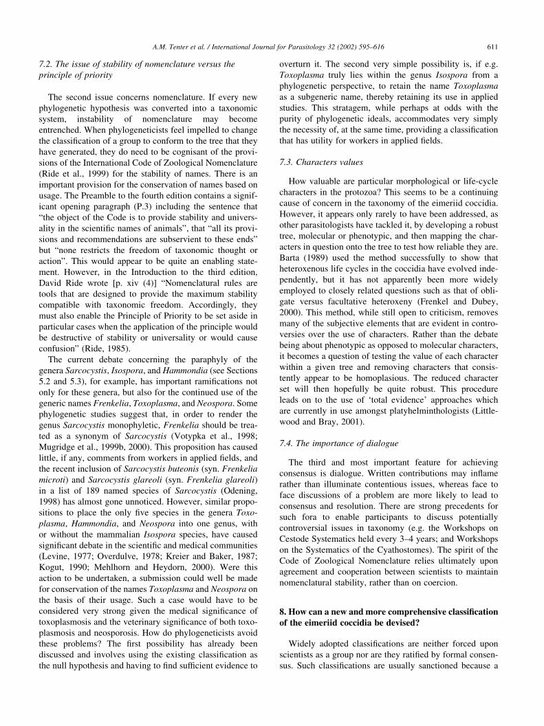
7.2. The issue of stability of nomenclature versus the
principle of priority
The second issue concerns nomenclature. If every new
phylogenetic hypothesis was converted into a taxonomic
system, instability of nomenclature may become
entrenched. When phylogeneticists feel impelled to change
the classification of a group to conform to the tree that they
have generated, they do need to be cognisant of the provi-
sions of the International Code of Zoological Nomenclature
(Ride et al., 1999) for the stability of names. There is an
important provision for the conservation of names based on
usage. The Preamble to the fourth edition contains a signif-
icant opening paragraph (P.3) including the sentence that
“the object of the Code is to provide stability and univers-
ality in the scientific names of animals”, that “all its provi-
sions and recommendations are subservient to these ends”
but “none restricts the freedom of taxonomic thought or
action”. This would appear to be quite an enabling state-
ment. However, in the Introduction to the third edition,
David Ride wrote [p. xiv (4)] “Nomenclatural rules are
tools that are designed to provide the maximum stability
compatible with taxonomic freedom. Accordingly, they
must also enable the Principle of Priority to be set aside in
particular cases when the application of the principle would
be destructive of stability or universality or would cause
confusion” (Ride, 1985).
The current debate concerning the paraphyly of the
genera Sarcocystis, Isospora, and Hammondia (see Sections
5.2 and 5.3), for example, has important ramifications not
only for these genera, but also for the continued use of the
generic names Frenkelia, Toxoplasma, and Neospora. Some
phylogenetic studies suggest that, in order to render the
genus Sarcocystis monophyletic, Frenkelia should be trea-
ted as a synonym of Sarcocystis (Votypka et al., 1998;
Mugridge et al., 1999b, 2000). This proposition has caused
little, if any, comments from workers in applied fields, and
the recent inclusion of Sarcocystis buteonis (syn. Frenkelia
microti) and Sarcocystis glareoli (syn. Frenkelia glareoli)
in a list of 189 named species of Sarcocystis (Odening,
1998) has almost gone unnoticed. However, similar propo-
sitions to place the only five species in the genera Toxo-
plasma, Hammondia, and Neospora into one genus, with
or without the mammalian Isospora species, have caused
significant debate in the scientific and medical communities
(Levine, 1977; Overdulve, 1978; Kreier and Baker, 1987;
Kogut, 1990; Mehlhorn and Heydorn, 2000). Were this
action to be undertaken, a submission could well be made
for conservation of the names Toxoplasma and Neospora on
the basis of their usage. Such a case would have to be
considered very strong given the medical significance of
toxoplasmosis and the veterinary significance of both toxo-
plasmosis and neosporosis. How do phylogeneticists avoid
these problems? The first possibility has already been
discussed and involves using the existing classification as
the null hypothesis and having to find sufficient evidence to
overturn it. The second very simple possibility is, if e.g.
Toxoplasma truly lies within the genus Isospora from a
phylogenetic perspective, to retain the name Toxoplasma
as a subgeneric name, thereby retaining its use in applied
studies. This stratagem, while perhaps at odds with the
purity of phylogenetic ideals, accommodates very simply
the necessity of, at the same time, providing a classification
that has utility for workers in applied fields.
7.3. Characters values
How valuable are particular morphological or life-cycle
characters in the protozoa? This seems to be a continuing
cause of concern in the taxonomy of the eimeriid coccidia.
However, it appears only rarely to have been addressed, as
other parasitologists have tackled it, by developing a robust
tree, molecular or phenotypic, and then mapping the char-
acters in question onto the tree to test how reliable they are.
Barta (1989) used the method successfully to show that
heteroxenous life cycles in the coccidia have evolved inde-
pendently, but it has not apparently been more widely
employed to closely related questions such as that of obli-
gate versus facultative heteroxeny (Frenkel and Dubey,
2000). This method, while still open to criticism, removes
many of the subjective elements that are evident in contro-
versies over the use of characters. Rather than the debate
being about phenotypic as opposed to molecular characters,
it becomes a question of testing the value of each character
within a given tree and removing characters that consis-
tently appear to be homoplasious. The reduced character
set will then hopefully be quite robust. This procedure
leads on to the use of ‘total evidence’ approaches which
are currently in use amongst platyhelminthologists (Little-
wood and Bray, 2001).
7.4. The importance of dialogue
The third and most important feature for achieving
consensus is dialogue. Written contributions may inflame
rather than illuminate contentious issues, whereas face to
face discussions of a problem are more likely to lead to
consensus and resolution. There are strong precedents for
such fora to enable participants to discuss potentially
controversial issues in taxonomy (e.g. the Workshops on
Cestode Systematics held every 3–4 years; and Workshops
on the Systematics of the Cyathostomes). The spirit of the
Code of Zoological Nomenclature relies ultimately upon
agreement and cooperation between scientists to maintain
nomenclatural stability, rather than on coercion.
8. How can a new and more comprehensive classificationof the eimeriid coccidia be devised?
Widely adopted classifications are neither forced upon
scientists as a group nor are they ratified by formal consen-
sus. Such classifications are usually sanctioned because a
A.M. Tenter et al. / International Journal for Parasitology 32 (2002) 595–616 611
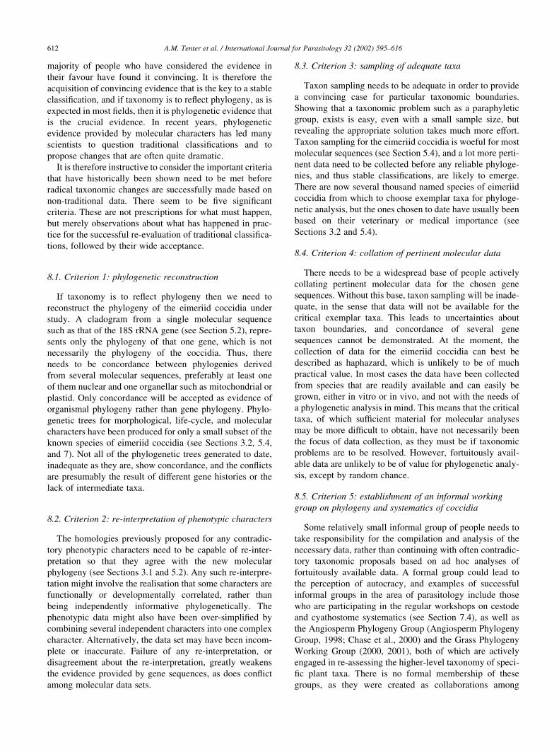
majority of people who have considered the evidence in
their favour have found it convincing. It is therefore the
acquisition of convincing evidence that is the key to a stable
classification, and if taxonomy is to reflect phylogeny, as is
expected in most fields, then it is phylogenetic evidence that
is the crucial evidence. In recent years, phylogenetic
evidence provided by molecular characters has led many
scientists to question traditional classifications and to
propose changes that are often quite dramatic.
It is therefore instructive to consider the important criteria
that have historically been shown need to be met before
radical taxonomic changes are successfully made based on
non-traditional data. There seem to be five significant
criteria. These are not prescriptions for what must happen,
but merely observations about what has happened in prac-
tice for the successful re-evaluation of traditional classifica-
tions, followed by their wide acceptance.
8.1. Criterion 1: phylogenetic reconstruction
If taxonomy is to reflect phylogeny then we need to
reconstruct the phylogeny of the eimeriid coccidia under
study. A cladogram from a single molecular sequence
such as that of the 18S rRNA gene (see Section 5.2), repre-
sents only the phylogeny of that one gene, which is not
necessarily the phylogeny of the coccidia. Thus, there
needs to be concordance between phylogenies derived
from several molecular sequences, preferably at least one
of them nuclear and one organellar such as mitochondrial or
plastid. Only concordance will be accepted as evidence of
organismal phylogeny rather than gene phylogeny. Phylo-
genetic trees for morphological, life-cycle, and molecular
characters have been produced for only a small subset of the
known species of eimeriid coccidia (see Sections 3.2, 5.4,
and 7). Not all of the phylogenetic trees generated to date,
inadequate as they are, show concordance, and the conflicts
are presumably the result of different gene histories or the
lack of intermediate taxa.
8.2. Criterion 2: re-interpretation of phenotypic characters
The homologies previously proposed for any contradic-
tory phenotypic characters need to be capable of re-inter-
pretation so that they agree with the new molecular
phylogeny (see Sections 3.1 and 5.2). Any such re-interpre-
tation might involve the realisation that some characters are
functionally or developmentally correlated, rather than
being independently informative phylogenetically. The
phenotypic data might also have been over-simplified by
combining several independent characters into one complex
character. Alternatively, the data set may have been incom-
plete or inaccurate. Failure of any re-interpretation, or
disagreement about the re-interpretation, greatly weakens
the evidence provided by gene sequences, as does conflict
among molecular data sets.
8.3. Criterion 3: sampling of adequate taxa
Taxon sampling needs to be adequate in order to provide
a convincing case for particular taxonomic boundaries.
Showing that a taxonomic problem such as a paraphyletic
group, exists is easy, even with a small sample size, but
revealing the appropriate solution takes much more effort.
Taxon sampling for the eimeriid coccidia is woeful for most
molecular sequences (see Section 5.4), and a lot more perti-
nent data need to be collected before any reliable phyloge-
nies, and thus stable classifications, are likely to emerge.
There are now several thousand named species of eimeriid
coccidia from which to choose exemplar taxa for phyloge-
netic analysis, but the ones chosen to date have usually been
based on their veterinary or medical importance (see
Sections 3.2 and 5.4).
8.4. Criterion 4: collation of pertinent molecular data
There needs to be a widespread base of people actively
collating pertinent molecular data for the chosen gene
sequences. Without this base, taxon sampling will be inade-
quate, in the sense that data will not be available for the
critical exemplar taxa. This leads to uncertainties about
taxon boundaries, and concordance of several gene
sequences cannot be demonstrated. At the moment, the
collection of data for the eimeriid coccidia can best be
described as haphazard, which is unlikely to be of much
practical value. In most cases the data have been collected
from species that are readily available and can easily be
grown, either in vitro or in vivo, and not with the needs of
a phylogenetic analysis in mind. This means that the critical
taxa, of which sufficient material for molecular analyses
may be more difficult to obtain, have not necessarily been
the focus of data collection, as they must be if taxonomic
problems are to be resolved. However, fortuitously avail-
able data are unlikely to be of value for phylogenetic analy-
sis, except by random chance.
8.5. Criterion 5: establishment of an informal working
group on phylogeny and systematics of coccidia
Some relatively small informal group of people needs to
take responsibility for the compilation and analysis of the
necessary data, rather than continuing with often contradic-
tory taxonomic proposals based on ad hoc analyses of
fortuitously available data. A formal group could lead to
the perception of autocracy, and examples of successful
informal groups in the area of parasitology include those
who are participating in the regular workshops on cestode
and cyathostome systematics (see Section 7.4), as well as
the Angiosperm Phylogeny Group (Angiosperm Phylogeny
Group, 1998; Chase et al., 2000) and the Grass Phylogeny
Working Group (2000, 2001), both of which are actively
engaged in re-assessing the higher-level taxonomy of speci-
fic plant taxa. There is no formal membership of these
groups, as they were created as collaborations among
A.M. Tenter et al. / International Journal for Parasitology 32 (2002) 595–616612
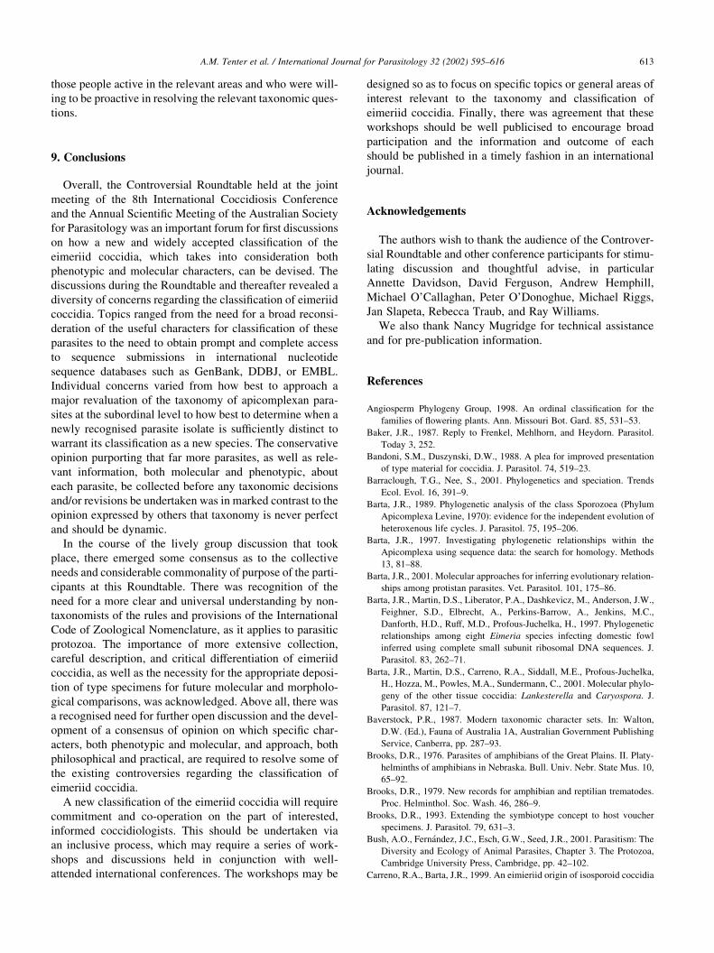
those people active in the relevant areas and who were will-
ing to be proactive in resolving the relevant taxonomic ques-
tions.
9. Conclusions
Overall, the Controversial Roundtable held at the joint
meeting of the 8th International Coccidiosis Conference
and the Annual Scientific Meeting of the Australian Society
for Parasitology was an important forum for first discussions
on how a new and widely accepted classification of the
eimeriid coccidia, which takes into consideration both
phenotypic and molecular characters, can be devised. The
discussions during the Roundtable and thereafter revealed a
diversity of concerns regarding the classification of eimeriid
coccidia. Topics ranged from the need for a broad reconsi-
deration of the useful characters for classification of these
parasites to the need to obtain prompt and complete access
to sequence submissions in international nucleotide
sequence databases such as GenBank, DDBJ, or EMBL.
Individual concerns varied from how best to approach a
major revaluation of the taxonomy of apicomplexan para-
sites at the subordinal level to how best to determine when a
newly recognised parasite isolate is sufficiently distinct to
warrant its classification as a new species. The conservative
opinion purporting that far more parasites, as well as rele-
vant information, both molecular and phenotypic, about
each parasite, be collected before any taxonomic decisions
and/or revisions be undertaken was in marked contrast to the
opinion expressed by others that taxonomy is never perfect
and should be dynamic.
In the course of the lively group discussion that took
place, there emerged some consensus as to the collective
needs and considerable commonality of purpose of the parti-
cipants at this Roundtable. There was recognition of the
need for a more clear and universal understanding by non-
taxonomists of the rules and provisions of the International
Code of Zoological Nomenclature, as it applies to parasitic
protozoa. The importance of more extensive collection,
careful description, and critical differentiation of eimeriid
coccidia, as well as the necessity for the appropriate deposi-
tion of type specimens for future molecular and morpholo-
gical comparisons, was acknowledged. Above all, there was
a recognised need for further open discussion and the devel-
opment of a consensus of opinion on which specific char-
acters, both phenotypic and molecular, and approach, both
philosophical and practical, are required to resolve some of
the existing controversies regarding the classification of
eimeriid coccidia.
A new classification of the eimeriid coccidia will require
commitment and co-operation on the part of interested,
informed coccidiologists. This should be undertaken via
an inclusive process, which may require a series of work-
shops and discussions held in conjunction with well-
attended international conferences. The workshops may be
designed so as to focus on specific topics or general areas of
interest relevant to the taxonomy and classification of
eimeriid coccidia. Finally, there was agreement that these
workshops should be well publicised to encourage broad
participation and the information and outcome of each
should be published in a timely fashion in an international
journal.
Acknowledgements
The authors wish to thank the audience of the Controver-
sial Roundtable and other conference participants for stimu-
lating discussion and thoughtful advise, in particular
Annette Davidson, David Ferguson, Andrew Hemphill,
Michael O’Callaghan, Peter O’Donoghue, Michael Riggs,
Jan Slapeta, Rebecca Traub, and Ray Williams.
We also thank Nancy Mugridge for technical assistance
and for pre-publication information.
References
Angiosperm Phylogeny Group, 1998. An ordinal classification for the
families of flowering plants. Ann. Missouri Bot. Gard. 85, 531–53.
Baker, J.R., 1987. Reply to Frenkel, Mehlhorn, and Heydorn. Parasitol.
Today 3, 252.
Bandoni, S.M., Duszynski, D.W., 1988. A plea for improved presentation
of type material for coccidia. J. Parasitol. 74, 519–23.
Barraclough, T.G., Nee, S., 2001. Phylogenetics and speciation. Trends
Ecol. Evol. 16, 391–9.
Barta, J.R., 1989. Phylogenetic analysis of the class Sporozoea (Phylum
Apicomplexa Levine, 1970): evidence for the independent evolution of
heteroxenous life cycles. J. Parasitol. 75, 195–206.
Barta, J.R., 1997. Investigating phylogenetic relationships within the
Apicomplexa using sequence data: the search for homology. Methods
13, 81–88.
Barta, J.R., 2001. Molecular approaches for inferring evolutionary relation-
ships among protistan parasites. Vet. Parasitol. 101, 175–86.
Barta, J.R., Martin, D.S., Liberator, P.A., Dashkevicz, M., Anderson, J.W.,
Feighner, S.D., Elbrecht, A., Perkins-Barrow, A., Jenkins, M.C.,
Danforth, H.D., Ruff, M.D., Profous-Juchelka, H., 1997. Phylogenetic
relationships among eight Eimeria species infecting domestic fowl
inferred using complete small subunit ribosomal DNA sequences. J.
Parasitol. 83, 262–71.
Barta, J.R., Martin, D.S., Carreno, R.A., Siddall, M.E., Profous-Juchelka,
H., Hozza, M., Powles, M.A., Sundermann, C., 2001. Molecular phylo-
geny of the other tissue coccidia: Lankesterella and Caryospora. J.
Parasitol. 87, 121–7.
Baverstock, P.R., 1987. Modern taxonomic character sets. In: Walton,
D.W. (Ed.), Fauna of Australia 1A, Australian Government Publishing
Service, Canberra, pp. 287–93.
Brooks, D.R., 1976. Parasites of amphibians of the Great Plains. II. Platy-
helminths of amphibians in Nebraska. Bull. Univ. Nebr. State Mus. 10,
65–92.
Brooks, D.R., 1979. New records for amphibian and reptilian trematodes.
Proc. Helminthol. Soc. Wash. 46, 286–9.
Brooks, D.R., 1993. Extending the symbiotype concept to host voucher
specimens. J. Parasitol. 79, 631–3.
Bush, A.O., Fernandez, J.C., Esch, G.W., Seed, J.R., 2001. Parasitism: The
Diversity and Ecology of Animal Parasites, Chapter 3. The Protozoa,
Cambridge University Press, Cambridge, pp. 42–102.
Carreno, R.A., Barta, J.R., 1999. An eimieriid origin of isosporoid coccidia
A.M. Tenter et al. / International Journal for Parasitology 32 (2002) 595–616 613
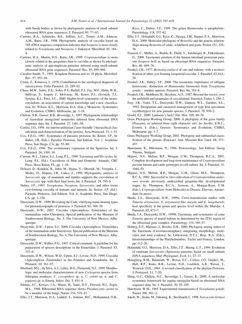
with Stieda bodies as shown by phylogenetic analysis of small subunit
ribosomal RNA gene sequences. J. Parasitol. 85, 77–83.
Carreno, R.A., Schnitzler, B.E., Jeffries, A.C., Tenter, A.M., Johnson,
A.M., Barta, J.R., 1998. Phylogenetic analysis of coccidia based on
18S rDNA sequence comparison indicates that Isospora is most closely
related to Toxoplasma and Neospora. J. Eukaryot. Microbiol. 45, 184–
8.
Carreno, R.A., Martin, D.S., Barta, J.R., 1999. Cryptosporidium is more
closely related to the gregarines than to coccidia as shown by phyloge-
netic analysis of apicomplexan parasites inferred using small-subunit
ribosomal RNA gene sequences. Parasitol. Res. 85, 899–904.
Cavalier-Smith, T., 1993. Kingdom Protozoa and its 18 phyla. Microbiol.
Rev. 57, 953–94.
Cerna, Z., Kolarova, I., 1978. Contribution to the serological diagnosis of
sarcocystosis. Folia Parasitol. 25, 289–92.
Chase, M.W., Soltis, D.E., Soltis, P.S., Rudall, P.J., Fay, M.F., Hahn, W.H.,
Sullivan, S., Jospeh, J., Molvaray, M., Kores, P.J., Givinish, T.J.,
Sytsma, K.J., Pires, J.C., 2000. Higher-level systematics of the mono-
cotyledons: an assessment of current knowledge and a new classifica-
tion. In: Wilson, K.L., Morrison, D.A. (Eds.), Monocots: Systematics
and Evolution, CSIRO, Melbourne, pp. 3–16.
Chilton, N.B., Gasser, R.B., Beveridge, I., 1997. Phylogenetic relationships
of Australian strongyloid nematodes inferred from ribosomal DNA
sequence data. Int. J. Parasitol. 27, 1481–94.
Corliss, J.O., 1994. An interim utilitarian (‘user-friendly’) hierarchical clas-
sification and characterization of the protists. Acta Protozool. 33, 1–51.
Cox, F.E.G., 1991. Systematics of parasitic protozoa. In: Kreier, J.P., In:
Baker, J.R. (Eds.). Parasitic Protozoa, 2nd Edition, Vol. 1. Academic
Press, San Diego, CA, pp. 55–80.
Cox, F.E.G., 1994. The evolutionary expansion of the Sporozoa. Int. J.
Parasitol. 24, 1301–16.
Current, W.L., Upton, S.J., Long, P.L., 1990. Taxonomy and life cycles. In:
Long, P.L. (Ed.), Coccidiosis of Man and Domestic Animals, CRC
Press, Boca Raton, FL, pp. 1–16.
Dolezel, D., Koudela, B., Jirku, M., Hypsa, V., Obornik, M., Votypka, J.,
Modry, D., Slapeta, J.R., Lukes, J., 1999. Phylogenetic analysis of
Sarcocystis spp. of mammals and reptiles supports the coevolution of
Sarcocystis spp. with their final hosts. Int. J. Parasitol. 29, 795–8.
Dubey, J.P., 1993. Toxoplasma, Neospora, Sarcocystis, and other tissue
cyst-forming coccidia of humans and animals. In: Kreier, J.P. (Ed.).
Parasitic Protozoa, 2nd Edition, Vol. 6. Academic Press, San Diego,
CA, pp. 1–158.
Duszynski, D.W., 1999. Revisiting the Code: clarifying name-bearing types
for photomicrographs of protozoa. J. Parasitol. 85, 769–70.
Duszynski, D.W., 2002. Coccidia (Apicomplexa: Eimeriidae) of the
mammalian order Chiroptera. Special publication of the Museum of
Southwestern Biology, No. 5, The University of New Mexico, Albu-
querque.
Duszynski, D.W., Upton, S.J., 2000. Coccidia (Apicomplexa: Eimeriidae)
of the mammalian order Insectivora. Special publication of the Museum
of Southwestern Biology, No. 4, The University of New Mexico, Albu-
querque.
Duszynski, D.W., Wilber, P.G., 1997. Critical comment. A guideline for the
preparation of species descriptions in the Eimeriidae. J. Parasitol. 83,
333–6.
Duszynski, D.W., Wilson, W.D., Upton, S.J., Levine, N.D., 1999. Coccidia
(Apicomplexa: Eimeriidae) in the Primates and Scandentia. Int. J.
Primatol. 20, 761–97.
Eberhard, M.L., da Silva, A.J., Lilley, B.G., Pieniazek, N.J., 1999. Morpho-
logic and molecular characterization of new Cyclospora species from
Ethiopian monkeys: C. cercopitheci sp. n. C. colobi sp. n. and C.
papionis sp. n. Emerg. Infect. Dis. 5, 651–8.
Edman, J.C., Kovacs, J.A., Masur, H., Santi, D.V., Elwood, H.J., Sogin,
M.L., 1988. Ribosomal RNA sequence shows Pneumocystis carinii to
be a member of the fungi. Nature 334, 519–22.
Ellis, J.T., Morrison, D.A., Liddell, S., Jenkins, M.C., Mohammed, O.B.,
Ryce, C., Dubey, J.P., 1999. The genus Hammondia is paraphyletic.
Parasitology 118, 357–62.
Ellis, J.T., Holmdahl, O.J., Ryce, C., Njenga, J.M., Harper, P.A., Morrison,
D.A., 2000. Molecular phylogeny of Besnoitia and the genetic relation-
ships among Besnoitia of cattle, wildebeest and goats. Protist 151, 329–
36.
Franzen, C., Muller, A., Bialek, R., Diehl, V., Salzberger, B., Fatkenheuer,
G., 2000. Taxonomic position of the human intestinal protozoan para-
site Isospora belli as based on ribosomal RNA sequences. Parasitol.
Res. 86, 669–76.
Frenkel, J.K., 1977. Besnoitia wallacei of cats and rodents: with a reclassi-
fication of other cyst-forming isosporoid coccidia. J. Parasitol. 63, 611–
28.
Frenkel, J.K., Dubey, J.P., 2000. The taxonomic importance of obligate
heteroxeny: distinction of Hammondia hammondi from Toxoplasma
gondii – another opinion. Parasitol. Res. 86, 783–6.
Frenkel, J.K., Mehlhorn, H., Heydorn, A.O., 1987. Beyond the oocyst: over
the molehills and mountains of coccidialand. Parasitol. Today 3, 250–2.
Frey, J.K., Yates, T.L., Duszynski, D.W., Gannon, W.L., Gardner, S.L.,
1992. Designation and curatorial management of type host specimens
(symbiotypes) for new parasite species. J. Parasitol. 78, 930–2.
Gould, S.J., 2000. Linnaeus’s luck? Nat. Hist. 109, 48–76.
Grass Phylogeny Working Group, 2000. A phylogeny of the grass family
(Poaceae), as inferred from eight character sets. In: Jacobs, S.W.L.,
Everett, J. (Eds.), Grasses: Systematics and Evolution, CSIRO,
Melbourne, pp. 3–7.
Grass Phylogeny Working Group, 2001. Phylogeny and subfamilial classi-
fication of the grasses (Poaceae). Ann. Missouri Bot. Gard. 88, 373–
457.
Hausmann, K., Hulsmann, N., 1996. Protozoology, 2nd Edition. Georg
Thieme, Stuttgart.
Hijjawi, N.S., Meloni, B.P., Morgan, U.M., Thompson, R.C.A., 2001.
Complete development and long-term maintenance of Cryptosporidium
parvum human and cattle genotypes in cell culture. Int. J. Parasitol. 31,
1048–55.
Hijjawi, N.S., Meloni, B.P., Morgan, U.M., Olson, M.E., Thompson,
R.C.A., 2002. Successful in vitro cultivation of Cryptosporidium ander-
soni reveals previously undescribed gregarine-like developmental
stages. In: Thompson, R.C.A., Armson, A., Morgan-Ryan, U.M.
(Eds.), Cryptosporidium: from Molecules to Disease, Elsevier, Amster-
dam (in press).
Hnida, J.A., Duszynski, D.W., 1999a. Cross-transmission studies with
Eimeria arizonensis, E. arizonensis-like oocysts and E. langebarteli:
host specificity at the genus and species level within the Muridae. J.
Parasitol. 85, 873–7.
Hnida, J.A., Duszynski, D.W., 1999b. Taxonomy and systematics of some
Eimeria species of murid rodents as determined by the ITS1 region of
the ribosomal gene complex. Parasitology 119, 349–57.
Hoberg, E.P., Mariaux, J., Brooks, D.R., 2000. Phylogeny among orders of
the Eucestoda (Cercomeromorphae): integrating morphology, mole-
cules and total evidence. In: Littlewood, D.T.J., Bray, R.A. (Eds.),
Interrelationships of the Platyhelminthes, Taylor and Francis, London,
pp. 112–26.
Holmdahl, O.J., Morrison, D.A., Ellis, J.T., Huong, L.T., 1999. Evolution
of ruminant Sarcocystis (Sporozoa) parasites based on small subunit
rDNA sequences. Mol. Phylogenet. Evol. 11, 27–37.
Honigberg, B.M., Balamuth, W., Bovee, E.C., Corliss, J.O., Gojdics, M.,
Hall, R.P., Kudo, R.R., Levine, N.D., Loeblich, A.R., Weiser, J.,
Wenrich, D.H., 1964. A revised classification of the phylum Protozoa.
J. Protozool. 11, 7–20.
Hung, G.C., Chilton, N.C., Beveridge, I., Gasser, R., 2000. A molecular
systematic framework for equine strongyles based on ribosomal DNA
sequence data. Int. J. Parasitol. 30, 95–105.
Hutchison, W.M., 1965. Experimental transmission of Toxoplasma gondii.
Nature 206, 961–2.
Jakob, W., Stolte, M., Odening, K., Bockhardt, I., 1998. Sarcocystis kirmsei
A.M. Tenter et al. / International Journal for Parasitology 32 (2002) 595–616614
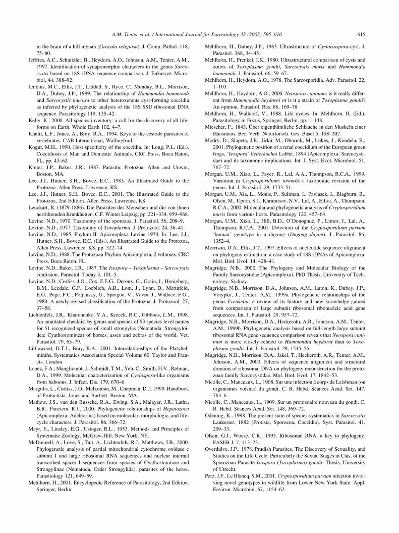
in the brain of a hill mynah (Gracula religiosa). J. Comp. Pathol. 118,
75–80.
Jeffries, A.C., Schnitzler, B., Heydorn, A.O., Johnson, A.M., Tenter, A.M.,
1997. Identification of synapomorphic characters in the genus Sarco-
cystis based on 18S rDNA sequence comparison. J. Eukaryot. Micro-
biol. 44, 388–92.
Jenkins, M.C., Ellis, J.T., Liddell, S., Ryce, C., Munday, B.L., Morrison,
D.A., Dubey, J.P., 1999. The relationship of Hammondia hammondi
and Sarcocystis mucosa to other heteroxenous cyst-forming coccidia
as inferred by phylogenetic analysis of the 18S SSU ribosomal DNA
sequence. Parasitology 119, 135–42.
Kelly, K., 2000. All species inventory: a call for the discovery of all life-
forms on Earth. Whole Earth 102, 4–7.
Khalil, L.F., Jones, A., Bray, R.A., 1994. Keys to the cestode parasites of
vertebrates. CAB International, Wallingford.
Kogut, M.H., 1990. Host specificity of the coccidia. In: Long, P.L. (Ed.),
Coccidiosis of Man and Domestic Animals, CRC Press, Boca Raton,
FL, pp. 43–62.
Kreier, J.P., Baker, J.R., 1987. Parasitic Protozoa. Allen and Unwin,
Boston, MA.
Lee, J.J., Hutner, S.H., Bovee, E.C., 1985. An Illustrated Guide to the
Protozoa. Allen Press, Lawrence, KS.
Lee, J.J., Hutner, S.H., Bovee, E.C., 2001. The Illustrated Guide to the
Protozoa, 2nd Edition. Allen Press, Lawrence, KS.
Leuckart, R. (1879-1886). Die Parasiten des Menschen und die von ihnen
herruhrenden Krankheiten. C.F. Winter Leipzig, pp. 221–334, 959–968.
Levine, N.D., 1970. Taxonomy of the sporozoa. J. Parasitol. 56, 208–9.
Levine, N.D., 1977. Taxonomy of Toxoplasma. J. Protozool. 24, 36–41.
Levine, N.D., 1985. Phylum II. Apicomplexa Levine 1970. In: Lee, J.J.,
Hutner, S.H., Bovee, E.C. (Eds.), An Illustrated Guide to the Protozoa,
Allen Press, Lawrence. KS, pp. 322–74.
Levine, N.D., 1988. The Protozoan Phylum Apicomplexa, 2 volumes. CRC
Press, Boca Raton, FL.
Levine, N.D., Baker, J.R., 1987. The Isospora – Toxoplasma – Sarcocystis
confusion. Parasitol. Today 3, 101–5.
Levine, N.D., Corliss, J.O., Cox, F.E.G., Deroux, G., Grain, J., Honigberg,
B.M., Leedale, G.F., Loeblich, A.R., Lom, J., Lynn, D., Merinfeld,
E.G., Page, F.C., Poljansky, G., Sprague, V., Vavra, J., Wallace, F.G.,
1980. A newly revised classification of the Protozoa. J. Protozool. 27,
37–58.
Lichtenfels, J.R., Kharchenko, V.A., Krecek, R.C., Gibbons, L.M., 1998.
An annotated checklist by genus and species of 93 species level names
for 51 recognized species of small strongyles (Nematoda: Strongyloi-
dea: Cyathostominea) of horses, asses and zebras of the world. Vet.
Parasitol. 79, 65–79.
Littlewood, D.T.J., Bray, R.A., 2001. Interrelationships of the Platyhel-
minths, Systematics Association Special Volume 60. Taylor and Fran-
cis, London.
Lopez, F.A., Manglicmot, J., Schmidt, T.M., Yeh, C., Smith, H.V., Relman,
D.A., 1999. Molecular characterization of Cyclospora-like organisms
from baboons. J. Infect. Dis. 179, 670–6.
Margulis, L., Corliss, J.O., Melkonian, M., Chapman, D.J., 1990. Handbook
of Protoctista. Jones and Bartlett, Boston, MA.
Mathew, J.S., van den Bussche, R.A., Ewing, S.A., Malayer, J.R., Latha,
B.R., Panciera, R.J., 2000. Phylogenetic relationships of Hepatozoon
(Apicomplexa: Adeleorina) based on molecular, morphologic, and life-
cycle characters. J. Parasitol. 86, 366–72.
Mayr, E., Linsley, E.G., Usinger, R.L., 1953. Methods and Principles of
Systematic Zoology. McGraw-Hill, New York, NY.
McDonnell, A., Love, S., Tait, A., Lichtenfels, R.J., Matthews, J.B., 2000.
Phylogenetic analysis of partial mitochondrial cytochrome oxidase c
subunit I and large ribosomal RNA sequences and nuclear internal
transcribed spacer I sequences from species of Cyathostominae and
Strongylinae (Nematoda, Order Strongylida), parasites of the horse.
Parasitology 121, 649–59.
Mehlhorn, H., 2001. Encyclopedic Reference of Parasitology, 2nd Edition.
Springer, Berlin.
Mehlhorn, H., Dubey, J.P., 1983. Ultrastructure of Cystoisospora-cyst. J.
Parasitol. 368, 34–45.
Mehlhorn, H., Frenkel, J.K., 1980. Ultrastructural comparison of cysts and
zoites of Toxoplasma gondii, Sarcocystis muris and Hammondia
hammondi. J. Parasitol. 66, 59–67.
Mehlhorn, H., Heydorn, A.O., 1978. The Sarcosporidia. Adv. Parasitol. 22,
1–103.
Mehlhorn, H., Heydorn, A.O., 2000. Neospora caninum: is it really differ-
ent from Hammondia heydorni or is it a strain of Toxoplasma gondii?
An opinion. Parasitol. Res. 86, 169–78.
Mehlhorn, H., Walldorf, V., 1988. Life cycles. In: Mehlhorn, H. (Ed.),
Parasitology in Focus, Springer, Berlin, pp. 1–148.
Miescher, F., 1843. Uber eigenthumliche Schlauche in den Muskeln einer
Hausmaus. Ber. Verh. Naturforsch. Ges. Basel 5, 198–202.
Modry, D., Slapeta, J.R., Jirku, M., Obornik, M., Lukes, J., Koudela, B.,
2001. Phylogenetic position of a renal coccidium of the European green
frogs, ‘Isospora’ lieberkuehni Labbe, 1894 (Apicomplexa: Sarcocysti-
dae) and its taxonomic implications. Int. J. Syst. Evol. Microbiol. 51,
767–72.
Morgan, U.M., Xiao, L., Fayer, R., Lal, A.A., Thompson, R.C.A., 1999.
Variation in Cryptosporidium: towards a taxonomic revision of the
genus. Int. J. Parasitol. 29, 1733–51.
Morgan, U.M., Xia, L., Monis, P., Suliman, I., Pavlasek, I., Blagburn, B.,
Olsen, M., Upton, S.J., Khramtsov, N.V., Lal, A., Elliot, A., Thompson,
R.C.A., 2000. Molecular and phylogenetic analysis of Cryptosporidium
muris from various hosts. Parasitology 120, 457–64.
Morgan, U.M., Xiao, L., Hill, B.D., O’Donoghue, P., Limor, J., Lal, A.,
Thompson, R.C.A., 2001. Detection of the Cryptosporidium parvum
‘human’ genotype in a dugong (Dugong dugon). J. Parasitol. 86,
1352–4.
Morrison, D.A., Ellis, J.T., 1997. Effects of nucleotide sequence alignment
on phylogeny estimation: a case study of 18S rDNAs of Apicomplexa.
Mol. Biol. Evol. 14, 428–41.
Mugridge, N.B., 2002. The Phylogeny and Molecular Biology of the
Family Sarcocystidae (Apicomplexa). PhD Thesis, University of Tech-
nology, Sydney.
Mugridge, N.B., Morrison, D.A., Johnson, A.M., Luton, K., Dubey, J.P.,
Votypka, J., Tenter, A.M., 1999a. Phylogenetic relationships of the
genus Frenkelia: a review of its history and new knowledge gained
from comparison of large subunit ribosomal ribonucleic acid gene
sequences. Int. J. Parasitol. 29, 957–72.
Mugridge, N.B., Morrison, D.A., Heckeroth, A.R., Johnson, A.M., Tenter,
A.M., 1999b. Phylogenetic analysis based on full-length large subunit
ribosomal RNA gene sequence comparison reveals that Neospora cani-
num is more closely related to Hammondia heydorni than to Toxo-
plasma gondii. Int. J. Parasitol. 29, 1545–56.
Mugridge, N.B., Morrison, D.A., Jakel, T., Heckeroth, A.R., Tenter, A.M.,
Johnson, A.M., 2000. Effects of sequence alignment and structural
domains of ribosomal DNA on phylogeny reconstruction for the proto-
zoan family Sarcocystidae. Mol. Biol. Evol. 17, 1842–53.
Nicolle, C., Manceaux, L., 1908. Sur une infection a corps de Leishman (ou
organismes voisins) du gondi. C. R. Hebd. Seances Acad. Sci. 147,
763–6.
Nicolle, C., Manceaux, L., 1909. Sur un protozoaire nouveau du gondi. C.
R. Hebd. Seances Acad. Sci. 148, 369–72.
Odening, K., 1998. The present state of species-systematics in Sarcocystis
Lankester, 1882 (Protista, Sporozoa, Coccidia). Syst. Parasitol. 41,
209–33.
Olsen, G.J., Woese, C.R., 1993. Ribosomal RNA: a key to phylogeny.
FASEB J. 7, 113–23.
Overdulve, J.P., 1978. Prudish Parasites. The Discovery of Sexuality, and
Studies on the Life Cycle, Particularly the Sexual Stages in Cats, of the
Sporozoan Parasite Isospora (Toxoplasma) gondii. Thesis, University
of Utrecht.
Perz, J.F., Le Blancq, S.M., 2001. Cryptosporidium parvum infection invol-
ving novel genotypes in wildlife from Lower New York State. Appl.
Environ. Microbiol. 67, 1154–62.
A.M. Tenter et al. / International Journal for Parasitology 32 (2002) 595–616 615
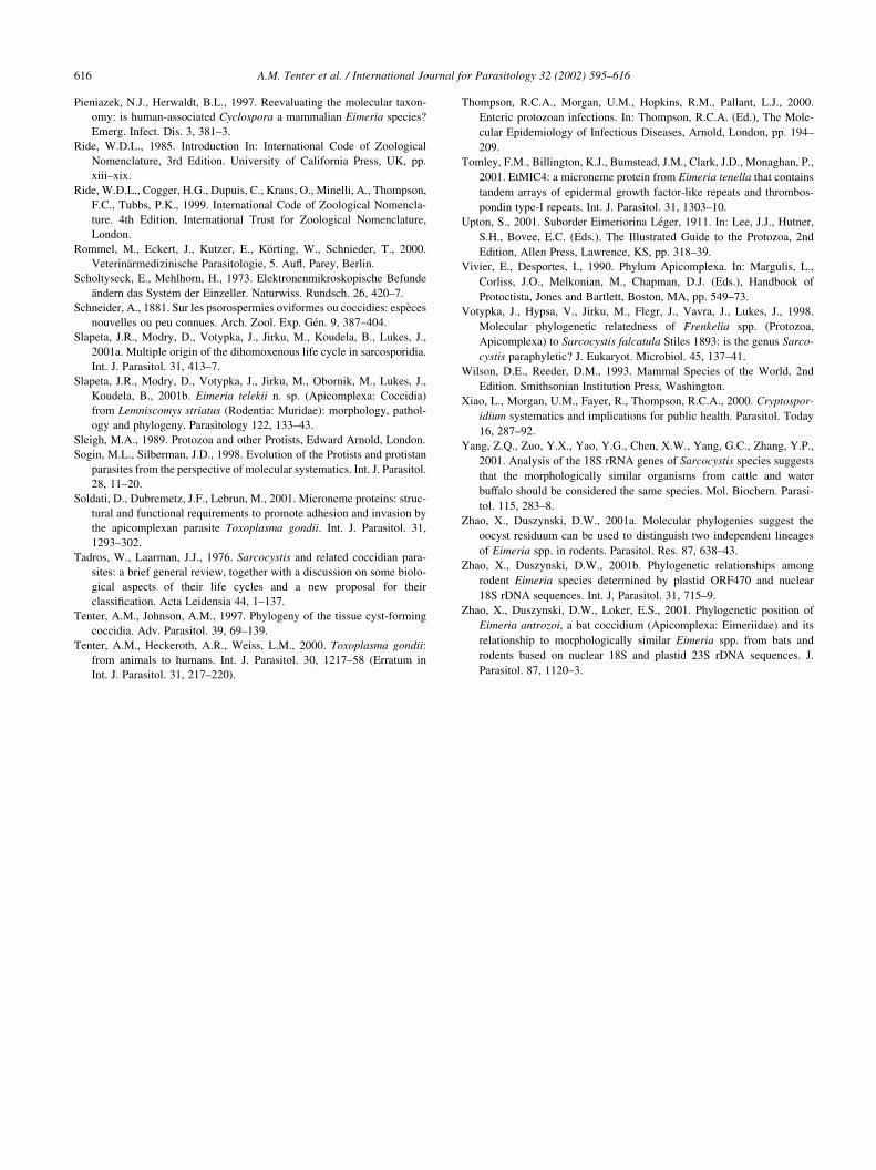
Pieniazek, N.J., Herwaldt, B.L., 1997. Reevaluating the molecular taxon-
omy: is human-associated Cyclospora a mammalian Eimeria species?
Emerg. Infect. Dis. 3, 381–3.
Ride, W.D.L., 1985. Introduction In: International Code of Zoological
Nomenclature, 3rd Edition. University of California Press, UK, pp.
xiii–xix.
Ride, W.D.L., Cogger, H.G., Dupuis, C., Kraus, O., Minelli, A., Thompson,
F.C., Tubbs, P.K., 1999. International Code of Zoological Nomencla-
ture. 4th Edition, International Trust for Zoological Nomenclature,
London.
Rommel, M., Eckert, J., Kutzer, E., Korting, W., Schnieder, T., 2000.
Veterinarmedizinische Parasitologie, 5. Aufl. Parey, Berlin.
Scholtyseck, E., Mehlhorn, H., 1973. Elektronenmikroskopische Befunde
andern das System der Einzeller. Naturwiss. Rundsch. 26, 420–7.
Schneider, A., 1881. Sur les psorospermies oviformes ou coccidies: especes
nouvelles ou peu connues. Arch. Zool. Exp. Gen. 9, 387–404.
Slapeta, J.R., Modry, D., Votypka, J., Jirku, M., Koudela, B., Lukes, J.,
2001a. Multiple origin of the dihomoxenous life cycle in sarcosporidia.
Int. J. Parasitol. 31, 413–7.
Slapeta, J.R., Modry, D., Votypka, J., Jirku, M., Obornik, M., Lukes, J.,
Koudela, B., 2001b. Eimeria telekii n. sp. (Apicomplexa: Coccidia)
from Lemniscomys striatus (Rodentia: Muridae): morphology, pathol-
ogy and phylogeny. Parasitology 122, 133–43.
Sleigh, M.A., 1989. Protozoa and other Protists, Edward Arnold, London.
Sogin, M.L., Silberman, J.D., 1998. Evolution of the Protists and protistan
parasites from the perspective of molecular systematics. Int. J. Parasitol.
28, 11–20.
Soldati, D., Dubremetz, J.F., Lebrun, M., 2001. Microneme proteins: struc-
tural and functional requirements to promote adhesion and invasion by
the apicomplexan parasite Toxoplasma gondii. Int. J. Parasitol. 31,
1293–302.
Tadros, W., Laarman, J.J., 1976. Sarcocystis and related coccidian para-
sites: a brief general review, together with a discussion on some biolo-
gical aspects of their life cycles and a new proposal for their
classification. Acta Leidensia 44, 1–137.
Tenter, A.M., Johnson, A.M., 1997. Phylogeny of the tissue cyst-forming
coccidia. Adv. Parasitol. 39, 69–139.
Tenter, A.M., Heckeroth, A.R., Weiss, L.M., 2000. Toxoplasma gondii:
from animals to humans. Int. J. Parasitol. 30, 1217–58 (Erratum in
Int. J. Parasitol. 31, 217–220).
Thompson, R.C.A., Morgan, U.M., Hopkins, R.M., Pallant, L.J., 2000.
Enteric protozoan infections. In: Thompson, R.C.A. (Ed.), The Mole-
cular Epidemiology of Infectious Diseases, Arnold, London, pp. 194–
209.
Tomley, F.M., Billington, K.J., Bumstead, J.M., Clark, J.D., Monaghan, P.,
2001. EtMIC4: a microneme protein from Eimeria tenella that contains
tandem arrays of epidermal growth factor-like repeats and thrombos-
pondin type-I repeats. Int. J. Parasitol. 31, 1303–10.
Upton, S., 2001. Suborder Eimeriorina Leger, 1911. In: Lee, J.J., Hutner,
S.H., Bovee, E.C. (Eds.). The Illustrated Guide to the Protozoa, 2nd
Edition, Allen Press, Lawrence, KS, pp. 318–39.
Vivier, E., Desportes, I., 1990. Phylum Apicomplexa. In: Margulis, L.,
Corliss, J.O., Melkonian, M., Chapman, D.J. (Eds.), Handbook of
Protoctista, Jones and Bartlett, Boston, MA, pp. 549–73.
Votypka, J., Hypsa, V., Jirku, M., Flegr, J., Vavra, J., Lukes, J., 1998.
Molecular phylogenetic relatedness of Frenkelia spp. (Protozoa,
Apicomplexa) to Sarcocystis falcatula Stiles 1893: is the genus Sarco-
cystis paraphyletic? J. Eukaryot. Microbiol. 45, 137–41.
Wilson, D.E., Reeder, D.M., 1993. Mammal Species of the World, 2nd
Edition. Smithsonian Institution Press, Washington.
Xiao, L., Morgan, U.M., Fayer, R., Thompson, R.C.A., 2000. Cryptospor-
idium systematics and implications for public health. Parasitol. Today
16, 287–92.
Yang, Z.Q., Zuo, Y.X., Yao, Y.G., Chen, X.W., Yang, G.C., Zhang, Y.P.,
2001. Analysis of the 18S rRNA genes of Sarcocystis species suggests
that the morphologically similar organisms from cattle and water
buffalo should be considered the same species. Mol. Biochem. Parasi-
tol. 115, 283–8.
Zhao, X., Duszynski, D.W., 2001a. Molecular phylogenies suggest the
oocyst residuum can be used to distinguish two independent lineages
of Eimeria spp. in rodents. Parasitol. Res. 87, 638–43.
Zhao, X., Duszynski, D.W., 2001b. Phylogenetic relationships among
rodent Eimeria species determined by plastid ORF470 and nuclear
18S rDNA sequences. Int. J. Parasitol. 31, 715–9.
Zhao, X., Duszynski, D.W., Loker, E.S., 2001. Phylogenetic position of
Eimeria antrozoi, a bat coccidium (Apicomplexa: Eimeriidae) and its
relationship to morphologically similar Eimeria spp. from bats and
rodents based on nuclear 18S and plastid 23S rDNA sequences. J.
Parasitol. 87, 1120–3.
A.M. Tenter et al. / International Journal for Parasitology 32 (2002) 595–616616