Embed Size (px)
Citation preview

IOL OpacificationR. Khoramnia
[email protected] www.ivcrc.com www.djapplelab.com
International Vision Correction Research Centre (IVCRC)The David J. Apple International Laboratory for Ocular Pathology Department of OphthalmologyRuprecht-Karls-University of HeidelbergChairman: G. U. Auffarth, MD, PhD, FEBO

University Eye Clinic Heidelberg
IVCRC / DJ Apple Laboratory: Financial Disclosures 2018/2019
1 = Research Grants; 2 = Travel Expenses; 3 = Lecture Fees; 4 = Consulting
Acufocus1
Alcon/Novartis1,2,3,4
Alimera1,2
AMO/Johnson&Johnson1,2,3,4
Bausch+Lomb2,3
SIFI3Carl Zeiss Meditec1,2,3
Contamac1
Glaukos1
Hoya1,2,3
Kowa1,2,3
Oculentis1,2,3
Oculus1,2,3
Physiol1Presbia2,3,4
Rayner1,2,3
Santen1
SIFI1,2,3
Ursapharm1,2,3

University Eye Clinic Heidelberg
David J. Apple Laboratory
Analysis of explanted IOLs, Biomaterial analysis of new IOLs,
Analysis of the optical quality of IOLs
University Eye Clinic Heidelberg
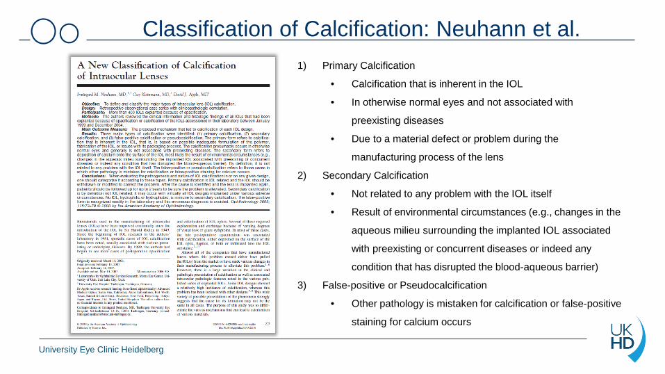
Classification of Calcification: Neuhann et al. 1) Primary Calcification
• Calcification that is inherent in the IOL
• In otherwise normal eyes and not associated with
preexisting diseases
• Due to a material defect or problem during the
manufacturing process of the lens
2) Secondary Calcification
• Not related to any problem with the IOL itself
• Result of environmental circumstances (e.g., changes in the
aqueous milieu surrounding the implanted IOL associated
with preexisting or concurrent diseases or indeed any
condition that has disrupted the blood-aqueous barrier)
3) False-positive or Pseudocalcification
• Other pathology is mistaken for calcification or false-positive
staining for calcium occurs

University Eye Clinic Heidelberg
Opcacification of hydrophilic acrylic IOLs

University Eye Clinic Heidelberg
Classification of Calcification: Neuhann et al. 1) Primary Calcification
• Calcification that is inherent in the IOL
• In otherwise normal eyes and not associated with
preexisting diseases
• Due to a material defect or problem during the
manufacturing process of the lens
2) Secondary Calcification
• Not related to any problem with the IOL itself
• Result of environmental circumstances (e.g., changes in the
aqueous milieu surrounding the implanted IOL associated
with preexisting or concurrent diseases or indeed any
condition that has disrupted the blood-aqueous barrier)
3) False-positive or Pseudocalcification
• Other pathology is mistaken for calcification or false-positive
staining for calcium occurs

University Eye Clinic Heidelberg
Opacified hydrophilic IOL: Aquasense
*[Opacification of a hydrophilic intraocular lens 4 years after cataract surgery. A biomaterial analysis]. Khoramnia R, Salgado JP, Auffarth GU, Schmidt S, Wegner A, Kobuch KA, Winkler von Mohrenfels C. Ophthalmologe. 2012 May;109(5):483-6.

University Eye Clinic Heidelberg
Opacified hydrophilic IOL: Euromaxx (Argonoptics)*
*Tandogan T, Khoramnia R, Choi CY, Scheuerle A, Wenzel M, Hugger P, Auffarth GU. Optical and material analysis of opacified hydrophilicintraocular lenses after explantation: a laboratory study. BMC Ophthalmol. 2015 Nov 25;15:170. doi: 10.1186/s12886-015-0149-1.

University Eye Clinic Heidelberg
Opacified hydrophilic IOL: Euromaxx (Argonoptics)*
Light and scanning electron microscopy as well as X-ray spectroscopy in opacified Euromaxx ALI313Y and ALI313 IOLs
*Tandogan T, Khoramnia R, Choi CY, Scheuerle A, Wenzel M, Hugger P, Auffarth GU.Optical and material analysis of opacified hydrophilicintraocular lenses after explantation: a laboratory study. BMC Ophthalmol. 2015 Nov 25;15:170. doi: 10.1186/s12886-015-0149-1.

University Eye Clinic Heidelberg
Material & Methods*− Explantation of 6 IOLs (Euromaxx ALI313Y and ALI313) that had opacified
− Light microscopy: Staining with Alizarin red and von Kossa
− Scanning electron microscopy
− X-ray spectroscopy (EDX): Chemical composition of the deposits
− Optical bench (OptiSpheric, Trioptics): Optical quality assessment
*Tandogan T, Khoramnia R, Choi CY, Scheuerle A, Wenzel M, Hugger P, Auffarth GU.Optical and material analysis of opacified hydrophilic intraocular lenses after explantation: a laboratory study. BMC Ophthalmol. 2015 Nov 25;15:170. doi: 10.1186/s12886-015-0149-1.

University Eye Clinic Heidelberg
Results – Gross examination*
*Tandogan T, Khoramnia R, Choi CY, Scheuerle A, Wenzel M, Hugger P, Auffarth GU.Optical and material analysis of opacified hydrophilic intraocular lenses after explantation: a laboratory study. BMC Ophthalmol. 2015 Nov 25;15:170. doi: 10.1186/s12886-015-0149-1.

University Eye Clinic Heidelberg
Results – Light microscopy*Staining with Alizarin red
*Tandogan T, Khoramnia R, Choi CY, Scheuerle A, Wenzel M, Hugger P, Auffarth GU.Optical and material analysis of opacified hydrophilic intraocular lenses after explantation: a laboratorystudy. BMC Ophthalmol. 2015 Nov 25;15:170. doi: 10.1186/s12886-015-0149-1.

University Eye Clinic Heidelberg
Von Kossa Staining
*Tandogan T, Khoramnia R, Choi CY, Scheuerle A, Wenzel M, Hugger P, Auffarth GU.Optical and material analysis of opacified hydrophilic intraocular lenses after explantation: a laboratorystudy. BMC Ophthalmol. 2015 Nov 25;15:170. doi: 10.1186/s12886-015-0149-1.
Results – Light microscopy*
University Eye Clinic Heidelberg
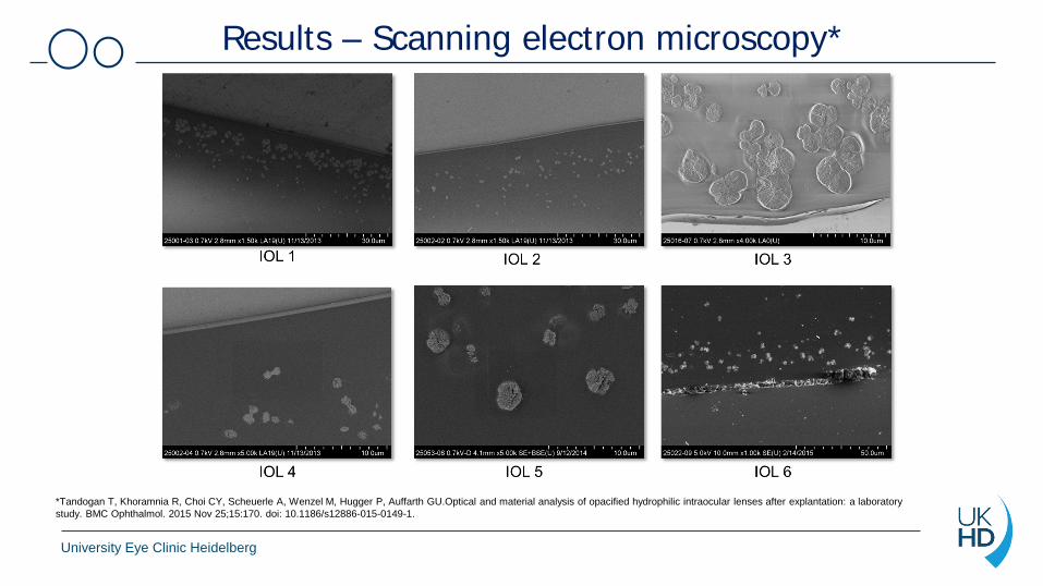
Results – Scanning electron microscopy*
*Tandogan T, Khoramnia R, Choi CY, Scheuerle A, Wenzel M, Hugger P, Auffarth GU.Optical and material analysis of opacified hydrophilic intraocular lenses after explantation: a laboratorystudy. BMC Ophthalmol. 2015 Nov 25;15:170. doi: 10.1186/s12886-015-0149-1.

University Eye Clinic Heidelberg
Results – EDX*Energy dispersive X-ray spectroscopy (EDX)
Calcium phosphate*Tandogan T, Khoramnia R, Choi CY, Scheuerle A, Wenzel M, Hugger P, Auffarth GU. Optical and material analysis of opacified hydrophilic intraocular lenses after explantation: a laboratorystudy. BMC Ophthalmol. 2015 Nov 25;15:170. doi: 10.1186/s12886-015-0149-1.

University Eye Clinic Heidelberg
Results – Element Mapping*
*Tandogan T, Khoramnia R, Choi CY, Scheuerle A, Wenzel M, Hugger P, Auffarth GU.Optical and material analysis of opacified hydrophilic intraocular lenses after explantation: a laboratorystudy. BMC Ophthalmol. 2015 Nov 25;15:170. doi: 10.1186/s12886-015-0149-1.

University Eye Clinic Heidelberg
Results – Optical Bench*Modulation Transfer Function (MTF)
Severe reduction of optical quality*Tandogan T, Khoramnia R, Choi CY, Scheuerle A, Wenzel M, Hugger P, Auffarth GU.Optical and material analysis of opacified hydrophilic intraocular lenses after explantation: a laboratorystudy. BMC Ophthalmol. 2015 Nov 25;15:170. doi: 10.1186/s12886-015-0149-1.

University Eye Clinic Heidelberg
Report to the authorities

University Eye Clinic Heidelberg
Recall of IOL

University Eye Clinic Heidelberg
Classification of Calcification: Neuhann et al. 1) Primary Calcification
• Calcification that is inherent in the IOL
• In otherwise normal eyes and not associated with
preexisting diseases
• Due to a material defect or problem during the
manufacturing process of the lens
2) Secondary Calcification
• Not related to any problem with the IOL itself
• Result of environmental circumstances (e.g., changes in
the aqueous milieu surrounding the implanted IOL
associated with preexisting or concurrent diseases or
indeed any condition that has disrupted the blood-
aqueous barrier)
3) False-positive or Pseudocalcification
• Other pathology is mistaken for calcification or false-positive
staining for calcium occurs

University Eye Clinic Heidelberg
IOL Opacification following Intracameral Injection of rtPA to Treat Inflammatory Membranes after Cataract Surgery
*Intraocular Lens Opacification following Intracameral Injection of Recombinant Tissue Plasminogen Activator to Treat Inflammatory Membranes after Cataract Surgery. Fung SS, Sykakis E, Islam NM, Zambarakji HJ, Khoramnia R, Auffarth GU, Parmar DN. J Ophthalmol. 2015;2015:975075. doi: 10.1155/2015/975075.

University Eye Clinic Heidelberg
*Intraocular Lens Opacification following Intracameral Injection of Recombinant Tissue Plasminogen Activator to Treat Inflammatory Membranes after Cataract Surgery. Fung SS, Sykakis E, Islam NM, Zambarakji HJ, Khoramnia R, Auffarth GU, Parmar DN. J Ophthalmol. 2015;2015:975075. doi: 10.1155/2015/975075.
IOL Opacification following Intracameral Injection of rtPA to Treat Inflammatory Membranes after Cataract Surgery

University Eye Clinic Heidelberg
Hydrophilic intraocular lens opacificationafter posterior lamellar keratoplasty*
*Giers BC, Tandogan T, Auffarth GU, Choi CY, Auerbach FN, Sel S, Mayer C, Khoramnia R. Hydrophilic intraocular lens opacification after posterior lamellar keratoplasty - a material analysiswith special reference to optical quality assessment. BMC Ophthalmol. 2017 Aug 22;17(1):150. doi: 10.1186/s12886-017-0546-8.

University Eye Clinic Heidelberg
*Giers BC, Tandogan T, Auffarth GU, Choi CY, Auerbach FN, Sel S, Mayer C, Khoramnia R. Hydrophilic intraocular lens opacification after posterior lamellar keratoplasty - a material analysiswith special reference to optical quality assessment. BMC Ophthalmol. 2017 Aug 22;17(1):150. doi: 10.1186/s12886-017-0546-8.
Hydrophilic IOL opacification after posterior lamellar keratoplasty*

University Eye Clinic Heidelberg
Light microscopy*
*Giers BC, Tandogan T, Auffarth GU, Choi CY, Auerbach FN, Sel S, Mayer C, Khoramnia R. Hydrophilic intraocular lens opacification after posterior lamellar keratoplasty - a material analysiswith special reference to optical quality assessment. BMC Ophthalmol. 2017 Aug 22;17(1):150. doi: 10.1186/s12886-017-0546-8.

University Eye Clinic Heidelberg
*Giers BC, Tandogan T, Auffarth GU, Choi CY, Auerbach FN, Sel S, Mayer C, Khoramnia R. Hydrophilic intraocular lens opacification after posterior lamellar keratoplasty - a material analysiswith special reference to optical quality assessment. BMC Ophthalmol. 2017 Aug 22;17(1):150. doi: 10.1186/s12886-017-0546-8.
Light microscopy*

University Eye Clinic Heidelberg
Scanning electron microscopy*
*Giers BC, Tandogan T, Auffarth GU, Choi CY, Auerbach FN, Sel S, Mayer C, Khoramnia R. Hydrophilic intraocular lens opacification after posterior lamellar keratoplasty - a material analysiswith special reference to optical quality assessment. BMC Ophthalmol. 2017 Aug 22;17(1):150. doi: 10.1186/s12886-017-0546-8.

University Eye Clinic Heidelberg
Element Mapping*
*Giers BC, Tandogan T, Auffarth GU, Choi CY, Auerbach FN, Sel S, Mayer C, Khoramnia R. Hydrophilic intraocular lens opacification after posterior lamellar keratoplasty - a material analysiswith special reference to optical quality assessment. BMC Ophthalmol. 2017 Aug 22;17(1):150. doi: 10.1186/s12886-017-0546-8.

University Eye Clinic Heidelberg
EDX*
*Giers BC, Tandogan T, Auffarth GU, Choi CY, Auerbach FN, Sel S, Mayer C, Khoramnia R. Hydrophilic intraocular lens opacification after posterior lamellar keratoplasty - a material analysiswith special reference to optical quality assessment. BMC Ophthalmol. 2017 Aug 22;17(1):150. doi: 10.1186/s12886-017-0546-8.

University Eye Clinic Heidelberg
Optical quality*
*Giers BC, Tandogan T, Auffarth GU, Choi CY, Auerbach FN, Sel S, Mayer C, Khoramnia R. Hydrophilic intraocular lens opacification after posterior lamellar keratoplasty - a material analysiswith special reference to optical quality assessment. BMC Ophthalmol. 2017 Aug 22;17(1):150. doi: 10.1186/s12886-017-0546-8.

University Eye Clinic Heidelberg
Hydrophilic IOL opacification after posterior lamellar keratoplasty: Straylight*
*Assessment of straylight and the modulation transfer function of intraocular lenses with centrally localized opacification associated with the intraocular injection of gas. Łabuz G, Yildirim TM, van den Berg TJTP, Khoramnia R, Auffarth GU. J Cataract Refract Surg. 2018 May;44(5):615-622.
Severeincrease of straylight

University Eye Clinic Heidelberg
Hydrophilic IOL opacification after PpV*
*Material Analysis and Optical Quality Assessment of Opacified Hydrophilic Acrylic Intraocular Lenses after Pars Plana Vitrectomy. Yildirim TM, Auffarth GU, Łabuz G, Bopp S, Son HS, Khoramnia R. Am J Ophthalmol. 2018 Sep;193:10-19.

University Eye Clinic Heidelberg
*Opacification of hydrophilic intraocular lenses associated with vitrectomy and injection of intraocular gas. Marcovich AL, Tandogan T, Bareket M, Etting E, Kaplan-Ashiri I, Bukelman A, Auffarth GU, Khoramnia R. BMJ Open Ophthalmol. 2018 Dec 15;3(1):e000157.
Hydrophilic IOL opacification after PpV*

University Eye Clinic Heidelberg
Hydrophilic IOL opacification after PpV*
*Opacification of hydrophilic intraocular lenses associated with vitrectomy and injection of intraocular gas. Marcovich AL, Tandogan T, Bareket M, Etting E, Kaplan-Ashiri I, Bukelman A, Auffarth GU, Khoramnia R. BMJ Open Ophthalmol. 2018 Dec 15;3(1):e000157.

University Eye Clinic Heidelberg
Opacification of hydrophilic intraocular lens after multiple injections of bevacizumab*
*Opacification of hydrophilic intraocular lens after multiple injections of bevacizumab. Roland S., Khoramnia R., Auffarth G.U., Son H.-S., Yildirim T.M., Schoenherr U. In press

University Eye Clinic Heidelberg
Classification of Calcification: Neuhann et al. 1) Primary Calcification
• Calcification that is inherent in the IOL
• In otherwise normal eyes and not associated with
preexisting diseases
• Due to a material defect or problem during the
manufacturing process of the lens
2) Secondary Calcification
• Not related to any problem with the IOL itself
• Result of environmental circumstances (e.g., changes in the
aqueous milieu surrounding the implanted IOL associated
with preexisting or concurrent diseases or indeed any
condition that has disrupted the blood-aqueous barrier)
3) False-positive or Pseudocalcification
• Other pathology is mistaken for calcification or false-
positive staining for calcium occurs

University Eye Clinic Heidelberg
Liquefied Aftercataract*
*Giers BC, Khoramnia R, Tandogan T, Auffarth GU. Liquefied Aftercataract Mimicking IOL-Opacification. Klin Monbl Augenheilkd. 2017 Aug;234(8):988-990.

University Eye Clinic Heidelberg
Summary• In addition to good optical quality, material properties are important in IOLs.
• In contrast to IOLs from hydrophobic acrylic, IOLs made of hydrophilic material can calcify.
• Primary Calcification: Production errors may be responsible for IOL calcification (e.g. Aquasense and Euromaxx).
• Secondary Calcification: IOLs made of hydrophilic acrylic can opacify irrespective of themanufacturer during various interventions (e.g. posterior lamellar keratoplasty, rtPAinjection, PpV).
• IOL exchange in symptomatic patients is the only treatment option available. Since IOL exchange is associated with a high intraoperative complication rate, our results suggest to consider the risk of IOL calcification when implanting hydrophilic acrylic IOLs.

Thank you!