Embed Size (px)
Citation preview
Review article | Published 29 October 2015, doi:10.4414/smw.2015.14196
Cite this as: Swiss Med Wkly. 2015;145:w14196
Iron deficiency in sports – definition, influence onperformance and therapy
Consensus statement of the Swiss Society of Sports Medicine
German E. Clénina, Mareike Cordesa, Andreas Huberb, Yorck Olaf Schumacherc, Patrick Noackd, John Scalese, Susi Kriemlerf
a Sportmedizinisches Zentrum Ittigen bei Bern, Haus des Sports, Ittigen, Switzerlandb Zentrum für Labormedizin, Kantonsspital Aarau, Switzerlandc Aspetar Orthopedic and Sports Medicine Hospital, Doha, Quatard Zentrum für Medizin und Sport im Säntispark, Abtwil, Switzerlande Bannockburn Health Centre, Stirling, Scotlandf Epidemiology, Biostatistics and Prevention Institute, University of Zürich, Switzerland
Summary
Iron deficiency is frequent among athletes. All types of irondeficiency may affect physical performance and should betreated. The main mechanisms by which sport leads to irondeficiency are increased iron demand, elevated iron lossand blockage of iron absorption due to hepcidin bursts.As a baseline set of blood tests, haemoglobin, haematocrit,mean cellular volume, mean cellular haemoglobin and ser-um ferritin levels help monitor iron deficiency. In healthymale and female athletes >15 years, ferritin values <15 mcgare equivalent to empty, values from 15 to 30 mcg/l to lowiron stores. Therefore a cut-off of 30 mcg/l is appropriate.For children aged from 6–12 years and younger adoles-cents from 12–15 years, cut-offs of 15 and 20 mcg/l, re-spectively, are recommended. As an exception in adult elitesports, a ferritin value of 50 mcg/l should be attained inathletes prior to altitude training, as iron demands in thesesituations are increased.Treatment of iron deficiency consists of nutritional coun-selling, oral iron supplementation or, in specific cases, byintravenous injection. Athletes with repeatedly low ferritinvalues benefit from intermittent oral substitution. It is im-portant to follow up the athletes on an individual basis, re-peating the baseline blood tests listed above twice a year. Along-term daily oral iron intake or i.v. supplementation inthe presence of normal or even high ferritin values does notmake sense and may be harmful.
Key words: iron deficiency in sports; iron deficiency andperformance; diagnosis of iron deficiency; ferritin cut-off;dilutional pseudoanaemia in sports; treatment of irondeficiency
Introduction
Iron deficiency among athletes, in males and more oftenin females, is a commonly encountered condition for thesports medicine physician. Iron deficiency is one of themost common deficits globally with a clear predominancein adolescence and in menstruating females [1]. Data froma general Swiss population show frequencies for iron de-ficiency for menstruating females of 22.7%, for male mil-itary recruits of 7.2% and for iron deficiency anaemia of2.2% (females) and 0.1% (males) [2, 3]. In sports the rateof iron deficiency is distinctly higher, up to 52% in femaleadolescent athletes, and occurs more often in endurancesports and in disciplines with a high prevalence of eatingdisorders [4–6]. These abnormal findings need a carefulclinical look as iron deficiency affects many organ systemsof the body and not just oxygen transport, especially insports [7].On the other hand, Switzerland has recently experiencedsome kind of “iron hype” for various reasons. Not all ofthem seem to be rational [8, 9]. The problem is enhancedby the finding of an earlier study among Swiss top athletes,showing that iron supplements were consumed to some ex-tent uncritically and in excess [10, 11].Sports medicine physicians are often in charge of male andfemale athletes at their peak performance. In this role, theyare aware of the importance of adequately diagnosing andtreating iron deficiency in all its aspects, but at the sametime to prevent iron overload. This article is a consensusstatement of the Swiss Society of Sports Medicine andprovides an overview and practical guidelines for the dia-gnosis and treatment of iron deficiency in sports and shouldhelp clinicians in decision making.
Swiss Medical Weekly · PDF of the online version · www.smw.ch Page 1 of 15
Definition of iron deficiency andgeneral function of iron
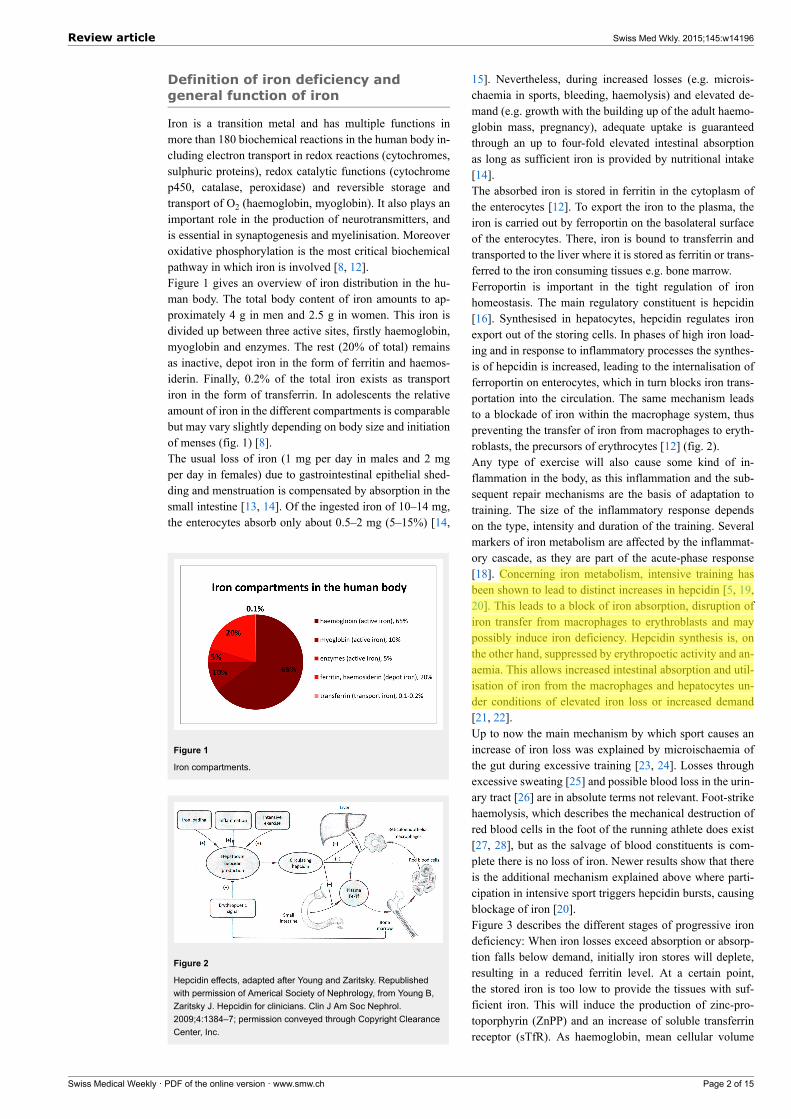
Iron is a transition metal and has multiple functions inmore than 180 biochemical reactions in the human body in-cluding electron transport in redox reactions (cytochromes,sulphuric proteins), redox catalytic functions (cytochromep450, catalase, peroxidase) and reversible storage andtransport of O2 (haemoglobin, myoglobin). It also plays animportant role in the production of neurotransmitters, andis essential in synaptogenesis and myelinisation. Moreoveroxidative phosphorylation is the most critical biochemicalpathway in which iron is involved [8, 12].Figure 1 gives an overview of iron distribution in the hu-man body. The total body content of iron amounts to ap-proximately 4 g in men and 2.5 g in women. This iron isdivided up between three active sites, firstly haemoglobin,myoglobin and enzymes. The rest (20% of total) remainsas inactive, depot iron in the form of ferritin and haemos-iderin. Finally, 0.2% of the total iron exists as transportiron in the form of transferrin. In adolescents the relativeamount of iron in the different compartments is comparablebut may vary slightly depending on body size and initiationof menses (fig. 1) [8].The usual loss of iron (1 mg per day in males and 2 mgper day in females) due to gastrointestinal epithelial shed-ding and menstruation is compensated by absorption in thesmall intestine [13, 14]. Of the ingested iron of 10–14 mg,the enterocytes absorb only about 0.5–2 mg (5–15%) [14,
Figure 1
Iron compartments.
Figure 2
Hepcidin effects, adapted after Young and Zaritsky. Republishedwith permission of Americal Society of Nephrology, from Young B,Zaritsky J. Hepcidin for clinicians. Clin J Am Soc Nephrol.2009;4:1384–7; permission conveyed through Copyright ClearanceCenter, Inc.
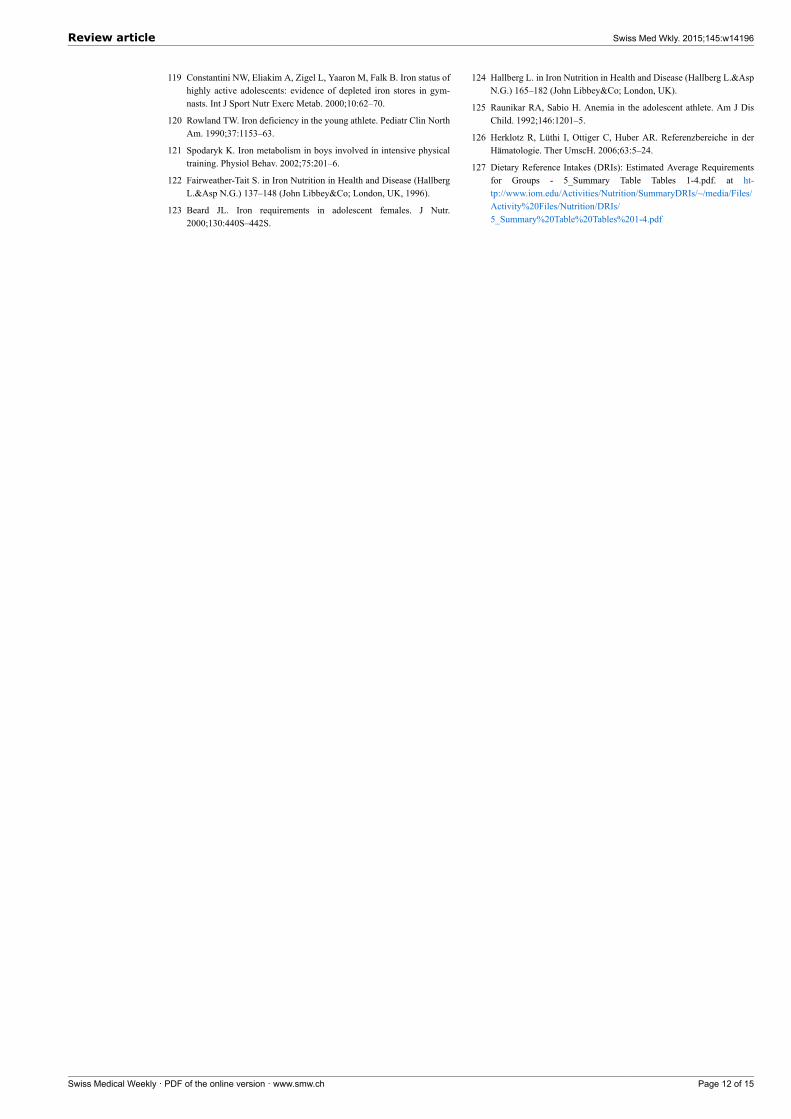
15]. Nevertheless, during increased losses (e.g. microis-chaemia in sports, bleeding, haemolysis) and elevated de-mand (e.g. growth with the building up of the adult haemo-globin mass, pregnancy), adequate uptake is guaranteedthrough an up to four-fold elevated intestinal absorptionas long as sufficient iron is provided by nutritional intake[14].The absorbed iron is stored in ferritin in the cytoplasm ofthe enterocytes [12]. To export the iron to the plasma, theiron is carried out by ferroportin on the basolateral surfaceof the enterocytes. There, iron is bound to transferrin andtransported to the liver where it is stored as ferritin or trans-ferred to the iron consuming tissues e.g. bone marrow.Ferroportin is important in the tight regulation of ironhomeostasis. The main regulatory constituent is hepcidin[16]. Synthesised in hepatocytes, hepcidin regulates ironexport out of the storing cells. In phases of high iron load-ing and in response to inflammatory processes the synthes-is of hepcidin is increased, leading to the internalisation offerroportin on enterocytes, which in turn blocks iron trans-portation into the circulation. The same mechanism leadsto a blockade of iron within the macrophage system, thuspreventing the transfer of iron from macrophages to eryth-roblasts, the precursors of erythrocytes [12] (fig. 2).Any type of exercise will also cause some kind of in-flammation in the body, as this inflammation and the sub-sequent repair mechanisms are the basis of adaptation totraining. The size of the inflammatory response dependson the type, intensity and duration of the training. Severalmarkers of iron metabolism are affected by the inflammat-ory cascade, as they are part of the acute-phase response[18]. Concerning iron metabolism, intensive training hasbeen shown to lead to distinct increases in hepcidin [5, 19,20]. This leads to a block of iron absorption, disruption ofiron transfer from macrophages to erythroblasts and maypossibly induce iron deficiency. Hepcidin synthesis is, onthe other hand, suppressed by erythropoetic activity and an-aemia. This allows increased intestinal absorption and util-isation of iron from the macrophages and hepatocytes un-der conditions of elevated iron loss or increased demand[21, 22].Up to now the main mechanism by which sport causes anincrease of iron loss was explained by microischaemia ofthe gut during excessive training [23, 24]. Losses throughexcessive sweating [25] and possible blood loss in the urin-ary tract [26] are in absolute terms not relevant. Foot-strikehaemolysis, which describes the mechanical destruction ofred blood cells in the foot of the running athlete does exist[27, 28], but as the salvage of blood constituents is com-plete there is no loss of iron. Newer results show that thereis the additional mechanism explained above where parti-cipation in intensive sport triggers hepcidin bursts, causingblockage of iron [20].Figure 3 describes the different stages of progressive irondeficiency: When iron losses exceed absorption or absorp-tion falls below demand, initially iron stores will deplete,resulting in a reduced ferritin level. At a certain point,the stored iron is too low to provide the tissues with suf-ficient iron. This will induce the production of zinc-pro-toporphyrin (ZnPP) and an increase of soluble transferrinreceptor (sTfR). As haemoglobin, mean cellular volume
Review article Swiss Med Wkly. 2015;145:w14196
Swiss Medical Weekly · PDF of the online version · www.smw.ch Page 2 of 15

(MCV), and mean cellular haemoglobin (MCH) are stillnormal, this condition is called nonanaemic iron deficiency(NAID). NAID is defined as a deficiency of iron withoutaffecting haematopoiesis.If iron balance remains negative, the youngest red cells willbe insufficiently haemoglobinised and thus appear as hypo-chromic and microcytic, with lower MCH and MCV thanthe entire cell population. If progression of iron deficiencycontinues, MCH and MCV will drop below the lower limitof the normal range (28 pg and 80 fl, respectively) and irondeficiency with microcytosis and/or hypochromia (IDMH)develops. IDMH is defined as an iron deficiency affectinghaematopoiesis. In this case, ferritin is <30 mcg/l, the redcell indices are mostly, but not always, affected and theconcentration of haemoglobin is still normal (men >140 g/l,women >120 g/l) [29].Ultimately, haemoglobin concentrations will drop belowthe lower limit of the normal range and frank iron deficientanaemia (IDA) is established [29]. In IDA ferritin andhaemoglobin are lowered and the red cell indices are re-duced or normal [29].A further particular situation is the functional iron defi-ciency encountered in patients with anaemia of chronic dis-order, or tumour or haemodialysis patients. The iron de-mand is higher than the iron supply out of the iron stores. Inconsequence hypochromic reticulocytes and erythrocytesare formed, the haemoglobin in reticulocytes (CHr or Ret-He, table 1) falls <28 pg [30, 31]. This situation is a resultof elevated interleukin-6 and hepcidin levels seriously im-pairing iron turnover and may therefore occur with normalor even elevated ferritin values, reflecting normal ironstores [21, 31].The basic measurements are shown in figure 4: these arehaemoglobin, haematocrit, and erythrocyte count, with cal-culation or measurement of the red cell indices MCV andMCH. Table 1 gives an overview and explains cut off val-ues of relevant tests to diagnose NAID, IDMH and IDA.Ferritin is the most widely used parameter in the evaluationof iron deficiency. Since ferritin also acts as an acute phaseprotein each inflammatory process should be excluded bythe patient history. Additionally, it should be consideredthat even heavy/powerful sporting activities might increaseacute phase reactants [37, 40–42]. Depending on durationand intensity of the activity, ferritin values may stay nor-mal, show a rise of 27% [40] with a return to baseline with-in a day or, in the case of ultramarathons, be double as highas the prerace value and return to baseline only after 6 days[42, 43].Free serum iron has a high daytime and high variabilitybetween one person and another. Morning values are at apeak more than twice as high as values measured 12 hourslater, so it cannot be used to represent iron in the body.Furthermore, free serum iron is lowered with acute phasereactions and elevated in cases of haemolysis after bloodsampling. Nowadays it is an obsolete marker and shouldbe used only to calculate transferrin saturation or in situ-ations of acute iron intoxication [29]. Transferrin saturationhas some benefit for classification of iron deficiency. A cutoff of 20% and below is accepted as definition of iron defi-ciency [29]. As free serum iron is needed for its calculation,one must be aware of this limitation, particularly in inflam-
mation where transferrin saturation may be between 10 and20% without iron deficiency.The indirect markers ZnPP [38, 39] and sTfR [29, 37]may be additionally useful to define IDMH and differenti-ate NAID from a functional iron deficiency. The sTfR wasthought to be an excellent parameter because it is not in-fluenced by inflammation and exercise. But it is mostly el-evated during increased erythropoesis, so it may be of onlyadditional value. ZnPP is reentering routine testing nowand gives additional information to judge the iron status oferythropoesis.Hepcidin has been established as a key regulator of iron ab-sorption from the enterocytes and release from the macro-phages; however, it has not yet entered routine testing [16,21].
Iron deficiency in sports – influenceon performance
Dilutional pseudoanaemiaAnaemia is defined by a lowered haemoglobin concentra-tion in a venous blood sample. This definition neglects the
Figure 3
Stages of iron deficiency.Hb = haemoglobin; IDA = iron deficiency anaemia; IDMH = irondeficiency with microcytosis and/or hypochromia; MCH = meancellular haemoglobin; MCV = mean cellular volume; NAID =nonanaemic iron deficiency; sTfr = soluble transferrin receptor;ZnPP = zinc protoporphyrin
Figure 4
Overview of diagnosis and indications for treatment in adults.CRP = C-reactive protein; Hb = haemoglobin; Hk = haematocrit;IDA= iron deficiency with anaemia; IDMH =iron deficiency withmicrocytosis and/or hypochromia; MCH = mean cellularhaemoglobin; MCV = mean cellular volume; NAID = nonanaemiciron deficiency; ; ZnPP = zinc protoporphyrin
Review article Swiss Med Wkly. 2015;145:w14196
Swiss Medical Weekly · PDF of the online version · www.smw.ch Page 3 of 15
fact that anaemia is the real reduction of the total haemo-globin mass in the body, a so-called absolute anaemia (seefig. 4). In contrast, relative or dilutional pseudoanaemia
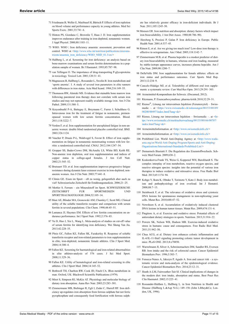
Figure 5
Total blood volume of male and female untrained, top athletes andathletes with dilutional pseudoanaemia in ml [44, 46, 49].APA = athlete with dilutional pseudoanaemia; TA = top athlete; UT= untrainedIn the column of the red cell mass the haematocrit in percentage isshown in white letters.
is defined as a lowered haemoglobin and haematocrit ow-ing to a distinctly elevated plasma volume with normal redcell mass and total haemoglobin mass. The other bloodparameters, particularly ferritin, MCV and MCH are withinthe normal range and performance is not affected. Thisplasma volume increase is considerable and is due to re-peated bouts of physical training over the years. It mag-nitude depends on the intensity and duration of the effort.In about 10–15% of mainly endurance athletes, especiallywhen training time exceeds 10 hours per week, this dilu-tional pseudoanaemia can be observed [44]. The same di-lutional effect is found in ultralong endurance disciplinesor sports events lasting several days (e.g. road cyclingtours). As seen in figure 5, in male and female athletes withdilutional pseudoanaemia the increase in plasma volumecauses a slight lowering of the haematocrit while the redcell mass stays high [42, 45–48].This is why top level endurance athletes show a high cor-relation between haemoglobin mass and endurance capa-city, but a low correlation between haematocrit or haemo-globin concentration and endurance capacity [49, 50].
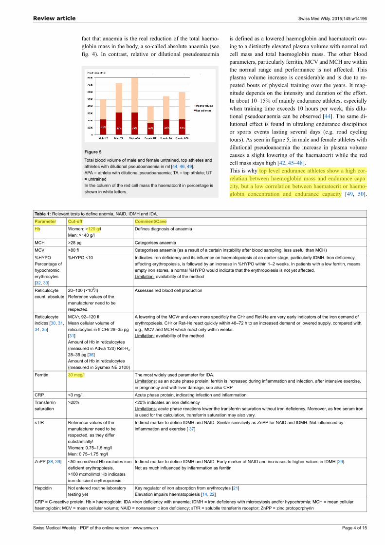
Table 1: Relevant tests to define anemia, NAID, IDMH and IDA.
Parameter Cut-off Comment/CaveHb Women: >120 g/l
Men: >140 g/lDefines diagnosis of anaemia
MCH >28 pg Categorises anaemia
MCV >80 fl Categorises anaemia (as a result of a certain instability after blood sampling, less useful than MCH)
%HYPOPercentage ofhypochromicerythrocytes[32, 33]
%HYPO <10 Indicates iron deficiency and its influence on haematopoiesis at an earlier stage, particularly IDMH. Iron deficiency,affecting erythropoiesis, is followed by an increase in %HYPO within 1–2 weeks. In patients with a low ferritin, meansempty iron stores, a normal %HYPO would indicate that the erythropoiesis is not yet affected.Limitation: availability of the method
Reticulocytecount, absolute
20–100 (×109/l)Reference values of themanufacturer need to berespected.
Assesses red blood cell production
Reticulocyteindices [30, 31,34, 35]
MCVr, 92–120 flMean cellular volume ofreticulocytes in fl CHr 28–35 pg[31]Amount of Hb in reticulocytes(measured in Advia 120) Ret-He
28–35 pg [36]Amount of Hb in reticulocytes(measured in Sysmex NE 2100)
A lowering of the MCVr and even more specificly the CHr and Ret-He are very early indicators of the iron demand oferythropoiesis. CHr or Ret-He react quickly within 48–72 h to an increased demand or lowered supply, compared with,e.g., MCV and MCH which react only within weeks.Limitation: availability of the method
Ferritin 30 mcg/l The most widely used parameter for IDA.Limitations: as an acute phase protein, ferritin is increased during inflammation and infection, after intensive exercise,in pregnancy and with liver damage, see also CRP
CRP <3 mg/l Acute phase protein, indicating infection and inflammation
Transferrinsaturation
>20% <20% indicates an iron deficiencyLimitations: acute phase reactions lower the transferrin saturation without iron deficiency. Moreover, as free serum ironis used for the calculation, transferrin saturation may also vary.
sTfR Reference values of themanufacturer need to berespected, as they differsubstantially!Woman: 0.75–1.5 mg/lMen: 0.75–1.75 mg/l
Indirect marker to define IDMH and NAID. Similar sensitivity as ZnPP for NAID and IDMH. Not influenced byinflammation and exercise [ 37]
ZnPP [38, 39] <50 mcmol/mol Hb excludes irondeficient erythropoiesis,>100 mcmol/mol Hb indicatesiron deficient erythropoiesis
Indirect marker to define IDMH and NAID. Early marker of NAID and increases to higher values in IDMH [29].Not as much influenced by inflammation as ferritin
Hepcidin Not entered routine laboratorytesting yet
Key regulator of iron absorption from erythrocytes [21]Elevation impairs haematopoiesis [14, 22]
CRP = C-reactive protein; Hb = haemoglobin; IDA =iron deficiency with anaemia; IDMH = iron deficiency with microcytosis and/or hypochromia; MCH = mean cellularhaemoglobin; MCV = mean cellular volume; NAID = nonanaemic iron deficiency; sTfR = solublle transferrin receptor; ZnPP = zinc protoporphyrin
Review article Swiss Med Wkly. 2015;145:w14196
Swiss Medical Weekly · PDF of the online version · www.smw.ch Page 4 of 15

Therefore, the haemoglobin mass is the important factorfor endurance capacity, while having a high plasma volume
accompanied by a low haematocrit is by no means a dis-advantage [51–53]. A larger intravascular volume has be-
Table 2: Randomised blinded interventional trials investigating NAID and performance.First author Performance
measurementn / Study population Inclusion criteria Intervention Results
Burden 2014[63]
VO2 maxTime to exhaustionRunning economyHaemoglobin mass
15Runners9 F, VO2 max 64.5 ml/kg·min
6 M VO2 max 76.7 ml/kgmin
Females:Hb >120 g/l Fer <30mcg/lMales:Hb >120 g/l Fer <40mcg/l
i.v. 500 mg Fe-carboxymaltose vsplacebo
Testing at BL, after 7days and after 4 wkBlood sampling asabove and one dayafter injection
– Fer ↑, traSat ↑, serum iron ↑ in Tx, Fer → in P– Hepcidin ↑ from day 1 up to four weeks in Tx– Hb mass →, VO2 max →, running economy →,time to exhaustion → in Tx and P
Garvican 2014[64]
Treadmill running withVO2 max, time toexhaustionHaemoglobin mass
27Highly trained distancerunners13 M, 14 F
4 randomisedtreatments: LG oral, LGi.v., CG oral, CG i.v.
Low group (LG)Fer <35 mcg/l and trSat<20% or Fer <15 mcg/l
Control group (CG)Fer <65 mcg/l
Oral: 105 g elementaliron 2×/d in LG; 1×/d inCGi.v.: 2–4 injections Fe-carboxymaltose due toiron status (mean i.v.CG 375 – mean i.v. LG550 mg)
BL testing and after 6wk
– Fer ↑ with oral and i.v. treatment, butsignificantly ↑ with i.v.– Hb → in all groups– In LG i.v.: Hb mass ↑, VO2 max ↑ and time toexhaustion ↑
Authors named CG SUB group, having asuboptimal iron status. But there was no changein Hb mass, VO2 max and time to exhaustion inthis group.
Della-Valle2014 [65]
4 km time trialVO2 peak
40Female rowersAt the beginning of aseason!
Hb >120 g/lFer <20 mcg/l
Oral 100 mg FeSO4/dvs placebo for 6 wk
Testing at BL and after6 wk of training
– Fat free mass ↑ and VO2 peak ↑ in Tx and P– Fer ↑ in Tx– Lower lactate response during first half of timetrial and after 5 min recovery in Tx– Energy expenditure in Tx ↑, lactate response ↓in first half of time trial in Tx, and 5 min after timetrial↓
Waldvogel2012 [66]
Chester step test (r =0.92 to VO2 max)Fatigue (VAS 10)
154Female blood donors
Hb >120 g/lFer ≤30 mcg/l
Oral 80 mg FeSO4/d vsplacebo for 4 wkBL (1 wk after donationof 450 ml blood) andafter 4 wk.
In Tx Hb ↑, Fer ↑ compared with P
No significant effect for fatigue, aerobic capacity (step test), mood disorder, quality of life
McClung 2009[67]
2 mile running timeProfile of mood state
219Female soldiers duringbasic combat training
Three groups:IDA (P 17 / Tx 18)ID (P 14 / Tx 14)Normal (P 51 / Tx 52)
No inclusion criteria.IDA defined as:Hb <120 g/land ≥2 of:Fer <12 mcg/l, traSat<16%, RDW >15%
ID defined as ≥2 of ironcrit.
Oral 100 mg FeSO4/dvs placebo
Testing at BL and after8 wk of basic combattraining
– RDW ↑, sTfr↑, Fer↓ in Tx and P.– Decrement in iron status ↓ in Tx– In soldiers with IDA and Tx– 2 mile running time↓ but not in P and Normal,– POMS↑ in Tx, P and Normal, but only in IDAvigour scores of POMS↑
Hinton 2007[68]
VO2 max60 min submax. cycleergometer test (at 60%VO2 max)
20Recreationally trained(3 M, 17 F)
Hb >120 g/l FHb >130 g/l M,Fer <16 mcg/l, sTfr >8mg/l, or sTfr / log Ferindex >4.5
Oral 30 mg elementaliron as FeSO4/d vsplacebo
Testing at BL and after6 wk
– Fer↑ in Tx, Hb and haematocrit →– In P ventilatory threshold↓, in Tx ventilatorythreshold→– Energetic efficiency during submaximal tes ↑ inTx
Brownlie 2004[69]
15 km time trial on cycleergometerVO2 max
41Untrained women
Training of 30 min/d,5×/wk for the final 4 wk
Hb >120 g/dl,Fer <16 mcg/l
Oral 100 mg FeSO4/dvs placebo
Testing at BL and after6 wk
Time in time trial↓, percentage of VO2 max ↓ ,work rate ↓ in Tx when sTfr >8 mg/lNo difference with normal sTfr
Brownlie 2002[70]
VO2 max 41Untrained womenTraining of 30 min/d,5×/wk for the final 4wk
Hb >120 g/dl,Fer <16 mcg/l
Oral 50 mg FeSO4/d vsplacebo for 6wk
Testing at BL and after6 wk
– Fer ↑, traSat ↑ in Tx– VO2 max ↑in Tx and P, but VO2 max wassignificantly higher in Txr
Friedman 2001[71]
Treadmill VO2 maxAnaerobic capacity (O2consumption)Haemoglobin mass
40Young elite athletes
Hb >135 g/l M,Hb >117 g/l FFer <20 mcg/l
Oral 200 mg elementaliron/d vs placebo for 12wk
Testing at BL and after12 wk
– Fer ↑ in Tx, whereas P →Venous Hb and Hb mass → in Tx and PVO2 max ↑ and O2 consumption ↑ inTx
Hinton 2000[72]
15 km time trial on cycleergometer
42WomenTraining of 30 min/d,5×/wk for the final 4 wk
Hb >120 g/lFer <16 mcg/l
Oral 100 mg FeSO4/dvs placebo for 6 wk
Testing at BL and after6 wk
– Fer ↑ and Hb ↑ in Tx– 15 km time ↓ in Tx and P, but significantly ↓ inTx (p = 0.04)
↑ = increased significantly; → = no change; ↓ = lowered significantly; BL = baseline; CG = control group; F = female; Fer = ferritin; Hb = haemoglobin; ID = iron deficiency;IDA = iron deficiency anaemia; LG = low group; M = male; NAID = nonanaemic iron deficiency; P = placebo group; POMS = profile of mood state; RDW = red celldistribution width; SUB = group with suboptimal iron status; traSat = transferrin saturation; Tx = treatment group; VAS = visual analogue scale
Review article Swiss Med Wkly. 2015;145:w14196
Swiss Medical Weekly · PDF of the online version · www.smw.ch Page 5 of 15
neficial effects on performance, as it improves thermoreg-ulation, increases cardiac preload and enhances the body´sability to dilute metabolites. The exact mechanism for thisadaptation remains to be explored, and shifts in proteinsand electrolytes are thought to play a role [54]. Exercise in-duced plasma volume shifts are usually reversible withindays after cessation of the physical effort [40].A dilutional pseudoanaemia cannot be ruled out by a ven-ous blood examination only, but in clinical practice serialmeasurements of the basic blood examination are helpful.These include measurements during the training pause atthe end of the season (haematocrit, Hk↑), during the season(Hk↓), once immediately after an intensive and long lastingtraining session (Hk↑) and 24–48 h after this session (Hk↓).If these serial measurements do not reveal the expectedvariations of the haematocrit (showing at least normal val-ues in the training pause at season’s end) the measurementof haemoglobin mass to confirm the dilutional pseudoan-aemia may be considered with, for example, the COrebreathing method [48–50].
Excessive haematocritsUnfortunately, top level road cyclists 15 years ago oftenshowed excessive haematocrit levels of over 50%, excep-tionally up to 60%. Based on the confessions of severalriders, these values could only be reached by mostly sys-tematic doping procedures with mainly erythropoietin,growth hormone and testosterone. During longer lastingcompetitions blood transfusions (retransfusion of ownblood, collected several months before) [55] were givento boost oxygen carrying capacity. Through awareness ofthese doping procedures, health control tests with bloodsampling were introduced, with upper limits of thehaematocrit (e.g. the 50% rule of the Union de CyclismeInternational [56]) or haemoglobin concentration (e.g. 170g/l for males, 160 g/l for females of the Fédération Inter-national de Ski [57]) to protect athletes from fatal throm-botic events. These were followed by newer doping tests(e.g. EPO-tests, growth hormone) and antidoping strategiessuch as the biological passport [58]. Several blood samplescollected over the year provide an individual blood profileand allow individual reference values to be established foreach athlete [59]. This biological passport proposed by theWADA (World antidoping agency) gives a much closerfollow-up and is used in cases of violation of the antidop-ing code [58].
Influence of iron on physical performanceIron is, in addition to its function in oxygen transport, akey component of the enzymatic system of the respiratorychain. These presumably distinct roles were investigated inearly animal studies [60–62]. In these studies, the authorsaimed at differentiating between a decline in performancedue to anaemia and a decline due to enzymatic impairment.Using an animal model in a cross-over setting with iron de-pletion and cross-over transfusion to correct for anaemia,it was shown that not only anaemia but also iron depletionwithout anaemia led to a significantly decreased numberof mitochondria and reduced activity of the respiratory en-zymes. The authors therefore postulated that iron depletion
without anaemia affected oxidative capacity, whereas an-aemia affected mostly oxygen transport [60–62].At present the cut-off for ferritin is still debated with valuesranging from 15 mcg/l from the World Health Organization[73] to 16–32 mcg/l [74, 75] in studies that used the “goldstandard procedure of bone marrow staining”. It has to benoted that studies investigating this matter were not all con-clusive and some presented controversial results [76, 77].If not only erythropoiesis but also clinical symptoms of aniron deficiency such as fatigue (but not yet performance)are considered, the cut-off may be slightly higher. RecentlyKrayenbuehl et al. showed in their double-blind random-ised study with intravenous iron administration in nonan-aemic premenopausal women with low ferritin and fatiguean improvement in mood state in the ferritin group <15mcg/l [78]. Earlier, Verdon et al. analysed fatigue and moodstate with oral therapy in a family doctor’s office setting.They found that nonanaemic women with ferritin concen-trations below 50 mcg/l improved with oral supplementa-tion [79]. Vaucher et al came to comparable results [80].But their proposed cut-off of 50 mcg/l must be interpretedwith caution because of methodological concerns (e.g. as-sessment of stool coloration by iron not blinded, definitionof iron deficiency based on a limited number of variables).A few studies have integrated a performance measure(table 2). McClung et al showed in female military recruitstreated with oral iron or placebo an improvement in run-ning performance in recruits with IDA. Those with NAIDshowed an improved mood state, but no change in per-formance. Since then, several other studies have shown aperformance impairment in nonanaemic iron-depleted en-durance athletes [68–70, 72, 81]. A limiting factor of allthese studies is the influence of training or mild changes inhaematological parameters of other origin, both of whichare difficult to quantify. Garvican et al very recently per-formed a study with 27 highly trained distance runners (13male and 14 female athletes) with measurement of haemo-globin mass and endurance performance. They comparedin a randomised controlled setting intravenous iron versus6 weeks of oral treatment with one group having clearly de-ficient iron stores (ferritin <15 mcg/l or ferritin <35 mcg/land transferrin saturation <20%) and the other with betteriron stores but a ferritin level of <65 mcg/l. Both forms ofsupplementation substantially increased ferritin levels, buthaemoglobin concentration did not change in any group.Haemoglobin mass increased in the iron deficiency groupwith intravenous treatment accompanied by an increase inVO2 max and run time to exhaustion, whereas the groupwith ferritin <65 mcg/l did not show any changes in theseparameters [53, 64].As multiple muscle enzymes are affected other functionsrelated to performance have been investigated. Brutsaert etal. found an increase in knee extensor strength after ironsupplementation in nonanaemic iron-depleted volunteersusing a randomised controlled study design, which pointstowards a role of iron in resistance to fatigue and adaptationto training [82]. Very recently Della Valle et al investigatedfemale rowers with NAID under oral iron supplementationfor 6 weeks in a double-blind design. They were able toshow an improvement of iron status and a better energeticefficiency in the iron group [65].
Review article Swiss Med Wkly. 2015;145:w14196
Swiss Medical Weekly · PDF of the online version · www.smw.ch Page 6 of 15

Based on the cited results and according to several reviewarticles, a ferritin cut-off of 30 mcg/l in adults seems to bemost plausible [8, 83–90].
Iron deficiency in sports – therapy
NutritionThe first step in therapy of iron deficiency is the correctionof the nutritional iron intake. In nutrition, haem iron(mainly in meat) and free iron as Fe2+ or Fe3+ exist. Oraluptake studies show that the uptake of haem iron is muchbetter than the uptake of free iron [91]. For the latter, up-take of Fe2+ is better than uptake of Fe3+ [92]. Meat, liv-er, poultry or fish contain haem iron as well as free iron. Avegetarian diet contains only free iron. The bioavailabilityof iron is very variable and depends much on actual ironstores [93], ranging from 5 to 15%. In the case of iron de-ficiency, a significant increase in iron bioavailability up to35% can be observed [94]. Furthermore, iron uptake in theintestinal tract is influenced by different nutritional factorsincluding enhancers and inhibitors. Substances enhancingiron uptake are vitamin C, peptides from partially digestedmuscle tissue, fermented food, organic acids such as malateor citrate. Substances inhibiting iron uptake are phytates,oxalates, polyphenols (black tea and coffee), peptides frompartially digested vegetable proteins and calcium [83, 84].The nutritional intake should be 14 mg per day. General re-commendations for an optimal dietary iron intake in sportsinclude an adequate energy intake, especially for athleteswith low body mass index as they suffer more frequentlyfrom iron deficiency [95]. Whether catabolism related tolow energy intake influences the hepcidin regulation anddown-regulates iron uptake remains open to debate. In gen-eral, regular consumption of meat, poultry or fish, at least5 times per week is recommended as it is the main con-tributor of nutritional iron intake. Complementary eatingof wholemeal products and daily legumes and green veget-ables is suggested. Furthermore, it is beneficial to replacetea and coffee by a glass of orange or citrus fruit juice withan iron containing meal as vitamin C enhances iron uptake[84, 94]. For vegetarians the goal is to reach a high loadof iron through their vegetable diet. Even if nutrition is im-portant in iron homeostasis in the human organism, IDAcannot be corrected by nutrition alone, as this would meaneating kilograms of iron containing products (e.g. liver).
Oral ironUsually dietary counselling and oral iron therapy are com-bined. Oral preparations differ in the amount and type ofiron (Fe2+ or Fe3+), their complex forming substrate and intheir galenical form. Novel products combine iron with vit-amin C. In a dose-finding study in elderly patients withIDA, Rimon et al compared three doses of oral iron: 15 mg,50 mg and 150 mg. They were able to show that iron sup-plementation at a level of the recommended daily allow-ances (RDA, 15 mg of elemental iron) already led to signi-ficant increases of the iron status. In these anaemic elderlypatients, the doses of 50 mg and 150 mg of elemental irondid not show further benefit, but had significantly moreside effects particularly in the highest dose group [96]. As
there is further evidence that oral iron loading increasescirculating hepcidin [97], the recommended dosage of oraliron should not be too high. In a recent comparison of oraliron supplementation in a randomised controlled trial look-ing at iron status and performance in active women, 100mg of FeSO4 (approximately 20 mg of elemental iron) wasshown to be effective [98]. We therefore recommend sup-plementation of 40 to 60 mg of elemental iron once daily.Oral iron is in general well tolerated and efficient [99].Side effects of oral therapy are usually not serious and in-clude nausea, dyspepsia, constipation or diarrhoea [83, 84,100]. Some individuals with an existing tendency to con-stipation benefit from drinking additional fruit juice to pre-vent heavy constipation. Otherwise compliance will be ser-iously affected.
Intravenous ironWhen oral therapy fails or immediate restoration is neededi.v. therapy should be considered. At the moment two dif-ferent preparations are available in Switzerland, compris-ing iron saccharose and ferric carboxymaltose [100]. Thedosage is dependent on the severity of the iron deficiency.In one application usually 200 mg Fe-saccharose, 500 to100 mg Fe-carboxymaltose can be administered.The main advantage of i.v. therapy is the immediate correc-tion of the iron deficiency and restoration of the empty ironstores. Generally, compliance with i.v. iron supplements isgood. Side effects may encompass a transient disturbanceof taste, headache, dizziness, myalgia and fever, but severeadverse reactions such as hypotonic and anaphylactoid re-actions, tachycardia and arrhythmia, dyspnoea and bron-chospasm may also be observed, albeit very rarely [100].Moreover, transient and usually asymptomatic hypophos-phataemia is frequently observed after the administrationof Fe-carboxymaltose [101].It is still under discussion whether hypophosphataemiamay possibly be a cofactor for cardiac events [101].Nowadays severe side effects are rare. For Fe-saccharoseand Fe-carboxymaltose there has been no fatal outcome re-ported in Switzerland. This is in contrast to the previouslyused iron-dextran products, which had a much higher rateof severe and fatal side effects. In 2013, another type of i.v.iron, Fe-oxytol, was withdrawn owing to severe hypersens-itivity reactions in four patients, including one fatal out-come, observed 9 months after approval by the regulatoryauthorities in Switzerland [102, 103]. Even with the use ofthe new preparations, severe adverse reactions cannot beexcluded, and the administration of i.v. iron preparations isonly recommended in settings where resuscitation skills areavailable and observation of the patient for 30 minutes afterthe end of the i.v. administration can be guaranteed [104,105].In general, the frequency of side effects seems to be lowerwhen the i.v. iron is applied by infusion instead of slowbolus injection. Importantly, the exact dilution given by themanufacturer needs to be respected. Further, the physicianneeds to respect the WADA antidoping regulations con-cerning administration of infusion in elite sports: “Intra-venous infusions and/or injections of more than 50 ml per6 hour period are prohibited except for those legitimately
Review article Swiss Med Wkly. 2015;145:w14196
Swiss Medical Weekly · PDF of the online version · www.smw.ch Page 7 of 15

received in the course of hospital admissions or clinical in-vestigations” [106].
Check of adequate iron stores
In sports a regular check of blood parameters is necessary,especially in endurance athletes. To monitor efficacy oftherapeutic measures we recommend repeating the basicblood tests 6 to 8 weeks after the start of the nutritionalmeasures, oral therapy or i.v. iron administration.Depending on the blood results, therapy will be continuedor modified with the aim of reaching or to keeping the ironstores in the regular range. Treatment approaches may becombined and athletes with repeatedly low iron stores maybenefit from intermittent oral substitution to preserve ironstores, e.g. substitution with 14 to 28 m elemental iron perday or 40–60 mg elemental iron two to three times perweek. In vegetarian athletes a similar approach to prevent-ing iron deficiency is recommended: 40 to 60 mg elementaliron three times per week instead of meat intake or a dailysupplementation with at least 14 or 28 mg/day of iron willusually cover iron demand.As iron homeostasis is exclusively and meticulously con-trolled by the iron uptake through the intestinal tract [107]and as there is no pathway to eliminate iron in cases ofoverload, iron supplementation has to be done always care-fully. In extreme cases, chronic iron overload may lead tosecondary haemochromatosis. Furthermore, excessive sup-plementation of oral or i.v. iron is thought to increase oxid-ative stress and production of free radicals [108, 109] andoxidative stress is suggested to play a role in cancer genesis[110, 111]. This must be critically appraised as acute exer-cise leads to oxidative stress [112, 113].It has been shown in mice that oral iron supplementationenhances colonic tumour development [114]. Data in hu-mans suggest that iron may increase the risk for colorectalcancer [115]. A recent meta-analysis showed on the onehand a tendency toward a positive association betweenhigh intake of haem iron and cancer risk. On the otherhand high levels of biomarkers of iron stores implied a
lower cancer risk [116]. Further prospective and experi-mental studies are needed to evaluate the possible influenceof iron in carcinogenesis.
Iron deficiency in the child andadolescent athlete
A British study reported an iron deficiency prevalence of21% in adolescents between 11–18 years [117, 118]. Al-though likely to be higher in the athletic population, dataabout young athletes concerning iron deficiency are sparse[119–121].Total iron requirements in children and adolescents are dis-tinctly increased because of additional iron needs for theexpansion of the total blood volume and mean haemo-globin mass, as well as for the enhancement in lean bodymass during growth [122]. In adolescent females the onsetof menarche is associated with an increase of iron require-ment. The mean total iron requirement for adolescentsreaches 1.8 mg/d for boys and 2.2 mg/d for girls (in femaleswith heavy periods it is considerably more), which corres-ponds to more than double that in the preadolescent peri-od [123, 124]. Otherwise risk factors for iron deficiency,both in younger and older athletes, are the same [20, 23, 24,125].Haematological normal values in children and adolescentsare different from adults and should always be considered(see table 3). We recommend defining the lower level ofnormal for ferritin as 15 mcg/l for children between 6–12years, 20 mcg/l between 12–15 years and 30 mcg/l for15–18 year old adolescents [126].As in adults, in a case of NAID dietary counselling is thefirst step, often combined with oral therapy. Iron require-ments (RDA) are 8 mg/d for 9–13 year-old children and 11mg/d for male and 15 mg/d for female adolescents olderthan 13 years. Careful management, especially of the men-struating teenage girl and the vegetarian athlete, is warran-ted [127]. If iron deficiency results in IDMH or IDA, fur-ther supplementation should be considered. Comparable tothe therapy in adults, oral substitution can be with either
Table 3: Critical cut-offs for differentiation of iron deficiency in children and adolescents. Adapted from Herklotz et al. [29].
Definition Haemoglobin MCV/MCH Ferritin CRPNAID Normal Normal
6–12y:MCV <76 fl or noMCH <25 pg or no
6–12y: <15 mcg/l
12–15 y:MCV <78 fl or noMCH <26 pg or no
12–15y: <20 mcg/l
IDMH Normal
15–18 y:MCV <79 fl or noMCH <26.5 pg or no
15–18y: <30 mcg/l
6–12 y:<112 g/l
6–12y:MCV <76 flMCH <25 pg
12–15y:M <125 g/lF <120 g/l
12–15y:MCV <78 fl or noMCH <26 pg or no
IDA
15–18y:M <130 g/lF <120 g/l
15–18 y:MCV <79 fl or noMCH <26.5 pg or no
Normal
CRP = C-reactive protein; IDA = iron deficiency anaemia; IDMH = iron deficiency with microcytosis and/or hypochromia; MCV = mean cellularhaemoglobin; MCH = mean corpuscular haemoglobin; NAID = nonanaemic iron deficiency
Review article Swiss Med Wkly. 2015;145:w14196
Swiss Medical Weekly · PDF of the online version · www.smw.ch Page 8 of 15

Fe2+ or Fe3+ preparations. The dosage for iron preparationsis 5 mg/kg/d for 3 months in two or three daily doses. TheFe2+ preparation is recommended as medication of choicebecause of better bioavailability. As in adult athletes, meas-urement of haemoglobin, red cell indices, ferritin and CRPafter 6–8 weeks of treatment is necessary in order to ob-serve responsiveness and compliance with the treatment.
Conclusions and recommendations
Iron deficiency in sports is frequent and relevant as allstages of iron deficiency ‒ IDA, IDMH and NAID ‒ affectphysical performance.To diagnose iron deficiency, haemoglobin, haematocrit,MCV, MCH and ferritin are first-line parameters to assess.For a valid interpretation of the results it is necessary toexclude acute-phase reactions that may interfere, such astraining sessions and infectious diseases (patient historyand measurement of CRP). In unclear situations a secondmeasurement of the same parameters or the additionalmeasurement of zinc-protoporphyrin, soluble transferrinreceptor and transferrin saturation may be helpful. In highlevel endurance athletes basic blood examinations shouldbe made two to three times a year.Ferritin values below <15 mcg/l are very specific for emptyiron stores. Ferritin values from 15 to 30 mcg/l correspondto low iron stores. A ferritin value of 30 mcg/l should betaken as a reasonable cut-off for adult men and women andolder adolescents (15 years and older). For younger adoles-cents from 12 to 15 years a cut-off of 20 mcg/l and forchildren from 6 to 12 years a cut-off of 15 mcg/l is re-commended. In adult elite sports altitude training repres-ents a special situation with an increased need for iron. Inthis case a ferritin value of 50 mcg/l prior to altitude train-ing should be aimed for. Every case of severe iron defi-ciency warrants an extended work up, and this is also truein sports.Therapy of iron deficiency in sports consists of the nutri-tional counselling including sufficient energy intake and5 times/week haem iron intake (meat, poultry, fish) withthe addition of legumes and green vegetables (e.g. spinach,fennel) usually combined with oral iron supplementation.Daily oral iron preparations in a dosage of 40 to 60 mgof elemental iron are appropriate, as the not severe butdisturbing gastrointestinal side effects are related to irondosage and preparation. Consider enhancers (vitamin C)and prevent inhibitors (coffee, black tea, phytates, calcium)of iron uptake to increase absorption. Athletes and patientswith repeatedly low ferritin values profit from intermittentoral substitution to preserve iron stores, e.g. two iron tab-lets per week (containing 40 to 60 mg elemental iron) asmaintenance therapy. In cases of plausible nonresponders,incompatibility with oral therapy, a concomitant disease(e.g. depression) or low iron stores before an altitude train-ing camp, i.v. iron therapy should be considered.Long-term daily oral iron intake or i.v. supplementation inthe presence of normal or even high ferritin values is notrecommended and may be harmful.
Acknowledgements: We thank all those who supported us towrite this article. These include Prof. Dr. med. Arno Schmidt-Trucksäss, Prof. Dr. med. Roland von Känel, Dr. med. KerstinWarnke and Dr. med. Christian Schlegel.Funding / potential competing interests: No financial supportand no other potential conflict of interest relevant to this articlewas reported.
Correspondence: German E. Clénin, Sportmedizinisches
Zentrum Ittigen bei Bern, Haus des Sports, CH-3063 Ittigen,
Switzerland, german.clenin[at]smzbi.ch
References
1 Proposed nutrient and energy intakes for the European community: a re-port of the Scientific Committee for Food of the European community.Nutr Rev. 1993;51:209–12.
2 Egli I. M. MARIA ANDERSSON*, INES M. EGLI*, MICHAELB. ZIMMERMANN. at <http://www.rosenfluh.ch/rosenfluh/articles/download/1318/Eisenmangel.pdf>
3 Schleiffenbaum BE, et al. Unexpected high prevalence of metabolicdisorders and chronic disease among young male draftees – the SwissArmy XXI experience. Swiss Med Wkly. 2006;136:175–84.
4 Sandström G, Börjesson M, Rödjer S. Iron deficiency in adolescent fe-male athletes – is iron status affected by regular sporting activity? ClinJ Sport Med. 2012;22:495–500.
5 Latunde-Dada G. O. Iron metabolism in athletes- achieving a goldstandard. Eur J Haematol. (2012). doi:10.1111/ejh.12026
6 Dubnov G, et al. High prevalence of iron deficiency and anemia in fe-male military recruits. Mil Med. 2006;171:866–9.
7 Cippa P, Krayenbühl P-A. Eisenmangel: Es geht nicht nur um Anämie.Schweiz Med Forum. 2014;11–12.
8 Martius F. Eisenmangel ohne Anämie – ein heisses Eisen. in SchweizMed Forum. 2009;9:294–9.
9 Streuli R. A. Ferrum bonum et laudabile (lucrosumque). in SchweizMed Forum. 2008;8:563.
10 Colombani PC, Mannhart C. Energie- und Nährstoffaufnahme im Sch-weizer Spitzensport – eine erste Bestandsaufnahme zu Beginn deszweiten Jahrtausends. 2003;7–16.
11 Mettler S, Zimmermann M. B. Iron excess in recreational marathon run-ners. Eur J Clin Nutr. 2010;64490–4.
12 Ganz T. Molecular Control of Iron Transport. JASN. 2007;18:394–400.
13 Löffler G. in S.416 ff (Springer Verlag).
14 Ganz T, Nemeth E. Iron metabolism: interactions with normal anddisordered erythropoiesis. Cold Spring Harb Perspect Med.2012;2:a011668.
15 Yip R. in Present Knowledge in Nutrition, Bowman BA, Russell RM311 – 328 (ILSI Press, Washington DC, 2001).
16 Nemeth E, Ganz T. Regulation of iron metabolism by hepcidin. AnnuRev Nutr. 2006;26:323–42.
17 Young B, Zaritsky J. Hepcidin for clinicians. Clin J Am Soc Nephrol.2009;4:1384–7.
18 Taylor C, et al. Hematologic, iron-related, and acute-phase proteinresponses to sustained strenuous exercise. J Appl Physiol.1987;62:464–9.
19 Peeling P, Dawson B, Goodman C, Landers G, Trinder D. Athletic in-duced iron deficiency: new insights into the role of inflammation, cy-tokines and hormones. Eur J Appl Physiol. 2008;103:381–91.
20 Peeling P. Exercise as a mediator of hepcidin activity in athletes. Eur JAppl Physiol. 2010;110:877–83.
21 Ganz T. Hepcidin, a key regulator of iron metabolism and mediator ofanemia of inflammation. Blood. 2003;102:783–8.
22 Newlin MK, et al. The effects of acute exercise bouts on hepcidin inwomen. Int J Sport Nutr Exerc Metab. 2012;22:79–88.
23 Gaudin C, Zerath E, Guezennec CY. Gastric lesions secondary to long-distance running. Dig Dis Sci. 1990;35:1239–43.
Review article Swiss Med Wkly. 2015;145:w14196
Swiss Medical Weekly · PDF of the online version · www.smw.ch Page 9 of 15

24 Peters HP, De Vries WR, Vanberge-Henegouwen GP, Akkermans LM.Potential benefits and hazards of physical activity and exercise on thegastrointestinal tract. Gut. 2001;48:435–9.
25 Waller MF, Haymes EM. The effects of heat and exercise on sweat ironloss. Med Sci Sports Exerc. 1996;28:197–203.
26 Jones GR, Newhouse I. Sport-related hematuria: a review. Clin J SportMed. 1997;7:119–25.
27 Miller BJ, Pate RR, Burgess W. Foot impact force and intravascularhemolysis during distance running. Int J Sports Med. 1988;9:56–60.
28 Telford RD, et al. Footstrike is the major cause of hemolysis during run-ning. J Appl Physiol. 2003;94:38–42.
29 Herklotz R, Huber A. Labordiagnose von Eisenstoffwechselstörungen.in Schweiz Med Forum. 2010;10:500–7.
30 Brugnara C, Laufer MR, Friedman AJ, Bridges K, Platt O. Reticulocytehemoglobin content (CHr): early indicator of iron deficiency and re-sponse to therapy. Blood. 1994;83:3100–1.
31 Thomas C, Thomas L. Biochemical markers and hematologic indicesin the diagnosis of functional iron deficiency. Clin Chem.2002;48:1066–76.
32 Macdougall IC. What is the most appropriate strategy to monitor func-tional iron deficiency in the dialysed patient on rhEPO therapy? Meritsof percentage hypochromic red cells as a marker of functional iron de-ficiency. Nephrol Dial Transplant. 1998;13:847–9.
33 Onofrio G d’, Zini G, Ricerca BM, Mancini S, Mango G. Automatedmeasurement of red blood cell microcytosis and hypochromia in irondeficiency and beta-thalassemia trait. Arch Pathol Lab Med.1992;116:84–9.
34 Onofrio G d’, et al. Simultaneous measurement of reticulocyte and redblood cell indices in healthy subjects and patients with microcytic andmacrocytic anemia. Blood. 1995;85:818–23.
35 Brugnara C, Zurakowski D, DiCanzio J, Boyd T, Platt O. Reticulocytehemoglobin content to diagnose iron deficiency in children. JAMA.1999;281:2225–30.
36 Franck S, Linssen J, Messinger M., Thomas L. Potential utility ofRet-Y in the diagnosis of iron-restricted erythropoiesis. Clin Chem.2004;50:1240–2.
37 Schumacher YO, Schmid A, König D, Berg A. Effects of exercise onsoluble transferrin receptor and other variables of the iron status. Br JSports Med. 2002;36:195–9.
38 Magge H, Sprinz P, Adams WG, Drainoni M-L, Meyers A. Zinc pro-toporphyrin and iron deficiency screening: trends and therapeutic re-sponse in an urban pediatric center. JAMA Pediatr. 2013;167:361–7.
39 Baart AM, et al. High prevalence of subclinical iron deficiency inwhole blood donors not deferred for low hemoglobin. Transfusion.2013;53:1670–7.
40 Voss SC, et al. Variability of serum markers of erythropoiesis during6 days of racing in highly trained cyclists. Int J Sports Med.2014;35:89–94.
41 Cordova A, Monserrat J, Villa G, Reyes E, Soto MA-M. Effects ofAM3 (Inmunoferon) on increased serum concentrations of interleuk-in-6 and tumour necrosis factor receptors I and II in cyclists. J SportsSci. 2006;24:565–73.
42 Dickson DN, Wilkinson RL, Noakes TD. Effects of ultra-marathontraining and racing on hematologic parameters and serum ferritin levelsin well-trained athletes. Int J Sports Med. 1982;3:111–7.
43 Fallon KE, Sivyer G, Sivyer K, Dare A. The biochemistry of runners ina 1600 km ultramarathon. Br J Sports Med. 1999;33:264–9.
44 Friedmann B. Standards der Sportmedizin Sportleranämie.DEUTSCHE ZEITSCHRIFT FÜR SPORTMEDIZIN 2001;52.
45 Shaskey DJ, Green GA. Sports haematology. Sports Med.2000;29:27–38.
46 Bärtsch P, Mairbäurl H, Friedmann B. Pseudo-anemia caused by sports.Ther Umsch. 1998;55:251–5.
47 Nichols AW. Nonorthopaedic problems in the aquatic athlete. ClinSports Med. 1999;18:395–411, viii.
48 Heinicke K, et al. Blood volume and hemoglobin mass in elite athletesof different disciplines. Int J Sports Med. 2001;22:504–12.
49 Steiner T, Wehrlin JP. Does hemoglobin mass increase from age 16to 21 and 28 in elite endurance athletes? Med Sci Sports Exerc.2011;43:1735–43.
50 Steiner T, Wehrlin JP. Comparability of haemoglobin mass measuredwith different carbon monoxide-based rebreathing procedures and cal-culations. Scand J Clin Lab Invest. 2011;71:19–29.
51 Hinrichs T, et al. Total hemoglobin mass, iron status, and endurancecapacity in elite field hockey players. J Strength Cond Res.2010;24:629–38.
52 Wehrlin JP. Live high-train low for 24 days increases hemoglobinmass and red cell volume in elite endurance athletes. J Appl Physiol.2006;100:1938–45.
53 Garvican LA, et al. Intravenous Iron Supplementation in Distance Run-ners with Low or Suboptimal Ferritin. Med Sci Sports Exerc. (2013).doi:10.1249/MSS.0b013e3182a53594.
54 Fellmann N. Hormonal and plasma volume alterations following endur-ance exercise. A brief review. Sports Med. 1992;13:37–49.
55 Hamilton T, et al. Die Radsport-Mafia und ihre schmutzigen Geschäfte:Der Insider-bericht über die Welt des Profiradsports: eine minutiöseBeichte, die erstmals das ganze ... Armstrongs Schlüsselrolle darinaufzeigt. (2012).
56 40_ans.pdf. at <http://oldsite.uci.ch/english/health_sante/docs/40_ans.pdf>
57 Bad blood : Nature News. at <http://www.nature.com//news/2006/060224/full/news060220-18.html>
58 Athlete Biological Passport (ABP) Operating Guidelines | World Anti-Doping Agency. at <https://www.wada-ama.org/en/resources/athlete-biological-passport/athlete-biological-passport-abp-operating-guidelines>
59 Saugy M, Lundby C, Robinson N. Monitoring of biological markersindicative of doping: the athlete biological passport. Br J Sports Med.2014;48:827–32.
60 Davies KJ, Maguire JJ, Brooks GA, Dallman PR, Packer L. Muscle mi-tochondrial bioenergetics, oxygen supply, and work capacity during di-etary iron deficiency and repletion. Am J Physiol. 1982;242:E418–427.
61 Davies KJ, et al. Distinguishing effects of anemia and muscle irondeficiency on exercise bioenergetics in the rat. Am J Physiol.1984;246:E535–543.
62 Finch CA, et al. Iron deficiency in the rat. Physiological and biochem-ical studies of muscle dysfunction. J Clin Invest. 1976;58:447–53.
63 Burden RJ, et al. Impact of Intravenous Iron on Aerobic Capacityand Iron Metabolism in Elite Athletes. Med Sci Sports Exerc. (2014).doi:10.1249/MSS.0000000000000568
64 Garvican LA, et al. Intravenous iron supplementation in distance run-ners with low or suboptimal ferritin. Med Sci Sports Exerc.2014;46:376–85.
65 DellaValle DM, Haas JD. Iron supplementation improves energeticefficiency in iron-depleted female rowers. Med Sci Sports Exerc.2014;46:1204–15.
66 Waldvogel S, et al. Clinical evaluation of iron treatment efficiencyamong non-anemic but iron-deficient female blood donors: a random-ized controlled trial. BMC Med. 2012;10;8.
67 McClung JP, et al. Randomized, double-blind, placebo-controlled trialof iron supplementation in female soldiers during military training: ef-fects on iron status, physical performance, and mood. Am J Clin Nutr.2009;90:124–31.
68 Hinton PS, Sinclair LM. Iron supplementation maintains ventilatorythreshold and improves energetic efficiency in iron-deficient nonanem-ic athletes. Eur J Clin Nutr. 2007;61:30–9.
69 Brownlie T, 4th, Utermohlen V, Hinton PS, Haas JD. Tissue irondeficiency without anemia impairs adaptation in endurance capacityafter aerobic training in previously untrained women. Am J Clin Nutr.2004;79:437–43.
70 Brownlie T, 4th, Utermohlen V, Hinton PS, Giordano C, Haas JD. Mar-ginal iron deficiency without anemia impairs aerobic adaptation amongpreviously untrained women. Am J Clin Nutr. 2002;75:734–42.
Review article Swiss Med Wkly. 2015;145:w14196
Swiss Medical Weekly · PDF of the online version · www.smw.ch Page 10 of 15
71 Friedmann B, Weller E, Mairbaurl H, Bärtsch P. Effects of iron repletionon blood volume and performance capacity in young athletes. Med SciSports Exerc. 2001;33:741–6.
72 Hinton PS, Giordano C, Brownlie T, Haas J. D. Iron supplementationimproves endurance after training in iron-depleted, nonanemic women.J Appl Physiol. 2000;88:1103–11.
73 WHO. WHO | Iron deficiency anaemia: assessment, prevention andcontrol. WHO at <http://www.who.int/nutrition/publications/micronu-trients/anaemia_iron_deficiency/WHO_NHD_01.3/en/>
74 Hallberg L, et al. Screening for iron deficiency: an analysis based onbone-marrow examinations and serum ferritin determinations in a pop-ulation sample of women. Br J Haematol. 1993;85:787–98.
75 van Tellingen O. The importance of drug-transporting P-glycoproteinsin toxicology. Toxicol Lett. 2001;120:31–41.
76 Magnusson B, Hallberg L, Rossander L, Swolin B. Iron metabolism and‘sports anemia’. I. A study of several iron parameters in elite runnerswith differences in iron status. Acta Med Scand. 1984,216:149–55.
77 Thomason RW, Almiski MS. Evidence that stainable bone marrow ironfollowing parenteral iron therapy does not correlate with serum ironstudies and may not represent readily available storage iron. Am J ClinPathol. 2009;131:580–5.
78 Krayenbuehl P-A, Battegay E, Breymann C, Furrer J, Schulthess G.Intravenous iron for the treatment of fatigue in nonanemic, premen-opausal women with low serum ferritin concentration. Blood.2011;118:3222–7.
79 Verdon F, et al. Iron supplementation for unexplained fatigue in non-an-aemic women: double blind randomised placebo controlled trial. BMJ.2003;326:1124.
80 Vaucher P, Druais P-L, Waldvogel S, Favrat B. Effect of iron supple-mentation on fatigue in nonanemic menstruating women with low fer-ritin: a randomized controlled trial. CMAJ. 2012;184:1247–54.
81 Gropper SS, Bader-Crowe DM, McAnulty LS, White BD, Keith RE.Non-anemic iron depletion, oral iron supplementation and indices ofcopper status in college-aged females. J Am Coll Nutr.2002;21:545–52.
82 Brutsaert TD, et al. Iron supplementation improves progressive fatigueresistance during dynamic knee extensor exercise in iron-depleted, non-anemic women. Am J Clin Nutr. 2003;77:441–8.
83 Clénin GE. Eisen im Sport – oft zu wenig, gelegentlich aber auch zuviel! Schweizerische Zeitschrift für Ernährungsmedizin. 2006;21–25.
84 Mettler S. Ferrum – ein Mineralstoff im Sport. SCHWEIZERISCHEZEITSCHRIFT FUR SPORTMEDIZIN UNDSPORTTRAUMATOLOGIE 2004;52:105–14.
85 Mast AE, Blinder MA, Gronowski AM, Chumley C, Scott MG. Clinicalutility of the soluble transferrin receptor and comparison with serumferritin in several populations. Clin Chem. 1998;44:45–51.
86 Lamanca JJ, Haymes EM. Effects of low ferritin concentration on en-durance performance. Int J Sport Nutr. 1992;2:376–85.
87 Yu D, Huo J, Xie L, Wang L. Meta-analysis of studies on cut-off valueof serum ferritin for identifying iron deficiency. Wei Sheng Yan Jiu.2013;42:228–35.
88 Pitsis GC, Fallon KE, Fallon SK, Fazakerley R. Response of solubletransferrin receptor and iron-related parameters to iron supplementationin elite, iron-depleted, nonanemic female athletes. Clin J Sport Med.2004;14:300–4.
89 Fallon KE. Screening for haematological and iron-related abnormalitiesin elite athletes-analysis of 576 cases. J Sci Med Sport.2008;11:329–36.
90 Fallon KE. Utility of hematological and iron-related screening in eliteathletes. Clin J Sport Med. 2004;14:145–52.
91 Bothwell TH, Charlton RW, Cook JD, Finch CA. IRon metabolism inman. Oxford, UK: Blackwell Scientific Publications (1979).
92 Miret S, Simpson RJ, McKie AT. Physiology and molecular biology ofdietary iron absorption. Annu Rev Nutr. 2003;23:283–301.
93 Zimmermann MB, Biebinger R, Egli I, Zeder C, Hurrell RF. Iron defi-ciency up-regulates iron absorption from ferrous sulphate but not ferricpyrophosphate and consequently food fortification with ferrous sulph-
ate has relatively greater efficacy in iron-deficient individuals. Br JNutr. 2011;105:1245–50.
94 Monsen ER. Iron nutrition and absorption: dietary factors which impactiron bioavailability. J Am Diet Assoc. 1988;88:786–90).
95 Hercberg S, Preziosi P, Galan P. Iron deficiency in Europe. PublicHealth Nutr. 2001;4:537–45.
96 Rimon E, et al. Are we giving too much iron? Low-dose iron therapy iseffective in octogenarians. Am J Med. 2005;118:1142–7.
97 Zimmermann M.B, et al. Plasma hepcidin is a modest predictor of diet-ary iron bioavailability in humans, whereas oral iron loading, measuredby stable-isotope appearance curves, increases plasma hepcidin. Am JClin Nutr. 2009;90:1280–7.
98 DellaValle DM. Iron supplementation for female athletes: effects oniron status and performance outcomes. Curr Sports Med Rep.2013;12:234–9.
99 Cancelo-Hidalgo MJ, et al. Tolerability of different oral iron supple-ments: a systematic review. Curr Med Res Opin. 2013;29:291–303.
100 Arzneimittel-Kompendium der Schweiz. (Documed, 2012).
101 Ritzmann, P. Eisencarboxymaltose. pharma-kritik 2010;32:29–31.
102 Rienso®, Lösung zur intravenösen Injektion (Ferumoxytol) – Swiss-medic –. at <https://www.swissmedic.ch/zulassungen/00153/00189/00200/00497/index.html?lang=de>
103 Rienso, Lösung zur intravenösen Injektion – Swissmedic –. at <ht-tps://www.swissmedic.ch/marktueberwachung/00135/00166/00707/index.html?lang=de>
104 Arzneimittelinformation. at <http://www.swissmedicinfo.ch/>
105 Arzneimittelinformation. at <http://www.swissmedicinfo.ch/>
106 Prohibited List. World Anti-Doping Agency at <http://www.wada-ama.org/en/World-Anti-Doping-Program/Sports-and-Anti-Doping-Organizations/International-Standards/Prohibited-List/>
107 Demarmels Biasiutti F. Die Regulation des Eisenstoffwechsels. Sch-weiz Med Forum. 2009;630–2.
108 Koskenkorva-Frank TS, Weiss G, Koppenol WH, Burckhardt S. Thecomplex interplay of iron metabolism, reactive oxygen species, andreactive nitrogen species: insights into the potential of various irontherapies to induce oxidative and nitrosative stress. Free Radic BiolMed. 2013;65:1174–94.
109 Kohgo Y, Ikuta K, Ohtake T, Torimoto Y, Kato J. Body iron metabol-ism and pathophysiology of iron overload. Int J Hematol.2008;88:7–15.
110 Steinboeck F, et al. The relevance of oxidative stress and cytotoxicDNA lesions for spontaneous mutagenesis in non-replicating yeastcells. Mutat Res. 2010;688:47–52.
111 Nowsheen S, et al. Accumulation of oxidatively induced clusteredDNA lesions in human tumor tissues. Mutat Res. 2009;674:131–6.
112 Pingitore A, et al. Exercise and oxidative stress: Potential effects ofantioxidant dietary strategies in sports. Nutrition. 2015;31:916–22.
113 Powers SK, Nelson WB, Hudson MB. Exercise-induced oxidativestress in humans: cause and consequences. Free Radic Biol Med.2011;51:942–50.
114 Chua ACG, et al. Dietary iron enhances colonic inflammation andIL-6/IL-11-Stat3 signaling promoting colonic tumor development inmice. PLoS ONE. 2013;8:e78850.
115 Wurzelmann JI, Silver A, Schreinemachers DM, Sandler RS, EversonRB. Iron intake and the risk of colorectal cancer. Cancer EpidemiolBiomarkers Prev. 1996;5:503–7.
116 Fonseca-Nunes A, Jakszyn P, Agudo A. Iron and cancer risk – a sys-tematic review and meta-analysis of the epidemiological evidence.Cancer Epidemiol Biomarkers Prev. 2014;23:12–31.
117 Heath A-LM, Fairweather-Tait SJ. Clinical implications of changes inthe modern diet: iron intake, absorption and status. Best Pract ResClin Haematol. 2002;15:225–41.
118 Rossander-Hulthen L, Hallberg L. in Iron Nutrition in Health andDisease (Hallberg L.&Asp N.G.) 149–156 (John Libbey&Co; Lon-don, UK, 1996).
Review article Swiss Med Wkly. 2015;145:w14196
Swiss Medical Weekly · PDF of the online version · www.smw.ch Page 11 of 15
119 Constantini NW, Eliakim A, Zigel L, Yaaron M, Falk B. Iron status ofhighly active adolescents: evidence of depleted iron stores in gym-nasts. Int J Sport Nutr Exerc Metab. 2000;10:62–70.
120 Rowland TW. Iron deficiency in the young athlete. Pediatr Clin NorthAm. 1990;37:1153–63.
121 Spodaryk K. Iron metabolism in boys involved in intensive physicaltraining. Physiol Behav. 2002;75:201–6.
122 Fairweather-Tait S. in Iron Nutrition in Health and Disease (HallbergL.&Asp N.G.) 137–148 (John Libbey&Co; London, UK, 1996).
123 Beard JL. Iron requirements in adolescent females. J Nutr.2000;130:440S–442S.
124 Hallberg L. in Iron Nutrition in Health and Disease (Hallberg L.&AspN.G.) 165–182 (John Libbey&Co; London, UK).
125 Raunikar RA, Sabio H. Anemia in the adolescent athlete. Am J DisChild. 1992;146:1201–5.
126 Herklotz R, Lüthi I, Ottiger C, Huber AR. Referenzbereiche in derHämatologie. Ther UmscH. 2006;63:5–24.
127 Dietary Reference Intakes (DRIs): Estimated Average Requirementsfor Groups - 5_Summary Table Tables 1-4.pdf. at ht-tp://www.iom.edu/Activities/Nutrition/SummaryDRIs/~/media/Files/Activity%20Files/Nutrition/DRIs/5_Summary%20Table%20Tables%201-4.pdf
Review article Swiss Med Wkly. 2015;145:w14196
Swiss Medical Weekly · PDF of the online version · www.smw.ch Page 12 of 15

Figures (large format)
Figure 1
Iron compartments.
Figure 2
Hepcidin effects, adapted after Young and Zaritsky 2009 [17].
Review article Swiss Med Wkly. 2015;145:w14196
Swiss Medical Weekly · PDF of the online version · www.smw.ch Page 13 of 15
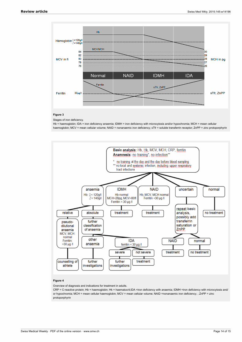
Figure 3
Stages of iron deficiency.Hb = haemoglobin; IDA = iron deficiency anaemia; IDMH = iron deficiency with microcytosis and/or hypochromia; MCH = mean cellularhaemoglobin; MCV = mean cellular volume; NAID = nonanaemic iron deficiency; sTfr = soluble transferrin receptor; ZnPP = zinc protoporphyrin
Figure 4
Overview of diagnosis and indications for treatment in adults.CRP = C-reactive protein; Hb = haemoglobin; Hk = haematocrit;IDA =iron deficiency with anaemia; IDMH =iron deficiency with microcytosis and/or hypochromia; MCH = mean cellular haemoglobin; MCV = mean cellular volume; NAID =nonanaemic iron deficiency; ; ZnPP = zincprotoporphyrin
Review article Swiss Med Wkly. 2015;145:w14196
Swiss Medical Weekly · PDF of the online version · www.smw.ch Page 14 of 15
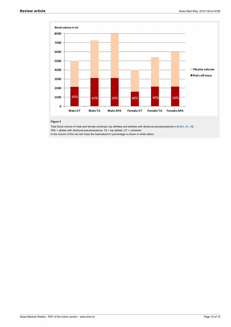
Figure 5
Total blood volume of male and female untrained, top athletes and athletes with dilutional pseudoanaemia in ml [44, 46, 49].APA = athlete with dilutional pseudoanaemia; TA = top athlete; UT = untrainedIn the column of the red cell mass the haematocrit in percentage is shown in white letters.
Review article Swiss Med Wkly. 2015;145:w14196
Swiss Medical Weekly · PDF of the online version · www.smw.ch Page 15 of 15