Embed Size (px)
Citation preview
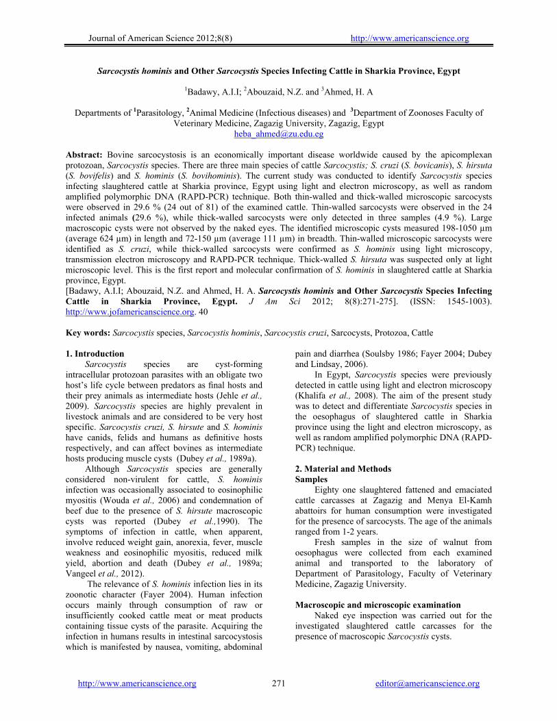
Journal of American Science 2012;8(8) http://www.americanscience.org
http://www.americanscience.org [email protected] 271
Sarcocystis hominis and Other Sarcocystis Species Infecting Cattle in Sharkia Province, Egypt
1Badawy, A.I.I; 2Abouzaid, N.Z. and 3Ahmed, H. A
Departments of 1Parasitology, 2Animal Medicine (Infectious diseases) and 3Department of Zoonoses Faculty of Veterinary Medicine, Zagazig University, Zagazig, Egypt
Abstract: Bovine sarcocystosis is an economically important disease worldwide caused by the apicomplexan protozoan, Sarcocystis species. There are three main species of cattle Sarcocystis; S. cruzi (S. bovicanis), S. hirsuta (S. bovifelis) and S. hominis (S. bovihominis). The current study was conducted to identify Sarcocystis species infecting slaughtered cattle at Sharkia province, Egypt using light and electron microscopy, as well as random amplified polymorphic DNA (RAPD-PCR) technique. Both thin-walled and thick-walled microscopic sarcocysts were observed in 29.6 % (24 out of 81) of the examined cattle. Thin-walled sarcocysts were observed in the 24 infected animals (29.6 %), while thick-walled sarcocysts were only detected in three samples (4.9 %). Large macroscopic cysts were not observed by the naked eyes. The identified microscopic cysts measured 198-1050 µm (average 624 µm) in length and 72-150 µm (average 111 µm) in breadth. Thin-walled microscopic sarcocysts were identified as S. cruzi, while thick-walled sarcocysts were confirmed as S. hominis using light microscopy, transmission electron microscopy and RAPD-PCR technique. Thick-walled S. hirsuta was suspected only at light microscopic level. This is the first report and molecular confirmation of S. hominis in slaughtered cattle at Sharkia province, Egypt. [Badawy, A.I.I; Abouzaid, N.Z. and Ahmed, H. A. Sarcocystis hominis and Other Sarcocystis Species Infecting Cattle in Sharkia Province, Egypt. J Am Sci 2012; 8(8):271-275]. (ISSN: 1545-1003). http://www.jofamericanscience.org. 40 Key words: Sarcocystis species, Sarcocystis hominis, Sarcocystis cruzi, Sarcocysts, Protozoa, Cattle 1. Introduction
Sarcocystis species are cyst-forming intracellular protozoan parasites with an obligate two host’s life cycle between predators as final hosts and their prey animals as intermediate hosts (Jehle et al., 2009). Sarcocystis species are highly prevalent in livestock animals and are considered to be very host specific. Sarcocystis cruzi, S. hirsute and S. hominis have canids, felids and humans as definitive hosts respectively, and can affect bovines as intermediate hosts producing muscle cysts (Dubey et al., 1989a).
Although Sarcocystis species are generally considered non-virulent for cattle, S. hominis infection was occasionally associated to eosinophilic myositis (Wouda et al., 2006) and condemnation of beef due to the presence of S. hirsute macroscopic cysts was reported (Dubey et al.,1990). The symptoms of infection in cattle, when apparent, involve reduced weight gain, anorexia, fever, muscle weakness and eosinophilic myositis, reduced milk yield, abortion and death (Dubey et al., 1989a; Vangeel et al., 2012).
The relevance of S. hominis infection lies in its zoonotic character (Fayer 2004). Human infection occurs mainly through consumption of raw or insufficiently cooked cattle meat or meat products containing tissue cysts of the parasite. Acquiring the infection in humans results in intestinal sarcocystosis which is manifested by nausea, vomiting, abdominal
pain and diarrhea (Soulsby 1986; Fayer 2004; Dubey and Lindsay, 2006).
In Egypt, Sarcocystis species were previously detected in cattle using light and electron microscopy (Khalifa et al., 2008). The aim of the present study was to detect and differentiate Sarcocystis species in the oesophagus of slaughtered cattle in Sharkia province using the light and electron microscopy, as well as random amplified polymorphic DNA (RAPD-PCR) technique. 2. Material and Methods Samples
Eighty one slaughtered fattened and emaciated cattle carcasses at Zagazig and Menya El-Kamh abattoirs for human consumption were investigated for the presence of sarcocysts. The age of the animals ranged from 1-2 years.
Fresh samples in the size of walnut from oesophagus were collected from each examined animal and transported to the laboratory of Department of Parasitology, Faculty of Veterinary Medicine, Zagazig University. Macroscopic and microscopic examination
Naked eye inspection was carried out for the investigated slaughtered cattle carcasses for the presence of macroscopic Sarcocystis cysts.
Journal of American Science 2012;8(8) http://www.americanscience.org
http://www.americanscience.org [email protected] 272
Small pieces of muscles (about 5x5 mm dimensions, 1-2 mm thickness) were compressed between two glass slides and examined microscopically for microscopic Sarcocystis cysts. Histopathological examination
Histopathological examination was performed for microscopically positive samples. About 0.5-1.0 cm from the oesophgeal samples were fixed in 10% neutral buffered formalin, processed by the standard histological techniques and stained with hematoxylin and eosin (Bancroft and Stevens, 1993). Electron microscopy
The thick-walled Sarcocystis cysts were processed for examination by transmission electron microscopy in order to differentiate S. hominis from S. hirsuta. Samples from the oesophageal muscles (2 mm x 2 mm) were fixed in 2.5% glutaraldehyde. Semi-thin sections were made and stained with toluidine blue, while ultra thin sections were stained with urinyl acetate and lead citrate and examined with the electron microscope (Jeol, JXA, 840A electron probe microanalyzer, Japan) at the national research center, Dokki, Egypt. RAPD-PCR
DNA was extracted from ethanol preserved heavily infested oesophageal samples proved positive for sarcocysts by light microscopy using Gene JETTM according to the manufacturer’s instructions. OSA-04 primer with the sequence of 5'-CCAGGGGAAGAGGCAT-3' (manufactured locally by Sigma, Egypt) was used to distinguish S. hominis from other Sarcocystis species of cattle (Güclü, et al. 2004). The RAPD-PCR protocol of Williams et al. (1990) was followed with minor modifications to optimize PCR conditions of the current study. PCR was performed in a total volume of 50 µl in a sterile 0.2 ml RNase free PCR tubes contained 25 µl of 2x reaction mix (Dream TaqTM PCR Master Mix, Fermentas), 40 pmole of the primer and 2 µl of template DNA, the reaction was then completed to 50 µl using nuclease free water. DNA was amplified using 45 cycles with a denaturation step at 94oC for one minute, primer annealing at 42oC for 1.5 minute and an extension step at 72oC for two minutes. Amplified PCR products were analyzed by electrophoresis in 1.5% ethidium bromide stained agarose gels at 100 volts for 40 minutes and visualized under UV transluminator.
3.Results
The prevalence of sarcocysts in slaughtered cattle carcasses used for human consumption in Zagazig city, Sharkia province was determined in the
current study using light and electron microscopy. RAPD-PCR technique was also used for the differentiation of S. hominis which has zoonotic importance.
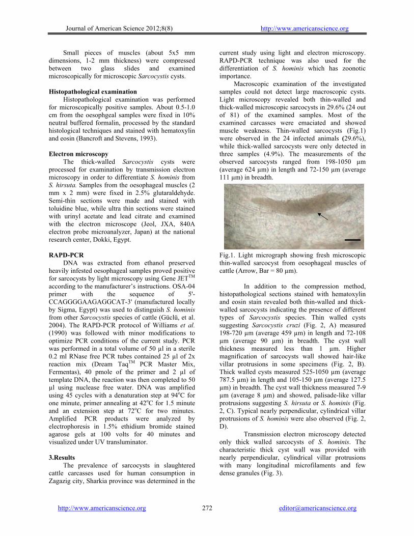
Macroscopic examination of the investigated samples could not detect large macroscopic cysts. Light microscopy revealed both thin-walled and thick-walled microscopic sarcocysts in 29.6% (24 out of 81) of the examined samples. Most of the examined carcasses were emaciated and showed muscle weakness. Thin-walled sarcocysts (Fig.1) were observed in the 24 infected animals (29.6%), while thick-walled sarcocysts were only detected in three samples (4.9%). The measurements of the observed sarcocysts ranged from 198-1050 µm (average 624 µm) in length and 72-150 µm (average 111 µm) in breadth.
Fig.1. Light micrograph showing fresh microscopic thin-walled sarcocyst from oesophageal muscles of cattle (Arrow, Bar = 80 µm).
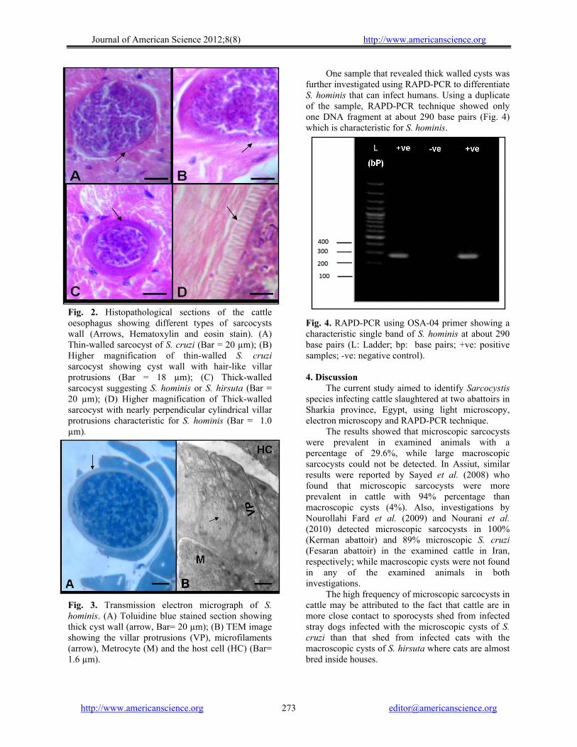
In addition to the compression method, histopathological sections stained with hematoxylin and eosin stain revealed both thin-walled and thick-walled sarcocysts indicating the presence of different types of Sarcocystis species. Thin walled cysts suggesting Sarcocystis cruzi (Fig. 2, A) measured 198-720 µm (average 459 µm) in length and 72-108 µm (average 90 µm) in breadth. The cyst wall thickness measured less than 1 µm. Higher magnification of sarcocysts wall showed hair-like villar protrusions in some specimens (Fig. 2, B). Thick walled cysts measured 525-1050 µm (average 787.5 µm) in length and 105-150 µm (average 127.5 µm) in breadth. The cyst wall thickness measured 7-9 µm (average 8 µm) and showed, palisade-like villar protrusions suggesting S. hirsuta or S. hominis (Fig. 2, C). Typical nearly perpendicular, cylindrical villar protrusions of S. hominis were also observed (Fig. 2, D).
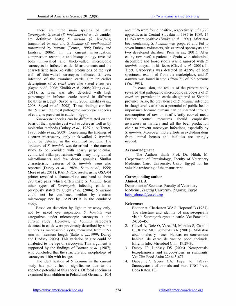
Transmission electron microscopy detected only thick walled sarcocysts of S. hominis. The characteristic thick cyst wall was provided with nearly perpendicular, cylindrical villar protrusions with many longitudinal microfilaments and few dense granules (Fig. 3).
Journal of American Science 2012;8(8) http://www.americanscience.org
http://www.americanscience.org [email protected] 273
Fig. 2. Histopathological sections of the cattle oesophagus showing different types of sarcocysts wall (Arrows, Hematoxylin and eosin stain). (A) Thin-walled sarcocyst of S. cruzi (Bar = 20 µm); (B) Higher magnification of thin-walled S. cruzi sarcocyst showing cyst wall with hair-like villar protrusions (Bar = 18 µm); (C) Thick-walled sarcocyst suggesting S. hominis or S. hirsuta (Bar = 20 µm); (D) Higher magnification of Thick-walled sarcocyst with nearly perpendicular cylindrical villar protrusions characteristic for S. hominis (Bar = 1.0 µm). Fig. 3. Transmission electron micrograph of S. hominis. (A) Toluidine blue stained section showing thick cyst wall (arrow, Bar= 20 µm); (B) TEM image showing the villar protrusions (VP), microfilaments (arrow), Metrocyte (M) and the host cell (HC) (Bar= 1.6 µm).
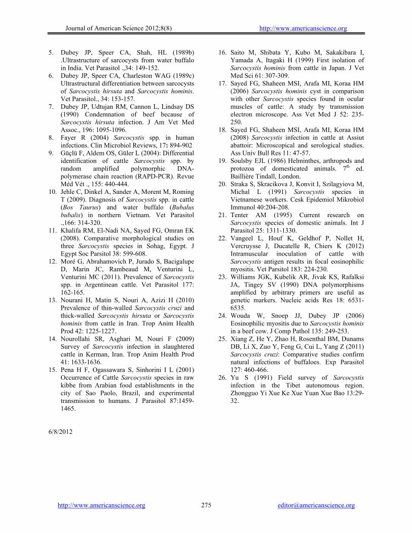
One sample that revealed thick walled cysts was further investigated using RAPD-PCR to differentiate S. hominis that can infect humans. Using a duplicate of the sample, RAPD-PCR technique showed only one DNA fragment at about 290 base pairs (Fig. 4) which is characteristic for S. hominis.
Fig. 4. RAPD-PCR using OSA-04 primer showing a characteristic single band of S. hominis at about 290 base pairs (L: Ladder; bp: base pairs; +ve: positive samples; -ve: negative control). 4. Discussion
The current study aimed to identify Sarcocystis species infecting cattle slaughtered at two abattoirs in Sharkia province, Egypt, using light microscopy, electron microscopy and RAPD-PCR technique.
The results showed that microscopic sarcocysts were prevalent in examined animals with a percentage of 29.6%, while large macroscopic sarcocysts could not be detected. In Assiut, similar results were reported by Sayed et al. (2008) who found that microscopic sarcocysts were more prevalent in cattle with 94% percentage than macroscopic cysts (4%). Also, investigations by Nourollahi Fard et al. (2009) and Nourani et al. (2010) detected microscopic sarcocysts in 100% (Kerman abattoir) and 89% microscopic S. cruzi (Fesaran abattoir) in the examined cattle in Iran, respectively; while macroscopic cysts were not found in any of the examined animals in both investigations.
The high frequency of microscopic sarcocysts in cattle may be attributed to the fact that cattle are in more close contact to sporocysts shed from infected stray dogs infected with the microscopic cysts of S. cruzi than that shed from infected cats with the macroscopic cysts of S. hirsuta where cats are almost bred inside houses.
Journal of American Science 2012;8(8) http://www.americanscience.org
http://www.americanscience.org [email protected] 274
There are three main species of cattle Sarcocystis; S. cruzi (S. bovicanis) of which canides are definitive hosts; S. hirsuta (S. bovifelis) transmitted by cats and S. hominis (S. bovihominis) transmitted by humans (Tenter, 1995; Dubey and Lindsay, 2006). In the current investigation, compression technique and histopathology revealed both thin-walled and thick-walled microscopic sarcocysts in infected cattle. Measurements and the characteristic hair-like villar protrusions of the cyst wall of thin-walled sarcocysts indicated S. cruzi infection of the examined cattle. Similar earlier descriptions of S. cruzi were also stated elsewhere (Sayed et al., 2006; Khalifa et al., 2008; Xiang et al., 2011). S. cruzi was also detected with high percentage in infected cattle reared in different localities in Egypt (Sayed et al., 2006; Khalifa et al., 2008; Sayed et al., 2008). These findings confirm that S. cruzi, the most pathogenic Sarcocystis species of cattle, is prevalent in cattle in Egypt.
Sarcocystis species can be differentiated on the basis of their specific cyst wall structure as well as by molecular methods (Dubey et al., 1989 a, b; Tenter, 1995; Jehle et al., 2009). Concerning the findings of electron microscopy, only thick-walled S. hominis could be detected in the examined sections. The structure of S. hominis was described in the current study to be provided with nearly perpendicular, cylindrical villar protrusions with many longitudinal microfilaments and few dense granules. Similar characteristic features of S. hominis were also reported (Dubey et al., 1989c; Saito et al., 1999; Moré et al., 2011). RAPD-PCR results using OSA-04 primer revealed a characteristic one band at about 290 base pairs which differentiate S. hominis from other types of Sarcocystis infecting cattle as previously stated by Güçlü et al. (2004). S. hirsuta could not be confirmed neither by electron microscopy nor by RAPD-PCR in the conduced study.
Based on detection by light microscopy only, not by naked eye inspection, S. hominis was categorized under microscopic sarcocysts in the current study. However, S. hominis sarcocysts detected in cattle were previously described by some authors as macroscopic cysts, measured from 1.2-7 mm in maximum length (Saito et al.,1999; Dubey and Lindsay, 2006). This variation in size could be attributed to the age of sarcocysts. This argument is supported by the findings of Böttner et al. (1987), who concluded that the structure and morphology of sarcocysts differ with its age.
The identification of S. hominis in the current study has public health significance due to the zoonotic potential of this species. Of fecal specimens examined from children in Poland and Germany, 10.4
and 7.3% were found positive, respectively. Of 1,228 apprentices in Central Slovakia in 1987 to 1989, 14 (1.1%) were positive (Straka et al., 1991). After raw beef containing S. hominis was prepared and fed to seven human volunteers, six excreted sporocysts and two developed diarrhea (Pena et al., 2001). After eating raw beef, a patient in Spain with abdominal discomfort and loose stools was diagnosed with S. hominis oocysts in his feces (Clavel et al., 2001). In Tibet, Sarcocystis was detected in 42.9% of beef specimens examined from the marketplace, and S. hominis was found in stools from 7% of 926 persons (Yu, 1991).
In conclusion, the results of the present study revealed that pathogenic microscopic sarcocysts of S. cruzi are prevalent in cattle slaughtered at Sharkia province. Also, the prevalence of S. hominis infection in slaughtered cattle has a potential of public health importance because humans can be infected through consumption of raw or insufficiently cooked meat. Further control measures should emphasize awareness in farmers and all the beef production chain to prevent sarcocysts infections, especially by S. hominis. Moreover, more efforts in excluding dogs from animal houses and facilities are strongly needed. Acknowledgment
The Authors thank Prof. Dr. Hilali, M. (Department of Parasitology, Faculty of Veterinary Medicine, Cairo University, Cairo, Egypt) for his valuable reviewing of the manuscript. Corresponding author Ahmed, H. A Department of Zoonoses Faculty of Veterinary Medicine, Zagazig University, Zagazig, Egypt [email protected]
References 1. Böttner A, Charleston WAG, Hopcroft D (1987).
The structure and identity of macroscopically visible Sarcocystis cysts in cattle. Vet Parasitol., 24: 35-45.
2. Clavel A, Doiz O, Varea M, Morales S, Castillo FJ, Rubio MC, Gomez-Lus R (2001) . Molestias abdominales y heces blandas en consumidor habitual de carne de vacuno poco cocinada. Enferm Infec Microbiol Clin., 19:29-30.
3. Dubey JP, Lindsay DS (2006). Neosporosis, toxoplasmosis and sarcocystosis in ruminants. Vet Clin Food Anim 22: 645-671.
4. Dubey JP, Speer CA, Fayer R (1989a). Sarcocystosis of animals and man. CRC Press, Boca Raton, FL.
Journal of American Science 2012;8(8) http://www.americanscience.org
http://www.americanscience.org [email protected] 275
5. Dubey JP, Speer CA, Shah, HL (1989b) .Ultrastructure of sarcocysts from water buffalo in India. Vet Parasitol .,34: 149-152.
6. Dubey JP, Speer CA, Charleston WAG (1989c) Ultrastructural differentiation between sarcocysts of Sarcocystis hirsuta and Sarcocystis hominis. Vet Parasitol., 34: 153-157.
7. Dubey JP, Udtujan RM, Cannon L, Lindsay DS (1990) Condemnation of beef because of Sarcocystis hirsuta infection. J Am Vet Med Assoc., 196: 1095-1096.
8. Fayer R (2004) Sarcocystis spp. in human infections. Clin Microbiol Reviews, 17: 894-902
9. Güçlü F, Aldem OS, Güler L (2004): Differential identification of cattle Sarcocystis spp. by random amplified polymorphic DNA-polymerase chain reaction (RAPD-PCR). Revue Méd Vét ., 155: 440-444.
10. Jehle C, Dinkel A, Sander A, Morent M, Roming T (2009). Diagnosis of Sarcocystis spp. in cattle (Bos Taurus) and water buffalo (Bubalus bubalis) in northern Vietnam. Vet Parasitol .,166: 314-320.
11. Khalifa RM, El-Nadi NA, Sayed FG, Omran EK (2008). Comparative morphological studies on three Sarcocystis species in Sohag, Egypt. J Egypt Soc Parsitol 38: 599-608.
12. Moré G, Abrahamovich P, Jurado S, Bacigalupe D, Marin JC, Rambeaud M, Venturini L, Venturini MC (2011). Prevalence of Sarcocystis spp. in Argentinean cattle. Vet Parasitol 177: 162-165.
13. Nourani H, Matin S, Nouri A, Azizi H (2010) Prevalence of thin-walled Sarcocystis cruzi and thick-walled Sarcocystis hirsuta or Sarcocystis hominis from cattle in Iran. Trop Anim Health Prod 42: 1225-1227.
14. Nourollahi SR, Asghari M, Nouri F (2009) Survey of Sarcocystis infection in slaughtered cattle in Kerman, Iran. Trop Anim Health Prod 41: 1633-1636.
15. Pena H F, Ogassawara S, Sinhorini I L (2001) Occurrence of Cattle Sarcocystis species in raw kibbe from Arabian food establishments in the city of Sao Paolo, Brazil, and experimental transmission to humans. J Parasitol 87:1459-1465.
16. Saito M, Shibata Y, Kubo M, Sakakibara I, Yamada A, Itagaki H (1999) First isolation of Sarcocystis hominis from cattle in Japan. J Vet Med Sci 61: 307-309.
17. Sayed FG, Shaheen MSI, Arafa MI, Koraa HM (2006) Sarcocystis hominis cyst in comparison with other Sarcocystis species found in ocular muscles of cattle: A study by transmission electron microscope. Ass Vet Med J 52: 235-250.
18. Sayed FG, Shaheen MSI, Arafa MI, Koraa HM (2008) Sarcocystis infection in cattle at Assiut abattoir: Microscopical and serological studies. Ass Univ Bull Res 11: 47-57.
19. Soulsby EJL (1986) Helminthes, arthropods and protozoa of domesticated animals. 7th ed. Baillière Tindall, London.
20. Straka S, Skracikova J, Konvit I, Szilagyiova M, Michal L (1991) Sarcocystis species in Vietnamese workers. Cesk Epidemiol Mikrobiol Immunol 40:204-208.
21. Tenter AM (1995) Current research on Sarcocystis species of domestic animals. Int J Parasitol 25: 1311-1330.
22. Vangeel L, Houf K, Geldhof P, Nollet H, Vercruysse J, Ducatelle R, Chiers K (2012) Intramuscular inoculation of cattle with Sarcocystis antigen results in focal eosinophilic myositis. Vet Parsitol 183: 224-230.
23. Williams JGK, Kubelik AR, Jivak KS, Rafalksi JA, Tingey SV (1990) DNA polymorphisms amplified by arbitrary primers are useful as genetic markers. Nucleic acids Res 18: 6531-6535.
24. Wouda W, Snoep JJ, Dubey JP (2006) Eosinophilic myositis due to Sarcocystis hominis in a beef cow. J Comp Pathol 135: 249-253.
25. Xiang Z, He Y, Zhao H, Rosenthal BM, Dunams DB, Li X, Zuo Y, Feng G, Cui L, Yang Z (2011) Sarcocystis cruzi: Comparative studies confirm natural infections of buffaloes. Exp Parasitol 127: 460-466.
26. Yu S (1991) Field survey of Sarcocystis infection in the Tibet autonomous region. Zhongguo Yi Xue Ke Xue Yuan Xue Bao 13:29-32.
6/8/2012