Embed Size (px)
Citation preview
C
Past
MLR
C
a
A
R
A
A
K
L
P
P
P
E
h2
j coloproctol (rio j). 2 0 1 4;3 4(4):265–268
Journal ofColoproctology
www.jco l .org .br
ase report
neumoretroperitoneum, pneumomediastinumnd subcutaneous emphysema after endoscopicubmucosal dissection of a rectal lateral spreadingumor
atheus M.M.M.D.E. Meyer ∗, Geraldo M.G. Cruz, Diego V. Sampaio, David De Lanna,uciana M.P. Costa, Ricardo G. Teixeira, Fernando J.C. Lavall Junior, Daniel A. Zanetti,oberta G.S. Lopes, Nayara S.R. Jardim, Eloah G. Lima
oloproctology Service, Santa Casa de Belo Horizonte, Belo Horizonte, MG, Brazil
r t i c l e i n f o
rticle history:
eceived 6 July 2014
ccepted 11 August 2014
vailable online 22 October 2014
eywords:
ateral spreading tumor
neumoretroperitoneum
neumomediastinum
neumoperitoneum
ndoscopic submucosal dissection
a b s t r a c t
Introduction: Endoscopic submucosal dissection (ESD) is an already established procedure in
the treatment of gastric and esophageal cancer in its early stages. Colorectal lesions, ini-
tially approached by endoscopic mucosal resection en bloc or in fragments, are the current
focus for submucosal approach, especially for superficial lateral spreading tumor of 20 mm-
diameter. The experience of Japanese centers, which are reference in therapeutic endoscopy,
demonstrates reduction in the rate of disease recurrence with this approach and, accord-
ing to specific histopathological criteria, may avoid colectomy in some cases of malignant
neoplasia.1–3
Case report: The patient was 50-year-old female. She underwent endoscopic submucosal
dissection of a rectal lateral spreading tumor measuring 50 mm, located 8 cm from the anal
margin. The procedure was performed without major complications, with just two points for
muscle layer detachment, without gross perforation and closed with metal clips. However,
the patient developed air leakage to the peritoneum, retroperitoneum, mediastinum and
subcutaneous tissue, being only treated with clinical procedures and without additional
intervention.
Conclusion: It is vital to know and be able to apply the technique of ESD, in addition to
addressing its complications, since despite the numerous benefits compared to surgery,
ESD can result in serious outcomes.4,5
© 2014 Sociedade Brasileira de Coloproctologia. Published by Elsevier Editora Ltda. All
rights reserved.
∗ Corresponding author.E-mail: [email protected] (M.M.M.M.D.E. Meyer).
ttp://dx.doi.org/10.1016/j.jcol.2014.08.008237-9363/© 2014 Sociedade Brasileira de Coloproctologia. Published by Elsevier Editora Ltda. All rights reserved.
266 j coloproctol (rio j). 2 0 1 4;3 4(4):265–268
Pneumorretroperitônio, pneumomediastino e enfisema subcutâneo apósdisseccão endoscópica da submucosa de lesão retal de crescimento lateral
Palavras-chave:
Lesão de crescimento lateral
Pneumorretroperitôneo
Pneumomediastino
Pneumoperitôneo
Disseccão submucosa
endoscópica
r e s u m o
Introducão: A disseccão endoscópica da submucosa (ESD) já é procedimento consagrado
no tratamento do câncer gástrico e esofagiano em suas fases precoces. As lesões colorre-
tais, inicialmente abordadas por mucossectomia, em bloco ou em fragmentos, são o foco
atual para a abordagem submucosa, principalmente para os tumores de crescimento lateral
superficial a partir de 20 mm de diâmetro. A experiência de centros japoneses, referências
em endoscopia terapêutica, demonstram reducão no índice de recidiva da doenca com esta
abordagem e, segundo critérios histopatológicos específicos, podem evitar uma colectomia
em alguns casos de neoplasia maligna.1–3
Relato de caso: Trata-se de paciente de 50 anos, submetida à disseccão endoscópica da sub-
mucosa de lesão de crescimento lateral, com 50 mm, localizada no reto, a 8 cm da margem
anal. O procedimento foi realizado sem maiores intercorrências, com apenas dois pontos
de afastamento da muscular, sem perfuracão grosseira, fechados com clipe. Entretanto, a
paciente evoluiu com escape aéreo para peritônio, retroperitônio, mediastino e subcútis,
sendo tratada sem intervencão adicional, apenas com manejo clínico.
Conclusão: É de fundamental importância conhecer e saber aplicar a técnica da ESD, além
de abordar suas complicacões, uma vez que, mesmo repleta de benefícios em relacão à
cirurgia, ela pode apresentar desfechos graves.4,5
© 2014 Sociedade Brasileira de Coloproctologia. Publicado por Elsevier Editora Ltda.
Todos os direitos reservados.
Introduction
Colonoscopy is widely used not only as a diagnostic proce-dure, but also with a therapeutic goal, being much prized byminimally invasive medicine.
ESD of early esophageal and gastric carcinomas is alreadypracticed worldwide. The same technique applied to not-invasive pre-malignant and malignant colorectal lesions isnot yet accepted as standard procedure. But this procedureis becoming increasingly feasible, to the extent that thetechnology extends the capabilities with tools appropriateto this procedure.1–3 Thus, ESD allows the required profes-sional training for a proper accomplishment of the method.Despite the prolonged surgical time and long learning curve,this method is superior to the piecemeal mucosal resectionand has a lower rate of local recurrence and greater healingpotential, besides allowing a histopathologic diagnosis for anaccurate disease staging.4,5
The perforations and bleeding are more common in thistechnique; but thus far the benefits conferred to the patientoutweigh the risks. Moreover, the literature shows that con-servative treatment of these complications has been possiblein most cases.6,7
Case report
A 50-year-old female patient was examined and who reportedabdominal cramping pain in hypogastrium, diarrhea alternat-ing with normal bowel habit and hematochezia for about ayear. The terminal ileum colonoscopy showed a type-II high
granular flat lesion, located about 8 cm from the anal mar-gin, measuring 50 mm in its greatest diameter. Our hospitaldid not have the needed equipment for colonoscopy imagingmagnification.
The patient was healthy, with criteria for cure of breastadenocarcinoma treated with left mastectomy, ipsilateral axil-lary lymphadenectomy and adjuvant treatment with radiationand chemotherapy 18 years ago. She reported a family his-tory of colorectal cancer in a first-degree relative (mother,age 70).
The patient was submitted to a colonoscopy with sub-mucosal dissection of the lesion under general anesthesia.Submucosal infiltration with glycerol 12% stained with indigocarmine for submucosal expansion and for better visualiza-tion of planes and vessels was performed. During dissection,0.1% carboxymethylcellulose was used, in order to keep forlonger the submucosal expansion. The muscle layer detach-ment was identified at two points; there was no grossperforation, with the biggest one measuring about 8 mm. Bothperforations were closed with metal clips, without subsequentintercurrences (Fig. 1).
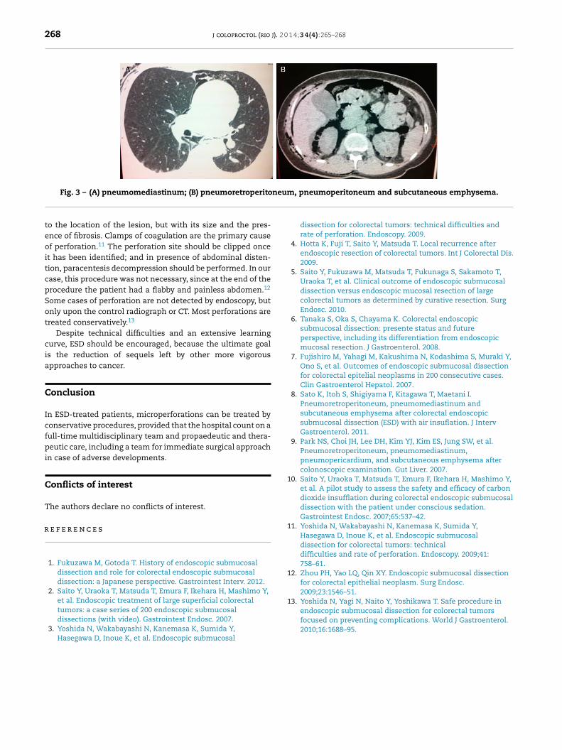
The patient remained hemodynamically stable, and wassuccessfully extubated at the end of colonoscopy. When theprocedure was over, an extensive subcutaneous emphysemathroughout the right half of the body was noted, but withoutclinical consequence. The abdomen was flaccid and painlessand the patient did not complain of dyspnea nor chest pain.An immediate workup was performed with chest radiography,
which showed pneumomediastinum, besides the extensivesubcutaneous emphysema. Still in the operative suite, theabdominal radiograph revealed retropneumoperitoneum andpneumoperitoneum (Fig. 2).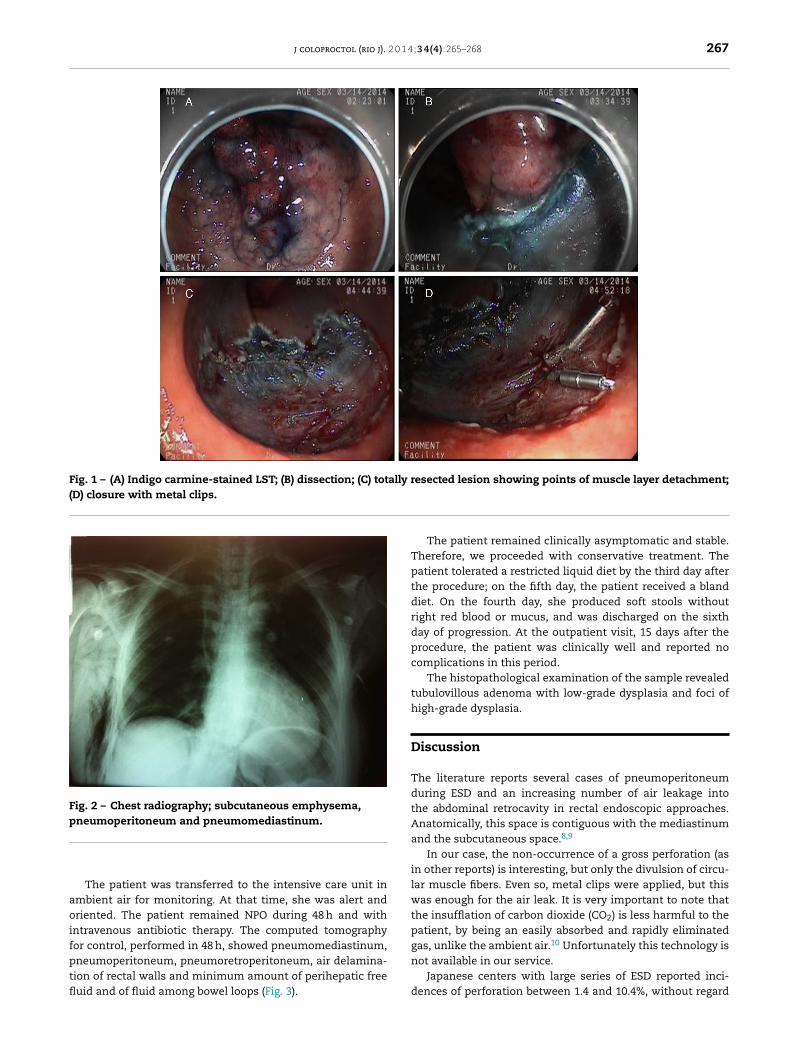
j coloproctol (rio j). 2 0 1 4;3 4(4):265–268 267
Fig. 1 – (A) Indigo carmine-stained LST; (B) dissection; (C) totally resected lesion showing points of muscle layer detachment;(D) closure with metal clips.
Fig. 2 – Chest radiography; subcutaneous emphysema,pneumoperitoneum and pneumomediastinum.
aoifptfl
gas, unlike the ambient air. Unfortunately this technology isnot available in our service.
The patient was transferred to the intensive care unit inmbient air for monitoring. At that time, she was alert andriented. The patient remained NPO during 48 h and with
ntravenous antibiotic therapy. The computed tomographyor control, performed in 48 h, showed pneumomediastinum,neumoperitoneum, pneumoretroperitoneum, air delamina-
ion of rectal walls and minimum amount of perihepatic freeuid and of fluid among bowel loops (Fig. 3).The patient remained clinically asymptomatic and stable.Therefore, we proceeded with conservative treatment. Thepatient tolerated a restricted liquid diet by the third day afterthe procedure; on the fifth day, the patient received a blanddiet. On the fourth day, she produced soft stools withoutright red blood or mucus, and was discharged on the sixthday of progression. At the outpatient visit, 15 days after theprocedure, the patient was clinically well and reported nocomplications in this period.
The histopathological examination of the sample revealedtubulovillous adenoma with low-grade dysplasia and foci ofhigh-grade dysplasia.
Discussion
The literature reports several cases of pneumoperitoneumduring ESD and an increasing number of air leakage intothe abdominal retrocavity in rectal endoscopic approaches.Anatomically, this space is contiguous with the mediastinumand the subcutaneous space.8,9
In our case, the non-occurrence of a gross perforation (asin other reports) is interesting, but only the divulsion of circu-lar muscle fibers. Even so, metal clips were applied, but thiswas enough for the air leak. It is very important to note thatthe insufflation of carbon dioxide (CO2) is less harmful to thepatient, by being an easily absorbed and rapidly eliminated
10
Japanese centers with large series of ESD reported inci-dences of perforation between 1.4 and 10.4%, without regard
268 j coloproctol (rio j). 2 0 1 4;3 4(4):265–268
neum
r
1
1
1
13. Yoshida N, Yagi N, Naito Y, Yoshikawa T. Safe procedure in
Fig. 3 – (A) pneumomediastinum; (B) pneumoretroperito
to the location of the lesion, but with its size and the pres-ence of fibrosis. Clamps of coagulation are the primary causeof perforation.11 The perforation site should be clipped onceit has been identified; and in presence of abdominal disten-tion, paracentesis decompression should be performed. In ourcase, this procedure was not necessary, since at the end of theprocedure the patient had a flabby and painless abdomen.12
Some cases of perforation are not detected by endoscopy, butonly upon the control radiograph or CT. Most perforations aretreated conservatively.13
Despite technical difficulties and an extensive learningcurve, ESD should be encouraged, because the ultimate goalis the reduction of sequels left by other more vigorousapproaches to cancer.
Conclusion
In ESD-treated patients, microperforations can be treated byconservative procedures, provided that the hospital count on afull-time multidisciplinary team and propaedeutic and thera-peutic care, including a team for immediate surgical approachin case of adverse developments.
Conflicts of interest
The authors declare no conflicts of interest.
e f e r e n c e s
1. Fukuzawa M, Gotoda T. History of endoscopic submucosaldissection and role for colorectal endoscopic submucosaldissection: a Japanese perspective. Gastrointest Interv. 2012.
2. Saito Y, Uraoka T, Matsuda T, Emura F, Ikehara H, Mashimo Y,et al. Endoscopic treatment of large superficial colorectal
tumors: a case series of 200 endoscopic submucosaldissections (with vídeo). Gastrointest Endosc. 2007.3. Yoshida N, Wakabayashi N, Kanemasa K, Sumida Y,Hasegawa D, Inoue K, et al. Endoscopic submucosal
, pneumoperitoneum and subcutaneous emphysema.
dissection for colorectal tumors: technical difficulties andrate of perforation. Endoscopy. 2009.
4. Hotta K, Fuji T, Saito Y, Matsuda T. Local recurrence afterendoscopic resection of colorectal tumors. Int J Colorectal Dis.2009.
5. Saito Y, Fukuzawa M, Matsuda T, Fukunaga S, Sakamoto T,Uraoka T, et al. Clinical outcome of endoscopic submucosaldissection versus endoscopic mucosal resection of largecolorectal tumors as determined by curative resection. SurgEndosc. 2010.
6. Tanaka S, Oka S, Chayama K. Colorectal endoscopicsubmucosal dissection: presente status and futureperspective, including its differentiation from endoscopicmucosal resection. J Gastroenterol. 2008.
7. Fujishiro M, Yahagi M, Kakushima N, Kodashima S, Muraki Y,Ono S, et al. Outcomes of endoscopic submucosal dissectionfor colorectal epitelial neoplasms in 200 consecutive cases.Clin Gastroenterol Hepatol. 2007.
8. Sato K, Itoh S, Shigiyama F, Kitagawa T, Maetani I.Pneumoretroperitoneum, pneumomediastinum andsubcutaneous emphysema after colorectal endoscopicsubmucosal dissection (ESD) with air insuflation. J IntervGastroenterol. 2011.
9. Park NS, Choi JH, Lee DH, Kim YJ, Kim ES, Jung SW, et al.Pneumoretroperitoneum, pneumomediastinum,pneumopericardium, and subcutaneous emphysema aftercolonoscopic examination. Gut Liver. 2007.
0. Saito Y, Uraoka T, Matsuda T, Emura F, Ikehara H, Mashimo Y,et al. A pilot study to assess the safety and efficacy of carbondioxide insufflation during colorectal endoscopic submucosaldissection with the patient under conscious sedation.Gastrointest Endosc. 2007;65:537–42.
1. Yoshida N, Wakabayashi N, Kanemasa K, Sumida Y,Hasegawa D, Inoue K, et al. Endoscopic submucosaldissection for colorectal tumors: technicaldifficulties and rate of perforation. Endoscopy. 2009;41:758–61.
2. Zhou PH, Yao LQ, Qin XY. Endoscopic submucosal dissectionfor colorectal epithelial neoplasm. Surg Endosc.2009;23:1546–51.
endoscopic submucosal dissection for colorectal tumorsfocused on preventing complications. World J Gastroenterol.2010;16:1688–95.