Embed Size (px)
Citation preview
This journal is©The Royal Society of Chemistry 2015 J. Mater. Chem. B, 2015, 3, 5569--5576 | 5569
Cite this: J.Mater. Chem. B, 2015,
3, 5569
A fast degradable citrate-based bone scaffoldpromotes spinal fusion
Jiajun Tang,†ab Jinshan Guo,†c Zhen Li,ab Cheng Yang,ab Denghui Xie,ab
Jian Chen,ab Shengfa Li,ab Shaolin Li,d Gloria B. Kim,c Xiaochun Bai,*ab
Zhongmin Zhang*a and Jian Yang*c
It is well known that high rates of fusion failure and pseudoarthrosis development (5–35%) are concomitant in
spinal fusion surgery, which was ascribed to the shortage of suitable materials for bone regeneration. Citrate
was recently recognized to play an indispensable role in enhancing osteoconductivity and osteoinductivity, and
promoting bone formation. To address the material challenges in spinal fusion surgery, we have synthesized
mechanically robust and fast degrading citrate-based polymers by incorporating N-methyldiethanolamine
(MDEA) into clickable poly(1,8-octanediol citrates) (POC-click), referred to as POC-M-click. The obtained
POC-M-click were fabricated into POC-M-click–HA matchstick scaffolds by forming composites with
hydroxyapatite (HA) for interbody spinal fusion in a rabbit model. Spinal fusion was analyzed by radiography,
manual palpation, biomechanical testing, and histological evaluation. At 4 and 8 weeks post surgery, POC-M-
click–HA scaffolds showed optimal degradation rates that facilitated faster new bone formation and higher spinal
fusion rates (11.2 � 3.7, 80 � 4.5 at week 4 and 8, respectively) than the poly(L-lactic acid)–HA (PLLA–HA) con-
trol group (9.3 � 2.4 and 71.1 � 4.4) (p o 0.05). The POC-M-click–HA scaffold-fused vertebrates possessed a
maximum load and stiffness of 880.8 � 14.5 N and 843.2 � 22.4 N mm�1, respectively, which were also much
higher than those of the PLLA–HA group (maximum: 712.0 � 37.5 N, stiffness: 622.5 � 28.4 N mm�1, p o0.05). Overall, the results suggest that POC-M-click–HA scaffolds could potentially serve as promising bone
grafts for spinal fusion applications.
1. Introduction
Bone transplantation is the second most common tissue transplantin the world following blood transfusion with over 2.2 millionprocedures performed annually worldwide.1,2 50% of bone trans-plantation procedures are spine fusion, which has become a routineprocedure in the field of spine surgery for the treatment of cervicalvertebra instability, lumbar degeneration, intervertebral disc injury,and spinal deformity diseases. Normally, spinal fusion surgery iseffective in achieving vertebral stability and nerve decompression.3
However, the rates of fusion failure and pseudarthrosis development
are reported to be as high as 5–35%. The choice of material asan intervertebral filler is extremely critical in spinal fusionsurgery in addition to the patient condition and the choice ofbone transplantation mode.4
The ideal bone substitute should be osteoconductive,osteoinductive, degradable and resorbable, non-immunogenic,risk-free from disease transmission, easy-to-use, mechanicallyrobust, and cost-effective. Up to now, autologous bones remainthe best filling material for intervertebral fusion due to their non-immunogenic properties and high intervertebral fusion rates com-pared to other materials. However, their use is quite limited due totheir associated disadvantages, including additional surgical trauma,increased risk of postoperative complications, and limited quantityof suitable autologous bones.5 Although the application of allograftand xenograft bones solves the problem of limited supply andavoids additional surgical trauma associated with autologousbone harvesting, it brings concerns such as immune rejectionand the risk of spread of bone disease.6–8 Thus, the developmentof engineered bone substitutes as fillers for intervertebral fusionis greatly encouraged. Examples of engineered bone substitutesinclude calcium-based and polymer-based synthetic bone substitutessuch as hydroxyapatite (HA), b-tricalcium phosphate (b-TCP),poly(L-lactic acid) (PLLA), poly(glycolic acid) (PGA) and their
a Academy of Orthopedics, Guangdong Province, Department of Orthopedic Surgery,
The Third Affiliated Hospital of Southern Medical University, Guangzhou, 510630,
China. E-mail: [email protected] Department of Cell Biology, School of Basic Medical Science,
Southern Medical University, Guangzhou 510515, China.
E-mail: [email protected] Department of Biomedical Engineering, Materials Research Institute,
The Huck Institutes of The Life Sciences, The Pennsylvania State University,
University Park, PA 16802, USA. E-mail: [email protected] Medical imaging department, Guangdong Province, The Third Affiliated Hospital
of Southern Medical University, Guangzhou, 510630, China
† These authors contribute equally to this work.
Received 2nd April 2015,Accepted 3rd June 2015
DOI: 10.1039/c5tb00607d
www.rsc.org/MaterialsB
Journal ofMaterials Chemistry B
PAPER
5570 | J. Mater. Chem. B, 2015, 3, 5569--5576 This journal is©The Royal Society of Chemistry 2015
copolymers, as well as polymer–HA or TCP composites.9–11
Unfortunately, the successful application of these materialshas been hampered by problems such as inherent brittleness,poor degradability, insufficient biocompatibility, low fusion rates,and unsatisfactory biomechanical properties.12,13 Therefore, thesearch for a biodegradable, cost-effective, biocompatible, osteo-conductive, and even osteoinductive bone substitute materialthat can be used to achieve a high spinal fusion rate and optimalbone regeneration has become the focus of extensive research.
Citrate, as an important intermediate in the Kreb’s cycle, ishighly concentrated in native bone (90% of body’s total citratecontent is located in the skeletal system) and is closely asso-ciated with bone metabolism and formation.2,14–17 Citrate notonly serves as a calcium-solubilizing agent, but also plays animportant role in the physical binding and thickness control ofbone apatite nanocrystals.15–17 Our recent exciting findings furthershowed that exogenous citrates enhance alkaline phosphatase (ALP)and osterix (OSX) gene expression in C2C12 cells, a mouse myoblastcell line that can differentiate into osteoblasts,18 and promote themineralization of osteoblastic differentiated human mesenchymalstem cells (hMSCs).19 A series of citrate-based biodegradablecomposites have recently been developed for bone regeneration,such as poly(1,8-octanediol citrate)–hydroxyapatite (POC–HA),20,21
clickable POC–HA (POC-click–HA),2,22 crosslinked urethane-dopedpolyesters–HA (CUPE–HA),2 citrate-based polymer blends–HA(CBPBHA),18 poly(ethylene glycol) maleate citrate–HA (PEGMC–HA),23 and injectable citrate-based mussel-inspired tissue bio-adhesives HA composites (iCMBA–HA).19 The above citrate-basedbiomaterials have demonstrated impressive in vivo performance invarious animal models for bone regeneration, such as POC-click–HAfor long segmental radial bone regeneration in rabbits,22 CUPE–HAand POC-click–HA for calvarial regeneration in rats,2 and iCMBA–HAfor comminuted radial bone regeneration in rabbits.19 However,none of the citrate-based biodegradable composites have beenoptimized and evaluated for spinal fusion applications.
Abundant free carboxyl groups provided by citrate polymerscan enhance polymer–HA interactions by calcium chelating. Upto 65 wt% HA can be effectively incorporated in citrate-basedpolymers (i.e. POC) to mimic the composition of native bonesin contrast to conventional biodegradable polymers (i.e. PLLA),into which only up to 35% of HA can be incorporated before thecomposites become too brittle. This attribute is a remarkableadvantage of citrate-based polymers compared to other degradablepolymers, but the mechanical strengths of citrate-based polymer–HAcomposites still need to be improved to meet the requirements forbone applications. To further improve the mechanical strength ofcitrate-based polymers without sacrificing the valuable free carboxylgroups on citrate, clickable POC (POC-click) with robustlyenhanced mechanical strength was recently developed in ourlab24 by employing click chemistry (azide–alkyne cycloaddition)as an additional cross-linking mechanism.
The composites of POC-click with HA did show significantlyimproved mechanical strengths and enhanced calvarial regenerationin rats.2,22 However, click reaction resulted in rigid triazole rings,which delayed the degradation of POC-click as compared withnormal POC.24–26
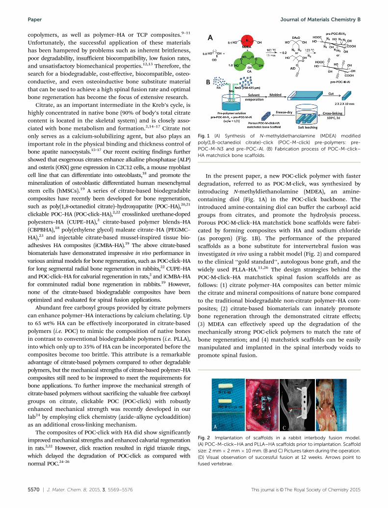
In the present paper, a new POC-click polymer with fasterdegradation, referred to as POC-M-click, was synthesized byintroducing N-methyldiethanolamine (MDEA), an amine-containing diol (Fig. 1A) in the POC-click backbone. Theintroduced amine-containing diol can buffer the carboxyl acidgroups from citrates, and promote the hydrolysis process.Porous POC-M-click–HA matchstick bone scaffolds were fabri-cated by forming composites with HA and sodium chloride(as porogen) (Fig. 1B). The performance of the preparedscaffolds as a bone substitute for intervertebral fusion wasinvestigated in vivo using a rabbit model (Fig. 2) and comparedto the clinical ‘‘gold standard’’, autologous bone graft, and thewidely used PLLA–HA.11,26 The design strategies behind thePOC-M-click–HA matchstick spinal fusion scaffolds are asfollows: (1) citrate polymer–HA composites can better mimicthe citrate and mineral compositions of nature bone comparedto the traditional biodegradable non-citrate polymer–HA com-posites; (2) citrate-based biomaterials can innately promotebone regeneration through the demonstrated citrate effects;(3) MDEA can effectively speed up the degradation of themechanically strong POC-click polymers to match the rate ofbone regeneration; and (4) matchstick scaffolds can be easilymanipulated and implanted in the spinal interbody voids topromote spinal fusion.
Fig. 1 (A) Synthesis of N-methyldiethanolamine (MDEA) modifiedpoly(1,8-octanediol citrate)-click (POC-M-click) pre-polymers: pre-POC-M-N3 and pre-POC-Al. (B) Fabrication process of POC-M-click–HA matchstick bone scaffolds.
Fig. 2 Implantation of scaffolds in a rabbit interbody fusion model.(A) POC-M-click–HA and PLLA–HA scaffolds prior to implantation. Scaffoldsize: 2 mm� 2 mm� 10 mm. (B and C) Pictures taken during the operation.(D) Visual observation of successful fusion at 12 weeks. Arrows point tofused vertebrae.
Paper Journal of Materials Chemistry B
This journal is©The Royal Society of Chemistry 2015 J. Mater. Chem. B, 2015, 3, 5569--5576 | 5571
2. Results and discussion
Citrate is an essential constituent in native bone and was foundto play an indispensable role in the formation and thicknessregulation of the nanocrystalline structure of bone apatite.15–17
The most recent finding further indicated that citrate incorporation(‘‘citration’’) in concert with mineralization must be included in theprocess of bone formation.27 The existing findings in our groupshowed that exogenous citrate, in the forms of free citrate salt orcitrate-based polymers, can enhance the expression of genes relatedto bone formation, including alkaline phosphatase (ALP) and osterix(OSX), and promote the mineralization of osteoblastic-committedhuman mesenchymal stem cells (hMSCs).18,19 These evolvingfindings strongly suggest that citrate should be involved inbone substitute design to mimic the structure and function ofnative bone.
On the other hand, proper mechanical strength, sufficientpolymer–ceramic binding, and suitable degradation rates arethree key factors for the successful application of biodegradablepolymer–bioceramic composites in bone regeneration. Althoughtraditional commercially available biodegradable polymers, suchas PLLA, are widely used as bone substitutes, they suffer fromineffective binding to inorganic particles and long degradationtimes. Complete degradation of PLLA needs more than one year(B52 weeks),28 which would hinder the bone-healing process byinhibiting new bone and vascular ingrowth.19
The existence of abundant free carboxyl groups and theapplication of click chemistry in POC-click polymers providedthem with sufficient binding to bioceramic particles and enhancedmechanical properties.24 However, the rigid triazole rings incrosslinked POC-click delayed the degradation. To preserve highermechanical strength and generate faster degradation rate, anamine-containing diol, N-methyldiethanol-amine (MDEA), wasintroduced into POC-click pre-polymers to obtain pre-POC-M-N3
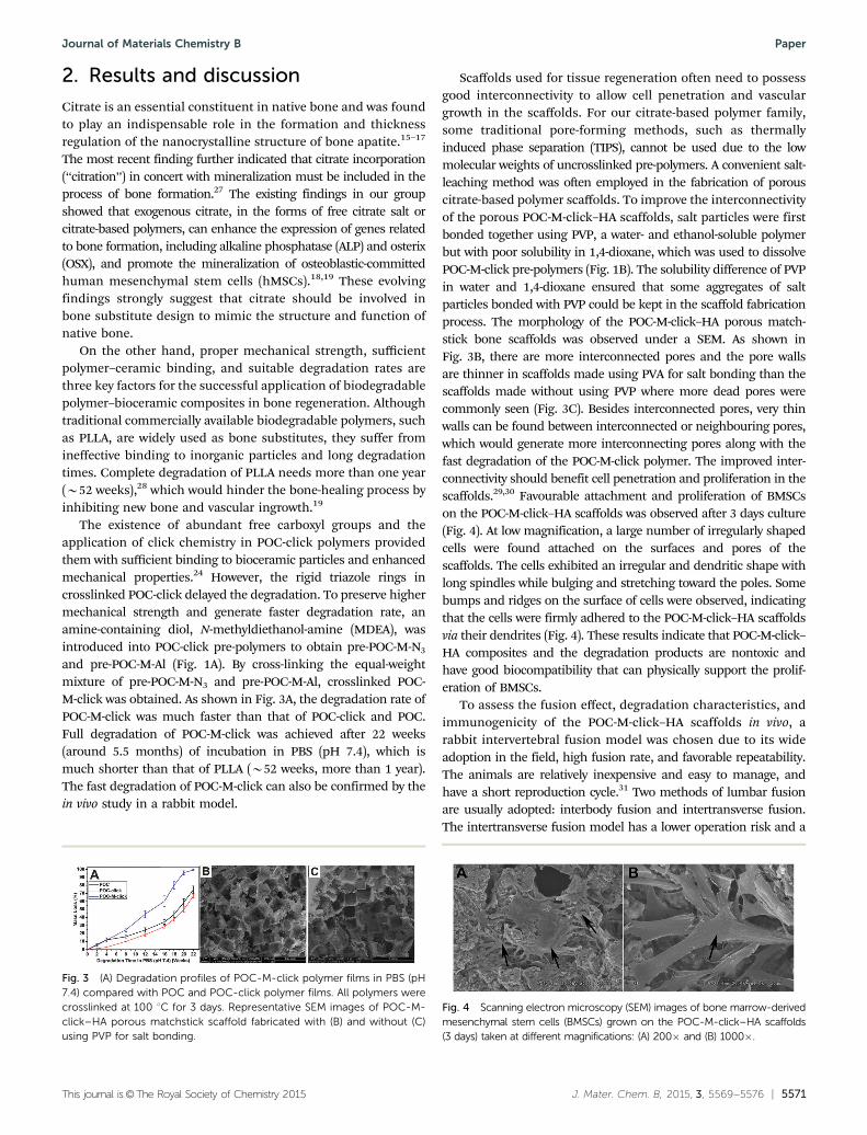
and pre-POC-M-Al (Fig. 1A). By cross-linking the equal-weightmixture of pre-POC-M-N3 and pre-POC-M-Al, crosslinked POC-M-click was obtained. As shown in Fig. 3A, the degradation rate ofPOC-M-click was much faster than that of POC-click and POC.Full degradation of POC-M-click was achieved after 22 weeks(around 5.5 months) of incubation in PBS (pH 7.4), which ismuch shorter than that of PLLA (B52 weeks, more than 1 year).The fast degradation of POC-M-click can also be confirmed by thein vivo study in a rabbit model.
Scaffolds used for tissue regeneration often need to possessgood interconnectivity to allow cell penetration and vasculargrowth in the scaffolds. For our citrate-based polymer family,some traditional pore-forming methods, such as thermallyinduced phase separation (TIPS), cannot be used due to the lowmolecular weights of uncrosslinked pre-polymers. A convenient salt-leaching method was often employed in the fabrication of porouscitrate-based polymer scaffolds. To improve the interconnectivityof the porous POC-M-click–HA scaffolds, salt particles were firstbonded together using PVP, a water- and ethanol-soluble polymerbut with poor solubility in 1,4-dioxane, which was used to dissolvePOC-M-click pre-polymers (Fig. 1B). The solubility difference of PVPin water and 1,4-dioxane ensured that some aggregates of saltparticles bonded with PVP could be kept in the scaffold fabricationprocess. The morphology of the POC-M-click–HA porous match-stick bone scaffolds was observed under a SEM. As shown inFig. 3B, there are more interconnected pores and the pore wallsare thinner in scaffolds made using PVA for salt bonding than thescaffolds made without using PVP where more dead pores werecommonly seen (Fig. 3C). Besides interconnected pores, very thinwalls can be found between interconnected or neighbouring pores,which would generate more interconnecting pores along with thefast degradation of the POC-M-click polymer. The improved inter-connectivity should benefit cell penetration and proliferation in thescaffolds.29,30 Favourable attachment and proliferation of BMSCson the POC-M-click–HA scaffolds was observed after 3 days culture(Fig. 4). At low magnification, a large number of irregularly shapedcells were found attached on the surfaces and pores of thescaffolds. The cells exhibited an irregular and dendritic shape withlong spindles while bulging and stretching toward the poles. Somebumps and ridges on the surface of cells were observed, indicatingthat the cells were firmly adhered to the POC-M-click–HA scaffoldsvia their dendrites (Fig. 4). These results indicate that POC-M-click–HA composites and the degradation products are nontoxic andhave good biocompatibility that can physically support the prolif-eration of BMSCs.
To assess the fusion effect, degradation characteristics, andimmunogenicity of the POC-M-click–HA scaffolds in vivo, arabbit intervertebral fusion model was chosen due to its wideadoption in the field, high fusion rate, and favorable repeatability.The animals are relatively inexpensive and easy to manage, andhave a short reproduction cycle.31 Two methods of lumbar fusionare usually adopted: interbody fusion and intertransverse fusion.The intertransverse fusion model has a lower operation risk and a
Fig. 3 (A) Degradation profiles of POC-M-click polymer films in PBS (pH7.4) compared with POC and POC-click polymer films. All polymers werecrosslinked at 100 1C for 3 days. Representative SEM images of POC-M-click–HA porous matchstick scaffold fabricated with (B) and without (C)using PVP for salt bonding.
Fig. 4 Scanning electron microscopy (SEM) images of bone marrow-derivedmesenchymal stem cells (BMSCs) grown on the POC-M-click–HA scaffolds(3 days) taken at different magnifications: (A) 200� and (B) 1000�.
Journal of Materials Chemistry B Paper
5572 | J. Mater. Chem. B, 2015, 3, 5569--5576 This journal is©The Royal Society of Chemistry 2015
larger bone graft bed, allowing it to accommodate relatively largerimplants. However, the fusion rate for the larger defect in thismodel is low because of the poor vascular conditions around thetransverse processes.32,33 Compared to the intertransverse fusionmodel, the interbody fusion model has advantages due to the richvascular supply at the defect site, which is beneficial for new bonegrowth. In addition, the results obtained in the interbody fusionmodel can better represent the clinical situation, as the clinicalapplication of interbody fusion is much more common thanintertransverse fusion.34,35 In the present study, we sought toimprove the traditional surgical method by using an anterolateralincision for accessing the target disk and fixation of the vertebralbodies with a specific steel plate (Fig. 2B and C).36–38 On the otherhand, this method simplifies the operative course and preventstranslocation of the implant. Moreover, it can provide a relativelystable environment for the fusion, avoiding the interfering effectsof rabbits’ irregular movements.36,39
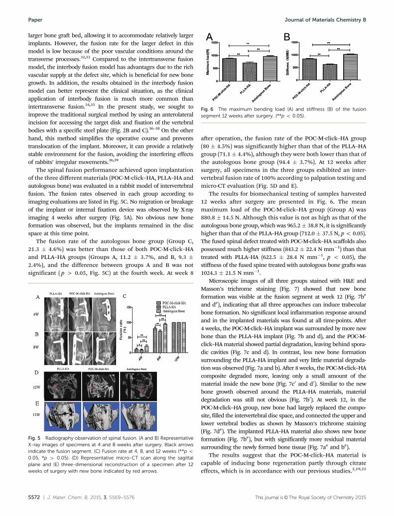
The spinal fusion performance achieved upon implantationof the three different materials (POC-M-click–HA, PLLA–HA andautologous bone) was evaluated in a rabbit model of intervertebralfusion. The fusion rates observed in each group according toimaging evaluations are listed in Fig. 5C. No migration or breakageof the implant or internal fixation device was observed by X-rayimaging 4 weeks after surgery (Fig. 5A). No obvious new boneformation was observed, but the implants remained in the discspace at this time point.
The fusion rate of the autologous bone group (Group C,21.3 � 4.6%) was better than those of both POC-M-click–HAand PLLA–HA groups (Groups A, 11.2 � 3.7%, and B, 9.3 �2.4%), and the difference between groups A and B was notsignificant ( p 4 0.05, Fig. 5C) at the fourth week. At week 8
after operation, the fusion rate of the POC-M-click–HA group(80 � 4.5%) was significantly higher than that of the PLLA–HAgroup (71.1 � 4.4%), although they were both lower than that ofthe autologous bone group (94.4 � 3.7%). At 12 weeks aftersurgery, all specimens in the three groups exhibited an inter-vertebral fusion rate of 100% according to palpation testing andmicro-CT evaluation (Fig. 5D and E).
The results for biomechanical testing of samples harvested12 weeks after surgery are presented in Fig. 6. The meanmaximum load of the POC-M-click–HA group (Group A) was880.8 � 14.5 N. Although this value is not as high as that of theautologous bone group, which was 965.2� 38.8 N, it is significantlyhigher than that of the PLLA–HA group (712.0 � 37.5 N, p o 0.05).The fused spinal defect treated with POC-M-click–HA scaffolds alsopossessed much higher stiffness (843.2 � 22.4 N mm�1) than thattreated with PLLA–HA (622.5 � 28.4 N mm�1, p o 0.05), thestiffness of the fused spine treated with autologous bone grafts was1024.3 � 21.5 N mm�1.
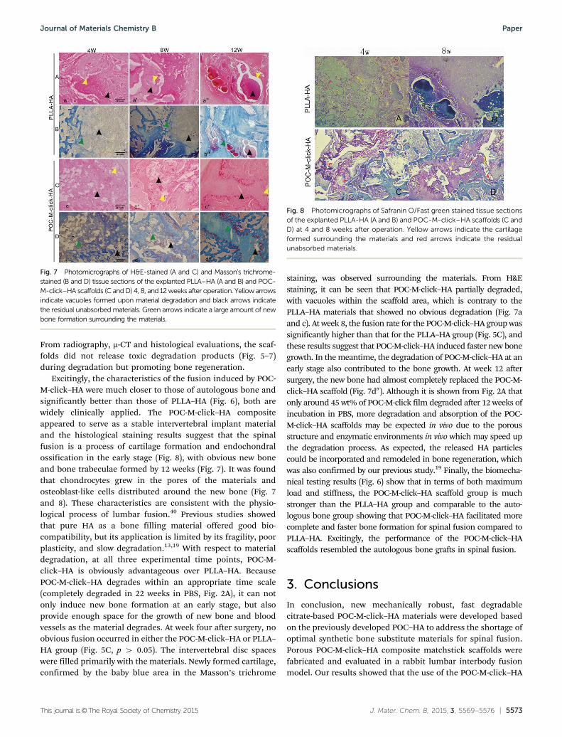
Microscopic images of all three groups stained with H&E andMasson’s trichrome staining (Fig. 7) showed that new boneformation was visible at the fusion segment at week 12 (Fig. 7b00
and d00), indicating that all three approaches can induce trabecularbone formation. No significant local inflammation response aroundand in the implanted materials was found at all time-points. After4 weeks, the POC-M-click–HA implant was surrounded by more newbone than the PLLA–HA implant (Fig. 7b and d), and the POC-M-click–HA material showed partial degradation, leaving behind spora-dic cavities (Fig. 7c and d). In contrast, less new bone formationsurrounding the PLLA–HA implant and very little material degrada-tion was observed (Fig. 7a and b). After 8 weeks, the POC-M-click–HAcomposite degraded more, leaving only a small amount of thematerial inside the new bone (Fig. 7c0 and d0). Similar to the newbone growth observed around the PLLA–HA materials, materialdegradation was still not obvious (Fig. 7b0). At week 12, in thePOC-M-click–HA group, new bone had largely replaced the compo-site, filled the intervertebral disc space, and connected the upper andlower vertebral bodies as shown by Masson’s trichrome staining(Fig. 7d00). The implanted PLLA–HA material also shows new boneformation (Fig. 7b00), but with significantly more residual materialsurrounding the newly formed bone tissue (Fig. 7a00 and b00).
The results suggest that the POC-M-click–HA material iscapable of inducing bone regeneration partly through citrateeffects, which is in accordance with our previous studies.2,19,22
Fig. 5 Radiography observation of spinal fusion. (A and B) RepresentativeX-ray images of specimens at 4 and 8 weeks after surgery. Black arrowsindicate the fusion segment. (C) Fusion rate at 4, 8, and 12 weeks (**p o0.05, *p 4 0.05). (D) Representative micro-CT scan along the sagittalplane and (E) three-dimensional reconstruction of a specimen after 12weeks of surgery with new bone indicated by red arrows.
Fig. 6 The maximum bending load (A) and stiffness (B) of the fusionsegment 12 weeks after surgery. (**p o 0.05).
Paper Journal of Materials Chemistry B
This journal is©The Royal Society of Chemistry 2015 J. Mater. Chem. B, 2015, 3, 5569--5576 | 5573
From radiography, m-CT and histological evaluations, the scaf-folds did not release toxic degradation products (Fig. 5–7)during degradation but promoting bone regeneration.
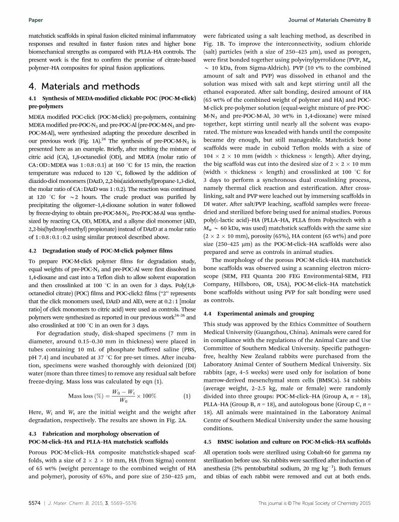
Excitingly, the characteristics of the fusion induced by POC-M-click–HA were much closer to those of autologous bone andsignificantly better than those of PLLA–HA (Fig. 6), both arewidely clinically applied. The POC-M-click–HA compositeappeared to serve as a stable intervertebral implant materialand the histological staining results suggest that the spinalfusion is a process of cartilage formation and endochondralossification in the early stage (Fig. 8), with obvious new boneand bone trabeculae formed by 12 weeks (Fig. 7). It was foundthat chondrocytes grew in the pores of the materials andosteoblast-like cells distributed around the new bone (Fig. 7and 8). These characteristics are consistent with the physio-logical process of lumbar fusion.40 Previous studies showedthat pure HA as a bone filling material offered good bio-compatibility, but its application is limited by its fragility, poorplasticity, and slow degradation.13,19 With respect to materialdegradation, at all three experimental time points, POC-M-click–HA is obviously advantageous over PLLA–HA. BecausePOC-M-click–HA degrades within an appropriate time scale(completely degraded in 22 weeks in PBS, Fig. 2A), it can notonly induce new bone formation at an early stage, but alsoprovide enough space for the growth of new bone and bloodvessels as the material degrades. At week four after surgery, noobvious fusion occurred in either the POC-M-click–HA or PLLA–HA group (Fig. 5C, p 4 0.05). The intervertebral disc spaceswere filled primarily with the materials. Newly formed cartilage,confirmed by the baby blue area in the Masson’s trichrome
staining, was observed surrounding the materials. From H&Estaining, it can be seen that POC-M-click–HA partially degraded,with vacuoles within the scaffold area, which is contrary to thePLLA–HA materials that showed no obvious degradation (Fig. 7aand c). At week 8, the fusion rate for the POC-M-click–HA group wassignificantly higher than that for the PLLA–HA group (Fig. 5C), andthese results suggest that POC-M-click–HA induced faster new bonegrowth. In the meantime, the degradation of POC-M-click–HA at anearly stage also contributed to the bone growth. At week 12 aftersurgery, the new bone had almost completely replaced the POC-M-click–HA scaffold (Fig. 7d00). Although it is shown from Fig. 2A thatonly around 45 wt% of POC-M-click film degraded after 12 weeks ofincubation in PBS, more degradation and absorption of the POC-M-click–HA scaffolds may be expected in vivo due to the porousstructure and enzymatic environments in vivo which may speed upthe degradation process. As expected, the released HA particlescould be incorporated and remodeled in bone regeneration, whichwas also confirmed by our previous study.19 Finally, the biomecha-nical testing results (Fig. 6) show that in terms of both maximumload and stiffness, the POC-M-click–HA scaffold group is muchstronger than the PLLA–HA group and comparable to the auto-logous bone group showing that POC-M-click–HA facilitated morecomplete and faster bone formation for spinal fusion compared toPLLA–HA. Excitingly, the performance of the POC-M-click–HAscaffolds resembled the autologous bone grafts in spinal fusion.
3. Conclusions
In conclusion, new mechanically robust, fast degradablecitrate-based POC-M-click–HA materials were developed basedon the previously developed POC–HA to address the shortage ofoptimal synthetic bone substitute materials for spinal fusion.Porous POC-M-click–HA composite matchstick scaffolds werefabricated and evaluated in a rabbit lumbar interbody fusionmodel. Our results showed that the use of the POC-M-click–HA
Fig. 7 Photomicrographs of H&E-stained (A and C) and Masson’s trichrome-stained (B and D) tissue sections of the explanted PLLA–HA (A and B) and POC-M-click–HA scaffolds (C and D) 4, 8, and 12 weeks after operation. Yellow arrowsindicate vacuoles formed upon material degradation and black arrows indicatethe residual unabsorbed materials. Green arrows indicate a large amount of newbone formation surrounding the materials.
Fig. 8 Photomicrographs of Safranin O/Fast green stained tissue sectionsof the explanted PLLA-HA (A and B) and POC-M-click–HA scaffolds (C andD) at 4 and 8 weeks after operation. Yellow arrows indicate the cartilageformed surrounding the materials and red arrows indicate the residualunabsorbed materials.
Journal of Materials Chemistry B Paper
5574 | J. Mater. Chem. B, 2015, 3, 5569--5576 This journal is©The Royal Society of Chemistry 2015
matchstick scaffolds in spinal fusion elicited minimal inflammatoryresponses and resulted in faster fusion rates and higher bonebiomechanical strengths as compared with PLLA–HA controls. Thepresent work is the first to confirm the promise of citrate-basedpolymer–HA composites for spinal fusion applications.
4. Materials and methods4.1 Synthesis of MEDA-modified clickable POC (POC-M-click)pre-polymers
MDEA modified POC-click (POC-M-click) pre-polymers, containingMDEA modified pre-POC-N3 and pre-POC-Al (pre-POC-M-N3 and pre-POC-M-Al), were synthesized adapting the procedure described inour previous work (Fig. 1A).24 The synthesis of pre-POC-M-N3 ispresented here as an example. Briefly, after melting the mixture ofcitric acid (CA), 1,8-octanediol (OD), and MDEA (molar ratio ofCA : OD : MDEA was 1 : 0.8 : 0.1) at 160 1C for 15 min, the reactiontemperature was reduced to 120 1C, followed by the addition ofdiazido-diol monomers (DAzD, 2,2-bis(azidomethyl)propane-1,3-diol,the molar ratio of CA : DAzD was 1 : 0.2). The reaction was continuedat 120 1C for B2 hours. The crude product was purified byprecipitating the oligomer–1,4-dioxane solution in water followedby freeze-drying to obtain pre-POC-M-N3. Pre-POC-M-Al was synthe-sized by reacting CA, OD, MDEA, and a alkyne diol monomer (AlD,2,2-bis(hydroxyl-methyl) propionate) instead of DAzD at a molar ratioof 1 : 0.8 : 0.1 : 0.2 using similar protocol described above.
4.2 Degradation study of POC-M-click polymer films
To prepare POC-M-click polymer films for degradation study,equal weights of pre-POC-N3 and pre-POC-Al were first dissolved in1,4-dioxane and cast into a Teflon dish to allow solvent evaporationand then crosslinked at 100 1C in an oven for 3 days. Poly(1,8-octanediol citrate) (POC) films and POC-click2 films (‘‘2’’ representsthat the click monomers used, DAzD and AlD, were at 0.2 : 1 [molarratio] of click monomers to citric acid) were used as controls. Thesepolymers were synthesized as reported in our previous work24–26 andalso crosslinked at 100 1C in an oven for 3 days.
For degradation study, disk-shaped specimens (7 mm indiameter, around 0.15–0.30 mm in thickness) were placed intubes containing 10 mL of phosphate buffered saline (PBS,pH 7.4) and incubated at 37 1C for pre-set times. After incuba-tion, specimens were washed thoroughly with deionized (DI)water (more than three times) to remove any residual salt beforefreeze-drying. Mass loss was calculated by eqn (1).
Mass loss ð%Þ ¼W0 �Wt
W0� 100% (1)
Here, Wi and Wt are the initial weight and the weight afterdegradation, respectively. The results are shown in Fig. 2A.
4.3 Fabrication and morphology observation ofPOC-M-click–HA and PLLA–HA matchstick scaffolds
Porous POC-M-click–HA composite matchstick-shaped scaf-folds, with a size of 2 � 2 � 10 mm, HA (from Sigma) contentof 65 wt% (weight percentage to the combined weight of HAand polymer), porosity of 65%, and pore size of 250–425 mm,
were fabricated using a salt leaching method, as described inFig. 1B. To improve the interconnectivity, sodium chloride(salt) particles (with a size of 250–425 mm), used as porogen,were first bonded together using polyvinylpyrrolidone (PVP, Mw
B 10 kDa, from Sigma-Aldrich). PVP (10 v% to the combinedamount of salt and PVP) was dissolved in ethanol and thesolution was mixed with salt and kept stirring until all theethanol evaporated. After salt bonding, desired amount of HA(65 wt% of the combined weight of polymer and HA) and POC-M-click pre-polymer solution (equal-weight mixture of pre-POC-M-N3 and pre-POC-M-Al, 30 wt% in 1,4-dioxane) were mixedtogether, kept stirring until nearly all the solvent was evapo-rated. The mixture was kneaded with hands until the compositebecame dry enough, but still manageable. Matchstick bonescaffolds were made in cuboid Teflon molds with a size of104 � 2 � 10 mm (width � thickness � length). After drying,the big scaffold was cut into the desired size of 2 � 2 � 10 mm(width � thickness � length) and crosslinked at 100 1C for3 days to perform a synchronous dual crosslinking process,namely thermal click reaction and esterification. After cross-linking, salt and PVP were leached out by immersing scaffolds inDI water. After salt/PVP leaching, scaffold samples were freeze-dried and sterilized before being used for animal studies. Porouspoly(L-lactic acid)–HA (PLLA–HA, PLLA from Polyscitech with aMw B 60 kDa, was used) matchstick scaffolds with the same size(2 � 2 � 10 mm), porosity (65%), HA content (65 wt%) and poresize (250–425 mm) as the POC-M-click–HA scaffolds were alsoprepared and serve as controls in animal studies.
The morphology of the porous POC-M-click–HA matchstickbone scaffolds was observed using a scanning electron micro-scope (SEM, FEI Quanta 200 FEG Environmental-SEM, FEICompany, Hillsboro, OR, USA), POC-M-click–HA matchstickbone scaffolds without using PVP for salt bonding were usedas controls.
4.4 Experimental animals and grouping
This study was approved by the Ethics Committee of SouthernMedical University (Guangzhou, China). Animals were cared forin compliance with the regulations of the Animal Care and UseCommittee of Southern Medical University. Specific pathogen-free, healthy New Zealand rabbits were purchased from theLaboratory Animal Center of Southern Medical University. Sixrabbits (age, 4–5 weeks) were used only for isolation of bonemarrow-derived mesenchymal stem cells (BMSCs). 54 rabbits(average weight, 2–2.5 kg, male or female) were randomlydivided into three groups: POC-M-click–HA (Group A, n = 18),PLLA–HA (Group B, n = 18), and autologous bone (Group C, n =18). All animals were maintained in the Laboratory AnimalCentre of Southern Medical University under the same housingconditions.
4.5 BMSC isolation and culture on POC-M-click–HA scaffolds
All operation tools were sterilized using Cobalt-60 for gamma raysterilization before use. Six rabbits were sacrificed after induction ofanesthesia (2% pentobarbital sodium, 20 mg kg�1). Both femursand tibias of each rabbit were removed and cut at both ends.
Paper Journal of Materials Chemistry B
This journal is©The Royal Society of Chemistry 2015 J. Mater. Chem. B, 2015, 3, 5569--5576 | 5575
Intramedullary contents were extracted using a syringe and dis-persed in complete Dulbecco’s modified Eagle’s medium (DMEM,with 10% fetal bovine serum (FBS)) and centrifuged at 1000 rpm for5 minutes, followed by the removal of supernatants. The collectedbone marrow-derived mesenchymal stem cells (BMSCs) werere-suspended in complete DMEM medium and transferred to Petridishes. The cell culture media were replaced for the first time after24 hours and every 2 days thereafter. After reaching 80–90%confluence, the cells were detached with 0.25% trypsin (incubatedabout 3 minutes) and passaged. The original generation was notedas P0, followed by P1, P2, P3, and so on accordingly.41 Cells at thethird generation were used for culture on the sterilized POC-M-click–HA scaffolds.
BMSCs were first seeded on POC-M-click–HA scaffolds in a6-well plate with a cell density of 1� 104 cells per mL (for 1 scaffoldwith a size of 2� 2� 10 mm, using 3 mL), the cell-seeded scaffoldswere then moved to a new 6-well plate the next day and cultured foranother 7 days in complete DMEM media. The cell culture mediawere replaced every other day. To observe cell morphology, the cellson the POC-M-click–HA scaffolds were fixed with 4% glutaralde-hyde solution for 3 hours, washed with PBS solution, and againfixed in 2% osmic acid, and dried for 1 hour. Then the constructswere dehydrated using a graded ethanol series and then transferredto isoamyl acetate, dried at CO2 critical point, and observed under ascanning electron microscope (SEM, Dutch PHILIP TEcNAI-10).
4.6 Surgical method
Prior to surgery, rabbits were fasted for 24 hours. The rabbits weresedated with injection of 2% sodium pentobarbital (30 mg kg�1) andprepared for surgery as per standard practice. For autologous bonegrafting, an approximately 50 mm3 bone block was taken from theiliac crest.
For spinal fusion surgery, the rabbits were placed in a lateralposition. The L4–L5 transverse processes were exposed andremoved through anterolateral surgical intervention to revealthe L4/L5 discs. The L4/L5 discs were then resected. Afteraddressing minor bleeding from the dissected ends, POC-M-click–HA or PLLA–HA matchstick scaffolds were filled in thedefects and the L4 and L5 vertebras were then fixed with screwsand connected with steel plate (Fig. 2). The wounds weresutured after being washed and tamponed with a gelatin sponge.POC-M-click–HA and PLLA–HA matchstick scaffolds were sterilizedby exposing to ethylene oxide overnight before animal surgeries. Allanimals were given penicillin (50 000 U kg�1) intramuscularly for 3consecutive days to prevent infection. The rabbits were allowedaccess to food 24 hours after surgery. Wound healing conditionsand hind leg movements were closely observed.
4.7 General observation, X-ray imaging, and micro-computedtomography (micro-CT) evaluation
At 4, 8, and 12 weeks post operation, the rabbits were anaesthetizedto obtain lumbar radiographs of the interested area in various views(AP view, lateral view and Flexion-Extension position view) using anX-ray machine (Philips, Netherlands). The position of the tissueconstruct and spinal fusion for each specimen were evaluated bythree advanced radiologists based on the radiographs and
stretching palpation tests in a double-blind style based on theSuk’s system.42 Solid union was defined as an obvious interverteb-ral bone bridge formed when the intervertebral range of motion(ROM) on flexion-extension radiographs was o41. Probable unionwas confirmed when subtle intervertebral bone bridge was formed,but with o41 intervertebral ROM on flexion-extension radiographs.Fusion nonunion was defined as little or no bone formationbetween vertebra and the spine, and the motion was beyond 41on flexion-extension radiographs. At each time point, the numberof specimens meeting the standards of solid and probable unionsin the three groups was recorded, and the fusion rate for eachgroup at a certain time point was calculated by eqn (2):
Fusion rate ð%Þ ¼ Nt �Nnon
Nt� 100 (2)
Here, Nt is the total number of specimens tested and Nnon is thenumber of nonunion specimens. For each sample, the fusion ratevalues obtained by the three experienced radiologists were averaged.
At 12 weeks post operation, all rabbits were sacrificed toobtain the lumbar specimens, which were set for a micro-computertomography (m-CT) test and histological examination. m-CT analysiswas conducted using a Micro-CT imaging system (ZKKS-MCT-Sharp-III scanner, Caskaisheng, China) following standard protocols.19 Thescanning system was set to 70 kV, 30 W, and 429 mA. The three-dimensional (3D) images were reconstructed with ZKKS-MicroCT 3.0software. The spinal fusion performance and bone mass formedbetween upper and lower endplates were evaluated based on sagittalplane view and 3D reconstructed images.
4.8 Biomechanical testing
The L4–L5 vertebral bodies and intervertebral discs at each timepoint were removed and stored at �20 1C. Before mechanicaltesting, the specimens were warmed to room temperature and bothends of the specimen were embedded in poly(methyl methacrylate)(PMMA) to make sure the plane of disc is vertical to the compressiondirection. The specimens were then fixed on a mechanical tester(Bose Electro Force 3510, USA), and the load was applied at a fixedrate of 0.008 mm s�1 for compression tests. The changes ofdisplacement and pressure were recorded. Then values for spinalstiffness and maximum load were calculated.22
4.9 Hematoxylin and eosin (H&E) staining and Masson’strichrome staining
Histological examination was performed at pre-determinedtime points (4, 8, 12 week post-operation) according to previousprotocols.19 After fixation in 4% paraformaldehyde for 1 week,lumbar spine specimens were soaked in ethylenediaminetetraaceticacid (EDTA) decalcified liquid for 4 weeks and then embedded inparaffin for later sections at a thickness of 5–8 mm using a SP2500microtome (Leica Microsystems, Germany). Sections were thenstained by the hematoxylin and eosin (H&E), Masson’s trichrome,Safranin O/Fast green staining methods following standard proto-cols. For both types of stained sections, bone histomorphometricanalysis was performed under a semi-automated digitizing imageanalyzer system consisting of an Olympus BX51 microscope(Center Valley, PA, USA), a computer-coupled QImagingRetigaEXi
Journal of Materials Chemistry B Paper
5576 | J. Mater. Chem. B, 2015, 3, 5569--5576 This journal is©The Royal Society of Chemistry 2015
camera (Surrey, Canada), and BioQuantOsteo 2009 software(Nashville, TN, USA)
4.10 Statistical analysis
All data are presented as mean � standard deviation (SD) anddifferences were tested using the t-test. Values of p o 0.05 wereconsidered to indicate significant differences between the twogroups. Data were analyzed using SPSS 13.0 statistical software(SPSS, Inc., Chicago, IL, USA). The main paragraph text followsdirectly on here.
Acknowledgements
This work was supported in part by National Institutes ofHealth (NIH) Awards (EB012575, CA182670, and HL118498),National Science Foundation (NSF) Awards (DMR1313553,CMMI 1266116), and a National Natural Sciences Foundationof China Award (31228007).
Notes and references
1 J. J. Reid, J. S. Johnson and J. C. Wang, J. Biomech., 2011,44, 213.
2 D. Sun, Y. Chen, R. T. Tran, S. Xu, D. Xie, C. Jia, Y. Wang,Y. Guo, J. Guo, Z. Zhang, J. Yang, D. Jin and X. Bai, Sci. Rep.,2014, 4, 6912.
3 Z. Ghogawala, R. G. Whitemore, W. C. Watters 3rd,A. Sharan, P. V. Mummaneni, A. T. Dailey, T. F. Choudhri,J. C. Eck, M. W. Groff, J. C. Wang, D. K. Resnick, S. S. Dhalland M. G. Kaiser, J. Neurosurg. Spine, 2014, 21, 14.
4 B. R. Ransom, E. Neale, M. Henkart, P. N. Bullock andP. G. Nelson, J. Neurophysiol., 1977, 40, 1132.
5 R. Dimitriou, G. I. Mataliotakis, A. G. Angoules,N. K. Kanakaris and P. V. Giannoudis, Injury, 2011, 42, S3.
6 T. Nakajima, H. Iizuka, S. Tsutsumi, M. Kayakabe andK. Takagishi, Spine, 2007, 32, 2432.
7 J. D. Kretlow and A. G. Mikos, Tissue Eng., 2007, 13, 927.8 C. Hou, R. Yang and S. Hou, J. Hosp. Infect., 2005, 59, 41.9 M. F. Moreau, Y. Gallois, M. F. Basle and D. Chappard,
Biomaterials, 2000, 21, 369.10 J. J. Li, D. L. Kaplanb and H. Zreiqat, J. Mater. Chem. B, 2014,
2, 7272.11 T. Lou, X. Wang, G. Song, Z. Gu and Z. Yang, Int. J. Biol.
Macromol., 2014, 69, 464.12 W. Yang, S. K. Both, G. L. van Osch, Y. Wang, J. A. Jansen
and F. Yang, Eur. Cells Mater., 2014, 27, 350.13 M. Stewart, J. F. Welter and V. M. Goldberg, J. Biomed. Mater.
Res., Part A, 2004, 69, 1.14 L. C. Costello, R. B. Franklin, M. A. Reynolds and M. Chellaiah,
Open Bone J., 2012, 4, DOI: 10.2174/1876525401204010027.15 Y. Y. Hu, A. Rawal and K. Schmidt-Rohr, Proc. Natl. Acad.
Sci. U. S. A., 2010, 107, 22425.16 Y. Y. Hu, X. Liu, X. Ma, A. Rawal, T. Prozorov, M. Akinc,
S. K. Mallapragada and K. Schmidt-Rohr, Chem. Mater.,2011, 23, 2481.
17 B. Xie and G. H. Nancollas, Proc. Natl. Acad. Sci. U. S. A.,2010, 107, 22369.
18 R. T. Tran, L. Wang, C. Zhang, M. Huang, W. Tang,C. Zhang, Z. Zhang, D. Jin, B. Banik, J. L. Brown, X. Baiand J. Yang, J. Biomed. Mater. Res., Part A, 2014, 102, 2521.
19 D. Xie, J. Guo, M. R. Mehdizadeh, R. T. Tran, R. Chen,D. Sun, G. Qian, D. Jin, X. Bai and J. Yang, J. Mater. Chem. B,2015, 3, 387.
20 H. Qiu, J. Yang, P. Kodali, J. Koh and G. A. Ameer, Biomater-ials, 2006, 27, 5845.
21 E. J. Chung, P. Kodali, W. Laskin, J. L. Koh and G. A. Ameer,J. Mater. Sci.: Mater. Med., 2011, 22, 2131.
22 Y. Guo, R. T. Tran, D. Xie, Y. Wang, D. Y. Nguyen,E. Gerhard, J. Guo, J. Tang, Z. Zhang, X. Bai and J. Yang,J. Biomed. Mater. Res., Part A, 2015, 103, 772.
23 D. Gyawali, P. Nair, H. K. Kim and J. Yang, Biomater. Sci.,2013, 1, 52.
24 J. Guo, Z. Xie, R. T. Tran, D. Xie, D. Jin, X. Bai and J. Yang,Adv. Mater., 2014, 26, 1906.
25 J. Yang, A. R. Webb, S. J. Pickerill, G. Hageman andG. A. Ameer, Biomaterials, 2006, 27, 1889.
26 J. Yang, A. R. Webb and G. A. Ameer, Adv. Mater., 2004,16, 511.
27 L. C. Costello, M. Chellaiah, J. Zou, R. B. Franklin andM. A. Reynolds, J. Regener. Med. Tissue Eng., 2014, 3, 4, DOI:10.7243/2050-1218-3-4.
28 V. Arias, A. Hoglund, K. Odelius and A. C. Albertsson,Biomacromolecules, 2014, 15, 391.
29 X. Miao and D. Sun, Materials, 2010, 3, 26.30 M. Schumacher, U. Deisinger, R. Detsch and G. Ziegler,
J. Mater. Sci.: Mater. Med., 2010, 21, 3119.31 W. R. Walsh, F. Vizesi, G. B. Cornwall, D. Bell, R. Oliver and
Y. Yu, Eur. Spine J., 2009, 18, 1610.32 M. Palumbo, M. Valdes, A. Robertson, S. Sheikh and
P. Lucas, Spine J., 2004, 4, 287.33 M. Valdes, M. Palumbo, A. J. Appel, S. McAllister and
M. Ehrlich, Spine J., 2004, 4, 293.34 P. Ragni and S. T. Lindlholm, Clin. Orthop., 1991, 272, 292.35 N. M. Raizman, J. R. O’Brien, K. L. Poehling-Monaghan and
W. D. Yu, J. Am. Acad. Orthop. Surg., 2009, 17, 494.36 M. Bezer, Y. Yildirim, B. Erol and O. Guven, Eur. Spine J.,
2005, 14, 227.37 Y. Gu, L. Chen, H. L. Yang, Z. P. Luo and T. S. Tang,
J. Biomed. Mater. Res., Part A, 2011, 97, 177.38 L. Jin, Y. Wan, A. L. Shimer, F. H. Shen and X. J. Li, Tissue
Eng., 2012, 3, 2041731412454420, DOI: 10.1177/2041731412454420.
39 W. E. G. Muller, E. Tolba, H. C. Schroder, M. Neufurth,S. Wang, T. Link, B. Al-Nawasc and X. Wang, J. Mater. Chem.B, 2015, 3, 1722.
40 S. D. Boden, Tissue Eng., 2000, 6, 383.41 J. W. Larson 3rd, E. A. Levicoff, L. G. Gilbertson and
J. D. Kang, J. Bone Jt. Surg., Am. Vol., 2006, 88(suppl 2), 83.42 S. I. Suk, C. K. Lee, W. J. Kim, J. H. Lee, K. J. Cho and
H. J. Kim, Spine, 1997, 1, 210.
Paper Journal of Materials Chemistry B